


Abstract
Background: Robotic-assisted total hip arthroplasty (RA-THA) may improve component positioning but adds workflow demands during adoption. We evaluated the learning curve for total operative and robotic workflow time during the initial implementation of a CT-based robotic system. Methods: We retrospectively analyzed 108 consecutive adults undergoing primary RA-THA between January 2024 and June 2025. We recorded skin-to-skin and robotic workflow times for each procedure. Segmented regression was fitted to individual procedure times, with model selection by Bayesian information criterion. We used CUSUM sensitivity analyses and LOESS smoothing to describe time trends. Results: For both endpoints, a one-breakpoint model was favored, with the estimated change in slope at case 12. Mean skin-to-skin time was 80.8 ± 24.9 min in cases 1–12 and 48.1 ± 12.7 min in cases 13–108. Corresponding robotic workflow times were 46.3 ± 18.8 and 28.2 ± 9.7 min. The CUSUM peak varied with the reference value, and the analysis of individual times did not support a distinct late optimization phase. Conclusions: Operative times decreased most markedly during the first approximately 12 cases and more gradually thereafter. These findings describe operative efficiency in a single-surgeon cohort; further studies should examine clinical outcomes and component-positioning accuracy alongside time trends.
1. Introduction
Total hip arthroplasty (THA) is widely regarded as one of the most successful procedures in modern orthopedic surgery, consistently providing substantial improvements in pain relief, mobility, and quality of life for patients with end-stage hip osteoarthritis [1,2].
Large registry-based and systematic analyses show excellent long-term survivorship of modern implants [2].
Despite these excellent outcomes, technical factors such as component positioning, restoration of hip biomechanics, and accurate reconstruction of leg length and femoral offset remain essential determinants of implant longevity and patient satisfaction [3,4,5,6]. Malposition of the acetabular component has been associated with complications including instability, impingement, accelerated wear, and implant failure [6,7,8]. Consequently, improving the accuracy and reproducibility of implant placement has become a major focus in contemporary hip arthroplasty.
Robotic-assisted surgical systems have been introduced in joint arthroplasty to enhance surgical precision and reproducibility. In THA, CT-based robotic platforms enable surgeons to perform detailed preoperative planning and intraoperative execution with haptic guidance, potentially improving acetabular component orientation and biomechanical restoration [9,10,11,12,13,14]. Several studies and meta-analyses have shown that robotic-assisted THA may reduce variability in component positioning and increase the proportion of implants placed within target orientation zones compared with conventional techniques [10,11,12,13,14,15,16].
However, as with most newly introduced surgical technologies, adopting robotic systems involves a learning curve. During the early stages of implementation, surgeons and operating room staff must adapt to additional procedural steps such as preoperative planning, anatomical registration, system calibration, and robotic-guided acetabular preparation [9,17,18,19,20]. These workflow adjustments may initially increase operative time and influence operating room efficiency.
In robotic-assisted THA specifically, the available literature remains limited. Several studies have suggested learning curves ranging from approximately 12 to 35 cases for operative time and workflow integration when adopting robotic hip arthroplasty platforms [17,18,19,20,21,22]. A clearer understanding of the learning trajectory associated with robotic THA is important for surgeons and institutions considering implementing robotic technology, as it may influence operative scheduling, resource allocation, and training requirements.
Cumulative summation (CUSUM) analysis is often used to visualize sequential changes in operative performance. However, the shape and apparent turning points of a CUSUM curve depend on the selected reference value [23,24]. Previous studies evaluating robotic-assisted arthroplasty have demonstrated relatively short learning curves for operative efficiency. In robotic knee arthroplasty, operative time learning curves range from approximately 7 to 9 cases depending on the robotic system and surgeon experience [25,26].
This study evaluated changes in skin-to-skin and robotic workflow times across consecutive robotic-assisted total hip arthroplasty procedures using segmented regression of individual case times, supplemented by descriptive CUSUM sensitivity analyses and LOESS visualization.
2. Materials and Methods
We conducted a retrospective analysis of 108 consecutive patients who underwent primary robotic-arm-assisted total hip arthroplasty (RA-THA) using a CT-based robotic system (MAKO; Stryker, Kalamazoo, MI, USA) between January 2024 and June 2025. A single surgeon performed all procedures and was experienced in conventional THA but had no prior experience with robotic-assisted THA before the study period. Inclusion criteria were adult patients with end-stage primary hip osteoarthritis undergoing primary RA-THA. Patients with secondary osteoarthritis related to fracture or developmental dysplasia, as well as those requiring complex reconstruction, conversion of previous hip procedures, or treatment in the presence of previous hardware, were excluded.
The study cohort consisted of 58 males (53.7%) and 50 females (46.3%), with a mean age of 68.9 ± 7.6 years. We treated 54 right hips (50%) and 54 left hips (50%) (Table 1).
Table 1.
Baseline characteristics of the study cohort. Data are presented as mean ± standard deviation or n (%). The overall mean age and standard deviation were obtained from the original cohort summary. Individual patient ages were not available in the dataset used for the revised analysis, preventing age comparisons between phases. BMI was not recorded.
Operative time data were recorded in the MAKO case registry for every procedure and subsequently extracted for analysis. Skin-to-skin operative time was defined as the interval between the first skin incision and final wound closure. Robotic workflow time was recorded separately and included all robot-dependent steps, including anatomical landmark acquisition, system calibration and verification, intraoperative adjustment of the surgical plan, robot-assisted bone preparation, and final confirmation of implant positioning. Robotic workflow time was entirely within the skin-to-skin interval and did not add to operative time. Complete time data were available for all 108 procedures.
2.1. Surgical Technique
All procedures were performed using a standardized posterolateral approach for total hip arthroplasty. Patients were positioned in the lateral decubitus position with appropriate pelvic supports to ensure stability throughout the procedure. A preoperative CT scan was used for 3D modeling and robotic planning. A standard posterolateral incision was made, followed by splitting of the gluteus maximus and detachment of the short external rotators with capsulotomy to expose the hip joint. After femoral head dislocation and neck osteotomy, pelvic registration pins were inserted into the iliac crest, and optical tracker arrays were mounted. The MAKO probe registered anatomical landmarks to align the patient’s anatomy with the preoperative CT-based plan.
The robotic arm was then utilized for acetabular reaming within haptic boundaries, ensuring precise inclination and anteversion according to the preoperative plan. The acetabular component was impacted under robotic guidance, followed by liner placement. The surgeon manually performed femoral preparation using broaches to establish canal fit and version. Trial components were inserted, and leg length, offset, and stability were assessed with real-time navigation feedback. Final implants were placed once proper biomechanics and stability were confirmed. After reduction, repair of the capsule and short external rotators was performed when feasible. Meticulous irrigation, hemostasis, and layered closure completed the procedure.
2.2. Data Analysis
CUSUM curves were recalculated separately for skin-to-skin operative time and robotic workflow time using fixed reference values. Because no external or prospectively specified reference was available for both endpoints, sensitivity analyses used reference values of 45, 50, 55, and 60 min for skin-to-skin time and 25, 30, 35, and 40 min for robotic workflow time. For a cohort-window sensitivity analysis, we recalculated CUSUM curves after restricting the series to the first 80, 90, 100, and 108 consecutive cases. We selected these values after data collection; CUSUM was interpreted descriptively and was not used to determine phase boundaries. We estimated breakpoints by segmented regression of individual procedure times [27]. For each endpoint, we fitted linear models with zero, one, or two breakpoints to individual procedure times in chronological order. Candidate breakpoints were constrained to leave at least 10 cases in each segment. We selected the number of breakpoints using the Bayesian information criterion (BIC), with estimated breakpoint locations included in the model penalty. We assessed breakpoint uncertainty using 600 residual bootstrap samples. We assessed serial dependence in the residuals of the selected segmented regression models, rather than in the cumulative CUSUM series. Residual diagnostics included lag-1 autocorrelation, the Durbin–Watson statistic, and Ljung–Box tests at lags 5 and 10. We preserved the chronological case order when fitting the models and calculating residual diagnostics. We calculated a Davies test for a change in slope at each endpoint using 30 evaluation points between cases 10 and 98, with a Python (version 3.12.14) implementation of the Davies upper-bound calculation. We visualized trends in individual procedure times using LOESS smoothing with a span of 0.30 and degree 1; we estimated 95% confidence bands using 400 residual-bootstrap samples. We summarized continuous variables as mean ± standard deviation and range, and categorical variables as counts and percentages. Sex and laterality were compared between phases using Fisher’s exact test. We performed the revised segmented regression and bootstrap analyses in Python using NumPy version 2.3.5 and SciPy version 1.17.0.
3. Results
Learning Curve Analysis
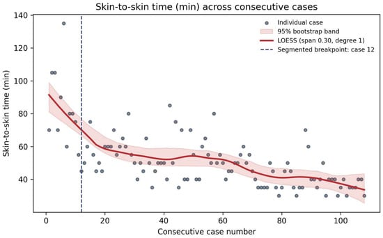
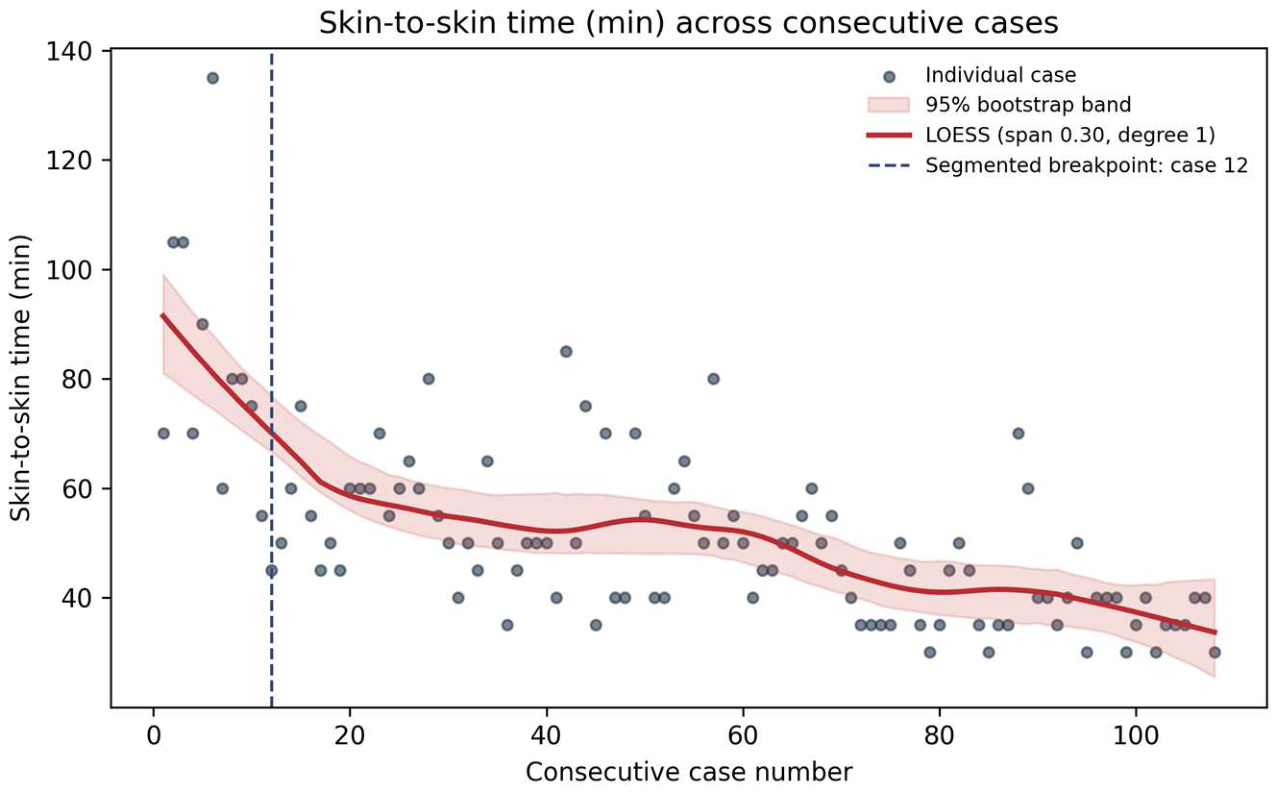
We analyzed 108 robotic-assisted THA procedures. For skin-to-skin operative time, segmented regression of the individual procedure times favored a one-breakpoint model at case 12 (BIC 555.6), compared with models without a breakpoint (BIC 571.2) or with two breakpoints (BIC 563.5). The 95% residual-bootstrap interval for the breakpoint was cases 10–22. The estimated slope was −3.61 min per case before the breakpoint and −0.26 min per case after the breakpoint. Mean skin-to-skin time was 80.8 ± 24.9 min for cases 1–12 and 48.1 ± 12.7 min for cases 13–108. These results indicate a more pronounced initial reduction followed by a gradual decrease in operative time. The Davies test supported a change in slope (p = 0.000013).
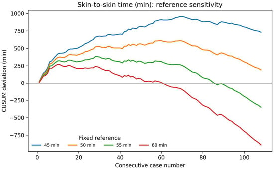
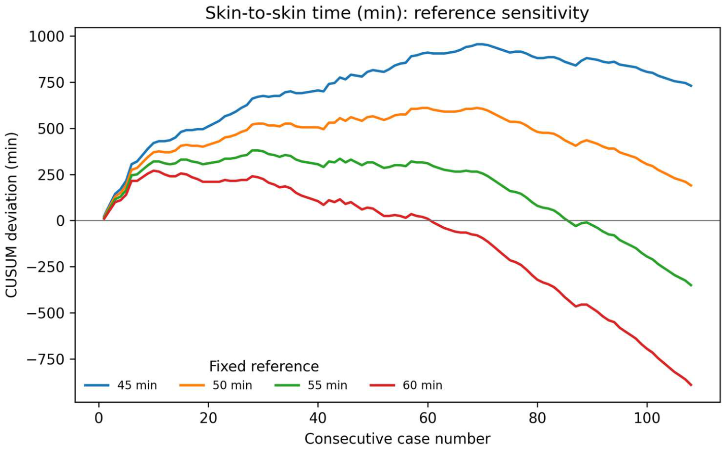
Across the complete series, mean skin-to-skin operative time was 51.8 ± 17.7 min. The CUSUM peak location depended on the fixed reference value: it occurred at case 69 with a 45-min reference, case 59 with 50 min, case 28 with 55 min, and case 10 with 60 min. Figure 1 shows the CUSUM sensitivity analysis for skin-to-skin operative time. For each fixed reference value, the CUSUM peak location did not change when the series was restricted to the first 80, 90, 100, or 108 procedures. Thus, the CUSUM curves did not provide a stable independent basis for defining a late-phase boundary. Figure 2 shows the individual procedure times and their smoothed trend.
Figure 1.
Sensitivity analysis of the cumulative sum (CUSUM) of skin-to-skin operative time across 108 consecutive robotic-assisted total hip arthroplasty procedures. Curves use fixed reference values of 45, 50, 55, and 60 min, selected after data collection. The location of the CUSUM peak varies with the reference value; CUSUM was therefore interpreted descriptively and was not used to define phase boundaries.
Figure 2.
Skin-to-skin operative time across 108 consecutive procedures. Points represent individual procedure times; the solid line shows a locally weighted regression (LOESS; span 0.30, degree 1), with a 95% residual-bootstrap confidence band based on 400 samples. The dashed vertical line marks the breakpoint at case 12, estimated independently by segmented regression of the individual times. LOESS was used for visualization and did not determine the breakpoint.
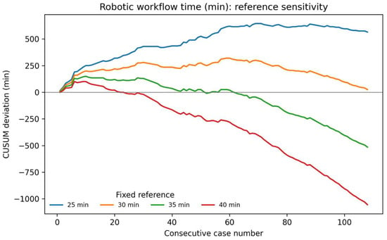
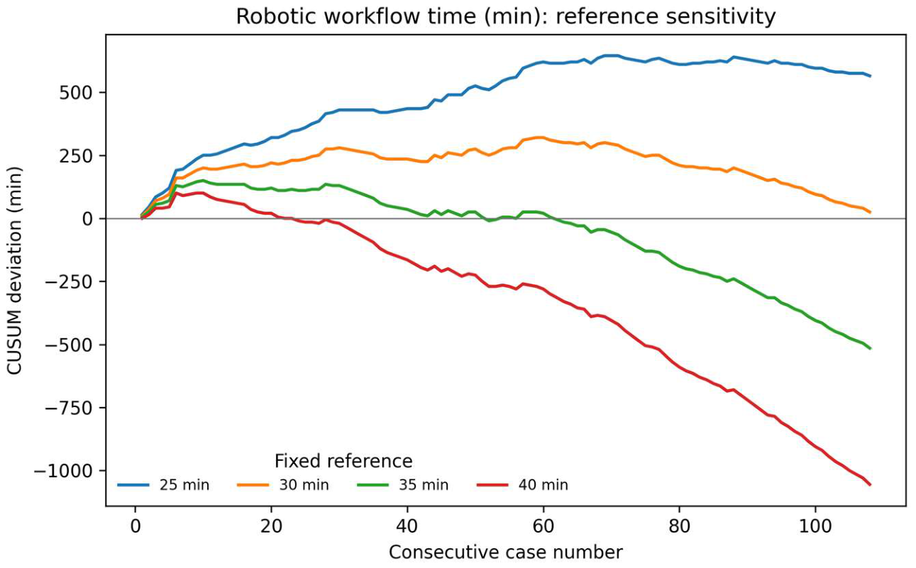
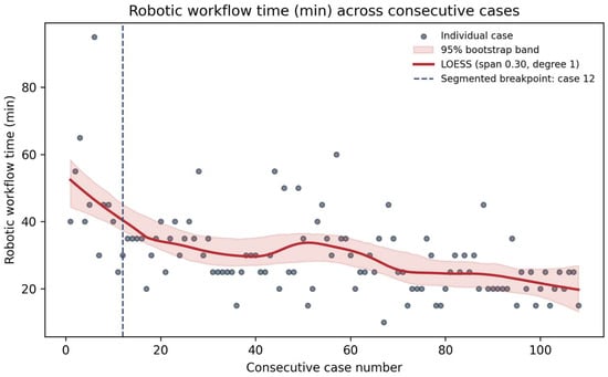
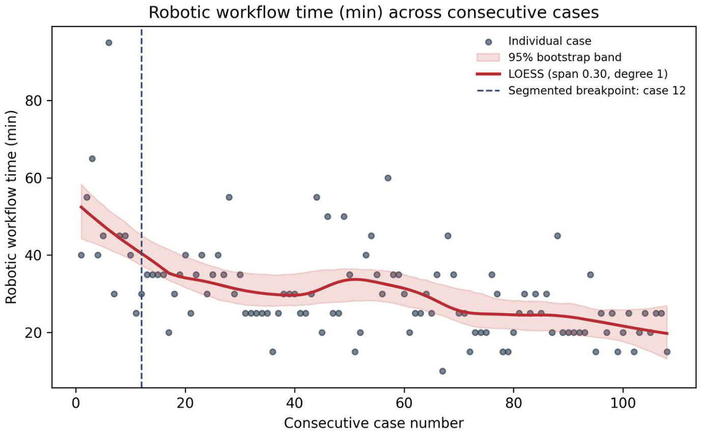
Robotic workflow time showed a similar pattern. Segmented regression of individual times favored a one-breakpoint model at case 12 (BIC 515.3), compared with models without a breakpoint (BIC 518.1) or with two breakpoints (BIC 522.9). The 95% residual-bootstrap interval for the breakpoint was cases 10–46, indicating greater uncertainty than for skin-to-skin time. The estimated slope was −2.03 min per case before the breakpoint and −0.15 min per case after the breakpoint. Mean robotic workflow time was 46.3 ± 18.8 min for cases 1–12 and 28.2 ± 9.7 min for cases 13–108; the overall mean was 30.2 ± 12.4 min (Table 2). In the CUSUM sensitivity analysis, the peak occurred at cases 69, 59, 10, and 6 when reference values of 25, 30, 35, and 40 min were used, respectively. Figure 3 shows the corresponding CUSUM sensitivity analysis for robotic workflow time. For each fixed reference value, the CUSUM peak location did not change across the 80-, 90-, 100-, and 108-case windows. The Davies test also supported a change in slope for robotic workflow time (p = 0.0050). Figure 4 shows the individual robotic workflow times and their smoothed trend. Residual diagnostics found no evidence of remaining serial autocorrelation at the examined lags. For skin-to-skin time, lag-1 residual autocorrelation was −0.012, and the Durbin–Watson statistic was 1.962; Ljung–Box tests yielded p = 0.886 at lag 5 and p = 0.954 at lag 10. Corresponding values for robotic workflow time were −0.088 and 2.143, with Ljung–Box p = 0.544 and p = 0.890, respectively.
Table 2.
Operative times and available case-mix variables by phase. Sex and laterality were compared using Fisher’s exact test. Only the overall cohort summary was available for age, and BMI was not recorded; therefore, neither variable could be compared between phases. No separate hypothesis test was applied to phase-specific mean times, as the phases were defined from those same outcomes.
Figure 3.
Sensitivity analysis of the cumulative sum (CUSUM) of robotic workflow time across 108 consecutive robotic-assisted total hip arthroplasty procedures. Curves use fixed reference values of 25, 30, 35, and 40 min, selected after data collection. The location of the CUSUM peak varies with the reference value; CUSUM was interpreted descriptively and was not used to define phase boundaries.
Figure 4.
Robotic workflow time across 108 consecutive procedures. Points represent individual procedure times; the solid line shows a locally weighted regression (LOESS; span 0.30, degree 1), with a 95% residual-bootstrap confidence band based on 400 samples. The dashed vertical line marks the breakpoint at case 12, estimated independently by segmented regression of the individual times. LOESS was used for visualization and did not determine the breakpoint.
The robotic registry also contained planned and intraoperative component-positioning parameters. We did not include these parameters as learning-curve endpoints or compare them between phases because the intraoperative values had not been independently validated against postoperative imaging and therefore could not establish component-positioning accuracy. Moreover, combined anteversion and femoral stem version were incompletely recorded. Consequently, the present analysis was restricted to operative efficiency.
4. Discussion
The main finding is that operative times decreased across 108 consecutive robotic-assisted THA procedures. Segmented regression of individual procedure times favored an early change in slope at approximately case 12 for both skin-to-skin and robotic workflow time, followed by a more gradual reduction. The analysis did not support a distinct late transition to a third phase. Because robotic workflow time formed part of the total skin-to-skin operative time, the similar trends in these two measures should not be regarded as independent evidence of the learning-curve pattern. The timing of the early change broadly aligns with previous reports of workflow adaptation in robotic-assisted THA, although estimates vary by surgical approach, platform, surgeon experience, and endpoint [17,18,19,20,21,22].
The early reduction in operative time may reflect increasing familiarity with registration, calibration, surgical planning, and robotic-assisted acetabular preparation. Continued improvement thereafter may also reflect refinements in the broader surgical workflow and coordination within the operating-room team. These explanations remain hypotheses: the registry recorded total and robotic workflow times but did not measure the duration of each procedural step or separately quantify surgeon and team contributions.
These findings also align conceptually with prior studies in robotic knee arthroplasty, where investigators have described a short early learning phase for operating-room integration followed by continued efficiency gains over time [25,26]. This supports the broader view that robotic technology in arthroplasty is associated with a measurable but manageable learning curve, particularly for surgeons already experienced in conventional joint replacement.
Previous studies have linked longer operative time to postoperative adverse events after total joint arthroplasty, including infection and readmission [28,29,30,31]. These findings provide context for studying operative efficiency during robotic implementation. However, we did not evaluate postoperative complications in the present cohort; therefore, the observed reductions in operative time cannot be interpreted as evidence of reduced clinical risk.
Previous studies have reported improved reproducibility of acetabular component positioning with robotic-assisted THA compared with conventional techniques [11,12,13,14,15,16]. These findings provide context for adopting robotic systems, but this study did not independently assess component-positioning accuracy. Accordingly, our data address operative efficiency and do not establish whether changes in implant positioning or clinical outcomes accompanied the observed time trends.
This study has several limitations. It describes a single surgeon at one institution, so the time trends may differ with other surgeons, teams, approaches, or robotic platforms. Although the overall mean age and standard deviation were available from the original cohort summary, individual patient ages were unavailable for the revised analysis, and BMI had not been recorded. Consequently, neither age nor BMI could be compared across the estimated phases, and sex and laterality alone cannot exclude differences in case complexity. The breakpoint is an estimate rather than a fixed threshold for proficiency, particularly for robotic workflow time, for which its confidence interval was wide. Although residual diagnostics did not identify serial autocorrelation at the examined lags, these findings do not establish complete independence between consecutive procedures. Unmeasured changes in case complexity, operating-room conditions, or team composition may still have influenced the observed time trends. Robotic workflow time was contained within skin-to-skin time, so these endpoints were not independent. The registry did not separately record the duration of individual workflow steps, and the contribution of team experience could not be isolated from that of the surgeon. Finally, we did not evaluate postoperative complications, patient-reported outcomes, and independently measured component-positioning accuracy. The results should therefore be interpreted as changes in operative efficiency within this cohort, rather than evidence of improved clinical outcomes or technical accuracy.
Overall, this series suggests that the largest reduction in operative time occurred during approximately the first 12 procedures, followed by more gradual improvement. The data do not establish a discrete threshold at which the surgeon or team achieved full proficiency, nor do they support a separate late optimization phase.
5. Conclusions
In this single-surgeon series of 108 robotic-assisted total hip arthroplasties, skin-to-skin and robotic workflow times decreased with case experience. The most pronounced reduction occurred during the first approximately 12 cases, followed by a more gradual decrease; analysis of individual procedure times did not support a distinct later threshold for optimized performance. These findings describe operative efficiency within this cohort and should not be taken as evidence of improved component-positioning accuracy or clinical outcomes. Future multicenter studies should assess whether similar time trends occur across surgeons and teams, account for patient complexity, and examine positioning accuracy, complications, and patient-reported outcomes alongside operative time.
Author Contributions
Conceptualization, C.D.C. and F.B.; methodology, F.N.; software, M.C.; validation, C.B., C.D.C., and F.N.; formal analysis, M.C.; investigation, M.V.; resources, M.V.; data curation, F.N.; writing—original draft preparation, F.B.; writing—review and editing, G.R.; visualization, C.B.; supervision, F.G.; project administration, F.G.; funding acquisition, G.R. All authors have read and agreed to the published version of the manuscript.
Funding
This research received no external funding.
Institutional Review Board Statement
Not applicable.
Informed Consent Statement
Not applicable.
Data Availability Statement
The case-level dataset used in this study, including chronological case order, skin-to-skin operative time, robotic workflow time, sex, and surgical laterality, was extracted from the institutional MAKO case registry. A de-identified version of the analytical dataset is available from the corresponding author upon reasonable request.
Conflicts of Interest
The authors declare no conflicts of interest.
Abbreviations
The following abbreviations are used in this manuscript:
| CT | Computed Tomography |
| CUSUM | Cumulative Sum |
| LOESS | Locally Estimated Scatterplot Smoothing |
| RA-THA | Robotic-Assisted Total Hip Arthroplasty |
| THA | Total Hip Arthroplasty |
| 3D | Three-Dimensional |
References
- Learmonth, I.D.; Young, C.; Rorabeck, C. The operation of the century: Total hip replacement. Lancet 2007, 370, 1508–1519. [Google Scholar] [CrossRef] [Scilit]
- Evans, J.T.; Evans, J.P.; Walker, R.W.; Blom, A.W.; Whitehouse, M.R.; Sayers, A. How long does a hip replacement last? A systematic review and meta-analysis of case series and national registry reports with more than 15 years of follow-up. Lancet 2019, 393, 647–654. [Google Scholar] [CrossRef] [Scilit]
- Bhaskar, D.; Rajpura, A.; Board, T. Current concepts in acetabular positioning in total hip arthroplasty. Indian J. Orthop. 2017, 51, 386–396. [Google Scholar] [CrossRef] [Scilit]
- Flecher, X.; Ollivier, M.; Argenson, J.N. Lower limb length and offset in total hip arthroplasty. Orthop. Traumatol. Surg. Res. 2016, 102, S9–S20. [Google Scholar] [CrossRef] [Scilit]
- Desai, A.S.; Dramis, A.; Board, T.N. Leg length discrepancy after total hip arthroplasty: A review of literature. Curr. Rev. Musculoskelet. Med. 2013, 6, 336–341. [Google Scholar] [CrossRef] [Scilit]
- Oommen, A.T. Offset restoration in total hip arthroplasty: Important: A current review. World J. Orthop. 2024, 15, 696–703. [Google Scholar] [CrossRef] [Scilit]
- Lewinnek, G.E.; Lewis, J.L.; Tarr, R.; Compere, C.L.; Zimmerman, J.R. Dislocations after total hip-replacement arthroplasties. J. Bone Jt. Surg. Am. 1978, 60, 217–220. [Google Scholar] [CrossRef] [Scilit]
- Seagrave, K.G.; Troelsen, A.; Malchau, H.; Husted, H.; Gromov, K. Acetabular cup position and risk of dislocation in primary total hip arthroplasty. Acta Orthop. 2017, 88, 10–17. [Google Scholar] [CrossRef] [Scilit]
- Kayani, B.; Konan, S.; Ayuob, A.; Ayyad, S.; Haddad, F.S. The current role of robotics in total hip arthroplasty. EFORT Open Rev. 2019, 4, 618–625. [Google Scholar] [CrossRef] [Scilit]
- Bullock, E.K.C.; Brown, M.J.; Clark, G.; Plant, J.G.A.; Blakeney, W.G. Robotics in total hip arthroplasty: Current concepts. J. Clin. Med. 2022, 11, 6674. [Google Scholar] [CrossRef] [Scilit]
- Redmond, J.M.; Gupta, A.; Hammarstedt, J.E.; Petrakos, A.E.; Stake, C.E.; Domb, B.G. Accuracy of component placement in robotic-assisted total hip arthroplasty. Orthopedics 2016, 39, 193–199. [Google Scholar] [CrossRef] [Scilit]
- Illgen, R.L.; Bukowski, B.R.; Abiola, R.; Anderson, P.; Chughtai, M.; Khlopas, A.; Mont, M.A. Robotic-assisted total hip arthroplasty: Outcomes at minimum two-year follow-up. Surg. Technol. Int. 2017, 30, 365–372. [Google Scholar]
- Emara, A.K.; Samuel, L.T.; Acuña, A.J.; Kuo, A.; Khlopas, A.; Kamath, A.F. Robotic-arm assisted versus manual total hip arthroplasty: Systematic review and meta-analysis of radiographic accuracy. Int. J. Med. Robot. Comput. Assist. Surg. 2021, 17, e2332. [Google Scholar] [CrossRef] [Scilit]
- Llombart-Blanco, R.; Mariscal, G.; Barrios, C.; Vera, P.; Llombart-Ais, R. MAKO robot-assisted total hip arthroplasty: A comprehensive meta-analysis of efficacy and safety outcomes. J. Orthop. Surg. Res. 2024, 19, 698. [Google Scholar] [CrossRef] [Scilit]
- Singh, A.; Kumar, P.; Kalyan, K.; Gundalli, A.C.; Mane, S.S.; Swarnkar, H.; Singh, L. Accuracy of acetabular cup positioning in robotic-assisted total hip arthroplasty: A CT-based evaluation. SICOT-J 2024, 10, 57. [Google Scholar] [CrossRef] [Scilit]
- Hayashi, S.; Hashimoto, S.; Kamenaga, T.; Nakano, N.; Matsumoto, T.; Ishida, K.; Shibanuma, N.; Kamenaga, T.; Kuroda, R. Accuracy of cup position following robot-assisted total hip arthroplasty may be associated with surgical approach and pelvic tilt. Sci. Rep. 2021, 11, 7578. [Google Scholar] [CrossRef] [Scilit]
- Buchan, G.B.J.; Hecht, C.J.I.I.; Lawrie, C.M.; Sculco, P.K.; Kamath, A.F. The learning curve for a novel, fluoroscopy-based robotic-assisted total hip arthroplasty system. Int. J. Med. Robot. Comput. Assist. Surg. 2023, 19, e2518. [Google Scholar] [CrossRef] [Scilit]
- Schwartz, J.M.; Grant, A.R.; Bhadra, A.K. The learning curve for robotic-assisted total hip arthroplasty in low-, medium-, and high-volume surgeons. J. Orthop. 2024, 55, 163–168. [Google Scholar] [CrossRef] [Scilit]
- Hecht, C.J.; Porto, J.R.; Sanghvi, P.A.; Homma, Y.; Sculco, P.K.; Kamath, A.F. Contemporary analysis of the learning curve for robotic-assisted total hip arthroplasty: Emerging technologies. J. Robot. Surg. 2024, 18, 160. [Google Scholar] [CrossRef] [Scilit]
- Sun, H.; Lu, H.; Xiao, Q.; Ding, Z.; Luo, Z.; Zhou, Z. The learning curve of a novel seven-axis robot-assisted total hip arthroplasty system: A randomized controlled trial. BMC Musculoskelet. Disord. 2024, 25, 342. [Google Scholar] [CrossRef] [Scilit]
- Tian, R.; Duan, X.; Kong, N.; Wang, K.; Yang, P. Precise acetabular positioning, discrepancy in leg length, and hip offset using a new seven-axis robot-assisted total hip arthroplasty system requires no learning curve: A retrospective study. J. Orthop. Surg. Res. 2023, 18, 236. [Google Scholar] [CrossRef] [Scilit]
- Kamath, A.G.; Gill, S.S.; Namireddy, S.R.; Krkovic, M. Learning curves associated with robotic total hip arthroplasty: A scoping review. Orthop. Surg. 2025, 17, 2529–2540. [Google Scholar] [CrossRef] [Scilit]
- Chaput de Saintonge, D.M.; Vere, D.W. Why don’t doctors use cusums? Lancet 1974, 1, 120–121. [Google Scholar] [CrossRef] [Scilit]
- Page, E.S. Continuous inspection schemes. Biometrika 1954, 41, 100–115. [Google Scholar] [CrossRef] [Scilit]
- Kayani, B.; Konan, S.; Huq, S.S.; Tahmassebi, J.; Haddad, F.S. Robotic-arm assisted total knee arthroplasty has a learning curve of seven cases for integration into the surgical workflow but no learning curve effect for accuracy of implant positioning. Knee Surg. Sports Traumatol. Arthrosc. 2019, 27, 1132–1141. [Google Scholar] [CrossRef] [Scilit]
- Pagan, C.A.; Karasavvidis, T.; Siljander, B.; Debbi, E.M.; DeCook, C.A.; Vigdorchik, J. Operative time learning curve for an image-free robotic arm assisted total knee arthroplasty: A cumulative sum analysis. Arthroplast. Today 2024, 31, 101588. [Google Scholar] [CrossRef] [Scilit]
- Muggeo, V.M.R. Estimating regression models with unknown break-points. Stat. Med. 2003, 22, 3055–3071. [Google Scholar] [CrossRef] [Scilit]
- Bohl, D.D.; Ondeck, N.T.; Darrith, B.; Hannon, C.P.; Fillingham, Y.A.; Della Valle, C.J. Impact of operative time on adverse events following primary total joint arthroplasty. J. Arthroplast. 2018, 33, 2256–2262.e4. [Google Scholar] [CrossRef] [Scilit]
- George, J.; Mahmood, B.; Sultan, A.A.; Sodhi, N.; Mont, M.A.; Higuera, C.A.; Stearns, K.L. How fast should a total knee arthroplasty be performed? An analysis of 140,199 surgeries. J. Arthroplast. 2018, 33, 2616–2622. [Google Scholar] [CrossRef] [Scilit]
- Wang, Q.; Goswami, K.; Shohat, N.; Aalirezaie, A.; Manrique, J.; Parvizi, J. Longer operative time results in a higher rate of subsequent periprosthetic joint infection in patients undergoing primary joint arthroplasty. J. Arthroplast. 2019, 34, 947–953. [Google Scholar] [CrossRef] [Scilit]
- Namba, R.S.; Inacio, M.C.S.; Paxton, E.W. Risk factors associated with deep surgical site infections after primary total knee arthroplasty: An analysis of 56,216 knees. J. Bone Jt. Surg. Am. 2013, 95, 775–782. [Google Scholar] [CrossRef] [Scilit]

Disclaimer/Publisher’s Note: The statements, opinions and data contained in all publications are solely those of the individual author(s) and contributor(s) and not of MDPI and/or the editor(s). MDPI and/or the editor(s) disclaim responsibility for any injury to people or property resulting from any ideas, methods, instructions or products referred to in the content. |
© 2026 by the authors. Licensee MDPI, Basel, Switzerland. This article is an open access article distributed under the terms and conditions of the Creative Commons Attribution (CC BY) license.