


Abstract
Brucella canis is a recognized zoonotic pathogen associated with human infection in a variety of epidemiological settings; however, the magnitude of its impact on human health remains uncertain because of its nonspecific clinical presentation, limited availability of species-specific diagnostic methods, and insufficient awareness among physicians, veterinarians, microbiologists, and public health professionals. Unlike classical human brucellosis caused by smooth Brucella species, B. canis infection may be overlooked, potentially leading to delayed diagnosis and incomplete case detection. This narrative review summarizes current evidence on the epidemiology, transmission pathways, occupational and non-occupational risk factors, clinical manifestations, diagnostic approaches, treatment, and public health implications of human B. canis infection. Particular emphasis is placed on published human cases, laboratory-acquired infections, household clusters, cases reported in different age and immune-status groups, and the biological and epidemiological characteristics of canine infection that facilitate zoonotic transmission. The review further examines the limitations of currently available microbiological, serological, and molecular diagnostic methods, the lack of harmonized surveillance systems, and the consequences of inadequate recognition from a One Health perspective. Collectively, the available evidence confirms the zoonotic potential of B. canis but remains insufficient to define the true burden of human infection. Improving clinical awareness, strengthening collaboration between veterinary and human health sectors, expanding access to standardized diagnostic tools, and implementing integrated One Health surveillance may improve recognition and reduce zoonotic risk.
1. Introduction
Brucella canis (B. canis) is recognized as one of the principal bacterial causes of reproductive disease in dogs and has gained increasing attention because of its zoonotic potential and public health relevance [1,2,3]. It is a facultative intracellular, non-motile, non-encapsulated, non-spore-forming, Gram-negative coccobacillus [4,5] that circulates primarily within domestic dog populations, which represent its main reservoir [4,6]. In canine hosts, infection is classically associated with reproductive disorders, including infertility, abortion, epididymitis, orchitis, and prostatitis [6]. These manifestations closely resemble the reproductive syndrome caused by the smooth Brucella species (B. abortus, B. melitensis, and B. suis) in livestock. However, the clinical spectrum extends beyond reproductive disease and may include prolonged bacteremia, splenitis, nephritis, discospondylitis, endocarditis, uveitis, and chorioretinitis [7,8]. Importantly, a substantial proportion of infected dogs remain clinically asymptomatic while continuing to harbor and intermittently shed the organism over extended periods [4,9]. This combination of chronic infection, asymptomatic carriage, and prolonged bacterial shedding may facilitate persistence and dissemination of B. canis within breeding kennels, shelters, rescue organizations, multi-dog households, and other settings where dogs are maintained in close contact, thereby increasing opportunities for transmission among animals and potential zoonotic exposure [10,11]. Evidence from studies of stray dog populations also indicates circulation of Brucella spp., including B. canis, outside owned and breeding dog populations [12].
Human infection occurs primarily through direct contact with infected dogs or exposure to contaminated biological materials, particularly reproductive materials associated with abortion [13]. B. canis may also be shed in reproductive secretions and urine, which can represent potential sources of exposure [14]. Laboratory-acquired infection with B. canis was documented in early reports, emphasizing the occupational risk associated with handling cultures and clinical specimens [15]. Human B. canis infection has been documented following direct or occupational exposure to infected dogs and contaminated biological materials [16,17,18,19]. In humans, infection most commonly presents as a nonspecific febrile illness characterized by fever, chills, malaise, peripheral lymphadenopathy, and splenomegaly, although severe complications, including infective endocarditis and central nervous system involvement, have also been reported [19]. Although documented human cases remain relatively uncommon, infections have been reported in both Europe and the Americas, predominantly among individuals with close household or occupational contact with infected dogs [3,16,17,18,19]. Recent One Health and veterinary investigations from Brazil and Italy further illustrate the geographical heterogeneity of canine B. canis circulation, exposure settings, and surveillance findings [20,21].
Owing to its nonspecific clinical presentation and the limited availability of diagnostic methods specifically validated for human B. canis infection, the true frequency of human infection remains uncertain. Evidence from published case reports, occupational and laboratory exposure investigations, outbreak reports, and limited serological studies indicates that the magnitude of potential underdiagnosis is difficult to quantify [1,13,19]. Recognition is further complicated by limited surveillance, low clinical suspicion, and diagnostic constraints. Despite growing recognition of its zoonotic potential, canine brucellosis is subject to heterogeneous surveillance and reporting practices across countries, limiting direct comparisons of its occurrence and public health significance [21,22]. Consequently, the available evidence confirms the zoonotic potential of B. canis but remains insufficient to determine the true burden of human infection [3,23].
Several recent reviews have comprehensively addressed canine brucellosis, the microbiology and epidemiology of B. canis, and its increasing importance in veterinary medicine and within the One Health framework [3,24,25]. However, the available evidence on human B. canis infection remains fragmented across individual case reports, occupational exposure investigations, laboratory-acquired infections, outbreak reports, and public health communications. The epidemiological, clinical, diagnostic, and surveillance-related evidence concerning human infection has therefore rarely been considered together within an integrated One Health framework [3].
The present narrative review integrates evidence from the veterinary, medical, and public health literature to provide a comprehensive assessment of the zoonotic implications of B. canis infection. By bringing together published human cases, documented exposure events, diagnostic challenges in both dogs and humans, and the veterinary characteristics that facilitate zoonotic transmission, this review critically examines the available evidence on human B. canis infection and the factors that complicate its recognition. Particular emphasis is placed on the interplay between clinical awareness, diagnostic limitations, and surveillance gaps within a One Health framework [26]. By synthesizing evidence across these complementary disciplines, the review identifies priorities for improving diagnosis, biosafety, reporting, surveillance, and interdisciplinary collaboration, thereby supporting evidence-based strategies to reduce human exposure and strengthen prevention at the animal–human interface.
2. Literature Search Strategy
A literature search was conducted to support this narrative review and to identify publications addressing the zoonotic and public health aspects of Brucella canis infection. Electronic searches were performed in PubMed/MEDLINE, Scopus, Web of Science, and Google Scholar using combinations of the following keywords: “Brucella canis”, “human infection”, “zoonosis”, “laboratory-acquired infection”, “occupational exposure”, “case report”, “family cluster”, “children”, “immunocompromised”, “diagnosis”, “surveillance”, and “One Health”. The literature was last updated on 1 September 2026.
Original research articles, case reports, outbreak investigations, surveillance reports, public health communications, and relevant review articles were considered according to their relevance to the scope of this narrative review. Veterinary studies were included when their findings directly informed zoonotic transmission, human exposure, diagnostic challenges, or prevention at the animal–human interface. Reference lists of relevant publications were also examined to identify additional pertinent studies.
A narrative approach was selected as this review objective was to integrate a heterogenous body of evidence spanning human infection, veterinary epidemiology, diagnostic methods, molecular investigations, surveillance, and regulatory frameworks within a One Health perspective, rather than to address a single predefined outcome or quantitatively synthesize a uniform group of studies. This approach also enabled incorporation of evidence that has emerged since recent reviews, including newly reported human cases and molecular investigations from 2024 to 2026, a detailed evaluation of the performance and validation limitations of diagnostic assays and reagents, as well as recent developments in surveillance and regulatory approaches, including the introduction of mandatory pre-import B. canis testing for certain commercially imported dogs in Great Britain in 2025. The review additionally considers B. canis within the current European Union regulatory framework and the implications of its absence from harmonized disease-specific surveillance and control measures. In this respect, the present review complements the 2025 scoping review of human B. canis infection [3] by providing an updated and broader cross-disciplinary synthesis of the available evidence.
The literature was selected on the basis of relevance to the stated objective of this narrative review and did not follow a systematic study-selection protocol; consequently, no formal risk-of-bias assessment, evidence-quality scoring or meta-analysis was performed. This approach may be subject to selection and publication bias and limits quantitative comparison across studies. The findings should therefore be interpreted as a critical synthesis of a heterogeneous evidence base rather than as quantitative estimates of incidence, prevalence, comparative risk, diagnostic performance, or therapeutic efficacy.
3. Veterinary Characteristics of Brucella canis Infection Relevant to Zoonotic Transmission
3.1. Clinical Complexity of Canine Brucellosis
Although canine brucellosis is classically regarded as a reproductive disease, its clinical spectrum extends beyond the reproductive tract. Similar to brucellosis caused by the smooth Brucella species (B. abortus, B. melitensis, and B. suis) in livestock, B. canis infection is commonly associated with abortion, embryonic death, infertility, epididymitis, orchitis, and prostatitis [2,22,27,28,29]. Extra-reproductive manifestations, including discospondylitis, polyarthritis, uveitis, lymphadenopathy, and endocarditis, have also been reported and may occur in the absence of reproductive abnormalities [2,22,27,28,29,30,31,32].
From a zoonotic perspective, this clinical variability is relevant because infected dogs without characteristic reproductive signs may not immediately raise suspicion of canine brucellosis. B. canis infection should therefore be considered in dogs with compatible clinical findings when supported by epidemiological risk factors, such as breeding activity, kennel residence, or recent importation [2,22,29]. Recognition of these presentations is important for initiating appropriate diagnostic testing and reducing opportunities for exposure of other dogs and humans.
3.2. Biological and Epidemiological Characteristics Facilitating Silent Transmission
Several biological and epidemiological features distinguish B. canis from many other bacterial pathogens affecting dogs and contribute to its long-term persistence within canine populations. Following infection, prolonged bacteremia may persist for several months and, in some animals, recur intermittently thereafter, providing repeated opportunities for bacterial dissemination [22,27,28].
Equally important is the fact that infected dogs may remain clinically asymptomatic. These animals may remain undetected for prolonged periods while intermittently shedding B. canis through reproductive secretions, semen, urine, and blood [4,14,27,28]. In contrast, abortion and associated infected materials, including aborted fetuses, placental tissues, and vaginal discharges, represent clinically apparent reproductive events that may result in substantial environmental contamination and exposure rather than silent transmission [33]. Consequently, apparently healthy dogs represent an important source of infection within breeding kennels, commercial breeding facilities, shelters, rescue organizations, and multi-dog households, where they contribute to the silent maintenance and spread of the pathogen [1,27,28,34,35]. At the population level, transmission risk is strongly influenced by epidemiological context, including the prevalence of infection in the source population, breeding practices, dog density, testing policies, and the introduction of infected animals into previously unaffected populations [34,35,36,37]. In addition, the growing movement of dogs through breeding programs, rescue organizations, shelters, and international adoption networks facilitates the geographic dissemination of the pathogen, including its introduction into new regions [1,36,37].
Recent molecular epidemiological investigations have provided additional insight into the genetic diversity and geographic relationships of B. canis isolates. Importantly, genomic findings reported from China originate from different host and epidemiological sources and should not be interpreted as evidence of a human transmission chain. Whole-genome analysis of the first human B. canis isolate reported from Yunnan Province demonstrated close phylogenetic relatedness to previously characterized strains from Zhejiang and Beijing [38]. In a separate veterinary investigation, a B. canis isolate recovered from an aborted canine fetus in Inner Mongolia clustered closely with strains from Zhejiang and Jiangsu, as well as with isolates from Japan and South Korea [39]. Earlier molecular epidemiological investigations of B. canis strains from China further demonstrated genetic diversity among isolates and provided evidence of geographic relationships among circulating strains [40].
Molecular investigations in other geographic settings have similarly demonstrated the value of genomic and molecular typing for characterizing canine B. canis populations and tracing their epidemiological relationships. Genomic characterization of isolates from kennel, household, and stray dogs in Chile provided insight into the diversity and population structure of circulating strains [41], while investigation of a kennel outbreak in Sweden demonstrated the utility of genetic markers for source tracing [42]. Molecular epidemiological analysis in Costa Rica revealed widespread canine B. canis infection together with evidence of the introduction of foreign strains [43]. In addition, genomic investigation of a canine kennel outbreak demonstrated that genetic variation may arise through within-host evolution during persistent infection [44].
Taken together, these studies demonstrate the value of WGS and complementary molecular typing approaches for investigating the genetic diversity, geographic relationships, and epidemiological origins of B. canis. However, phylogenetic relatedness between isolates does not by itself establish direct transmission between individual hosts or demonstrate a human transmission chain. Host species, epidemiological source, geographic origin, and available exposure information should therefore be considered together when interpreting molecular relationships among B. canis isolates [38,39,40,41,42,43,44].
3.3. Diagnostic Challenges in Dogs and Their Public Health Implications
Accurate diagnosis of canine brucellosis remains challenging because no single diagnostic test provides optimal sensitivity and specificity throughout the course of infection. The selection and interpretation of diagnostic tests should therefore take into account the stage of infection, the dynamics of bacteremia and bacterial shedding, as well as the host immune response. Serological methods, including the Rapid Slide Agglutination Test (RSAT), 2-mercaptoethanol RSAT (2ME-RSAT), agar gel immunodiffusion (AGID), and enzyme-linked immunosorbent assay (ELISA), are widely used for screening and diagnostic confirmation [45]. However, their performance varies according to the stage of infection, and they may be affected by false-positive reactions, making confirmatory testing essential for an accurate diagnosis [22,27,28,45].
Direct diagnostic methods, including blood culture, semen culture, vaginal secretion culture, and polymerase chain reaction (PCR), provide microbiological confirmation of B. canis infection but are also associated with important limitations [27,28,45,46,47,48,49]. Culture sensitivity may be affected by intermittent bacteremia and bacterial shedding, while isolation of viable Brucella requires appropriate biosafety precautions because of the occupational risk associated with laboratory handling [27,28,49]. PCR enables rapid and specific detection of B. canis DNA in different clinical specimens [45,46,47]; however, differences in sample type, assay design, and laboratory protocols limit direct comparison and broader standardization of molecular testing [45,46,47,48,49].
These diagnostic limitations have important implications from a One Health perspective. Delayed or missed diagnosis in dogs may allow infected animals to remain undetected within breeding kennels, shelters, rescue organizations, and households, increasing opportunities for transmission to other dogs and potential human exposure. Appropriate selection and interpretation of veterinary diagnostic tests are therefore important not only for disease control in dogs but also for reducing exposure at the animal–human interface [1,26,49].
4. Published Human Cases: An Integrated Epidemiological Analysis
4.1. Characteristics of Documented Human Brucella canis Infection
Compared with human brucellosis caused by Brucella melitensis, B. abortus, or B. suis, documented human infections with B. canis remain relatively uncommon. However, the number of published cases cannot be considered a reliable estimate of incidence because available evidence is derived largely from case reports, outbreak investigations, and limited exposure studies [3,50,51,52,53,54]. Accordingly, documented infection should be distinguished from serological evidence of exposure and from suspected but unconfirmed underdiagnosis.
Published evidence identifies occupational contact with dogs and canine biological materials as an important setting for human exposure [50,51,52]. Individuals at increased risk include veterinarians, veterinary nurses and technicians, kennel personnel, dog breeders, animal shelter employees, laboratory personnel, and other professionals who have frequent contact with dogs or canine biological specimens as part of their occupational activities [1,17,50,51,52,55].
Human infection may occur through direct contact with infected dogs or exposure to contaminated biological materials, particularly blood and reproductive materials, including vaginal discharges, semen, aborted fetuses, and placental tissues [51,52,54]. These exposure pathways reflect important characteristics of canine infection, including prolonged bacteremia and bacterial shedding [4,14,27,28].
Human infection is not restricted to occupational settings. Documented cases and outbreak investigations also demonstrate transmission associated with close contact with infected companion dogs [19,51,54]. Thus, the available evidence supports considering the nature and intensity of exposure to infected dogs and their biological materials rather than occupational status alone when assessing zoonotic risk. Exposure events have also been documented in unusual public settings, including contact with an infected pregnant dog during international air travel [56].
4.2. Laboratory-Associated Infections: Occupational Risk and Biosafety Implications
Brucellosis is widely recognized as one of the most frequently reported laboratory-acquired bacterial infections worldwide [57,58]. Although most laboratory-associated infections are caused by the smooth Brucella species (B. melitensis, B. abortus, and B. suis), occupational exposure to B. canis has also been documented, demonstrating that this species likewise poses a laboratory hazard [15,16,17,18,59].
Laboratory personnel represent an occupational group at particular risk because Brucella spp. can establish infection following exposure to very small inoculum, particularly through infectious aerosols generated during routine microbiological procedures. Manipulation of blood cultures, centrifugation, preparation of bacterial suspensions, pipetting, subculturing, biochemical identification, and other laboratory procedures may generate aerosols before Brucella is recognized as the causative agent. Consequently, occupational exposure may occur during the routine processing of unsuspected clinical specimens rather than during the handling of confirmed Brucella isolates, when appropriate biosafety measures are already in place [59,60].
Early laboratory-acquired B. canis infections were described by Morisset et al. in two laboratory workers exposed while preparing B. canis antigen. Although the presumed route of infection was inoculation through contaminated skin abrasions, the two cases differed markedly in clinical severity, ranging from mild fatigue to systemic illness with persistent bacteremia, lymphadenopathy, and splenomegaly. Both patients recovered following tetracycline therapy. These early observations provided evidence of the zoonotic potential of B. canis and illustrated the variable clinical presentation of laboratory- associated infection [15].
More than three decades later, Wallach et al. reported another laboratory-acquired B. canis infection in a technician who manipulated viable bacteria outside a biological safety cabinet and without appropriate personal protective equipment. The patient developed a febrile systemic illness, and the diagnosis was confirmed by blood culture and species-specific serology. This case illustrated the occupational risk associated with inadequate biosafety practices during manipulation of viable B. canis [16].
More recent reports indicate that the greatest laboratory hazard often arises not from the manipulation of recognized Brucella cultures but from the delayed identification of unsuspected clinical isolates. Dentinger et al. described a large laboratory exposure following the diagnosis of pediatric B. canis infection in New York City. Before Brucella was suspected, the child’s positive blood cultures were processed according to routine laboratory procedures on an open bench, resulting in the potential exposure of 31 laboratory personnel [17]. Although no secondary laboratory-acquired infections occurred, the incident required extensive public health intervention, including risk assessment, post-exposure prophylaxis for high-risk personnel, and six months of active symptom surveillance. This investigation highlighted the substantial biosafety and public health consequences of delayed laboratory recognition of Brucella spp. [17].
Similarly, Ahmed-Bentley et al. reported a high-risk laboratory exposure after a B. canis isolate was manipulated on an open laboratory bench before the organism had been correctly identified. Seventeen clinical microbiology personnel were classified as having high-risk exposure, and one laboratory worker subsequently had a positive serological result for B. canis. Although no clinically apparent disease occurred, the investigation reinforced the importance of prompt identification of the organism, appropriate biosafety practices, and clinical and serological follow-up for personnel involved in high-risk laboratory incidents [18].
Collectively, these reports demonstrate that laboratory-associated B. canis exposure may occur through multiple routes, including direct manipulation of viable cultures, accidental inoculation, contact with contaminated materials, and generation of infectious aerosols during routine laboratory procedures. Because laboratory confirmation of brucellosis involves multiple manipulations, including culture, subculture, biochemical characterization, and species identification, minimizing occupational risk requires strict adherence to biosafety protocols, prompt recognition of suspected Brucella isolates, and effective communication between clinicians, veterinarians, and microbiology laboratories [60].
4.3. Beyond Occupational Exposure: Broader Epidemiological Settings
Beyond established occupational settings, evolving patterns of dog ownership and the increasing movement of dogs through rescue and adoption networks may expand the range of individuals potentially exposed to B. canis [35,36,37].
Increasing public involvement in dog rescue, fostering, shelter activities, and international adoption may result in close contact with dogs of unknown infectious status. Such contact may include exposure to potentially infectious biological materials, particularly reproductive secretions, blood, urine, aborted fetuses, and placental tissues. Because infected dogs may remain clinically asymptomatic, exposure can occur before canine brucellosis is recognized [1,3,10,19,35,36,37]. A recent report from China further illustrates this risk. Ge et al. described probable zoonotic transmission from an asymptomatic adopted dog to its owner following unprotected handling of aborted tissues. Serological, qPCR, metagenomic, and genomic investigations detected B. canis and B. melitensis genetic material in the dog, highlighting both the zoonotic implications of companion-animal exposure and the diagnostic complexity of Brucella infections [61].
An illustrative example of this epidemiological context was provided by Schiavo et al. (2024), who investigated B. canis infection among individuals with animal hoarding disorder and their dogs using a One Health approach [20]. Although no serological evidence of human infection was detected, serological and molecular testing confirmed the circulation of B. canis in several households. The study therefore documents a setting in which prolonged human contact with infected dog populations may occur, while also illustrating that evidence of canine infection or exposure opportunity should not be interpreted as evidence of human infection [20].
Collectively, these observations indicate that potential zoonotic exposure to B. canis is not confined to traditional occupational settings and may also occur in domestic, rescue, foster-care, shelter, and adoption contexts [7,11,20,35,36,37]. Recent surveillance data from Zhejiang Province further emphasize the broader importance of non-occupational exposure in brucellosis control, with 499 non-occupational cases reported during 2018–2024 and multiple exposure pathways identified; however, these data were not specific to B. canis [62].
These epidemiological settings illustrate the close relationship between canine infection and opportunities for human exposure and support consideration of B. canis within an integrated One Health framework (Figure 1). Coordinated veterinary, medical, laboratory, and public health approaches are therefore relevant to surveillance and prevention at the animal–human interface [1,20,26].
Figure 1.
One Health framework illustrating the epidemiology of Brucella canis infection. The figure summarizes the links between canine infection, routes of human exposure, public health consequences, and coordinated One Health interventions aimed at improving surveillance, diagnosis, biosafety, and prevention.
4.4. Household Clusters and Unusual Exposure Events
Although most documented infections involve isolated individuals, household transmission has also been reported. Lucero et al. described a family cluster involving three adults and three children, demonstrating that prolonged exposure to a single infected dog was associated with multiple human infections within the same household [54]. More recently, Williams et al. described an unusual exposure event following the abortion of an infected dog during an international commercial flight. The subsequent multidisciplinary public health investigation highlighted the challenges of managing unexpected B. canis exposure events in public settings and underscored the importance of coordinated responses involving veterinary, laboratory, and public health authorities [56]. A recent scoping review identified 24 studies describing clinical B. canis infection in 68 individuals; among cases with a known or suspected exposure source, 80% were associated with dogs and 20% with laboratory exposure [3].
Table 1 summarizes the human cases and reports identified in the literature reviewed here and should not be interpreted as a systematic or exhaustive registry of all human B. canis infections.
Table 1.
Summary of published human Brucella canis infections and their principal epidemiological and clinical characteristics.
Among the 21 reports summarized in Table 1, nine originated from the United States, four from Argentina, three from Japan, and one each from Iran, Canada, the Netherlands, South Africa, and Chile [74,75]. The recent Chilean series included 10 patients diagnosed by ME-RSAT serology, with dog exposure reported in most cases; importantly, blood cultures were negative in the patients tested, illustrating the heterogeneity in the level of diagnostic evidence across published reports [75]. Most reports described direct household or occupational exposure to dogs or canine biological materials, whereas laboratory-associated exposure was documented in early reports. Cases involved children and adolescents, adults, and older individuals. Culture contributed to diagnostic confirmation in 16 of the 21 reports, whereas several reports relied on serological evidence alone or included mixed levels of diagnostic evidence. Nonspecific febrile illness and bacteremia were common clinical presentations, while severe focal manifestations, including endocarditis, osteoarticular involvement, and neurobrucellosis, were also reported.
Although individual case reports remain relatively uncommon, the temporal distribution of published reports shows that human B. canis infection has been documented over more than five decades (Figure 2). Early reports were sporadic and were primarily associated with laboratory exposure or direct contact with infected dogs. More recent reports include cases from multiple geographical regions and a broader range of epidemiological and clinical settings, including severe focal manifestations, pediatric cases, household clusters, and infections in immunocompromised individuals. However, the available literature consists predominantly of case reports, case series, and exposure investigations and therefore does not allow assessment of temporal trends in incidence. The apparent increase in the number and diversity of published reports may reflect greater clinical awareness, improved diagnostic capacity, and increased reporting rather than a true increase in human infection. Consequently, the available evidence supports the occurrence of human B. canis infection across diverse settings but remains insufficient to quantify its population-level burden [13,19].
Figure 2.
Timeline illustrating the evolution of reported human Brucella canis infections from the 1960s to the 2020s. The figure summarizes the temporal distribution of published reports, the broadening geographical range of reported cases, and the diversity of clinical presentations described in the literature.
5. Clinical Manifestations and Disease Spectrum
Human B. canis infection lacks a pathognomonic clinical presentation and most commonly manifests as a nonspecific febrile illness. Patients typically present with flu-like symptoms, including intermittent or undulating fever, chills, malaise, fatigue, headache, and generalized weakness, frequently accompanied by peripheral lymphadenopathy, hepatomegaly, and splenomegaly [3,19,53]. Because these manifestations overlap with those of numerous viral, bacterial, and inflammatory diseases, B. canis infection may not be considered in the initial differential diagnosis unless a history of contact with infected dogs or relevant occupational exposure is specifically identified. As a result, diagnosis may be delayed, postponing microbiological confirmation and the initiation of appropriate antimicrobial therapy [19,53].
An additional challenge in estimating the true burden of human B. canis infection is that exposure does not invariably result in clinically apparent disease. Evidence from serological surveys indicates that antibodies against B. canis may be detected in asymptomatic individuals or in those with only mild, nonspecific illness that does not prompt medical evaluation. This pattern has been documented among veterinarians and other individuals with frequent occupational exposure to infected dogs, in whom antibodies against B. canis have been detected despite the absence of compatible clinical manifestations [50].
Furthermore, estimates of human serological exposure and infection are strongly influenced by the serological methods used [50]. In the study by Krueger et al., seroprevalence among occupationally exposed individuals was 10.8% when screened using the Rapid Slide Agglutination Test (RSAT) but decreased to 3.6% after confirmation with the more specific 2-mercaptoethanol Rapid Slide Agglutination Test (2-ME RSAT or ME-RSAT) [50]. This discrepancy reflects differences in test specificity and highlights the importance of interpreting serological findings within the context of the diagnostic algorithm employed. Consequently, serological evidence of exposure and the limitations of currently available serological assays complicate the interpretation of seroprevalence as a measure of human infection [50,53]. Although many reported human B. canis infections have been mild or nonspecific, severe invasive disease has also been documented. Published cases include persistent bacteremia [70], infective endocarditis [67], septic arthritis [3], and central nervous system involvement, including neurobrucellosis and meningoencephalitis [73]. These reports illustrate the broad clinical spectrum of human B. canis infection and the potential for serious focal or systemic manifestations.
Published evidence suggests that certain population groups may warrant particular clinical attention following exposure to B. canis. Although the number of documented human cases remains limited, clinically significant infections have been reported in children [17,54], immunocompromised individuals, including patients with HIV infection [69,76], and older adults [13,18,73]. However, the available evidence is derived primarily from individual case reports and is insufficient to determine whether these groups are at increased risk of infection or severe disease.
Children are among the populations in which B. canis infection has been documented following close contact with household pets, particularly puppies [17,54]. The pediatric cases reported to date demonstrate that B. canis infection may initially mimic common childhood illnesses, potentially delaying diagnosis. These reports also illustrate the potential role of infected puppies acquired through commercial breeding or pet trade networks as a source of household transmission [17].
Immunocompromised individuals are another group in whom B. canis infection has been reported. Although only a few cases have been described, the available evidence is insufficient to determine whether impaired immunity increases susceptibility to infection or the risk of severe disease [69,76]. Lucero et al. reported B. canis infection in a 36-year-old man with advanced HIV infection who presented with prolonged fever, asthenia, malaise, headache, and respiratory symptoms [69]. Because the clinical presentation was initially compatible with a more common bacterial respiratory infection, empirical antimicrobial therapy was started before blood cultures identified B. canis. Following microbiological confirmation, targeted antimicrobial treatment resulted in complete clinical recovery [69]. This case illustrates how the nonspecific presentation of B. canis infection in an immunocompromised patient may mimic other infectious diseases, potentially delaying diagnosis [69].
Clinically significant B. canis infection has also been reported in older adults. Although relatively few cases have been reported, published evidence demonstrates clinical heterogeneity, ranging from prolonged nonspecific febrile illness to severe systemic and neurological involvement. Majzoobi et al. reported B. canis infection in a 68-year-old woman whose prolonged febrile illness was initially treated as urosepsis [13]. Routine serological tests for brucellosis were negative, and the diagnosis was established only after blood culture and polymerase chain reaction (PCR) confirmed B. canis infection. This case highlights the limitations of conventional serological testing in human B. canis infection [13].
More recently, Ishihara et al. reported a rare case of neurobrucellosis in a 68-year-old man presenting with meningoencephalomyelitis. The diagnosis was established by B. canis serology in combination with abnormal cerebrospinal fluid findings. This case illustrates that central nervous system involvement can represent a severe manifestation of human B. canis infection [73].
Ahmed-Bentley et al. also reported B. canis infection in a 70-year-old woman receiving immunomodulatory therapy for psoriatic arthritis. The diagnosis was established by blood culture after 3.5 days of incubation, and the patient responded well to targeted antimicrobial therapy. This case further illustrates the diagnostic challenges associated with human B. canis infection [18].
Collectively, these reports indicate that older adults may present with prolonged febrile illness and nonspecific systemic manifestations that may mimic more common infectious, inflammatory, or malignant conditions [13,18,73]. Delayed diagnosis and negative routine serological findings have been described in published cases [3,13]. Although the available evidence remains limited, B. canis infection should be considered in the differential diagnosis of older patients with persistent febrile illness and relevant epidemiological exposure, particularly when initial diagnostic investigations are inconclusive [3,13,18,73].
Taken together, the available evidence suggests that the clinical presentation of B. canis infection may be influenced by the intensity and nature of exposure and by host-related factors. While exposure to infected dogs or contaminated canine materials is a major epidemiological context for transmission, age, immune status, and underlying health conditions have been described in patients with clinically significant disease but their contribution to disease severity and complications remains uncertain. Recognition of these clinical and epidemiological contexts suggests that the clinical presentation of B. canis infection may be influenced by host-related factors [3,69,73].
6. Laboratory Diagnosis of Human Brucella canis Infection
Laboratory confirmation of human B. canis infection can be more challenging than the diagnosis of classical human brucellosis caused by the smooth Brucella species [3]. These challenges arise from the nonspecific clinical presentation of the disease, limited clinical awareness, the absence of standardized diagnostic algorithms, and the restricted availability of diagnostic assays specifically developed for B. canis. Consequently, laboratory confirmation often requires the integration of microbiological, serological, and molecular methods, each with distinct strengths and limitations [3].
Three complementary diagnostic approaches are currently available: (i) direct isolation of the organism by culture, (ii) indirect diagnosis through the detection of anti-B. canis antibodies, including agglutination-based methods described for human testing [77], and (iii) molecular detection using polymerase chain reaction (PCR)-based assays [78] (Table 2).
Table 2.
Advantages and limitations of the principal laboratory methods relevant to the diagnosis of human Brucella canis infection.
Isolation of B. canis from blood culture remains the reference method for confirming human infection because it provides direct microbiological evidence and enables species identification. Several published human cases have been diagnosed using blood culture [16,17]. However, the diagnostic sensitivity of blood culture is limited by the intermittent or low-grade bacteremia that frequently characterizes B. canis infection, particularly in prolonged or chronic infection, when circulating bacterial levels may be reduced [78]. In addition, manipulation of viable isolates poses a recognized laboratory biosafety risk, emphasizing the importance of appropriate biosafety procedures and early recognition of suspected Brucella isolates [59,78].
Serological testing remains the most widely used diagnostic approach for human brucellosis. However, most commercially available assays were developed using smooth Brucella antigens. Smooth Brucella species possess an immunodominant O-polysaccharide (O-chain) as part of their lipopolysaccharide, whereas the rough species B. canis naturally lacks this O-chain. Consequently, conventional serological assays based on smooth Brucella lipopolysaccharide have limited utility for detecting B. canis infection. Several alternative serological assays using B. canis-relevant antigens have been evaluated. Lucero et al. (2005) reported that an indirect ELISA achieved 100% sensitivity in 17 sera from patients with culture-confirmed B. canis infection or close contact with culture-positive dogs [53].
More recently, Sánchez-Jiménez et al. (2020) demonstrated that an iELISA combining the recombinant PdhB and Tuf antigens achieved a sensitivity of 98% and a specificity of 73% when compared with PCR in the human serum samples evaluated, highlighting the potential of recombinant antigen-based assays to improve the serological diagnosis of human B. canis infection [79]. Importantly, seropositivity should be interpreted as immunological evidence of exposure and, in the absence of compatible clinical findings or direct microbiological confirmation, should not be considered sufficient on its own to establish active B. canis infection [50,53]. PCR-based methods provide rapid and highly specific detection of Brucella DNA and may facilitate species identification, particularly when culture is negative, or antimicrobial therapy has already been initiated. Diagnostic performance depends substantially on the genomic target and assay design. Common targets used for Brucella detection include bcsp31, the insertion sequence IS711, and outer-membrane protein loci such as omp2a/omp2b [78]. The bcsp31 gene is among the most widely used genus-level targets, whereas IS711-based real-time PCR may provide greater analytical sensitivity because this insertion sequence is present in multiple copies, although copy-number and sequence variability can affect assay performance [75]. Accordingly, diagnostic performance may vary according to the molecular target, assay format, and clinical specimen, and findings from human brucellosis caused predominantly by smooth Brucella species should not be extrapolated directly to human B. canis infection [78]. PCR assays specifically designed for B. canis have also been developed, but their validation has largely involved canine samples rather than human clinical specimens [45,46,47]. Their routine clinical use remains limited by the lack of standardized protocols, commercially available assays, and large-scale clinical validation studies [59,78].
Matrix-assisted laser desorption/ionization time-of-flight mass spectrometry (MALDI-TOF MS) can provide rapid identification of cultured Brucella isolates, but its performance depends strongly on the reference database used. In human B. canis infection, MALDI-TOF MS may identify the isolate at the genus level but may fail to reliably distinguish B. canis from closely related Brucella species, requiring confirmatory molecular or other species-level testing [52,59].
Based on the reviewed literature, a proposed diagnostic decision framework for B. canis is summarized in Figure 3.
Figure 3.
Conceptual diagnostic decision framework for Brucella canis derived from the diagnostic approaches discussed in the reviewed literature; the framework has not been clinically validated as a diagnostic algorithm.
This framework should be interpreted as a conceptual synthesis of the available literature rather than as a clinically validated diagnostic algorithm. Serological positivity alone indicates immunological evidence of exposure and does not necessarily establish active infection, particularly in asymptomatic individuals. Conversely, a negative culture does not exclude infection because bacteremia may be intermittent or low-grade and bacterial growth may require prolonged incubation. Similarly, a negative PCR result should be interpreted in the context of specimen type, timing of sampling, and assay design, as molecular methods remain incompletely standardized for human B. canis infection. Diagnostic interpretation should therefore integrate clinical findings, exposure history, and complementary laboratory results [3,53,78].
Collectively, the available evidence does not identify a single laboratory method that currently provides optimal sensitivity and specificity for the diagnosis of human Brucella canis infection. Blood culture remains the reference standard but is limited by variable sensitivity, prolonged incubation, and biosafety concerns. Serological diagnosis requires careful interpretation because most conventional assays were developed for the smooth Brucella species, whereas molecular methods remain insufficiently standardized and have limited clinical validation specifically for human B. canis infection. Together, these diagnostic challenges may contribute to delayed recognition of human B. canis infection and complicate estimation of its true frequency [3,59,78]. Improving laboratory diagnosis will therefore require greater clinical awareness, the development of standardized and clinically validated B. canis-specific diagnostic assays, and closer collaboration between clinicians, microbiologists, veterinarians, and public health professionals within a One Health framework [26].
7. Treatment of Human Brucella canis Infection and Its Outcome
No evidence-based treatment guidelines specific for human B. canis infection are currently available, and therapeutic approaches are largely based on regimens used for human brucellosis caused by the smooth Brucella species. Thus, therapy generally relies on antimicrobials active against intracellular bacteria, namely tetracyclines such as doxycycline administered alone or, preferably, combined with rifampicin or an aminoglycoside such as streptomycin or gentamicin [3]. The antimicrobial treatment is typically continued for several weeks in connection with Brucella’s potential intracellular persistence leading to therapy failure or disease relapse.
The recent scoping review by Weese and Weese [3] identified considerable variation in the antimicrobial management of human B. canis infection. Specific treatment information was available for only 30 individuals; tetracyclines, alone or in combination, were the most frequently used antimicrobials (21/30, 70%), followed by rifampin (9/30, 30%), trimethoprim-sulfamethoxazole (7/30, 23%), and aminoglycosides (7/30, 23%). Reported combinations included doxycycline with rifampin, streptomycin, or gentamicin, among several other therapeutic approaches [3]. This diversity reflects both the absence of standardized recommendations for human B. canis infection and the heterogeneous clinical circumstances in which these cases have been recognized. Treatment duration also varies and cannot be generalized from the available cases. Although 6-week regimens have been successfully used in several culture-confirmed infections [70,72], substantially longer therapy may be required in complicated or focal disease. Nomura et al. [70] reported two patients with blood culture-confirmed B. canis infection who received doxycycline-based treatment for 6 weeks, combined with streptomycin during the initial 2 weeks in one patient and with rifampin throughout treatment in the other. In contrast, in the case of B. canis meningoencephalomyelitis reported by Ishihara et al. [73], doxycycline was administered for 96 days, with streptomycin during the initial 16 days. The prolonged course pointed out the severity of neurological involvement and the gradual improvement of cerebrospinal fluid and serological findings; the patient ultimately achieved clinical and cerebrospinal fluid improvement without neurological sequelae during long-term follow-up [73]. Thus, although 6-week treatment courses have been successful in several reported infections, the current evidence does not support a uniform 6–8-week duration for human B. canis infection, particularly in patients with focal or complicated disease.
Primary case reports further underline the variability of therapeutic approaches while generally indicating favorable clinical responses. Lucero et al. [69] described an HIV-positive patient with culture-confirmed B. canis infection who was successfully treated with doxycycline combined with ciprofloxacin. Treatment may require further adaptation in pregnancy, when commonly used tetracycline-based regimens are unsuitable. In the pregnancy-associated case reported by Dunn et al. [72], ceftriaxone, rifampin, and gentamicin were initially administered during the third trimester. Gentamicin was discontinued after 7 days, and rifampin was subsequently withdrawn because of elevated liver enzymes; following delivery, treatment was modified to doxycycline and ceftriaxone. The patient completed a total 6-week course calculated from the first negative blood cultures, with a favorable maternal outcome and no reported infection in the infant [72]. These cases illustrate that successful treatment has been achieved using different antimicrobial combinations, but they represent individualized clinical experiences rather than evidence of comparative efficacy between regimens.
Unlike canine brucellosis, which is characterized by persistent infection and frequent therapeutic failure despite prolonged antimicrobial treatment [27,80], human B. canis infection has generally shown favorable clinical outcomes [3]. In the 2025 scoping review, outcome information was available for 35 individuals, all of whom were reported to have a clinical response, and no deaths were identified [3]. Also, published case reports indicate generally favorable clinical outcomes following antimicrobial treatment in both uncomplicated infections and severe manifestations, including persistent bacteremia [70], neurobrucellosis [73], and infective endocarditis [65].
Nevertheless, these findings require cautious interpretation as the evidence consists predominantly of individual case reports and small case series, treatment regimens and durations are heterogeneous, and microbiological and long-term follow-up are inconsistently reported. Moreover, clinical recovery should not necessarily be considered equivalent to microbiological eradication. Persistent subclinical bacteremia has been documented despite clinical resolution [3]. This apparent contrast between canine and human infection should not be interpreted as evidence that B. canis is intrinsically more readily eradicated in humans. The number of documented human infections is small, microbiological clearance has not been systematically assessed, follow-up is inconsistent, and persistent bacteremia has also been documented in humans. Differences in case ascertainment, clinical presentation, host response, treatment practices, and post-treatment monitoring may therefore contribute to the apparently more favorable outcomes reported in humans.
Importantly, there are also no controlled studies directly comparing therapeutic response in human B. canis infection with brucellosis caused by smooth Brucella species, particularly B. melitensis, despite the fact that treatment of B. canis infection is largely extrapolated from experience with these organisms. Consequently, the available evidence does not establish whether B. canis differs from other Brucella species in microbiological clearance, treatment response, or recurrence in humans. The generally favorable outcomes reported to date are encouraging, but the absence of randomized controlled trials, standardized treatment protocols, and systematic long-term follow-up prevents definition of an optimal antimicrobial combination or duration of therapy. Further clinical evidence is therefore required to establish criteria for microbiological cure, determine the risk of persistence or recurrence, and develop species-specific recommendations for the treatment and follow-up of human B. canis infection.
Recognition of human B. canis infection is affected by several interconnected factors rather than a single diagnostic or epidemiological limitation (Figure 4). Low clinical awareness may reduce suspicion among healthcare providers, particularly because infection often presents with nonspecific manifestations and may occur outside recognized occupational exposure settings [1,2,3]. These awareness gaps are compounded by important diagnostic limitations, including the lack of standardized human diagnostic algorithms and limited validation of available serological and molecular assays [3,53,78,79]. In parallel, heterogeneous surveillance and reporting practices make the population-level frequency of human infection difficult to estimate [1,3], while limited integration between veterinary and public health sectors remains an important One Health challenge [26,81]. Together, these three gaps may contribute to delayed diagnosis and incomplete case detection.
Figure 4.
Major gaps limiting recognition of human Brucella canis infection. Reduced clinical awareness, diagnostic limitations, and fragmented surveillance may interact to contribute to underrecognition and uncertainty regarding the population-level frequency of human infection.
8. Regulatory and Surveillance Frameworks
Regulatory and surveillance approaches to Brucella canis differ substantially between jurisdictions. Within the European Union, Regulation (EU) 2016/429 provides the overarching framework for the prevention and control of transmissible animal diseases, while Directive 2003/99/EC establishes a framework for monitoring zoonoses and zoonotic agents [82,83]. However, the current EU list of diseases subject to specific Animal Health Law rules identifies infection with B. abortus, B. melitensis, and B. suis, but does not specifically list B. canis [84]. Consequently, these instruments do not themselves establish a harmonized EU-wide surveillance or control programme specifically for canine or human B. canis infection.
National approaches may therefore differ. In Great Britain, detection of B. canis in dogs became reportable during 2021 [85]. More recently, Great Britain introduced mandatory pre-import B. canis testing for commercially imported dogs originating in or dispatched from Romania, effective from 7 October 2025, illustrating how surveillance and import measures may be adapted in response to changing national risk assessments [86]. These differences highlight the absence of a uniform international approach and the importance of interpreting surveillance data within the regulatory context in which they are generated.
9. Conclusions
The available evidence indicates that the principal challenges associated with Brucella canis are no longer limited to recognition of its zoonotic potential, but increasingly concern the ability to detect, monitor, and manage infection consistently across veterinary and human health settings. A key priority is the standardization and validation of B. canis-specific diagnostic reagents and assays, particularly for serological and molecular testing, together with improved comparability of diagnostic results across laboratories.
Important surveillance gaps also remain. Within the European Union, B. canis is not specifically included among the listed diseases subject to harmonized Animal Health Law control measures, limiting the development of coordinated EU-wide surveillance and control strategies [84]. Greater attention should therefore be given to risk-based monitoring of companion animals involved in non-occupational exposure settings, including household dogs and animals moving through rescue, adoption, breeding, and international transport networks. Integration of veterinary screening, human exposure assessment, laboratory capacity, and information exchange within a One Health framework will be essential to improve detection and provide a stronger evidence base for future surveillance and prevention policies [81].
Author Contributions
Conceptualization, M.N., G.C.N. and H.A.; methodology, M.N., S.C., V.M. and I.K.; software, P.C.P.; validation, M.N., G.C.N. and H.A.; formal analysis, M.N., S.C. and G.C.N.; resources, M.N. and P.C.P.; writing—original draft preparation, M.N., G.C.N., S.C., S.L.O., V.M., I.K. and H.A.; supervision, M.N. and H.A.; funding acquisition, M.N. and P.C.P. All authors have read and agreed to the published version of the manuscript.
Funding
This research was funded by the UASVM Cluj-Napoca project number 4064/03.03.2023.
Institutional Review Board Statement
Not applicable.
Informed Consent Statement
Not applicable.
Data Availability Statement
No new data were created or analyzed in this study. Data sharing is not applicable to this article.
Conflicts of Interest
Stephanie Leah Oren is employed by MDBiosciences Innovalora Ltd., Rehovot, Israel. Her employment is unrelated to the present work. The authors declare that the present review paper was conducted in the absence of any commercial or financial relationships that could be represent a potential conflict of interest.
References
- Djokic, V.; Freddi, L.; De Massis, F.; Lahti, E.; Van den Esker, M.H.; Whatmore, A.; Haughey, A.; Ferreira, A.C.; Garofolo, G.; Melzer, F.; et al. The emergence of Brucella canis as a public health threat in Europe: What we know and what we need to learn. Emerg. Microbes Infect. 2023, 12, 2249126. [Google Scholar] [CrossRef] [Scilit]
- Sebzda, M.K.; Kauffman, L.K. Update on Brucella canis: Understanding the past and preparing for the future. Vet. Clin. N. Am. Small Anim. Pract. 2023, 53, 1047–1062. [Google Scholar]
- Weese, J.S.; Weese, H.E. Brucellosis in humans caused by Brucella canis: A scoping review. Can. Vet. J. 2025, 66, 327–334. [Google Scholar]
- Santos, R.L.; Souza, T.D.; Mol, J.P.S.; Eckstein, C.; Paixão, T.A. Canine brucellosis: An update. Front. Vet. Sci. 2021, 8, 594291. [Google Scholar] [CrossRef] [Scilit]
- Chacón-Díaz, C.; Altamirano-Silva, P.; González-Espinoza, G.; Medina, M.C.; Alfaro-Alarcón, A.; Bouza-Mora, L.; Jiménez-Rojas, C.; Wong, M.; Barquero-Calvo, E.; Rojas, N.; et al. Brucella canis is an intracellular pathogen that induces a lower proinflammatory response than smooth zoonotic counterparts. Infect. Immun. 2015, 83, 4861–4870. [Google Scholar] [CrossRef] [Scilit]
- González-Espinoza, G.; Arce-Gorvel, V.; Mémet, S.; Gorvel, J.-P. Brucella: Reservoirs and niches in animals and humans. Pathogens 2021, 10, 186. [Google Scholar] [CrossRef] [Scilit]
- Ebani, V.V. Risks of Brucella spp. infection in dogs. Pathogens 2023, 12, 1209. [Google Scholar] [CrossRef] [Scilit]
- Cheng, Z.; Li, J.; Wang, Y.; Wang, J. Biological characteristics, human cell interaction mechanisms, and differential clinical outcomes of infection by Brucella melitensis, Brucella abortus, Brucella suis, and Brucella canis. Arch. Microbiol. 2026, 208, 224. [Google Scholar] [CrossRef] [Scilit]
- Silvestrini, A.R.; Silvestrini, G.R.; Silvestrini, I.R.; Gruenewald, A.P.C.; Reis, M. Problematização da brucelose canina: Relato de caso. PUBVET 2023, 17, e1491. [Google Scholar] [CrossRef] [Scilit]
- Woods, G. Brucella canis: A clinical review and rationalising risk. In Pract. 2024, 46, 118–129. [Google Scholar] [CrossRef] [Scilit]
- Rasool, A.; Kannan, P.; Thulasiraman, S. A comprehensive review of Brucella canis: Zoonotic risks and preventive strategies. Indian J. Anim. Reprod. 2023, 44, 8–13. [Google Scholar] [CrossRef] [Scilit]
- Sharma, V.; Sharma, R.; Aulakh, R.S.; Singh, B.B. Prevalence of Brucella species in stray cattle, dogs and cats: A systematic review. Prev. Vet. Med. 2023, 219, 106017. [Google Scholar] [CrossRef] [Scilit]
- Majzoobi, M.M.; Ghasemi Basir, H.R.; Arabestani, M.R.; Akbari, S.; Nazeri, H. Human brucellosis caused by Brucella canis: A rare case report. Arch. Clin. Infect. Dis. 2018, 13, e62776. [Google Scholar] [CrossRef] [Scilit]
- Perletta, F.; Di Pancrazio, C.; Rodomonti, D.; Di Febo, T.; Luciani, M.; Krasteva, I.M.; Maggetti, M.; Profeta, F.; Salini, R.; De Massis, F.; et al. Evaluation of three serological tests for diagnosis of canine brucellosis. Microorganisms 2023, 11, 2162. [Google Scholar] [CrossRef] [Scilit]
- Morisset, R.; Spink, W. Epidemic canine brucellosis due to a new species, Brucella canis. Lancet 1969, 294, 1000–1002. [Google Scholar] [CrossRef] [Scilit][Green Version]
- Wallach, J.C.; Giambartolomei, G.H.; Baldi, P.C.; Fossati, C.A. Human infection with M− strain of Brucella canis. Emerg. Infect. Dis. 2004, 10, 146–148. [Google Scholar] [CrossRef] [Scilit]
- Dentinger, C.M.; Jacob, K.; Lee, L.V.; Mendez, H.A.; Chotikanatis, K.; McDonough, P.L.; Chico, D.M.; De, B.K.; Tiller, R.V.; Traxler, R.M.; et al. Human Brucella canis infection and subsequent laboratory exposures associated with a puppy, New York City, 2012. Zoonoses Public Health 2015, 62, 407–414. [Google Scholar] [CrossRef] [Scilit]
- Ahmed-Bentley, J.; Roman, S.; Mirzanejad, Y.; Fraser, E.; Hoang, L.; Young, E.J.; Morshed, M.; Deans, G. Laboratory exposures from an unsuspected case of human infection with Brucella canis. Emerg. Infect. Dis. 2021, 27, 2489. [Google Scholar] [CrossRef] [Scilit]
- Hensel, M.E.; Negron, M.; Arenas-Gamboa, A.M. Brucellosis in dogs and public health risk. Emerg. Infect. Dis. 2018, 24, 1401–1406. [Google Scholar] [CrossRef] [Scilit]
- Schiavo, L.; Ribeiro, M.L.; De Almeida, M.B.; Da Cunha, G.R.; Espírito Santo, G.A.N.; Morikawa, V.M.; Vicente, A.F.; Ponsart, C.; De Santi, C.E.; Kmetiuk, L.B.; et al. One Health approach for Brucella canis: Serological and molecular detection in animal-hoarding individuals and their dogs. PLoS Negl. Trop. Dis. 2024, 18, e0011974. [Google Scholar] [CrossRef] [Scilit]
- Marenzoni, M.L.; Attura, S.; Favi, B.; Antognoni, M.T.; Conti, M.B.; Felici, A.; Maresca, C.; Scoccia, E.; Bonci, M.R.; Pistolesi, A.; et al. Absence of Brucella canis detection in dogs from Central Italy: Implications for regional surveillance and zoonotic risk. Epidemiologia 2025, 6, 71. [Google Scholar] [CrossRef] [Scilit]
- Kauffman, L.K.; Petersen, C.A. Canine brucellosis: Old foe and reemerging scourge. Vet. Clin. N. Am. Small Anim. Pract. 2019, 49, 763–779. [Google Scholar]
- Kazmierczak, J. Public Health Implications of Brucella canis Infections in Humans; National Association of State Public Health Veterinarians: Madison, WI, USA, 2012. [Google Scholar]
- Storoni, C.; Colognato, R.; Attili, A.R.; Preziuso, S.; Li, Y.; Cuteri, V. Brucella canis in dogs: An underrecognized zoonotic pathogen requiring a One Health response. One Health 2026, 23, 101506. [Google Scholar] [CrossRef] [Scilit]
- Dadar, M.; Fakhri, Y.; Shahali, Y.; Tittarelli, M.; Sacchini, F.; De Massis, F. Global epidemiology and diagnostic insights into canine brucellosis: A comprehensive meta-analysis and meta-regression. One Health 2025, 21, 101225. [Google Scholar] [CrossRef] [Scilit]
- World Health Organization; Food and Agriculture Organization of the United Nations; United Nations Environment Programme; World Organisation for Animal Health. One Health Joint Plan of Action (2022–2026): Working Together for the Health of Humans, Animals, Plants and the Environment; World Health Organization: Geneva, Switzerland, 2022. [Google Scholar]
- Wanke, M.M. Canine brucellosis. Anim. Reprod. Sci. 2004, 82–83, 195–207. [Google Scholar] [CrossRef] [Scilit]
- Hollett, R.B. Canine brucellosis: Outbreaks and compliance. Theriogenology 2006, 66, 575–587. [Google Scholar] [CrossRef] [Scilit]
- Cosford, K.L. Brucella canis: An Update on Research and Clinical Management. Can. Vet. J. 2018, 59, 74–81. [Google Scholar]
- Moeller, C.M.; Bartges, J.W.; Brust, K.D.; Kass, P.H.; Kelly, R.F.; Lashnits, E.W.; Li, C.F.; Merkel, L.K.; Pritchard, J.C.; Sykes, J.E. Clinical features that increase suspicion of Brucella canis infection in dogs with discospondylitis: A multicenter case-control study. J. Vet. Intern. Med. 2025, 39, e70112. [Google Scholar] [CrossRef] [Scilit]
- Long, C.; Burgers, E.; Copple, C.; Stainback, L.; Packer, R.A.; Kopf, K.; Schmidt, J.; Emch, S.; Windsor, R. Brucella canis discospondylitis in 33 dogs. Front. Vet. Sci. 2022, 9, 1043610. [Google Scholar] [CrossRef] [Scilit]
- Forbes, J.N.; Frederick, S.W.; Savage, M.Y.; Cross, A.R. Brucella canis sacroiliitis and discospondylitis in a dog. Can. Vet. J. 2019, 60, 1301–1304. [Google Scholar]
- De Souza, T.D.; De Carvalho, T.F.; Mol, J.P.S.; Lopes, J.V.M.; Silva, M.F.; Da Paixão, T.A.; Santos, R.L. Tissue distribution and cell tropism of Brucella canis in naturally infected canine foetuses and neonates. Sci. Rep. 2018, 8, 7203. [Google Scholar] [CrossRef] [Scilit]
- Whitten, T.V.; Brayshaw, G.; Patnayak, D.; Alvarez, J.; Larson, C.M.; Root Kustritz, M.; Holzbauer, S.M.; Torrison, J.; Scheftel, J.M. Seroprevalence of Brucella canis antibodies in dogs entering a Minnesota humane society, Minnesota, 2016–2017. Prev. Vet. Med. 2019, 168, 90–94. [Google Scholar] [CrossRef] [Scilit]
- Daly, R.; Willis, K.C.; Wood, J.; Brown, K.; Brown, D.; Beguin-Strong, T.; Smith, R.; Ruesch, H. Seroprevalence of Brucella canis in dogs rescued from South Dakota Indian reservations, 2015–2019. Prev. Vet. Med. 2020, 184, 105157. [Google Scholar] [CrossRef] [Scilit]
- Graham, H.; Van der Most, M.; Kampfraath, A.A.; Visser, V.; Dinkla, A.; Harders, F.; Ruuls, R.; Van Essen-Zandbergen, A.; Van den Esker, M.H.; Van der Heide, R.; et al. Transmission of Brucella canis in a canine kennel following introduction of an infected dog. Vet. Microbiol. 2024, 296, 110183. [Google Scholar] [CrossRef] [Scilit]
- Van Dijk, M.A.M.; Engelsma, M.Y.; Visser, V.X.N.; Keur, I.; Holtslag, M.E.; Willems, N.; Meij, B.P.; Willemsen, P.T.J.; Wagenaar, J.A.; Roest, H.I.J.; et al. Transboundary spread of Brucella canis through import of infected dogs, the Netherlands, November 2016–December 2018. Emerg. Infect. Dis. 2021, 27, 1783–1788. [Google Scholar] [CrossRef] [Scilit]
- Yang, Q.; Wang, P.; Yang, X.; Zhao, S.; Zhang, Q.; Yang, F.; Liu, Z.; Yu, B. Evolution and phylogenetic characteristics of the first Brucella canis strain isolated from a human patient in Yunnan Province, China. Front. Cell. Infect. Microbiol. 2026, 15, 1743711. [Google Scholar] [CrossRef] [Scilit]
- Zhao, F.; Yang, Q.; Liu, Z.; Li, Z.; Cao, X. Identification and phylogenetic analysis of a Brucella canis isolate from an aborted canine fetus. Vet. Microbiol. 2026, 312, 110810. [Google Scholar] [CrossRef] [Scilit]
- Liu, Z.G.; Wang, H.; Wang, M.; Li, Z.J. Investigation of the molecular epizootiological characteristics and tracking of the geographical origins of Brucella canis strains in China. Transbound. Emerg. Dis. 2020, 67, 834–843. [Google Scholar] [CrossRef] [Scilit]
- Galarce, N.; Escobar, B.; Martínez, E.; Alvarado, N.; Peralta, G.; Dettleff, P.; Dorner, J.; Martínez, V.; Borie, C. Prevalence and genomic characterization of Brucella canis strains isolated from kennels, household, and stray dogs in Chile. Animals 2020, 10, 2073. [Google Scholar] [CrossRef] [Scilit]
- Kaden, R.; Ågren, J.; Båverud, V.; Hallgren, G.; Ferrari, S.; Börjesson, J.; Lindberg, M.; Bäckman, S.; Wahab, T. Brucellosis outbreak in a Swedish kennel in 2013: Determination of genetic markers for source tracing. Vet. Microbiol. 2014, 174, 523–530. [Google Scholar] [CrossRef] [Scilit]
- Suárez-Esquivel, M.; Ruiz-Villalobos, N.; Hidalgo-Jara, W.; Chacón-Díaz, C.; Zúñiga-Pereira, A.M.; Masís-Mora, M.; Fernández-Fernández, E.; Hernández-Mora, G.; Barquero-Calvo, E.; Chaves-Olarte, E.; et al. Canine brucellosis in Costa Rica reveals widespread Brucella canis infection and the recent introduction of foreign strains. Vet. Microbiol. 2021, 257, 109072. [Google Scholar] [CrossRef] [Scilit]
- Gyuranecz, M.; Rannals, B.D.; Allen, C.A.; Jánosi, S.; Keim, P.S.; Foster, J.T. Within-host evolution of Brucella canis during a canine brucellosis outbreak in a kennel. BMC Vet. Res. 2013, 9, 76. [Google Scholar] [CrossRef] [Scilit]
- Mol, J.P.S.; Guedes, A.C.B.; Eckstein, C.; Quintal, A.P.N.; Souza, T.D.; Mathias, L.A.; Haddad, J.P.A.; Paixão, T.A.; Santos, R.L. Diagnosis of canine brucellosis: Comparison of various serologic tests and PCR. J. Vet. Diagn. Investig. 2020, 32, 77–86. [Google Scholar] [CrossRef] [Scilit]
- Keid, L.B.; Soares, R.M.; Vasconcellos, S.A.; Chiebao, D.P.; Megid, J.; Salgado, V.R.; Richtzenhain, L.J. A polymerase chain reaction for the detection of Brucella canis in semen of naturally infected dogs. Theriogenology 2007, 67, 1203–1210. [Google Scholar] [CrossRef] [Scilit]
- Aras, Z.; Uçan, U.S. Detection of Brucella canis from inguinal lymph nodes of naturally infected dogs by PCR. Theriogenology 2010, 74, 658–662. [Google Scholar] [CrossRef] [Scilit]
- Song, S.Z.; Li, Z.Y.; Liu, Y.Y.; Wu, Y.C.; Yu, K.Y.; He, Z. Establishment of a rapid method for the detection of Brucella canis based on recombinase-mediated thermostable nucleic acid amplification technology. Front. Cell. Infect. Microbiol. 2025, 14, 1493492. [Google Scholar] [CrossRef] [Scilit]
- De Massis, F.; Sacchini, F.; Petrini, A.; Bellucci, F.; Perilli, M.; Garofolo, G.; Savini, G.; Tittarelli, M. Canine brucellosis due to Brucella canis: Description of the disease and control measures. Vet. Ital. 2022, 58, 5–23. [Google Scholar] [CrossRef] [Scilit]
- Krueger, W.S.; Lucero, N.E.; Brower, A.; Heil, G.L.; Gray, G.C. Evidence for unapparent Brucella canis infections among adults with occupational exposure to dogs. Zoonoses Public Health 2014, 61, 509–518. [Google Scholar] [CrossRef] [Scilit]
- Johnson, C.A.; Carter, T.D.; Dunn, J.R.; Baer, S.R.; Schalow, M.M.; Bellay, Y.M.; Guerra, M.A.; Frank, N.A. Investigation and characterization of Brucella canis infections in pet-quality dogs and associated human exposures during a 2007–2016 outbreak in Michigan. J. Am. Vet. Med. Assoc. 2018, 253, 322–336. [Google Scholar] [CrossRef] [Scilit]
- Kolwijck, E.; Lutgens, S.P.; Visser, V.X.; van Apeldoorn, M.J.; Graham, H.; Koets, A.P.; Schrauwen, M.M.; Reubsaet, F.A.; Broens, E.M.; Kortbeek, L.M. First case of human Brucella canis infection in the Netherlands. Clin. Infect. Dis. 2022, 75, 2250–2252. [Google Scholar] [CrossRef] [Scilit]
- Lucero, N.E.; Escobar, G.I.; Ayala, S.M.; Jacob, N. Diagnosis of human brucellosis caused by Brucella canis. J. Med. Microbiol. 2005, 54, 457–461. [Google Scholar] [CrossRef] [Scilit]
- Lucero, N.E.; Corazza, R.; Almuzara, M.N.; Reynes, E.; Escobar, G.I.; Boeri, E.; Ayala, S.M. Human Brucella canis outbreak linked to infection in dogs. Epidemiol. Infect. 2010, 138, 280–285. [Google Scholar] [CrossRef] [Scilit]
- Pereira, C.R.; Cotrim de Almeida, J.V.F.; Cardoso de Oliveira, I.R.; Faria de Oliveira, L.; Pereira, L.J.; Zangerônimo, M.G.; Lage, A.P.; Dorneles, E.M.S. Occupational exposure to Brucella spp.: A systematic review and meta-analysis. PLoS Negl. Trop. Dis. 2020, 14, e0008164. [Google Scholar] [CrossRef] [Scilit]
- Williams, C.; Swisher, S.; Miller, N.; Pinn-Woodcock, T.; Austin, C.; Hsiao, S.H.; Arenas-Gamboa, A.M.; Tiller, R.; Thacker, T.; Taetzsch, S.; et al. Human exposures to Brucella canis from a pregnant dog during an international flight: Public health risks, diagnostic challenges and future considerations. Zoonoses Public Health 2024, 71, 629–641. [Google Scholar] [CrossRef] [Scilit]
- Robichaud, S.; Libman, M.; Behr, M.; Rubin, E. Prevention of laboratory-acquired brucellosis. Clin. Infect. Dis. 2004, 38, e119–e122. [Google Scholar] [CrossRef] [Scilit]
- Traxler, R.M.; Lehman, M.W.; Bosserman, E.A.; Guerra, M.A.; Smith, T.L. A literature review of laboratory-acquired brucellosis. J. Clin. Microbiol. 2013, 51, 3055–3062. [Google Scholar] [CrossRef] [Scilit]
- Yagupsky, P. Preventing laboratory-acquired brucellosis in the era of MALDI-TOF technology and molecular tests: A narrative review. Zoonotic Dis. 2022, 2, 172–182. [Google Scholar] [CrossRef] [Scilit]
- Centers for Disease Control and Prevention. Laboratory Risks for Brucellosis. Available online: https://www.cdc.gov/brucellosis/hcp/laboratory-risks/index.html (accessed on 4 July 2026).
- Ge, S.; Xu, J.; Shen, Q.; Ding, J.; Yang, X. Co-Infection of Brucella canis and Brucella melitensis in a Domestic Dog with Zoonotic Transmission to Human, China. Anim. Res. One Health 2026, 1–6. [Google Scholar]
- Shi, X.; Yang, Y.; Liu, Y.; Ren, J.; Zhang, R.; Guo, S.; Ma, H.; Sun, J. Epidemiological characteristics of brucellosis in non-occupational population in Zhejiang Province, China, 2018–2024: Epidemic trend and infection analysis. One Health Outlook 2026, 8, 23. [Google Scholar] [CrossRef] [Scilit]
- Swenson, R.M.; Carmichael, L.E.; Cundy, K.R. Human infection with Brucella canis. Ann. Intern. Med. 1972, 76, 435–438. [Google Scholar] [CrossRef] [Scilit]
- Munford, R.S.; Weaver, R.E.; Patton, C.; Feeley, J.C.; Feldman, R.A. Human disease caused by Brucella canis: A clinical and epidemiologic study of two cases. JAMA 1975, 231, 1267–1269. [Google Scholar] [CrossRef] [Scilit]
- Blankenship, R.M.; Sanford, J.P. Brucella canis: A cause of undulant fever. Am. J. Med. 1975, 59, 424–426. [Google Scholar] [CrossRef] [Scilit]
- Rifkin, G.D.; Supena, R.B.; Axelson, J.A. Brucella canis bacteremia: A case with negative B canis agglutinins. Am. J. Med. Sci. 1978, 276, 113–116. [Google Scholar]
- Ying, W.; Nguyen, M.Q.; Jahre, J.A. Brucella canis endocarditis: Case report. Clin. Infect. Dis. 1999, 29, 1593. [Google Scholar] [CrossRef] [Scilit]
- Piampiano, P.; McLeary, M.; Young, L.W.; Janner, D. Brucellosis: Unusual presentations in two adolescent boys. Pediatr. Radiol. 2000, 30, 355–357. [Google Scholar] [CrossRef] [Scilit]
- Lucero, N.E.; Maldonado, P.L.; Kaufman, S.; Escobar, G.I.; Boeri, E.; Jacob, N.R. Brucella canis causing infection in an HIV-infected patient. Vector-Borne Zoonotic Dis. 2010, 10, 527–529. [Google Scholar] [CrossRef] [Scilit]
- Nomura, A.; Imaoka, K.; Imanishi, H.; Shimizu, H.; Nagura, F.; Maeda, K.; Tomino, T.; Fujita, Y.; Kimura, M.; Stein, G. Human Brucella canis infections diagnosed by blood culture. Emerg. Infect. Dis. 2010, 16, 1183–1185. [Google Scholar] [CrossRef] [Scilit]
- Kawakami, N.; Wakai, Y.; Saito, K.; Imaoka, K. Chronic brucellosis in Japan. Intern. Med. 2019, 58, 3179–3183. [Google Scholar] [CrossRef] [Scilit]
- Dunn, A.M.; Coats, L.E.; Jhaveri, T.A.; Boodoo, K.; Williams, S.; Lutz, E.A.; Araji, S. Brucellosis in pregnancy: A case report. AME Case Rep. 2025, 9, 9. [Google Scholar] [CrossRef] [Scilit]
- Ishihara, M.; Abe, S.; Imaoka, K.; Nakagawa, T.; Kadota, K.; Oguro, H.; Nakajima, H.; Yamaguchi, S.; Nagai, A. Meningoencephalomyelitis caused by Brucella canis: A case report and literature review. Intern. Med. 2024, 63, 1823–1827. [Google Scholar] [CrossRef] [Scilit]
- Bamford, C.; Frean, J.; Hoek, K.; Lufundo, N.; Sekhoto, N.; Stolsie, K.; Rossouw, J. Case report: Brucella canis infection in a resident of the Eastern Cape Province, South Africa. IJID One Health 2025, 9, 100091. [Google Scholar] [CrossRef] [Scilit]
- Fica Cubillos, A.; Pinos, Y.; Delama, I.; Rojas, L.; Bastidas, L.; Teneb, E.; Pacheco, M.; Carrasco, L.; Velásquez, J.C.; Rosas, C.; et al. Serologic evidence of zoonotic infections by Brucella canis in Southern Chile: A neglected emerging disease. Rev. Med. Chile 2025, 153, 695–707. [Google Scholar] [CrossRef] [Scilit]
- Lawaczeck, E.; Toporek, J.; Cwikla, J.; Mathison, B.A. Brucella canis in an HIV-infected patient. Zoonoses Public Health 2011, 58, 150–152. [Google Scholar] [CrossRef] [Scilit]
- Polt, S.S.; Schaefer, J. A microagglutination test for human Brucella canis antibodies. Am. J. Clin. Pathol. 1982, 77, 740–744. [Google Scholar] [CrossRef] [Scilit]
- Di Bonaventura, G.; Angeletti, S.; Ianni, A.; Petitti, T.; Gherardi, G. Microbiological laboratory diagnosis of human brucellosis: An overview. Pathogens 2021, 10, 1623. [Google Scholar] [CrossRef] [Scilit]
- Sánchez-Jiménez, M.M.; De la Cuesta Zuluaga, J.J.; Garcia-Montoya, G.M.; Dabral, N.; Alzate, J.F.; Vemulapalli, R.; Olivera-Angel, M. Diagnosis of human and canine Brucella canis infection: Development and evaluation of indirect enzyme-linked immunosorbent assays using recombinant Brucella proteins. Heliyon 2020, 6, e04393. [Google Scholar] [CrossRef] [Scilit]
- Makloski, C.L. Canine brucellosis management. Vet. Clin. N. Am. Small Anim. Pract. 2011, 41, 1209–1219. [Google Scholar] [CrossRef] [Scilit]
- World Health Organization; Food and Agriculture Organization of the United Nations; United Nations Environment Programme; World Organisation for Animal Health. A Guide to Implementing the One Health Joint Plan of Action at National Level; World Health Organization: Geneva, Switzerland, 2023. [Google Scholar]
- European Parliament and Council. Regulation (EU) 2016/429 of the European Parliament and of the Council of 9 March 2016 on transmissible animal diseases and amending and repealing certain acts in the area of animal health (‘Animal Health Law’). Off. J. Eur. Union 2016, L84, 1–208. [Google Scholar]
- European Parliament and Council. Directive 2003/99/EC of the European Parliament and of the Council of 17 November 2003 on the monitoring of zoonoses and zoonotic agents, amending Council Decision 90/424/EEC and repealing Council Directive 92/117/EEC. Off. J. Eur. Union 2003, L325, 31–40. [Google Scholar]
- European Commission. Commission Implementing Regulation (EU) 2018/1882 of 3 December 2018 on the application of certain disease prevention and control rules to categories of listed diseases and establishing a list of species and groups of species posing a considerable risk for the spread of those listed diseases. Off. J. Eur. Union 2018, L308, 21–29. [Google Scholar]
- UK Health Security Agency. HAIRS Risk Assessment: Brucella canis. Published 18 September 2023. Available online: https://www.gov.uk/government/publications/hairs-risk-assessment-brucella-canis (accessed on 23 August 2026).
- Department for Environment, Food & Rural Affairs. Brucella canis: Testing Dogs before Import. Published 6 October 2025. Available online: https://www.gov.uk/guidance/brucella-canis-testing-dogs-before-import (accessed on 24 August 2026).
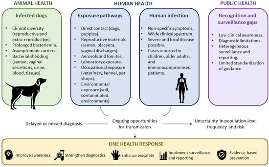
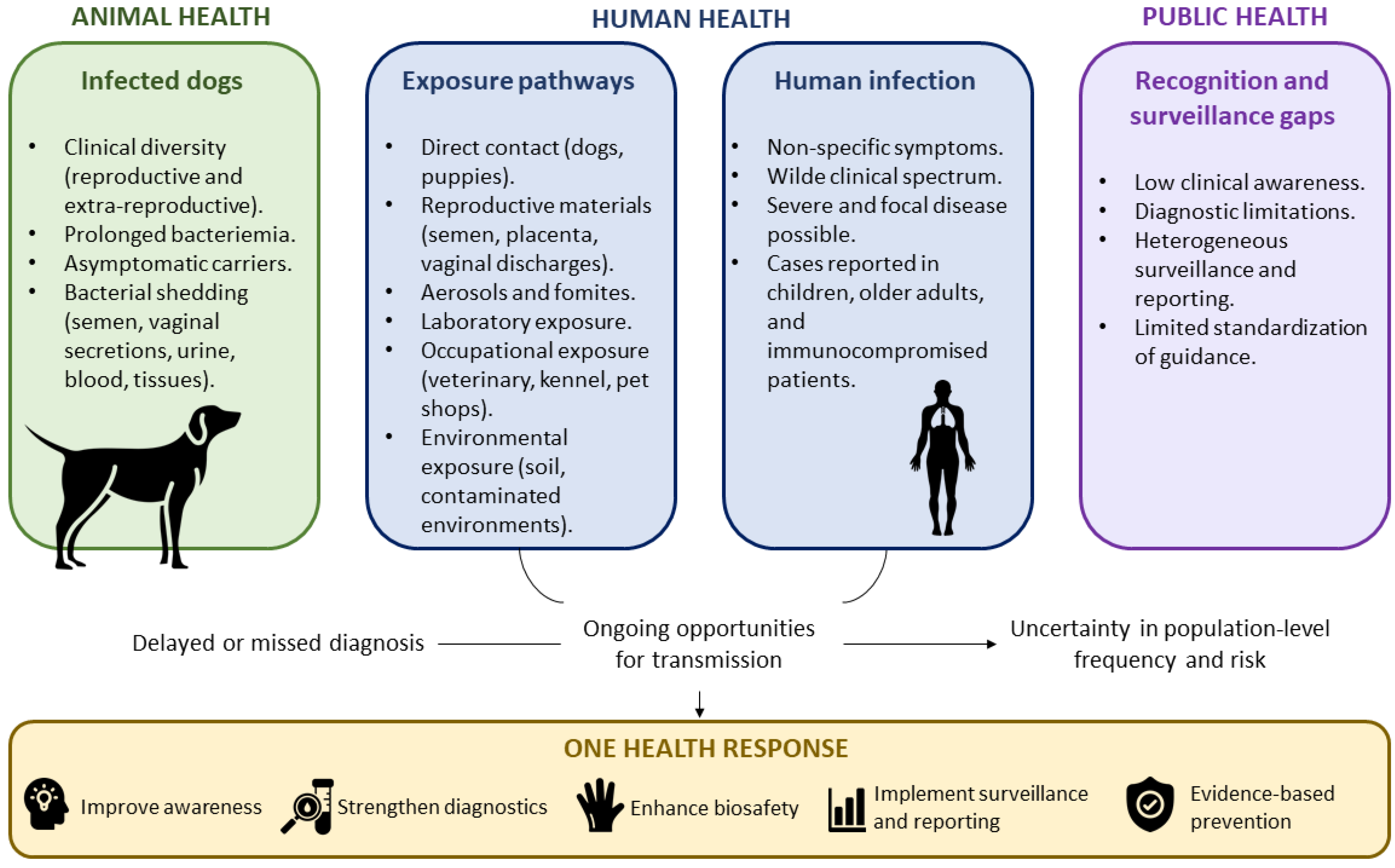
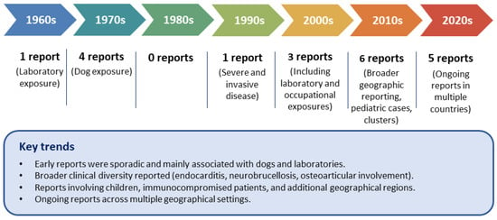
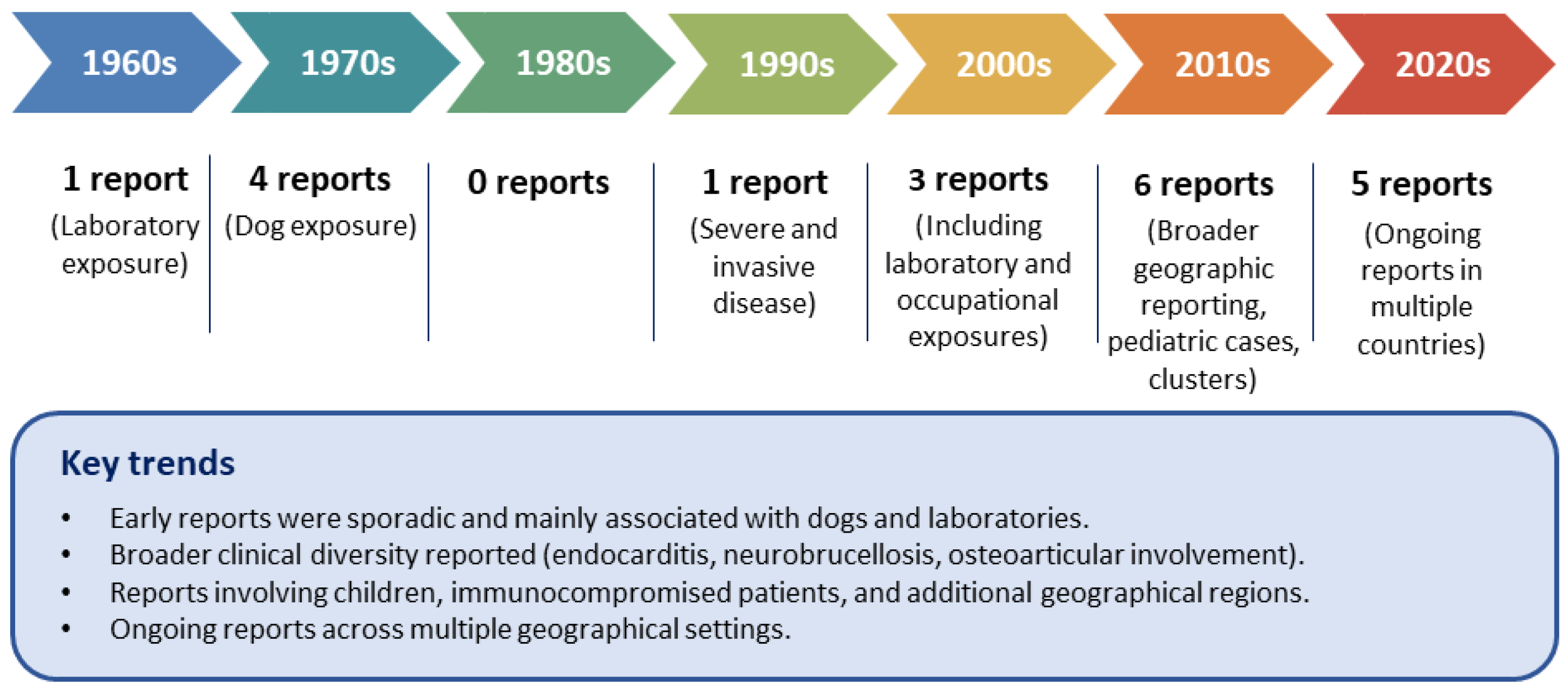
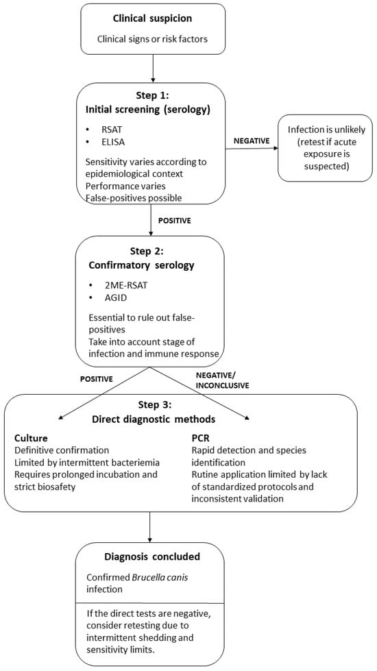
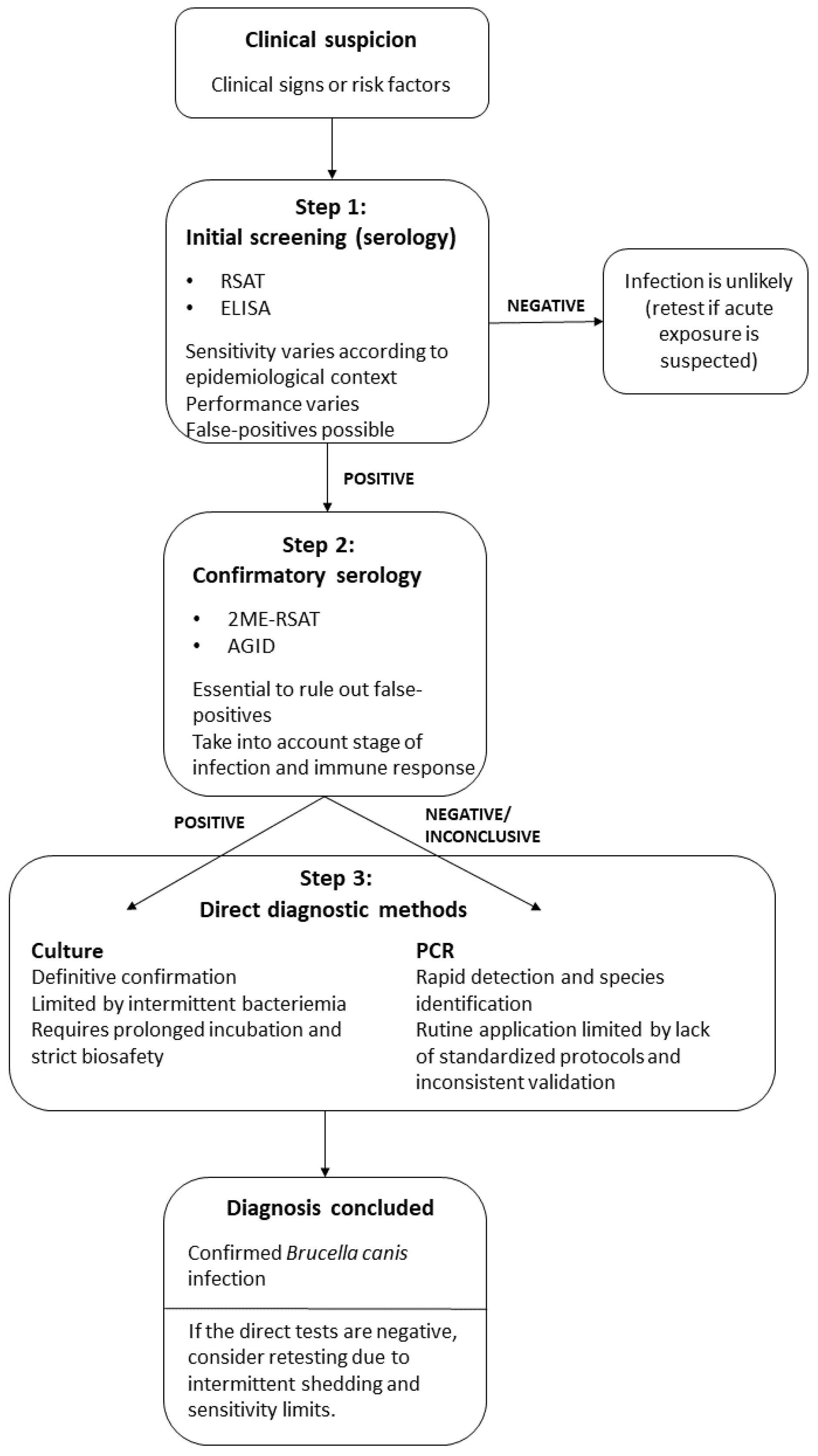
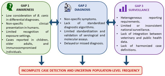
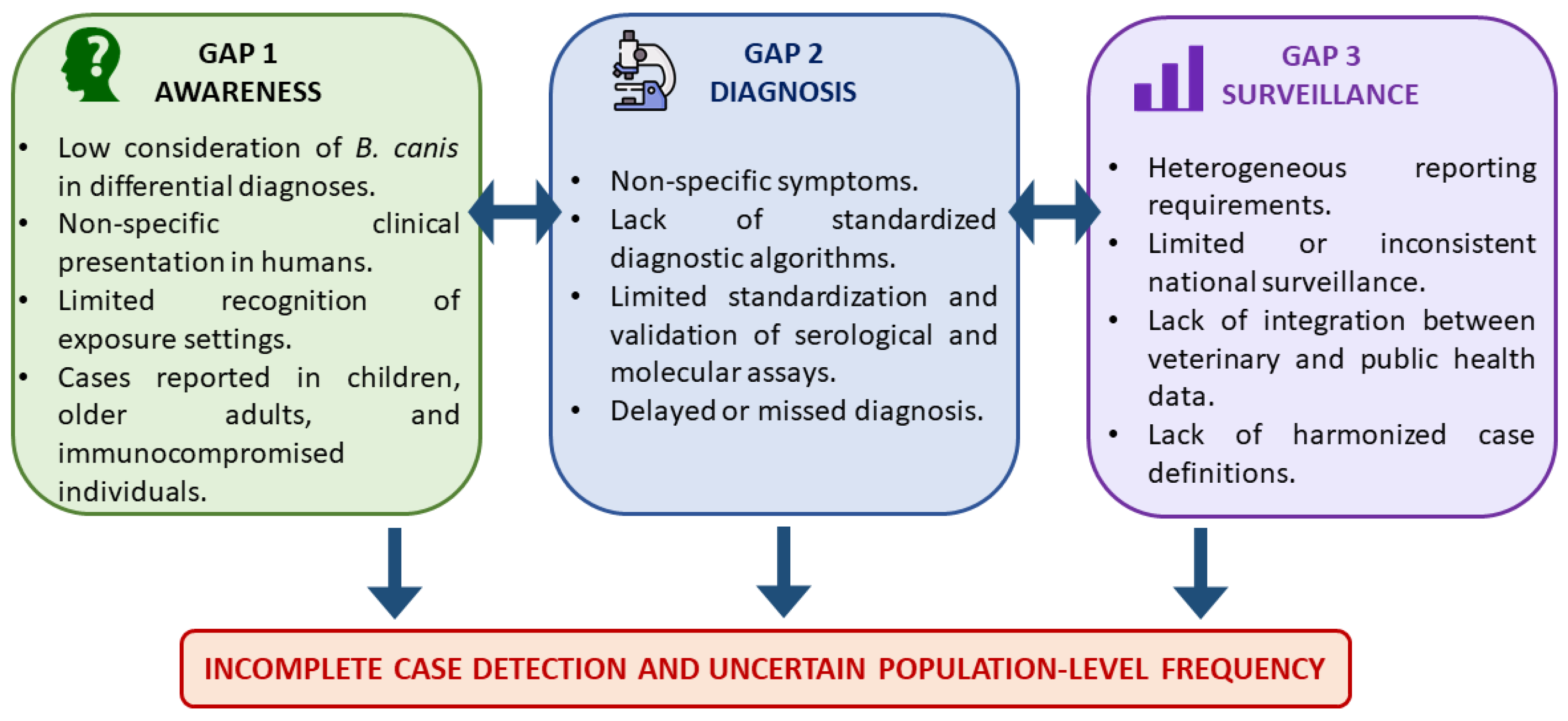

Disclaimer/Publisher’s Note: The statements, opinions and data contained in all publications are solely those of the individual author(s) and contributor(s) and not of MDPI and/or the editor(s). MDPI and/or the editor(s) disclaim responsibility for any injury to people or property resulting from any ideas, methods, instructions or products referred to in the content. |
© 2026 by the authors. Licensee MDPI, Basel, Switzerland. This article is an open access article distributed under the terms and conditions of the Creative Commons Attribution (CC BY) license.