


Abstract
Osteoarticular infections (OAIs) present a significant diagnostic and therapeutic challenge to paediatric clinical practice. When evaluating suspected paediatric OAIs, the principal pathogens commonly considered are Staphylococcus aureus, Streptococcus pyogenes, Streptococcus pneumoniae and Kingella kingae. However, advances in molecular diagnostic techniques, particularly polymerase chain reaction assays and next-generation sequencing, have considerably improved the detection of less commonly identified pathogens responsible for OAIs. Consequently, familiarity with fastidious, emerging, exposure-related and uncommon bacterial pathogens is essential to ensure accurate diagnoses and appropriate therapeutic interventions. This review summarises bacterial pathogens responsible for OAIs in immunocompetent children that are fastidious, emerging, exposure-related or otherwise less commonly encountered. We describe their microbiological characteristics, clinical phenotypes, diagnostic pitfalls and organism-specific diagnostic strategies.
1. Introduction
Paediatric osteoarticular infections (OAIs) are major diagnostic and therapeutic challenges as they can lead to prolonged morbidity, systemic complications, joint damage, growth disturbances and long-term functional impairment. Accurate identification of the causative organism is central to its effective management, as it confirms the microbiological diagnosis, guides targeted antimicrobial therapy and may improve clinical outcomes. Furthermore, precise pathogen identification enables detailed assessment of the antibiotic resistance profile, which is essential for guiding antimicrobial therapy.
Before the early 2000s, pathogen detection depended mainly on traditional cultures of blood, joint fluid or bone aspirates [1,2,3]. However, despite the appropriate sampling of paediatric OAIs, culture-negative results were common: 24–68% of cases of acute haematogenous osteomyelitis and 21–70% of cases of septic arthritis [1,2,3]. These culture-negative infections may have reflected the presence of fastidious organisms, low bacterial inoculum, prior antibiotic exposure or the intrinsic limitations of conventional culture-based methods. Over the past 20 years, the greater sensitivity and specificity of nucleic acid amplification assays (NAAAs) have greatly enhanced pathogen detection in cases of paediatric OAI. Beyond pathogen identification, some NAAAs can detect genes associated with antibiotic resistance mechanisms and virulence factors [4]. Next-generation sequencing represents the most advanced iteration of NAAA technology. As microbiology progressively shifts from phenotype-based approaches toward genomic characterization, these techniques may become increasingly important for paediatric. Plasma-based, metagenomic, next-generation sequencing is a particularly promising approach, as it may be able to detect the pathogens responsible for localised infections from circulating microbial DNA [5,6,7]. Taken together, these advances suggest that pathogen identification and resistance profiling may increasingly become integrated into the routine for paediatric OAIs. Nevertheless, important limitations remain, including restricted availability, cost, limited standardisation and the potential difficulty of interpreting positive molecular findings in the absence of clear clinical correlation. Furthermore, molecular results should always be interpreted within the appropriate clinical and microbiological context, as contamination, colonisation or detection of non-viable organisms may occasionally complicate interpretation. Consequently, molecular techniques should currently be considered complementary rather than complete replacements for conventional microbiological methods.
Clinicians are therefore increasingly likely to encounter a broader spectrum of uncommon, fastidious and emerging bacterial pathogens in community-acquired OAIs affecting otherwise healthy children. Due to clinicians’ limited experience with these atypical and rarely occurring pathogens, a heightened awareness of these infections is essential for accurately identifying and effectively managing them. This review examines bacterial pathogens responsible for paediatric OAIs in immunocompetent children, that are fastidious, emerging, exposure-related, culture-negative, or otherwise atypical from a diagnostic or clinical perspective, with an emphasis on their microbiological features, epidemiology, clinical phenotypes and diagnostic approaches. For clarity, pathogens discussed in this review are grouped according to their predominant microbiological characteristics, epidemiological context, diagnostic challenges and degree of clinical recognition, acknowledging that overlap between categories may exist.
2. Literature Search
This review was based on a structured literature search performed in PubMed, Embase and the Cochrane Library. The search terms covered studies published between January 1995 and December 2025 and combined terms related to paediatric osteoarticular infections (including “paediatric”, “children”, “osteomyelitis”, “septic arthritis”, “osteoarticular infection”) with terms related to uncommon pathogens (including “fastidious”, “atypical”, “uncommon”, “emerging”, and specific pathogen names). Reference lists of relevant articles were also manually screened to identify additional studies.
Studies eligible for inclusion included original clinical investigations, cohort studies, case series, and selected case reports addressing epidemiological, microbiological, diagnostic, or therapeutic aspects of osteoarticular infections caused by fastidious, emerging, exposure-related, culture-negative, or otherwise atypical bacterial pathogens in immunocompetent children. Priority was given to paediatric studies and clinically relevant microbiological reports. Animal studies, in vitro studies, conference abstracts, instructional lectures and publications without sufficient clinical data were excluded.
3. Microbiological Basis: Fastidious & Atypical Organisms
Fastidious microorganisms have complex or specific nutritional needs and will only grow when certain nutrients are present in their environment: they are thus difficult to cultivate using standard methods. Nevertheless, the concept of fastidiousness extends well beyond the simple notion of dependency on specific nutrients. It includes the requirement for a variety of chemical signals, many of which are produced by surrounding bacterial species. As early as 1974, Lewis Thomas noted in The Lives of a Cell [8] that most microbes could not be cultivated alone because they exist in dense, interdependent communities reliant on complex chemical signals. This insight highlighted the limitations of the classic culture-based methods used to detect certain microorganisms.
Atypical bacteria are prokaryotes that differ from standard bacteria due to features such as absent or atypical cell walls, uniquely specialised growth requirements or an obligate intracellular mode of replication. Conventional Gram staining may poorly visualise or inconsistently characterise them, for the same reasons. This group typically includes organisms such as Mycoplasma spp., which lack cell walls, Chlamydia spp., which are obligate intracellular pathogens, and Legionella spp., which require specialised culture conditions [9,10,11]. Although uncommon, atypical and fastidious bacteria have been increasingly recognised as potential causes of OAIs, particularly when conventional cultures are negative. Diagnoses often depend on molecular methods instead of routine cultures because these bacteria have strict growth requirements and may be intracellular [10,11].
A growing body of case reports and small series studies has identified a diverse array of fastidious and atypical microorganisms involved in paediatric OAIs, many of which were only identified thanks to extended culture techniques or molecular diagnostics. These include HACEK group microorganisms, anaerobes, slow-growing Gram-negative bacilli and atypical bacteria, all characterised by their low or negligible culture yields using conventional methods. Knowledge of the epidemiological and clinical characteristics of these less common pathogens is becoming increasingly important since current molecular diagnostic techniques will identify them more frequently. Clinicians managing paediatric OAIs will be increasingly likely to encounter uncommon pathogens for which routine orthopaedic and paediatric practice provides limited guidance. The main fastidious, emerging, exposure-related and rare bacterial pathogens associated with paediatric OAIs are summarised in Table 1. For clarity, pathogens are grouped according to their predominant microbiological, epidemiological and clinical context, while acknowledging that overlap between categories may exist.
Table 1.
Microbiological and clinical profiles of fastidious, emerging, exposure-related and rare bacterial pathogens causing paediatric OAIs, grouped according to their microbiological, epidemiological and clinical context.
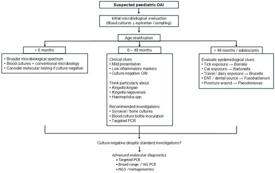
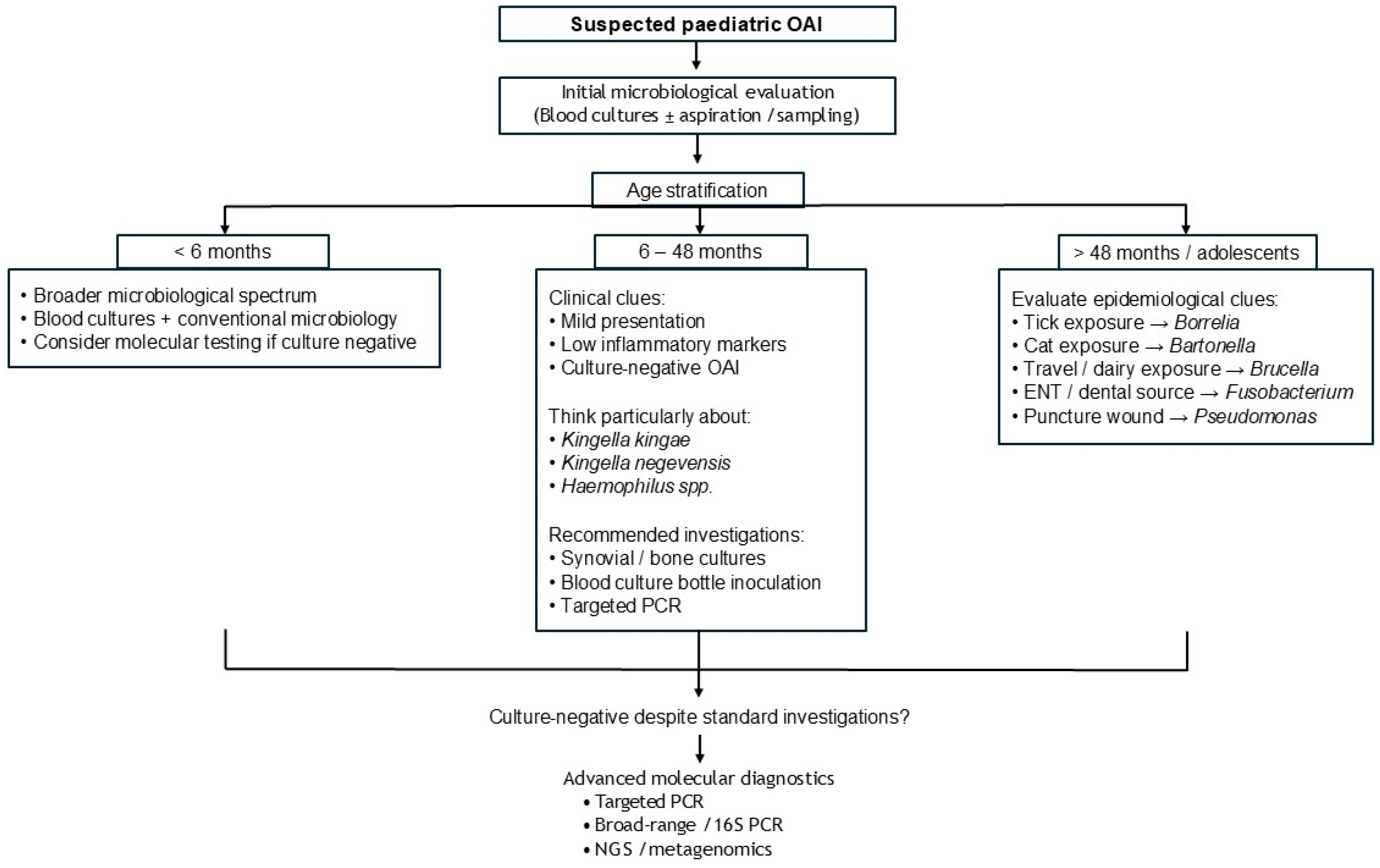
Figure 1 summarises a suggested diagnostic approach for children presenting with atypical or culture-negative paediatric OAIs.
Figure 1.
Suggested diagnostic approach for children presenting with atypical or culture-negative paediatric osteoarticular infections, integrating age-related epidemiology, clinical clues, exposure history and microbiological investigations.
4. Hacek Group Microorganisms
The acronym HACEK refers to a group of fastidious Gram-negative microorganisms, including Haemophilus spp., Aggregatibacter actinomycetemcomitans (formerly Actinobacillus actinomycetemcomitans), Cardiobacterium hominis, Eikenella corrodens and Kingella kingae. HACEK microorganisms grow slowly in standard blood cultures, often requiring extended incubation [12]. Usually found in the oropharynx, they are primarily known for causing infective endocarditis, but they can also lead to OAIs. In the context of this review, pathogens were included not solely according to their rarity, but based on one or more predefined characteristics, including fastidious microbiological behaviour, association with culture-negative infections, emerging recognition through molecular diagnostics, exposure-related epidemiology, or atypical clinical presentation in paediatric OAIs. Consequently, pathogen selection was based primarily on diagnostic and clinical relevance rather than exclusively on incidence.
4.1. Kingella kingae
OAIs caused by K. kingae are the prototypical example of fastidious, frequently culture-negative, Gram-negative musculoskeletal infections in young children. Since the 1980s, the reported number of cases of K. kingae OAIs has markedly increased, mainly due to the use of molecular identification methods [13,14,15,16,17]. K. kingae is thus now regarded as the leading bacterial cause of paediatric OAIs, especially in children under 48 months old [18,19]. In addition to classic acute haematogenous osteomyelitis and septic arthritis, this pathogen can cause atypical OAIs, including spondylodiscitis [20,21,22,23,24,25], subacute osteomyelitis [26,27,28], pyomyositis [29], bursitis [30] and tendon sheath infections [31,32,33].
K. kingae septic arthritis generally involves large weight-bearing joints such as hips, knees, ankles, shoulders or elbows [14,15,18,19,34,35,36]. Atypical joints, such as small metacarpophalangeal and metatarsophalangeal joints and sternoclavicular, acromioclavicular or tarsal joints, are over-represented in cases of K. kingae arthritis, compared with paediatric septic arthritis overall [13,14,15,18,19,34,35,36]. The anatomical sites involved in paediatric patients with K. kingae osteomyelitis include long bones such as the femur, tibia, humerus, radius and ulna [14,15,18,19,34,37]. A dedicated case series focusing on K. kingae-induced septic arthritis of the knee in young children emphasised its relatively mild clinical presentation and modest biological inflammatory response [38]. Nevertheless, any bone rarely infected by other pathogens, such as the calcaneum, talus, sternum or clavicle, can also be affected by K. kingae osteomyelitis [18,19,34,35,36]. The onset of a K. kingae OAI is generally insidious, and the disease is frequently diagnosed after a considerable delay, at a time when bone lesions are already subacute in character and can present as lytic lesions [31,32]. Finally, the clinical course is usually better for children with an OAI due to K. kingae rather than other typical pathogens, as evidenced by shorter hospitalisations and fewer adverse events [18,19,34,39].
4.2. Kingella negevensis
K. negevensis is a newly described Gram-negative bacterium closely related to K. kingae. Like K. kingae, K. negevensis can be isolated from the oropharynx of young children, and, in some cases, it is probably misidentified as K. kingae. Indeed, K. negevensis produces many of the same virulence factors as K. kingae, including a polysaccharide capsule, a secreted exopolysaccharide, a Knh-like trimeric autotransporter and type IV pili, suggesting that K. negevensis has significant pathogenic potential and can cause OAIs [40,41].
4.3. Non-Type-B Haemophilus influenzae, Unencapsulated H. influenzae and H. parainfluenzae
In the post-vaccination era, Haemophilus spp. rarely cause OAIs, with H. influenzae type b (Hib) now rare in countries with widespread immunisation programmes. However, some non-typeable H. influenzae (NTHi) and other encapsulated strains (particularly serotypes a, f and e) that are not targeted by immunisation continue to cause OAIs [42,43,44,45]. At Texas Children’s Hospital, from 2011 to 2018, non-typeable strains accounted for 39.3% of invasive H. influenzae infections [46,47,48,49], and type a accounted for 29.5% [50]. H. influenzae type a is the most common serotype, although types d, e and f are also reported. Another Haemophilus species, Haemophilus parainfluenzae, is an extremely rare cause of paediatric OAIs, with only a handful of cases reported in the literature [51,52,53].
These OAIs typically affect children younger than 4 years, with the highest prevalence between 7 and 24 months old [54]; septic arthritis is the most common manifestation. Indeed, a recent systematic review of the literature documented just 21 cases of acute septic arthritis due to non-type-b H. influenzae [46]. As was often the case with OAIs due to Hib, children tended to be under 2 years old and present with a monoarticular acute septic arthritis, usually affecting the knee [46]. Acute haematogenous osteomyelitis is much less common with non-type-b H. influenzae, and it may be concurrent with acute septic arthritis in up to 40% of cases [46].
Unencapsulated strains of H. influenzae (non-typeable H. influenzae) appear to be much rarer causes of OAIs than non-type-b H. influenzae, with only scattered reports in the literature, primarily describing acute septic arthritis [55]. From a practical viewpoint, given their rarity and potentially lower virulence, isolating an unencapsulated strain of H. influenzae should prompt an immunological evaluation of the child.
4.4. Aggregatibacter actinomycetemcomitans
Aggregatibacter actinomycetemcomitans is another member of the HACEK group. It primarily colonises the human oral cavity and is associated with aggressive periodontitis. It can also cause serious extraoral infections, including rare cases of OAIs like osteomyelitis and septic arthritis. Indeed, it has been implicated in osteomyelitis and epidural abscesses in children and adults [56], demonstrating that even slow-growing, Gram-negative microorganisms should be considered in the differential diagnosis of culture-negative OAIs, and emphasising the importance of using tissue cultures and NAAA technologies [57].
5. Emerging, Exposure-Related and Rare Pathogens
5.1. Emerging Pathogens
5.1.1. Neisseria meningitidis
While mostly known for causing meningitis, N. meningitidis can also cause osteoarticular disorders, such as septic arthritis, reactive arthritis and osteomyelitis. Arthritis can occur as a primary, isolated infection or can be secondary to sepsis. Joint arthritis may complicate acute meningococcal disease in 5–10% of cases [58,59,60] and commonly affects the knees and hips. Primary septic arthritis due to N. meningitidis without accompanying meningitis remains extremely rare and tends to be monoarticular [61,62,63]; it affects the larger joints in patients of all ages [58,59]. Primary meningococcal arthritis has been reported with serotypes W-135, B and C [58]. Alongside septic arthritis directly due to the pathogen, an immune-mediated arthritis may also develop, often appearing later in the disease course [58,60,64] and often being polyarthritic [60]. Given the ease with which this microorganism can be treated with antimicrobial therapy, its relative contribution to septic arthritis is probably underestimated, and some cases of immune-mediated arthritis probably involve eradicated septic arthritis. Acute haematogenous osteomyelitis due to N. meningitidis is less common than primary meningococcal arthritis, likely accounting for fewer than 2% of all cases of paediatric osteomyelitis [65,66]. In otherwise healthy children, it has been reported in the tibia [67], fibula [68], acromion [69], lumbar vertebrae [70] and humerus [71]. Serogroup B N. meningitidis (for which no vaccine existed until recently) predominates in the literature [67], although serogroups C and Y have also been described [67,68,69,70,71].
5.1.2. Fusobacterium spp.
Fusobacterium spp. are Gram-negative, anaerobic bacilli that are part of normal oral and gastrointestinal flora; they are usually opportunistic pathogens. OAIs due to anaerobic pathogens are rare, and, when present, Fusobacterium spp. are likely their most common cause in otherwise healthy children. Fusobacterium nucleatum and Fusobacterium necrophorum are the most frequently reported in the literature, although other species, such as Fusobacterium fusiforme, have been reported as well [72]. Fusobacterium spp. are the most common aetiological agents of Lemierre’s syndrome, which is characterised by bacteraemia involving anaerobic organisms (indeed, usually Fusobacterium spp.), internal jugular vein thrombosis, and a metastatic, suppurative spread of infection (abscess formation in various locations) [73]. Many reported cases of OAIs involving Fusobacterium spp. are associated with anaerobic sources of infection such as sinus disease, dental abscesses, mastoiditis or dog bites [74]. These infections are often polymicrobial [74], but an isolated disease, without the risk factors for an anaerobic source of infection, may also be observed, especially in children. Thus, Fusobacterium spp. may be present because of a metastatic spread of infection with acute septic arthritis [75,76] or acute haematogenous osteomyelitis [73,76]. Acute septic arthritis due to Fusobacterium spp. has been reported in hips [77,78,79] and knees [80], and acute haematogenous osteomyelitis has been reported in the pelvic region [81], hips [82] and femurs [83] without the presence of Lemierre’s syndrome or risk factors for anaerobic infection. Fusobacterium spp. have also been identified as the causative pathogens in cases of subacute osteomyelitis and pelvic OAIs with Brodie’s abscess in children [84,85]. Interestingly, patients with OAIs due to Fusobacterium spp. are often older than those with OAIs due to more typical organisms. Of the 23 cases of OAI involving Fusobacterium spp. identified in a narrative review, 19 patients (83%) were over 8 years old, suggesting that OAIs due to Fusobacterium spp. tend to occur in older children [86]. These cases are diagnostically challenging, as anaerobic cultures require specific conditions and results are frequently delayed or negative.
5.2. Exposure-Related Pathogens
5.2.1. Borrelia burgdorferi
Lyme borreliosis is a zoonotic disease caused by Borrelia burgdorferi, a member of the Spirochaetaceae family. Borrelia burgdorferi’s vectors are ticks of the Ixodes genus, which transmit the pathogen to human hosts while feeding on their blood. The typical clinical picture of Lyme disease includes symptoms involving the skin, joints, nervous system and, more rarely, the heart and eyes [87,88]. Lyme disease has three main stages: early localised, early disseminated and late disseminated infections. Musculoskeletal symptoms are common, but arthritis—characterised by swelling and pain in one or more large joints—is the main sign of Lyme borreliosis. Patients with confirmed borreliosis may also experience tendon, muscle or bone pain [89,90]. Joint involvement is typically asymmetrical and mostly in large joints, particularly the knees, but small joints may also be affected as components of oligoarthritis. Apart from knees, Lyme arthritis commonly also affects shoulder, elbow, wrist and ankle joints [87,89,90,91]. Lyme arthritis results from a strong inflammatory response (not from spirochete toxins) that causes subsequent host-driven aggrecan degradation and leads to cartilage damage and severe joint injury [87]. In the early localised stage of erythema migrans, almost half of patients experience migratory muscle and joint pain. The time between a tick bite and the onset of Lyme arthritis can vary greatly, ranging from a few days to several months [92,93]. If present for longer durations, Lyme arthritis can lead to the erosion and destruction of joint structures.
5.2.2. Brucella spp.
Brucellosis is a zoonotic infection caused by species of the Brucella genus. Brucella are small, aerobic, intracellular coccobacilli that preferentially localise in the reproductive organs of host animals (mainly ruminants), causing abortions and infertility in infected animals. The bacteria are transmitted from animals to humans through the ingestion of infected food (mainly unpasteurised dairy products), direct contact with infected animal products or viscera, or the inhalation of aerosols [94,95].
Brucellosis is primarily found among children living in endemic areas, but the disease has also been reported among returning travellers or individuals residing in regions bordering endemic areas [94,96]. Travel and globalisation both increase the likelihood that patients will present with a potential Brucella infection that has moved from an endemic to a non-endemic area. Alongside the classic clinical picture, which combines several non-specific systemic features (fever, fatigue, weight loss, hepatosplenomegaly), Brucella frequently presents together with arthritis or arthralgias (up to 40–80% in some series) that can mimic the presence of septic arthritis (with invasion of the organism into the joint space) [97,98]. However, arthritis is usually reactive in nature [99], polyarticular, additive and non-migratory, typically involving the sacroiliac region, knees or hips [94,95,97,100,101]. Alongside reactive arthritis, nearly 10% of individuals infected with Brucella spp. and presenting with osteoarticular complaints have their bacterial growth confirmed by synovial fluid culture [99]. However, this microorganism’s relatively fastidious nature may preclude its more frequent isolation in culture, leading to an underestimation of the true incidence of acute septic arthritis occurring with this pathogen [102]. Knees and hips are the joints most frequently affected by culture-proven acute septic arthritis due to Brucella spp. [103]. Though less commonly reported than acute septic arthritis, acute haematogenous osteomyelitis may also be caused by Brucella spp., with cases involving osteomyelitis of the calcaneus, the sternum and olecranon, and the femur described in the literature [100,103,104,105].
The direct inoculation of synovial fluid into blood culture bottles may improve culture yield and increase the isolation rates of Brucella spp. [102]. Real-time qPCR has emerged as a rapid, sensitive and specific tool for diagnosing brucellosis, overcoming many of the limitations of traditional culture and serological methods. While bacterial culture remains the gold standard assay, it is time-consuming and not very sensitive, making qPCR the preferred alternative for quick, genus-level identification of Brucella [106,107].
5.2.3. Salmonella spp.
OAIs caused by Salmonella spp. are rare but serious, usually manifesting as osteomyelitis or septic arthritis, often after bacteraemia in immunocompromised individuals or those with sickle cell disease. Non-typhoidal Salmonella can also cause invasive infections, but there are no risk factors in young children, especially under 1 year old [108]; a Taiwanese series found that over 60% of bacteraemia cases occurred in this age group [108]. However, the distribution of non-typhoidal Salmonella osteomyelitis in healthy children does not reveal a predominance of involvement among infants. Indeed, one series demonstrated that among the 46 cases of non-typhoidal Salmonella osteomyelitis reviewed in healthy children, only 19% were infants, with a median age of 9 years old and male predominance (61%) [109]. The most common serotypes were S. Enteritidis and S. Typhimurium. Complications were frequent and occurred in 41% of subjects, including relapses, multifocal disease or suppurative issues [109].
Acute septic arthritis with non-typhoidal Salmonella is rare. Sirinavin et al. described 82 invasive infections involving non-typhoidal Salmonella in immunocompetent children, only 2 (2.4%) of which involved acute septic arthritis [110], and Punpanich et al. reported that just 1.3% of 226 children with a culture-confirmed, invasive non-typhoidal Salmonella infection developed acute septic arthritis [111]. Acute septic arthritis due to non-typhoidal Salmonella has been reported in otherwise healthy children in elbows, sacroiliac joints, knees, metacarpophalangeal joints and shoulders [112,113,114,115,116]. Acute septic arthritis due to non-typhoidal Salmonella can be difficult to distinguish from reactive arthritis associated with gastrointestinal Salmonella infections [117,118]. Reactive arthritis due to Salmonella appears to have a slight female bias and is more common in adults than children [119]. Its incidence varied from 1.2–29.0% [119], presenting as either mono- or oligoarticular, and most commonly affecting the knee [117].
5.3. Rare, Exceptional and Diagnostically Challenging Pathogens
5.3.1. Actinomyces spp.
Actinomyces spp. are anaerobic Gram-positive bacteria that usually colonise the mouth, gut or genital tract. Infection occurs when they invade tissue during dental work, through poor oral hygiene or due to trauma. Actinomyces is therefore another anaerobic organism occasionally associated with OAIs. Actinomyces israelii is the most commonly reported species, though Actinomyces odontolyticus, Actinomyces viscosus, Actinomyces turicensis and Actinomyces naeslundii have also been mentioned [120,121,122]. Actinomyces OAIs are chronic in character and often follow a very indolent course, mimicking tumours or tuberculosis [121]. A systematic review of osteomyelitis cases due to Actinomyces spp. demonstrated a median time to presentation of 3 months, with three cases only being recognised after 2 years [121]. Actinomyces spp. are common flora in the oral cavity, which explains why over half of all Actinomyces infections involve the cervicofacial region [73]. Because of this tendency, the mandible is the most frequently affected bone in cases of osteomyelitis, before spondylitis of the C1 and C2 vertebrae [120,123]. Osteomyelitis of the iliac bone has also been encountered due to the disease spreading from the pelvis. Culture attempts are essential, but given the microorganism’s fastidious nature, NAAAs or a histological analysis may be required for diagnosis [120,121,123]. Histopathological analysis of Actinomyces infections often demonstrates infiltrative spread across tissue and fascial planes, sinus tract formation, sulphur granules, and Gram-positive branching organisms with acute-angle branching [86].
5.3.2. Other Rare Pathogens
Group C and G Streptococci (GCGS) OAIs in children are uncommon compared to other pathogens, with these microorganisms predominantly affecting elderly adults rather than paediatric populations. GCGS OAIs in humans generally produce a less acute clinical presentation than Group A Streptococcus [124]. Indeed, many GCGS in humans belong to the Streptococcus dysgalactiae subspecies equisimilis (SDSE), which shares many virulence factors with S. pyogenes through horizontal gene transfer [125,126,127]. Thus, GCGS have not been listed among the major pathogens in recent large prospective studies of paediatric OAIs [128,129].
A few cases of OAIs due to GCGS in otherwise healthy children have nevertheless been reported in the literature, with just one case of acute haematogenous osteomyelitis and six cases of acute septic arthritis [130,131,132,133,134]. Many laboratories are unable to identify isolates down to the species level (e.g., Streptococcus dysgalactiae, S. equi) and instead use the less specific Lancefield classification. Although data are too sparse to ascribe virulence differences to different species, and although all their isolates appear susceptible to penicillin, their susceptibility to non-beta-lactam agents (as may be required in penicillin-allergic patients) is less predictable and may vary by species or subspecies [91].
Staphylococcus caprae is a coagulase-negative staphylococcus rarely implicated in bone infections. Bone and joint infections due to S. caprae have been reported, but fortunately they remain rare [135,136,137,138]. Most S. caprae OAIs are the result of infected orthopaedic devices, especially infected joint prostheses and internal osteosynthesis devices [139,140,141,142]. Only very rare cases of primary S. caprae OAIs have been described, including osteitis [135,136], subacute osteomyelitis [138], arthritis [136] and spondylodiscitis [137,143].
Bartonella henselae OAIs caused by this Gram-negative bacterium are rare and atypical complications of cat scratch disease (CSD), which usually results from a scratch or bite from a young cat [144,145]. While typically presenting as a self-limiting lymphadenopathy, it can also cause osteomyelitis or septic arthritis. Even though cases of septic arthritis have been reported with this germ [146], OAIs due to B. henselae almost exclusively present as acute haematogenous osteomyelitis and demonstrate a strong tendency to affect the axial skeleton and pelvis, with nearly 75% of published cases localised to these areas [147,148]. Although osteomyelitis is an uncommon manifestation of CSD, it is a significant atypical presentation of paediatric bartonellosis and may be under-recognised in children with subacute axial or pelvic bone lesions. Furthermore, B. henselae has been increasingly recognised as a cause of atypical subacute osteomyelitis in children and adolescents [28]. Lymphadenopathy may be seen in approximately 25% of patients with CSD, which is not typical in cases of OAI [147]. Acute and convalescent serological studies and PCR testing may assist in diagnoses, given the microorganism’s fastidious nature [147,148,149]. In a similar vein, Vazquez et al. reported on a series of paediatric patients presenting with OAIs with atypical features, thus emphasising the need to use systematic serological and PCR testing appropriate to the epidemiological context [150]. One unknown remains regarding the treatment of these OAIs: given that antibiotic treatment of Bartonella lymphadenitis has not been shown to significantly improve long-term outcomes, it is unclear to what extent the treatment of OAIs hastens disease resolution [151]. However, given the lack of observational data for OAI disease processes, as well as the risk of morbidity in childhood OAIs, many authors and clinicians have continued to use antibiotic treatments.
Bordetella holmesii is a Gram-negative, rod-shaped bacterium of the Bordetella genus. Less well-known than its more famous relative Bordetella pertussis, this pathogen is considered to be an emerging bloodstream pathogen, more likely to affect immunocompromised children, such as asplenic patients [152]. However, native septic arthritis due to B. holmesii has been reported in adolescents, particularly in the setting of underlying haematological or immune conditions [153].
Similarly, Moraxella lacunata has also been identified as the causative agent of subacute osteomyelitis in a child, highlighting that even unusual members of the oropharyngeal microbiota can produce bone infections. Moraxella and Kingella are closely related Gram-negative bacteria in the Neisseriaceae family, often appearing as coccobacilli in pairs or short chains. Both are fastidious, non-motile, oxidase-positive and catalase-negative, and they commonly inhabit the human respiratory tract as opportunistic pathogens that can cause similar infections, such as endocarditis and OAIs.
Morganella morganii is a Gram-negative, facultatively anaerobic bacterium commonly found in the human intestinal tract and the environment, and as a commensal in many animals. It is typically considered an opportunistic pathogen and a significant cause of nosocomial infections. M. morganii has been reported as the causative agent of a Brodie’s abscess in a child’s talus, a presentation that highlights the expanding bacteriological spectrum of subacute OAIs [154].
Burkholderia pseudomallei can rarely cause isolated OAIs in otherwise healthy children, particularly in Southeast Asia and northern Australia [155]. The disease is more common in lower limbs, potentially because inoculation often occurs from walking barefoot through infected soil or water [155].
Pseudomonas aeruginosa is a common, highly adaptable, Gram-negative bacterium found in soil and water. It is usually implicated in infections following puncture wounds to the foot, manifesting with a sweetish odour and a greenish discolouration [156,157]. However, this pathogen can also be a rare cause of OAIs in the absence of trauma, occasionally as tibial osteomyelitis, pelvic osteomyelitis (potentially owing to its occasional presence in faecal flora) or, given its tendency to cause otic infections, as osteomyelitis of the skull associated with complications arising from mastoiditis [158,159,160].
6. Conclusions
Advances in nucleic acid amplification assay technologies have revolutionised diagnostics for infectious diseases. These advances have enabled better identification and resistance profiling of the causative pathogens responsible for paediatric osteoarticular infections (OAIs). It has also now been increasingly observed that otherwise healthy children may present with OAIs secondary to unanticipated pathogens. Although culture-negative results may become less frequent with the broader use of molecular diagnostics, clinicians are likely to encounter an expanding spectrum of uncommon, fastidious and emerging bacterial pathogens. Due to their infrequent occurrence and limited clinical experience with these atypical pathogens, heightened awareness is essential for the accurate identification and effective management of such infections. For paediatric orthopaedic surgeons, this evolving microbiological landscape also reinforces the need to consider uncommon pathogens in culture-negative, subacute OAIs, including those at atypical sites or resulting from atypical exposures.
Author Contributions
A.R. and D.C. conceptualized the review and defined its scope. A.R. drafted the initial manuscript. G.d.M., O.V., E.P., A.T., C.S., R.D. and D.C. contributed to the critical revision of the manuscript for important intellectual content. All authors have read and agreed to the published version of the manuscript.
Funding
This research received no external funding.
Institutional Review Board Statement
Not applicable. This article is a review and did not involve human participants, human data, or animal experiments.
Informed Consent Statement
Not applicable.
Data Availability Statement
Data sharing is not applicable to this article, as no datasets were generated or analyzed during the preparation of this review.
Conflicts of Interest
The authors declare that they have no competing interests.
Abbreviations
The following abbreviations are used in this manuscript:
| OAIs | Osteoarticular infections |
| PCR | Polymerase chain reaction |
| NAAAs | Nucleic acid amplification assays |
| Hib | H. influenzae type b |
| GCGS | Group C and G Streptococci |
| SDSE | Streptococcus dysgalactiae subspecies equisimilis |
| CSD | Cat scratch disease |
References
- Hunter, S.; Kioa, G.; Baker, J.F. Predictive Algorithms in the Diagnosis and Management of Pediatric Hip and Periarticular Infection. J. Bone Jt. Surg. Am. 2022, 104, 649–658. [Google Scholar] [CrossRef] [Scilit] [PubMed]
- Hunter, S.; Chan, H.; Crawford, H.; Baker, J.F. Appropriate Antibiotic Duration in Pediatric Bone and Joint Infection: A Systematic Review. J. Pediatr. Orthop. Soc. N. Am. 2023, 5, 736. [Google Scholar] [CrossRef] [Scilit] [PubMed]
- Lyon, R.M.; Evanich, J.D. Culture-Negative Septic Arthritis in Children. J. Pediatr. Orthop. 1999, 19, 655–659. [Google Scholar] [CrossRef] [Scilit]
- Banerjee, R.; Humphries, R. Rapid Antimicrobial Susceptibility Testing Methods for Blood Cultures and Their Clinical Impact. Front. Med. 2021, 8, 635831. [Google Scholar] [CrossRef] [Scilit] [PubMed]
- Han, D.; Li, R.; Shi, J.; Tan, P.; Zhang, R.; Li, J. Liquid Biopsy for Infectious Diseases: A Focus on Microbial Cell-Free DNA Sequencing. Theranostics 2020, 10, 5501–5513. [Google Scholar] [CrossRef] [Scilit] [PubMed]
- Han, D.; Li, Z.; Li, R.; Tan, P.; Zhang, R.; Li, J. mNGS in Clinical Microbiology Laboratories: On the Road to Maturity. Crit. Rev. Microbiol. 2019, 45, 668–685. [Google Scholar] [CrossRef] [Scilit] [PubMed]
- Ahmed, A.; Arun, A.; Lindner, M.; Sarmiento, A.; Del Valle Penella, A.; Laufer, P.; Sanchez-Vegas, C.; Cotilla, M.; Melish, M.; Trieu, C.; et al. Rapid, Non-Invasive Plasma-Based Microbial Cell-Free DNA next Generation Sequencing (the Karius Test) Outperforms the Standard of Care for the Detection of Kingella kingae Pediatric Spinal Infections. In Proceedings of the Pediatric Academic Societies (PAS) Meeting, Denver, CO, USA, 21–26 April 2022. [Google Scholar]
- Thomas, L. The Lives of a Cell: Notes of a Biology Watcher; Bantam Books: New York, NY, USA, 1974. [Google Scholar]
- Salton, M.R.J.; Kim, K.S. Structure. In Medical Microbiology, 4th ed.; Chapter 2; Baron, S., Ed.; University of Texas Medical Branch at Galveston: Galveston, TX, USA, 1996. Available online: https://www.ncbi.nlm.nih.gov/books/nbk8477/ (accessed on 15 April 2026).
- Nguyen, A.D.; Stamm, D.R.; Stankewicz, H.A. Atypical Bacterial Pneumonia. In StatPearls [Internet]; StatPearls Publishing: Treasure Island, FL, USA, 2026. Available online: https://www.ncbi.nlm.nih.gov/books/nbk532239/ (accessed on 5 May 2026).
- Saikku, P. Atypical Respiratory Pathogens. Clin. Microbiol. Infect. 1997, 3, 599–604. [Google Scholar] [CrossRef] [Scilit] [PubMed]
- Khaledi, M.; Sameni, F.; Afkhami, H.; Hemmati, J.; Dezfuli, A.A.Z.; Sanae, M.-J.; Validi, M. Infective Endocarditis by HACEK: A Review. J. Cardiothorac. Surg. 2022, 17, 185. [Google Scholar] [CrossRef] [Scilit] [PubMed]
- Ceroni, D.; Cherkaoui, A.; Ferey, S.; Kaelin, A.; Schrenzel, J. Kingella kingae Osteoarticular Infections in Young Children: Clinical Features and Contribution of a New Specific Real-Time PCR Assay to the Diagnosis. J. Pediatr. Orthop. 2010, 30, 301–304. [Google Scholar] [CrossRef] [Scilit] [PubMed]
- Chometon, S.; Benito, Y.; Chaker, M.; Boisset, S.; Ploton, C.; Bérard, J.; Vandenesch, F.; Freydiere, A.M. Specific Real-Time Polymerase Chain Reaction Places Kingella kingae as the Most Common Cause of Osteoarticular Infections in Young Children. Pediatr. Infect. Dis. J. 2007, 26, 377–381. [Google Scholar] [CrossRef] [Scilit] [PubMed]
- Ilharreborde, B.; Bidet, P.; Lorrot, M.; Even, J.; Mariani-Kurkdjian, P.; Liguori, S.; Vitoux, C.; Lefevre, Y.; Doit, C.; Fitoussi, F.; et al. New Real-Time PCR-Based Method for Kingella kingae DNA Detection: Application to Samples Collected from 89 Children with Acute Arthritis. J. Clin. Microbiol. 2009, 47, 1837–1841. [Google Scholar] [CrossRef] [Scilit] [PubMed]
- Rosey, A.-L.; Abachin, E.; Quesnes, G.; Cadilhac, C.; Pejin, Z.; Glorion, C.; Berche, P.; Ferroni, A. Development of a Broad-Range 16S rDNA Real-Time PCR for the Diagnosis of Septic Arthritis in Children. J. Microbiol. Methods 2007, 68, 88–93. [Google Scholar] [CrossRef] [Scilit] [PubMed]
- Verdier, I.; Gayet-Ageron, A.; Ploton, C.; Taylor, P.; Benito, Y.; Freydiere, A.-M.; Chotel, F.; Bérard, J.; Vanhems, P.; Vandenesch, F. Contribution of a Broad Range Polymerase Chain Reaction to the Diagnosis of Osteoarticular Infections Caused by Kingella kingae: Description of Twenty-Four Recent Pediatric Diagnoses. Pediatr. Infect. Dis. J. 2005, 24, 692–696. [Google Scholar] [CrossRef] [Scilit] [PubMed]
- Juchler, C.; Spyropoulou, V.; Wagner, N.; Merlini, L.; Dhouib, A.; Manzano, S.; Tabard-Fougère, A.; Samara, E.; Ceroni, D. The Contemporary Bacteriologic Epidemiology of Osteoarticular Infections in Children in Switzerland. J. Pediatr. 2018, 194, 190–196.E1. [Google Scholar] [CrossRef] [Scilit] [PubMed]
- Samara, E.; Spyropoulou, V.; Tabard-Fougère, A.; Merlini, L.; Valaikaite, R.; Dhouib, A.; Manzano, S.; Juchler, C.; Dayer, R.; Ceroni, D. Kingella kingae and Osteoarticular Infections. Pediatrics 2019, 144, e20191509. [Google Scholar] [CrossRef] [Scilit] [PubMed]
- Garron, E.; Viehweger, E.; Launay, F.; Guillaume, J.M.; Jouve, J.L.; Bollini, G. Nontuberculous Spondylodiscitis in Children. J. Pediatr. Orthop. 2002, 22, 321–328. [Google Scholar] [CrossRef] [Scilit]
- Ceroni, D.; Belaieff, W.; Kanavaki, A.; Della Llana, R.A.; Lascombes, P.; Dubois-Ferriere, V.; Dayer, R. Possible Association of Kingella kingae with Infantile Spondylodiscitis. Pediatr. Infect. Dis. J. 2013, 32, 1296–1298. [Google Scholar] [CrossRef] [Scilit] [PubMed]
- Ceroni, D.; Chargui, M.; De Marco, G.; Steiger, C.; Dayer, R. Better Comprehension of Primary Pyogenic Spinal Infections. Pediatr. Infect. Dis. J. 2023, 42, e39–e40. [Google Scholar] [CrossRef] [Scilit] [PubMed]
- Dayer, R.; Alzahrani, M.M.; Saran, N.; Ouellet, J.A.; Journeau, P.; Tabard-Fougère, A.; Martinez-Álvarez, S.; Ceroni, D. Spinal Infections in Children: A Multicentre Retrospective Study. Bone Jt. J. 2018, 100-B, 542–548. [Google Scholar] [CrossRef] [Scilit] [PubMed]
- Chargui, M.; Krzysztofiak, A.; Bernaschi, P.; De Marco, G.; Coulin, B.; Steiger, C.; Dayer, R.; Ceroni, D. Presumptive Bacteriological Diagnosis of Spondylodiscitis in Infants Less than 4 Years by Detecting K. Kingae DNA in Their Oropharynx: Data from a Preliminar Two Centers Study. Front. Pediatr. 2022, 10, 1046254. [Google Scholar] [CrossRef] [Scilit] [PubMed]
- Principi, N.; Esposito, S. Infectious Discitis and Spondylodiscitis in Children. Int. J. Mol. Sci. 2016, 17, 539. [Google Scholar] [CrossRef] [Scilit] [PubMed]
- Spyropoulou, V.; Dhouib Chargui, A.; Merlini, L.; Samara, E.; Valaikaite, R.; Kampouroglou, G.; Ceroni, D. Primary Subacute Hematogenous Osteomyelitis in Children: A Clearer Bacteriological Etiology. J. Child. Orthop. 2016, 10, 241–246. [Google Scholar] [CrossRef] [Scilit] [PubMed]
- Ceroni, D.; Belaieff, W.; Cherkaoui, A.; Lascombes, P.; Schrenzel, J.; de Coulon, G.; Dubois-Ferrière, V.; Dayer, R. Primary Epiphyseal or Apophyseal Subacute Osteomyelitis in the Pediatric Population: A Report of Fourteen Cases and a Systematic Review of the Literature. J. Bone Jt. Surg. Am. 2014, 96, 1570–1575. [Google Scholar] [CrossRef] [Scilit] [PubMed]
- Paris, E.; Marco, G.D.; Khan, A.A.; Tabard-Fougère, A.; Vazquez, O.; Steiger, C.; Dayer, R.; Ceroni, D. A Swiss Retrospective Case Series of Pediatric Primary Subacute Hematogenous Osteomyelitis. Microorganisms 2026, 14, 514. [Google Scholar] [CrossRef] [Scilit] [PubMed]
- Chargui, M.; De Marco, G.; Steiger, C.; Borner, B.; Habre, C.; Dayer, R.; Ceroni, D. Primary Pyomyositis Caused by Kingella kingae in a 21-Month-Old Infant: A Case Report. Pediatr. Infect. Dis. J. 2022, 41, e62–e63. [Google Scholar] [CrossRef] [Scilit] [PubMed]
- Pitts, C.C.; Smith, W.R.; Conklin, M.J. Pediatric Infectious Prepatellar Bursitis with Kingella kingae. Case Rep. Orthop. 2020, 2020, 6586517. [Google Scholar] [CrossRef] [Scilit] [PubMed]
- Lironi, C.; Steiger, C.; Juchler, C.; Spyropoulou, V.; Samara, E.; Ceroni, D. Pyogenic Tenosynovitis in Infants: A Case Series. Pediatr. Infect. Dis. J. 2017, 36, 1097–1099. [Google Scholar] [CrossRef] [Scilit] [PubMed]
- El Houmami, N.; Yagupsky, P.; Ceroni, D. Kingella kingae Hand and Wrist Tenosynovitis in Young Children. J. Hand Surg. Eur. 2018, 43, 1001–1004. [Google Scholar] [CrossRef] [Scilit] [PubMed]
- Ceroni, D.; Merlini, L.; Salvo, D.; Lascombes, P.; Dubois-Ferrière, V. Pyogenic Flexor Tenosynovitis of the Finger Due to Kingella kingae. Pediatr. Infect. Dis. J. 2013, 32, 702–703. [Google Scholar] [CrossRef] [Scilit] [PubMed]
- Coulin, B.; DeMarco, G.; Vazquez, O.; Spyropoulou, V.; Gavira, N.; Vendeuvre, T.; Tabard-Fougère, A.; Dayer, R.; Steiger, C.; Ceroni, D. Osteoarticular Infections in Children: Accurately Distinguishing between MSSA and Kingella kingae. Microorganisms 2022, 11, 11. [Google Scholar] [CrossRef] [Scilit] [PubMed]
- Dubnov-Raz, G.; Ephros, M.; Garty, B.-Z.; Schlesinger, Y.; Maayan-Metzger, A.; Hasson, J.; Kassis, I.; Schwartz-Harari, O.; Yagupsky, P. Invasive Pediatric Kingella kingae Infections: A Nationwide Collaborative Study. Pediatr. Infect. Dis. J. 2010, 29, 639–643. [Google Scholar] [CrossRef] [Scilit] [PubMed]
- Dubnov-Raz, G.; Scheuerman, O.; Chodick, G.; Finkelstein, Y.; Samra, Z.; Garty, B.-Z. Invasive Kingella kingae Infections in Children: Clinical and Laboratory Characteristics. Pediatrics 2008, 122, 1305–1309. [Google Scholar] [CrossRef] [Scilit] [PubMed]
- Noftal, F.; Mersal, A.; Yaschuk, Y.; Wedge, J.; Albritton, W. Osteomyelitis Due to Kingella kingae Infection. Can. J. Surg. 1988, 31, 21–22. [Google Scholar] [PubMed]
- Ramadani, A.; Coulin, B.; De Marco, G.; Vazquez, O.; Tabard-Fougère, A.; Gavira, N.; Steiger, C.N.; Dayer, R.; Ceroni, D. Clinical and Biologic Characteristics of Kingella kingae-Induced Septic Arthritis of the Knee in Young Children. Pediatr. Infect. Dis. J. 2023, 42, 195–198. [Google Scholar] [CrossRef] [Scilit] [PubMed]
- Ceroni, D.; Cherkaoui, A.; Combescure, C.; François, P.; Kaelin, A.; Schrenzel, J. Differentiating Osteoarticular Infections Caused by Kingella kingae from Those Due to Typical Pathogens in Young Children. Pediatr. Infect. Dis. J. 2011, 30, 906–909. [Google Scholar] [CrossRef] [Scilit] [PubMed]
- El Houmami, N.; Bakour, S.; Bzdrenga, J.; Rathored, J.; Seligmann, H.; Robert, C.; Armstrong, N.; Schrenzel, J.; Raoult, D.; Yagupsky, P.; et al. Isolation and Characterization of Kingella negevensis sp. Nov., a Novel Kingella Species Detected in a Healthy Paediatric Population. Int. J. Syst. Evol. Microbiol. 2017, 67, 2370–2376. [Google Scholar] [CrossRef] [Scilit] [PubMed]
- Porsch, E.A.; Yagupsky, P.; St Geme, J.W. Kingella Negevensis Shares Multiple Putative Virulence Factors with Kingella kingae. PLoS ONE 2020, 15, e0241511. [Google Scholar] [CrossRef] [Scilit] [PubMed]
- Collins, L.F.; Havers, F.P.; Tunali, A.; Thomas, S.; Clennon, J.A.; Wiley, Z.; Tobin-D’Angelo, M.; Parrott, T.; Read, T.D.; Satola, S.W.; et al. Invasive Nontypeable Haemophilus Influenzae Infection Among Adults with HIV in Metropolitan Atlanta, Georgia, 2008–2018. JAMA 2019, 322, 2399–2410. [Google Scholar] [CrossRef] [Scilit] [PubMed]
- Collins, S.; Vickers, A.; Ladhani, S.N.; Flynn, S.; Platt, S.; Ramsay, M.E.; Litt, D.J.; Slack, M.P.E. Clinical and Molecular Epidemiology of Childhood Invasive Nontypeable Haemophilus Influenzae Disease in England and Wales. Pediatr. Infect. Dis. J. 2016, 35, e76–e84. [Google Scholar] [CrossRef] [Scilit] [PubMed]
- Naito, S.; Takeuchi, N.; Ohkusu, M.; Takahashi-Nakaguchi, A.; Takahashi, H.; Imuta, N.; Nishi, J.; Shibayama, K.; Matsuoka, M.; Sasaki, Y.; et al. Clinical and Bacteriologic Analysis of Nontypeable Haemophilus Influenzae Strains Isolated from Children with Invasive Diseases in Japan from 2008 to 2015. J. Clin. Microbiol. 2018, 56, e00141-18. [Google Scholar] [CrossRef] [Scilit] [PubMed]
- Bertran, M.; D’Aeth, J.C.; Hani, E.; Amin-Chowdhury, Z.; Fry, N.K.; Ramsay, M.E.; Litt, D.J.; Ladhani, S.N. Trends in Invasive Haemophilus Influenzae Serotype a Disease in England from 2008-09 to 2021-22: A Prospective National Surveillance Study. Lancet Infect. Dis. 2023, 23, 1197–1206. [Google Scholar] [CrossRef] [Scilit] [PubMed]
- Pavlik, D.F.; Johnston, J.J.; Eldredge, J.D.; Dehority, W. Non-Type b Haemophilus Influenzae Septic Arthritis in Children. J. Pediatr. Infect. Dis. Soc. 2017, 6, e134–e139. [Google Scholar] [CrossRef] [Scilit]
- Bruce, M.G.; Zulz, T.; DeByle, C.; Singleton, R.; Hurlburt, D.; Bruden, D.; Rudolph, K.; Hennessy, T.; Klejka, J.; Wenger, J.D. Haemophilus Influenzae Serotype a Invasive Disease, Alaska, USA, 1983–2011. Emerg. Infect. Dis. 2013, 19, 932–937. [Google Scholar] [CrossRef] [Scilit] [PubMed]
- Brown, V.M.; Madden, S.; Kelly, L.; Jamieson, F.B.; Tsang, R.S.W.; Ulanova, M. Invasive Haemophilus Influenzae Disease Caused by Non-Type b Strains in Northwestern Ontario, Canada, 2002–2008. Clin. Infect. Dis. 2009, 49, 1240–1243. [Google Scholar] [CrossRef] [Scilit] [PubMed]
- McConnell, A.; Tan, B.; Scheifele, D.; Halperin, S.; Vaudry, W.; Law, B.; Embree, J. Invasive Infections Caused by Haemophilus Influenzae Serotypes in Twelve Canadian IMPACT Centers, 1996–2001. Pediatr. Infect. Dis. J. 2007, 26, 1025–1031. [Google Scholar] [CrossRef] [Scilit] [PubMed]
- Vallejo, J.G.; McNeil, J.C.; Hultén, K.G.; Sommer, L.M.; Dunn, J.J.; Kaplan, S.L. Invasive Haemophilus Influenzae Disease at Texas Children’s Hospital, 2011 to 2018. Pediatr. Infect. Dis. J. 2019, 38, 900–905. [Google Scholar] [CrossRef] [Scilit] [PubMed]
- Fukayama, H.; Myojin, S.; Funaki, T.; Fukuda, Y.; Nakamura, T.; Ishiguro, A.; Ogimi, C. Septic Arthritis Caused by Haemophilus parainfluenzae: A Pediatric Case Report and Literature Review. Pediatr. Infect. Dis. J. 2023, 42, e336–e340. [Google Scholar] [CrossRef] [Scilit] [PubMed]
- Hong, M.-J.; Kim, Y.-D.; Ham, H.-D. Acute Septic Arthritis of the Acromioclavicular Joint Caused by Haemophilus parainfluenzae: A Rare Causative Origin. Clin. Rheumatol. 2015, 34, 811–814. [Google Scholar] [CrossRef] [Scilit] [PubMed]
- Ho, M.-S.; Lin, C.-H.; Chen, S.-H.; Ding, Y.-S.; Chiang, C.-H. Haemophilus parainfluenzae Septic Arthritis Following Orthopedic Procedures in Immunocompetent Patients with Diverse Management: A Case Report of 2 Rare Cases. Medicine 2025, 104, e46433. [Google Scholar] [CrossRef] [Scilit] [PubMed]
- Peltola, H.; Pääkkönen, M. Acute Osteomyelitis in Children. N. Engl. J. Med. 2014, 370, 352–360. [Google Scholar] [CrossRef] [Scilit] [PubMed]
- Kim, J.H.; Muto, C.A.; Pasculle, A.W.; Vergis, E.N. Invasive Polyarticular Septic Arthritis Caused by Nontypeable Haemophilus Influenzae in a Young Adult: A Case Report and Literature Review. J. Clin. Rheumatol. 2011, 17, 380–382. [Google Scholar] [CrossRef] [Scilit] [PubMed]
- Patel, S.M.; Mo, J.H.; Walker, M.T.; Adley, B.; Noskin, G.A. Epidural Abscess and Osteomyelitis Due to Actinobacillus Actinomycetemcomitans. Diagn. Microbiol. Infect. Dis. 2004, 50, 283–285. [Google Scholar] [CrossRef] [Scilit] [PubMed]
- Sharma, K.; Mudgil, P.; Whitehall, J.S.; Gosbell, I. Aggregatibacter Actinomycetemcomitans Osteomyelitis in a 12 Year Old Boy: Case Report Emphasizing the Importance of Tissue Culture, and Review of Literature. Ann. Clin. Microbiol. Antimicrob. 2017, 16, 12. [Google Scholar] [CrossRef] [Scilit] [PubMed]
- Bilavsky, E.; Yarden-Bilavsky, H.; Zevit, N.; Amir, J. Primary Meningococcal Arthritis in a Child: Case Report and Literature Review. Scand. J. Infect. Dis. 2006, 38, 396–399. [Google Scholar] [CrossRef] [Scilit] [PubMed]
- Straticiuc, S.; Ignat, A.; Hanganu, E.; Lupu, V.V.; Ciubara, A.B.; Cretu, R. Neisseria meningitidis Serogroup C Causing Primary Arthritis in a Child: Case Report. Medicine 2016, 95, e2745. [Google Scholar] [CrossRef] [Scilit] [PubMed]
- Schaad, U.B. Arthritis in Disease Due to Neisseria meningitidis. Clin. Infect. Dis. 1980, 2, 880–888. [Google Scholar] [CrossRef] [Scilit] [PubMed]
- Dapueto, G.; Barrios, P.; Zabala, C.; Peluffo, G.; Giachetto, G. Primary Meningococcal Arthritis of the Hip Due to Serogroup W in a Pediatric Patient. Boletín Médico Hosp. Infant. México 2021, 78, 631–635. [Google Scholar] [CrossRef] [Scilit] [PubMed]
- O’Sullivan, D.; Linnane, B.; Mostyn, A.; Jonathan, N.; Lenihan, M.; O’Connell, N.H.; Dunne, C.P. Detection of Neisseria meningitidis in a Paediatric Patient with Septic Arthritis Using Multiplexed Diagnostic PCR Targeting Meningitis/Encephalitis (ME). Ann. Clin. Microbiol. Antimicrob. 2018, 17, 14. [Google Scholar] [CrossRef] [Scilit] [PubMed]
- Apfalter, P.; Hörler, R.; Nehrer, S. Neisseria meningitidis Serogroup W-135 Primary Monarthritis of the Hip in an Immunocompetent Child. Eur. J. Clin. Microbiol. Infect. Dis. 2000, 19, 475–476. [Google Scholar] [CrossRef] [Scilit] [PubMed]
- Masson-Behar, V.; Jacquier, H.; Richette, P.; Ziza, J.-M.; Zeller, V.; Rioux, C.; Coustet, B.; Dieudé, P.; Ottaviani, S. Arthritis Secondary to Meningococcal Disease: A Case Series of 7 Patients. Medicine 2017, 96, e7573. [Google Scholar] [CrossRef] [Scilit] [PubMed]
- Martin, A.C.; Anderson, D.; Lucey, J.; Guttinger, R.; Jacoby, P.A.; Mok, T.J.; Whitmore, T.J.; Whitewood, C.N.; Burgner, D.P.; Blyth, C.C. Predictors of Outcome in Pediatric Osteomyelitis: Five Years Experience in a Single Tertiary Center. Pediatr. Infect. Dis. J. 2016, 35, 387–391. [Google Scholar] [CrossRef] [Scilit] [PubMed]
- Howard-Jones, A.R.; Isaacs, D.; Gibbons, P.J. Twelve-Month Outcome Following Septic Arthritis in Children. J. Pediatr. Orthop. B 2013, 22, 486–490. [Google Scholar] [CrossRef] [Scilit] [PubMed]
- Buijze, G.A.; Snoep, A.W.H.; Brevoord, J. Serogroup C Meningococcal Osteomyelitis: A Case Report and Review of the Literature. Pediatr. Infect. Dis. J. 2009, 28, 929–930. [Google Scholar] [CrossRef] [Scilit] [PubMed]
- Percin, S.; Savk, O.; Icagasioglu, D. Meningococcal Osteomyelitis of the Fibula. Acta Orthop. Belg. 1993, 59, 219–221. [Google Scholar] [PubMed]
- Bannatyne, R.M.; Karmali, M.A. Meningococcal Osteomyelitis. Can. Med. Assoc. J. 1981, 125, 1313–1314. [Google Scholar] [PubMed]
- Freehafer, A.A.; Heiser, D.P.; Saunders, A.P. Infection of the Lower Lumbar Spine with Neisseria meningitidis. A Case Report. J. Bone Jt. Surg. Am. 1978, 60, 1001–1002. [Google Scholar] [CrossRef] [Scilit]
- Hammerschlag, M.R.; Baker, C.J. Meningococcal Osteomyelitis: A Report of Two Cases Associated with Septic Arthritis. J. Pediatr. 1976, 88, 519–520. [Google Scholar] [CrossRef] [Scilit] [PubMed]
- Ogden, J.A.; Light, T.R. Pediatric Osteomyelitis: III. Anaerobic Microorganisms. Clin. Orthop. Relat. Res. 1979, 145, 230–236. [Google Scholar] [CrossRef] [Scilit]
- Harris, J.D.; Kaeding, C.C.; Flanigan, D.C.; Naylor, A.R.; Ellis, T.J. Severe Musculoskeletal Infection Variant in Lemierre’s Syndrome. Orthopedics 2010, 33, 774. [Google Scholar] [CrossRef] [Scilit] [PubMed]
- Brook, I. Anaerobic Osteomyelitis in Children. Pediatr. Infect. Dis. J. 1986, 5, 550–556. [Google Scholar] [CrossRef] [Scilit] [PubMed]
- Vogel, L.C.; Boyer, K.M. Metastatic Complications of Fusobacterium necrophorum Sepsis. Two Cases of Lemierre’s Postanginal Septicemia. Am. J. Dis. Child. 1980, 134, 356–358. [Google Scholar] [CrossRef] [Scilit] [PubMed]
- Stahlman, G.C.; DeBoer, D.K.; Green, N.E. Fusobacterium Osteomyelitis and Pyarthrosis: A Classic Case of Lemierre’s Syndrome. J. Pediatr. Orthop. 1996, 16, 529–532. [Google Scholar] [CrossRef] [Scilit]
- Chryssagi, A.M.; Brusselmans, C.B.; Rombouts, J.J. Septic Arthritis of the Hip Due to Fusobacterium nucleatum. Clin. Rheumatol. 2001, 20, 229–231. [Google Scholar] [CrossRef] [Scilit] [PubMed]
- Beldman, T.F.; Teunisse, H.A.; Schouten, T.J. Septic Arthritis of the Hip by Fusobacterium necrophorum after Tonsillectomy: A Form of Lemierre Syndrome? Eur. J. Pediatr. 1997, 156, 856–857. [Google Scholar] [CrossRef] [Scilit] [PubMed]
- Kroon, E.; Arents, N.A.; Halbertsma, F.J. Septic Arthritis and Osteomyelitis in a 10-Year-Old Boy, Caused by Fusobacterium nucleatum, Diagnosed with PCR/16S Ribosomal Bacterial DNA Amplification. BMJ Case Rep. 2012, 2012, bcr1220115335. [Google Scholar] [CrossRef] [Scilit] [PubMed]
- Carrasco Cubero, C.; Zamora Red, P.; Salaberri Maestrojuan, J.J.; López Prieto, M.D. Septic Arthritis Due to Fusobacterium nucleatum in an Immunocompetent Patient. Reum. Clin. 2012, 8, 98–99. [Google Scholar] [CrossRef] [Scilit]
- Beauchamp, R.D.; Cimolai, N. Osteomyelitis of the Pelvis Due to Fusobacterium nucleatum. Can. J. Surg. 1991, 34, 618–620. [Google Scholar] [PubMed]
- Clark, J.R.; Trautwein, L.M.; Campbell, J.R. An Unusual Cause of Refusal to Walk. Semin. Pediatr. Infect. Dis. 2003, 14, 323–325. [Google Scholar] [CrossRef] [Scilit] [PubMed]
- Budd, E.; Johnson, D.S.; Thomas, E.; Sadarangani, M. Subacute Osteomyelitis of the Femur Due to Fusobacterium nucleatum in a 7-Year-Old Boy. Pediatr. Infect. Dis. J. 2015, 34, 324–326. [Google Scholar] [CrossRef] [Scilit] [PubMed]
- Almuzam, S.; Howard-Jones, A.R.; Birke, O.; Doyle, H.; Kesson, A.M.; Marais, B.J. Subacute Osteomyelitis Caused by Fusobacterium nucleatum in a Healthy Child. J. Paediatr. Child Health 2021, 57, 2010–2011. [Google Scholar] [CrossRef] [Scilit] [PubMed]
- Kokkonen, M.; Syvänen, J.; Raitio, A.; Ivaska, L.; Peltola, V.; Helenius, I. Fusobacterial Pelvic Osteomyelitis with Brodie’s Abscess in a 10-Year-Old Boy Requiring Surgical Evacuation: A Case Report. JBJS Case Connect. 2021, 11, e21. [Google Scholar] [CrossRef] [Scilit]
- Dehority, W. Atypical Causes of Bacterial Osteoarticular Infections in Immunocompetent Children. Minerva Pediatr. 2017, 69, 503–512. [Google Scholar] [CrossRef] [Scilit] [PubMed]
- Borchers, A.T.; Keen, C.L.; Huntley, A.C.; Gershwin, M.E. Lyme Disease: A Rigorous Review of Diagnostic Criteria and Treatment. J. Autoimmun. 2015, 57, 82–115. [Google Scholar] [CrossRef] [Scilit] [PubMed]
- Thompson, A.; Mannix, R.; Bachur, R. Acute Pediatric Monoarticular Arthritis: Distinguishing Lyme Arthritis from Other Etiologies. Pediatrics 2009, 123, 959–965. [Google Scholar] [CrossRef] [Scilit] [PubMed]
- Lantos, P.M.; Rumbaugh, J.; Bockenstedt, L.K.; Falck-Ytter, Y.T.; Aguero-Rosenfeld, M.E.; Auwaerter, P.G.; Baldwin, K.; Bannuru, R.R.; Belani, K.K.; Bowie, W.R.; et al. Clinical Practice Guidelines by the Infectious Diseases Society of America (IDSA), American Academy of Neurology (AAN), and American College of Rheumatology (ACR): 2020 Guidelines for the Prevention, Diagnosis, and Treatment of Lyme Disease. Arthritis Rheumatol. 2021, 73, 12–20. [Google Scholar] [CrossRef] [Scilit] [PubMed]
- Biesiada, G.; Czepiel, J.; Leśniak, M.R.; Garlicki, A.; Mach, T. Lyme Disease: Review. Arch. Med. Sci. 2012, 8, 978–982. [Google Scholar] [CrossRef] [Scilit] [PubMed]
- Pancewicz, S.A.; Rutkowski, R.; Rutkowski, K.; Zajkowska, J.M.; Kondrusik, M. Immunopathology of Lyme arthritis. Pol. Merkur. Lek. 2007, 23, 141–144. [Google Scholar]
- Steere, A.C.; Schoen, R.T.; Taylor, E. The Clinical Evolution of Lyme Arthritis. Ann. Intern. Med. 1987, 107, 725–731. [Google Scholar] [CrossRef] [Scilit] [PubMed]
- Herzer, P. Joint Manifestations of Lyme Borreliosis in Europe. Scand. J. Infect. Dis. Suppl. 1991, 77, 55–63. [Google Scholar] [PubMed]
- Zamani, A.; Kooraki, S.; Mohazab, R.A.; Zamani, N.; Matloob, R.; Hayatbakhsh, M.R.; Raeeskarami, S.R. Epidemiological and Clinical Features of Brucella Arthritis in 24 Children. Ann. Saudi Med. 2011, 31, 270–273. [Google Scholar] [CrossRef] [Scilit] [PubMed]
- Gotuzzo, E.; Seas, C.; Guerra, J.G.; Carrillo, C.; Bocanegra, T.S.; Calvo, A.; Castañeda, O.; Alarcón, G.S. Brucellar Arthritis: A Study of 39 Peruvian Families. Ann. Rheum. Dis. 1987, 46, 506–509. [Google Scholar] [CrossRef] [Scilit] [PubMed]
- Doyle, T.J.; Bryan, R.T. Infectious Disease Morbidity in the US Region Bordering Mexico, 1990–1998. J. Infect. Dis. 2000, 182, 1503–1510. [Google Scholar] [CrossRef] [Scilit] [PubMed]
- Hashemi, S.H.; Keramat, F.; Ranjbar, M.; Mamani, M.; Farzam, A.; Jamal-Omidi, S. Osteoarticular Complications of Brucellosis in Hamedan, an Endemic Area in the West of Iran. Int. J. Infect. Dis. 2007, 11, 496–500. [Google Scholar] [CrossRef] [Scilit] [PubMed]
- Bosilkovski, M.; Kirova-Urosevic, V.; Cekovska, Z.; Labacevski, N.; Cvetanovska, M.; Rangelov, G.; Cana, F.; Bogoeva-Tasevska, S. Osteoarticular Involvement in Childhood Brucellosis: Experience with 133 Cases in an Endemic Region. Pediatr. Infect. Dis. J. 2013, 32, 815–819. [Google Scholar] [CrossRef] [Scilit] [PubMed]
- Cerit, E.T.; Aydın, M.; Azap, A. A Case of Brucellar Monoarthritis and Review of the Literature. Rheumatol. Int. 2012, 32, 1465–1468. [Google Scholar] [CrossRef] [Scilit] [PubMed]
- al-Eissa, Y.A.; Kambal, A.M.; Alrabeeah, A.A.; Abdullah, A.M.; al-Jurayyan, N.A.; al-Jishi, N.M. Osteoarticular Brucellosis in Children. Ann. Rheum. Dis. 1990, 49, 896–900. [Google Scholar] [CrossRef] [Scilit] [PubMed]
- Geyik, M.F.; Gür, A.; Nas, K.; Cevik, R.; Saraç, J.; Dikici, B.; Ayaz, C. Musculoskeletal Involvement of Brucellosis in Different Age Groups: A Study of 195 Cases. Swiss Med. Wkly. 2002, 132, 98–105. [Google Scholar] [CrossRef] [Scilit] [PubMed]
- Yagupsky, P.; Peled, N.; Press, J. Use of BACTEC 9240 Blood Culture System for Detection of Brucella melitensis in Synovial Fluid. J. Clin. Microbiol. 2001, 39, 738–739. [Google Scholar] [CrossRef] [Scilit] [PubMed]
- Jalan, D.; Elhence, A.; Elhence, P.; Jain, P. A Case of Acute Septic Arthritis Hip Caused by Brucella melitensis in an Adolescent Child. BMJ Case Rep. 2015, 2015, bcr2015211678. [Google Scholar] [CrossRef] [Scilit] [PubMed]
- Hyun, D.Y.; Palazzi, D.L. Brucellosis of the Sternum and Olecranon in an Adolescent. Pediatr. Infect. Dis. J. 2007, 26, 92. [Google Scholar] [CrossRef] [Scilit] [PubMed]
- Taşdan, Y.; Alikaşifoğlu, M.; Midilli, K.; Ilter, O. Brucella Osteomyelitis of the Calcaneus. Pediatr. Infect. Dis. J. 1998, 17, 664–665. [Google Scholar] [CrossRef] [Scilit] [PubMed]
- Boschiroli, M.L.; Ouahrani-Bettache, S.; Foulongne, V.; Michaux-Charachon, S.; Bourg, G.; Allardet-Servent, A.; Cazevieille, C.; Liautard, J.P.; Ramuz, M.; O’Callaghan, D. The Brucella suis virB Operon Is Induced Intracellularly in Macrophages. Proc. Natl. Acad. Sci. USA 2002, 99, 1544–1549. [Google Scholar] [CrossRef] [Scilit] [PubMed]
- Yu, W.L.; Nielsen, K. Review of Detection of Brucella spp. by Polymerase Chain Reaction. Croat. Med. J. 2010, 51, 306–313. [Google Scholar] [CrossRef] [Scilit] [PubMed]
- Tsai, M.-H.; Huang, Y.-C.; Chiu, C.-H.; Yen, M.-H.; Chang, L.-Y.; Lin, P.-Y.; Lin, T.-Y. Nontyphoidal salmonella Bacteremia in Previously Healthy Children: Analysis of 199 Episodes. Pediatr. Infect. Dis. J. 2007, 26, 909–913. [Google Scholar] [CrossRef] [Scilit] [PubMed]
- Gill, A.N.; Muller, M.L.; Pavlik, D.F.; Eldredge, J.D.; Johnston, J.J.; Eickman, M.M.; Dehority, W. Nontyphoidal salmonella Osteomyelitis in Immunocompetent Children Without Hemoglobinopathies: A Case Series and Systematic Review of the Literature. Pediatr. Infect. Dis. J. 2017, 36, 910–912. [Google Scholar] [CrossRef] [Scilit] [PubMed]
- Sirinavin, S.; Jayanetra, P.; Thakkinstian, A. Clinical and Prognostic Categorization of Extraintestinal Nontyphoidal salmonella Infections in Infants and Children. Clin. Infect. Dis. 1999, 29, 1151–1156. [Google Scholar] [CrossRef] [Scilit] [PubMed]
- Punpanich, W.; Netsawang, S.; Thippated, C. Invasive Salmonellosis in Urban Thai Children: A Ten-Year Review. Pediatr. Infect. Dis. J. 2012, 31, e105–e110. [Google Scholar] [CrossRef] [Scilit] [PubMed]
- Al Nafeesah, A.S. Nontyphoidal salmonella Septic Arthritis of the Elbow in a Healthy Infant. Pan Afr. Med. J. 2015, 22, 357. [Google Scholar] [CrossRef] [Scilit] [PubMed]
- Feldman, L.S. Salmonella Septic Sacroiliitis: Case Report and Review. Pediatr. Infect. Dis. J. 2006, 25, 187–189. [Google Scholar] [CrossRef] [Scilit] [PubMed]
- Smith, S.P.; Thyoka, M.; Lavy, C.B.D.; Pitani, A. Septic Arthritis of the Shoulder in Children in Malawi. A Randomised, Prospective Study of Aspiration versus Arthrotomy and Washout. J. Bone Jt. Surg. Br. 2002, 84-B, 1167–1172. [Google Scholar] [CrossRef] [Scilit] [PubMed]
- Morgan, M.G.; Forbes, K.J.; Gillespie, S.G. Salmonella Septic Arthritis: A Case Report and Review. J. Infect. 1990, 21, 195–203. [Google Scholar] [CrossRef] [Scilit] [PubMed]
- Carroll, W.L.; Balistreri, W.F.; Brilli, R.; Parish, R.A.; Greenfield, D.J. Spectrum of Salmonella-Associated Arthritis. Pediatrics 1981, 68, 717–720. [Google Scholar] [CrossRef] [Scilit]
- Townes, J.M. Reactive Arthritis after Enteric Infections in the United States: The Problem of Definition. Clin. Infect. Dis. 2010, 50, 247–254. [Google Scholar] [CrossRef] [Scilit] [PubMed]
- Tuompo, R.; Hannu, T.; Mattila, L.; Siitonen, A.; Leirisalo-Repo, M. Reactive Arthritis Following Salmonella Infection: A Population-Based Study. Scand. J. Rheumatol. 2013, 42, 196–202. [Google Scholar] [CrossRef] [Scilit] [PubMed]
- Townes, J.M.; Deodhar, A.A.; Laine, E.S.; Smith, K.; Krug, H.E.; Barkhuizen, A.; Thompson, M.E.; Cieslak, P.R.; Sobel, J. Reactive Arthritis Following Culture-Confirmed Infections with Bacterial Enteric Pathogens in Minnesota and Oregon: A Population-Based Study. Ann. Rheum. Dis. 2008, 67, 1689–1696. [Google Scholar] [CrossRef] [Scilit] [PubMed]
- Catalano-Pons, C.; Raymond, J.; Chalumeau, M.; Armengaud, J.-B.; Kalifa, G.; Gendrel, D. Case 2: Paediatric Chronic Osteomyelitis: Report of Two Cases. Case 1 Diagnosis: Pulmonary TB Complicated by Pneumomediastinum. Case 2 Diagnosis: Osteomyelitis Caused by Actinomyces. Acta Paediatr. 2007, 96, 1849–1852. [Google Scholar] [CrossRef] [Scilit] [PubMed]
- Robinson, J.L.; Vaudry, W.L.; Dobrovolsky, W. Actinomycosis Presenting as Osteomyelitis in the Pediatric Population. Pediatr. Infect. Dis. J. 2005, 24, 365–369. [Google Scholar] [CrossRef] [Scilit]
- Saarinen, R.T.; Kolho, K.-L.; Kontio, R.; Saat, R.; Salo, E.; Pitkäranta, A. Mandibular Osteomyelitis in Children Mimicking Juvenile Recurrent Parotitis. Int. J. Pediatr. Otorhinolaryngol. 2011, 75, 811–814. [Google Scholar] [CrossRef] [Scilit] [PubMed]
- Prajapati, S.; Yoon, D.J.; Benitez, C.L.; Buyuk, A. Cervical Vertebral Actinomycosis Mimicking Malignancy in a Paediatric Patient. BMJ Case Rep. 2016, 2016, bcr2016214757. [Google Scholar] [CrossRef] [Scilit] [PubMed]
- Oppegaard, O.; Skrede, S.; Mylvaganam, H.; Kittang, B.R. Temporal Trends of β-Haemolytic Streptococcal Osteoarticular Infections in Western Norway. BMC Infect. Dis. 2016, 16, 535. [Google Scholar] [CrossRef] [Scilit] [PubMed]
- Xie, O.; Davies, M.R.; Tong, S.Y.C. Streptococcus dysgalactiae subsp. equisimilis Infection and Its Intersection with Streptococcus pyogenes. Clin. Microbiol. Rev. 2024, 37, e00175-23. [Google Scholar] [CrossRef] [Scilit] [PubMed]
- Baracco, G.J. Infections Caused by Group C and G Streptococcus (Streptococcus dysgalactiae subsp. equisimilis and Others): Epidemiological and Clinical Aspects. Microbiol. Spectr. 2019, 7, 10–1128. [Google Scholar] [CrossRef] [Scilit] [PubMed]
- Brandt, C.M.; Spellerberg, B.; Hughes, J.M.; Wilson, M.E. Human Infections Due to Streptococcus dysgalactiae subspecies equisimilis. Clin. Infect. Dis. 2009, 49, 766–772. [Google Scholar] [CrossRef] [Scilit] [PubMed]
- Ferroni, A.; Al Khoury, H.; Dana, C.; Quesne, G.; Berche, P.; Glorion, C.; Péjin, Z. Prospective Survey of Acute Osteoarticular Infections in a French Paediatric Orthopedic Surgery Unit. Clin. Microbiol. Infect. 2013, 19, 822–828. [Google Scholar] [CrossRef] [PubMed]
- Woods, C.R.; Bradley, J.S.; Chatterjee, A.; Kronman, M.P.; Arnold, S.R.; Robinson, J.; Copley, L.A.; Arrieta, A.C.; Fowler, S.L.; Harrison, C.; et al. Clinical Practice Guideline by the Pediatric Infectious Diseases Society (PIDS) and the Infectious Diseases Society of America (IDSA): 2023 Guideline on Diagnosis and Management of Acute Bacterial Arthritis in Pediatrics. J. Pediatr. Infect. Dis. Soc. 2024, 13, 1–59. [Google Scholar] [CrossRef] [Scilit] [PubMed]
- Brahmadathan, K.N.; Koshi, G. Importance of Group G Streptococci in Human Pyogenic Infections. J. Trop. Med. Hyg. 1989, 92, 35–38. [Google Scholar] [PubMed]
- Tuazon, C.U. Group G Streptococcus. Am. J. Med. Sci. 1980, 279, 121–124. [Google Scholar] [CrossRef] [Scilit] [PubMed]
- Bhavsar, S.M.; Shust, G.F. Invasive Group G Streptococcal Infection in a Paediatric Patient. BMJ Case Rep. 2016, 2016, bcr2016217790. [Google Scholar] [CrossRef] [Scilit] [PubMed]
- Nduaguba, A.M.; Flynn, J.M.; Sankar, W.N. Septic Arthritis of the Elbow in Children: Clinical Presentation and Microbiological Profile. J. Pediatr. Orthop. 2016, 36, 75–79. [Google Scholar] [CrossRef] [Scilit] [PubMed]
- Nakano, S.; Matsuoka, N.; Yamamoto, M.; Matsumura, Y.; Nagao, M.; Takikawa, K.; Ichiyama, S. Septic Arthritis Due to Streptococcus dysgalactiae subspecies equisimilis in a Healthy School Child. Pediatr. Infect. Dis. J. 2017, 36, 621–622. [Google Scholar] [CrossRef] [Scilit] [PubMed]
- Shuttleworth, R.; Behme, R.J.; McNabb, A.; Colby, W.D. Human Isolates of Staphylococcus caprae: Association with Bone and Joint Infections. J. Clin. Microbiol. 1997, 35, 2537–2541. [Google Scholar] [CrossRef] [Scilit] [PubMed]
- Darrieutort-Laffite, C.; André, V.; Leautez, S.; Tanguy, G.; Cormier, G. Arthrites Septiques à Staphylococcus caprae. Médecine Mal. Infect. 2013, 43, 131–132. [Google Scholar] [CrossRef] [Scilit] [PubMed]
- Seng, P.; Barbe, M.; Pinelli, P.O.; Gouriet, F.; Drancourt, M.; Minebois, A.; Cellier, N.; Lechiche, C.; Asencio, G.; Lavigne, J.P.; et al. Staphylococcus caprae Bone and Joint Infections: A Re-Emerging Infection? Clin. Microbiol. Infect. 2014, 20, O1052–O1058. [Google Scholar] [CrossRef] [Scilit] [PubMed]
- Vazquez, O.; De Marco, G.; Gavira, N.; Habre, C.; Bartucz, M.; Steiger, C.N.; Dayer, R.; Ceroni, D. Subacute Osteomyelitis Due to Staphylococcus caprae in a Teenager: A Case Report and Review of the Literature. World J. Clin. Cases 2023, 11, 4897–4902. [Google Scholar] [CrossRef] [Scilit] [PubMed]
- Allignet, J.; Galdbart, J.-O.; Morvan, A.; Dyke, K.G.H.; Vaudaux, P.; Aubert, S.; Desplaces, N.; Solh, N.E. Tracking Adhesion Factors in Staphylococcus caprae Strains Responsible for Human Bone Infections Following Implantation of Orthopaedic Material. Microbiology 1999, 145, 2033–2042. [Google Scholar] [CrossRef] [Scilit] [PubMed]
- Blanc, V.; Picaud, J.; Legros, E.; Bes, M.; Etienne, J.; Moatti, D.; Raynaud, M.F. Infection after total hip replacement by Staphylococcus caprae. Case report and review of the literature. Pathol. Biol. 1999, 47, 409–413. [Google Scholar] [PubMed]
- Campoccia, D.; Baldassarri, L.; An, Y.H.; Kang, Q.K.; Pirini, V.; Gamberini, S.; Pegreffi, F.; Montanaro, L.; Arciola, C.R. Automated Ribotyping to Distinguish the Differentnon Sau/non Sep Staphylococcal Emerging Pathogens in Orthopedic Implant Infections. Int. J. Artif. Organs 2006, 29, 421–429. [Google Scholar] [CrossRef] [Scilit] [PubMed]
- Arciola, C.R.; Campoccia, D.; An, Y.H.; Baldassarri, L.; Pirini, V.; Donati, M.E.; Pegreffi, F.; Montanaro, L. Prevalence and Antibiotic Resistance of 15 Minor Staphylococcal Species Colonizing Orthopedic Implants. Int. J. Artif. Organs 2006, 29, 395–401. [Google Scholar] [CrossRef] [Scilit] [PubMed]
- Gowda, A.; Pensiero, A.L.; Packer, C.D. Staphylococcus caprae: A Skin Commensal with Pathogenic Potential. Cureus 2018, 10, e3485. [Google Scholar] [CrossRef] [Scilit] [PubMed]
- Margileth, A.M. Cat Scratch Disease. Adv. Pediatr. Infect. Dis. 1993, 8, 1–21. [Google Scholar] [CrossRef] [Scilit]
- Lamps, L.W.; Scott, M.A. Cat-Scratch Disease: Historic, Clinical, and Pathologic Perspectives. Pathol. Patterns Rev. 2004, 121, S71–S80. [Google Scholar] [CrossRef] [Scilit] [PubMed]
- Lemos, A.P.; Domingues, R.; Gouveia, C.; de Sousa, R.; Brito, M.J. Atypical Bartonellosis in Children: What Do We Know? J. Paediatr. Child Health 2021, 57, 653–658. [Google Scholar] [CrossRef] [Scilit]
- Hajjaji, N.; Hocqueloux, L.; Kerdraon, R.; Bret, L. Bone Infection in Cat-Scratch Disease: A Review of the Literature. J. Infect. 2007, 54, 417–421. [Google Scholar] [CrossRef] [Scilit] [PubMed]
- Puri, K.; Kreppel, A.J.; Schlaudecker, E.P. Bartonella Osteomyelitis of the Acetabulum: Case Report and Review of the Literature. Vector Borne Zoonotic Dis. 2015, 15, 463–467. [Google Scholar] [CrossRef] [Scilit] [PubMed]
- Mirouse, G.; Journe, A.; Casabianca, L.; Moreau, P.E.; Pannier, S.; Glorion, C. Bartonella henselae Osteoarthritis of the Upper Cervical Spine in a 14-Year-Old Boy. Orthop. Traumatol. Surg. Res. 2015, 101, 519–522. [Google Scholar] [CrossRef] [Scilit] [PubMed]
- Vazquez, V.; Bermudez-Rivera, L.; Neto, A.; Perez, V.; Cadilla, A.; Sande, L. The Cruciality of Increasing Index of Suspicion for Atypical Bartonella henselae in Pediatric Patients: A Case Series. IDCases 2025, 40, e02192. [Google Scholar] [CrossRef] [Scilit] [PubMed]
- Bass, J.W.; Freitas, B.C.; Freitas, A.D.; Sisler, C.L.; Chan, D.S.; Vincent, J.M.; Person, D.A.; Claybaugh, J.R.; Wittler, R.R.; Weisse, M.E.; et al. Prospective Randomized Double Blind Placebo-Controlled Evaluation of Azithromycin for Treatment of Cat-Scratch Disease. Pediatr. Infect. Dis. J. 1998, 17, 447–452. [Google Scholar] [CrossRef] [Scilit] [PubMed]
- Tartof, S.Y.; Gounder, P.; Weiss, D.; Lee, L.; Cassiday, P.K.; Clark, T.A.; Briere, E.C.; on behalf of Bordetella holmesii Working Group. Bordetella holmesii Bacteremia Cases in the United States, April 2010–January 2011. Clin. Infect. Dis. 2014, 58, e39–e43. [Google Scholar] [CrossRef] [Scilit] [PubMed]
- Shepard, C.W.; Daneshvar, M.I.; Kaiser, R.M.; Ashford, D.A.; Lonsway, D.; Patel, J.B.; Morey, R.E.; Jordan, J.G.; Weyant, R.S.; Fischer, M. Bordetella holmesii Bacteremia: A Newly Recognized Clinical Entity among Asplenic Patients. Clin. Infect. Dis. 2004, 38, 799–804. [Google Scholar] [CrossRef] [Scilit] [PubMed]
- Harris, M.C.; DeRosa, D.C.; West, P.A. Subacute Osteomyelitis of the Pediatric Talus: A First Report of Brodie’s Abscess from Morganella morganii. Case Rep. Orthop. 2019, 2019, 7108047. [Google Scholar] [CrossRef] [Scilit] [PubMed]
- James, D.; Madhuri, V.; Gahukamble, A.D.; Choudhrie, L.; Pancharatnam, P. Burkholderia pseudomallei Osteomyelitis of the Metatarsal in an Infant. J. Foot Ankle Surg. 2013, 52, 370–373. [Google Scholar] [CrossRef] [Scilit] [PubMed]
- Truong, D.H.; Fontaine, J.L.; Malone, M.; Wukich, D.K.; Davis, K.E.; Lavery, L.A. A Comparison of Pathogens in Skin and Soft-Tissue Infections and Pedal Osteomyelitis in Puncture Wound Injuries Affecting the Foot. J. Am. Podiatr. Med. Assoc. 2022, 112, 20–206. [Google Scholar] [CrossRef] [Scilit] [PubMed]
- Haverstock, B.D.; Grossman, J.P. Puncture Wounds of the Foot. Evaluation and Treatment. Clin. Podiatr. Med. Surg. 1999, 16, 583–596. [Google Scholar] [CrossRef] [Scilit]
- Amir, A.Z.; Pomp, R.; Amir, J. Changes in Acute Mastoiditis in a Single Pediatric Tertiary Medical Center: Our Experience during 2008–2009 Compared with Data for 1983–2007. Scand. J. Infect. Dis. 2014, 46, 9–13. [Google Scholar] [CrossRef] [Scilit] [PubMed]
- Klein, J.D.; Leach, K.A. Pediatric Pelvic Osteomyelitis. Clin. Pediatr. 2007, 46, 787–790. [Google Scholar] [CrossRef] [Scilit] [PubMed]
- Dehority, W.; Silva, S.; Muller, M. Nosocomial Chronic Osteomyelitis of the Tibia in an Otherwise Healthy Adolescent: A Case Report. J. Orthop. Case Rep. 2019, 9, 71–75. [Google Scholar] [CrossRef] [PubMed]

Disclaimer/Publisher’s Note: The statements, opinions and data contained in all publications are solely those of the individual author(s) and contributor(s) and not of MDPI and/or the editor(s). MDPI and/or the editor(s) disclaim responsibility for any injury to people or property resulting from any ideas, methods, instructions or products referred to in the content. |
© 2026 by the authors. Licensee MDPI, Basel, Switzerland. This article is an open access article distributed under the terms and conditions of the Creative Commons Attribution (CC BY) license.