


Abstract
In Cambodia, livestock production plays an important role in the national economy and food security, yet tick-borne diseases remain an underrecognized constraint on animal health and productivity. Domestic animals may also serve as reservoirs of zoonotic pathogens in this predominantly rural setting. To address the lack of baseline molecular data on tick-borne pathogens in Cambodia, we conducted a cross-sectional study of ticks collected from November 2022 to April 2023 across 24 provinces. Ticks were collected from various hosts and environments, including cats, cattle, dogs, goats, pangolins, pythons, wild pigs, and bat cave floors, representing urban, rural, farm, wildlife rescue center, and forest fringe habitats. A total of 1526 ticks belonging to nine species were pooled into 352 samples and screened using conventional PCR (cPCR) targeting Anaplasma, Ehrlichia, Babesia, and Coxiella. Additionally, a subset of Rhipicephalus microplus ticks was analyzed using metatranscriptomic next-generation sequencing (NGS). Rhipicephalus microplus ticks collected from cattle tested positive for Anaplasma marginale (1.1% of pools) and Ehrlichia minasensis (0.9% of pools), whereas Rhipicephalus linnaei ticks collected from dogs were positive for Anaplasma platys (0.3% of pools) and Babesia canis (2.0% of pools). A high prevalence of Coxiella-like endosymbionts (15.6% of pools) was found in R. microplus from both cattle and goats. Metatranscriptomic analysis also identified six tick-associated viruses in R. microplus from cattle; with Guangdong tick manly virus being the most dominant (32.5% of samples); followed by Zhangzhou Totiv tick virus 1 (15.0%), Jingmen tick virus (5.0%), and Mogiana tick virus; Rhipicephalus-associated rhabdo-like virus; and Rhipicephalus-associated flavi-like virus; each at 2.5%. These findings provide the first molecular evidence of numerous bacterial, protozoal, and viral pathogens circulating in R. microplus and R. linnaei in Cambodia. The study highlights the need for integrated One Health surveillance to better understand, prevent, and control tick-borne diseases in the region.
1. Introduction
The public health burden of tick-borne diseases (TBDs) in Cambodia is critically underestimated, and their impact on veterinary health is likely equally significant [1]. Significant gaps in surveillance for both human and animal populations obscure the true diversity, distribution, and impact of tick-associated pathogens [2]. Ticks are major vectors for a wide range of bacterial, viral, and protozoal pathogens, transmitting them among wildlife, livestock, and companion animals, with considerable spillover potential to humans [3,4,5]. Infections from these pathogens can result in severe morbidity and mortality in both human and animal populations, placing a substantial burden on public health and livestock economies [6].
The family Ixodidae, commonly known as hard ticks, is part of the order Ixodida and includes some of the most important ectoparasitic vectors affecting both human and domestic hosts [7]. Among these ticks, species from the genus Rhipicephalus are particularly significant [8]. The brown dog tick, historically referred to as Rhipicephalus sanguineus, is now recognized as a species complex comprising multiple distinct lineages. In tropical Southeast Asia, including Cambodia, the lineage infesting dogs has been identified as Rhipicephalus linnaei [9,10]. Pathogens such as Anaplasma platys, Ehrlichia canis, and Babesia vogeli are frequently disseminated to companion animals via Rhipicephalus linnaei (the tropical lineage historically categorized within the R. sanguineus complex) [6,11,12,13,14]. Concurrently, livestock populations—predominantly cattle, but also including water buffaloes, sheep, and goats—face heavy exposure to Rhipicephalus microplus, an ectoparasite of profound economic consequence across the region [15]. Infections caused by Babesia bovis and B. bigemina have been detected in blood samples from cattle in Thailand [16], Malaysia [17], and Vietnam [18]. Additionally, Anaplasma marginale and Theileria annulata have been associated with R. microplus infestations from cattle in Laos, Thailand, and the Philippines [19,20,21,22]. These pathogens are of major veterinary importance, causing bovine anaplasmosis and theileriosis, respectively, and contributing to significant morbidity and production losses in livestock. In addition, parasites of the genus Babesia cause babesiosis in cattle, leading to significant economic losses worldwide ($13.9–$18.7 billion) due to reduced productivity and clinical signs such as anemia, hyperthermia, and hemoglobinuria [16,17,23].
Endemic across multiple Asian territories, Crimean-Congo hemorrhagic fever virus (CCHFV; family Nairoviridae, genus Orthonairovirus) is fundamentally maintained and transmitted by Hyalomma ticks [24,25,26]. CCHFV is mainly transmitted by ticks of the Hyalomma genus, which are the primary vectors and reservoirs for the Crimean-Congo hemorrhagic fever virus [27,28,29,30,31]. While ticks from the Rhipicephalus genus may occasionally test positive for viral RNA, this does not necessarily indicate that they are competent vectors [32]. The presence of viral RNA could simply be due to a recent blood meal from an infected host. Moreover, tick-borne encephalitis virus (TBEV), though its role in transmitting these viruses remains uncertain [33], which is spread at least by Ixodes ricinus and Ixodes persulcatus, is an increasing concern [34]. Another notable pathogen is the Severe fever with thrombocytopenia syndrome virus (SFTSV), belonging to the Bandavirus genus (Phenuiviridae family) [35]. This virus has been detected in Rhipicephalus sanguineus collected from dogs in Thailand [36], but humans infections are commonly associated with bites from Haemaphysalis longicornis [5,37]. In Cambodia, serological evidence shows human exposure to TBEV, CCHFV, SFTSV, Borrelia, Coxiella but active viral circulation in local ticks has not been detected [1]. Overall, ticks play a major role as vectors in the transmission of these viruses, representing a significant threat to both human and animal health [1,25].
Current knowledge of pathogen circulation in Cambodia relies heavily on inferences drawn from regional studies and is limited by a lack of direct, systematic surveillance. Direct molecular detection of pathogens in tick vectors is essential for understanding the prevalence and distribution of these pathogens; however, there is a critical lack of baseline data regarding which pathogens are currently circulating in tick populations infesting domestic and companion animals in Cambodia. To address this knowledge gap, this study aims to establish a comprehensive baseline overview of tick-associated microorganisms in Cambodia using two complementary molecular approaches. We conducted a targeted PCR survey to identify the occurrence and distribution of selected bacterial and protozoan pathogens (specifically Anaplasma spp., Ehrlichia spp., Babesia spp., and Coxiella spp.) in ticks collected from cattle, goats, and dogs across 24 provinces. At the same time, we incorporated a metagenomic analysis to provide an initial exploratory assessment of viral diversity within ticks, capturing a broader snapshot of the tick microbiome. While this viromic component was performed on a limited cohort of Rhipicephalus microplus ticks as a small-scale pilot study rather than a comprehensive prevalence assessment, its aim was to generate foundational data on tick-associated viruses in Cambodia and evaluate the feasibility of future metagenomic surveillance. Ultimately, by combining these targeted and exploratory molecular approaches, this study seeks to integrate various aspects of the tick microbiome into a unified baseline understanding of Cambodia’s tick-borne microbial diversity.
2. Materials and Methods
2.1. Ethical Approval
The study was approved and supported by the General Directorate of Animal Health and Production from the Ministry of Agriculture, Forestry, and Fisheries of Cambodia under permit No. 2981, signed on 1 October 2021.
2.2. Sample Collection
A cross-sectional tick survey was conducted across 24 provinces in Cambodia between November 2022 to April 2023. Ticks were collected from diverse hosts and environments, including domestic animals, wildlife, and vegetation.
Ticks were randomly selected from domestic and companion animals, specifically cattle, dogs, cats, and goats infested with ticks as determined through visual inspection and full-body palpation. Attached ticks were carefully removed using fine-tipped forceps or tick twisters to preserve specimen integrity. Wildlife-associated ticks were opportunistically collected from pangolins, pythons, and wild pigs. Questing ticks were sampled from vegetation in forest fringe areas using a standard flagging method with a 40 × 60 m white cloth attached to a 1.5 m pole, swiped over taller vegetation like bushes with frequent checks for ticks to prevent them from dropping off, for 30 min [38]. Additionally, bat-associated ticks were collected from cave floors.
All collected specimens were placed in 15 mL conical tubes containing 70% ethanol and labeled with details regarding host species and collection site.
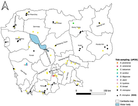
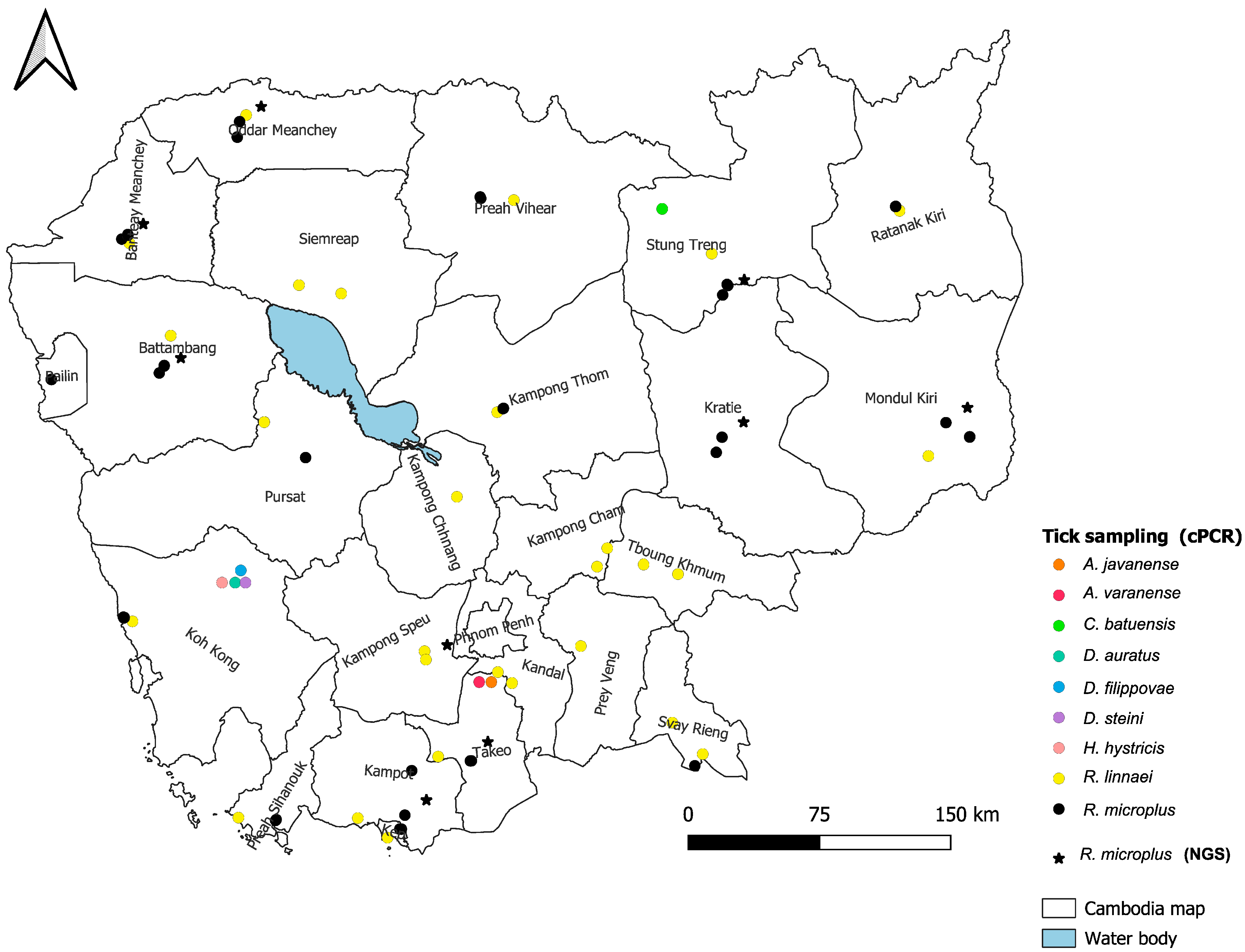
For molecular analyses, including conventional polymerase chain reaction (cPCR) and metatranscriptomic next-generation sequencing (mNGS), a subset of 1526 ticks was selected. These specimens were initially identified to the species level using morphological keys developed by the Medical and Veterinary Entomology Unit at the Institut Pasteur du Cambodge (IPC) [39,40,41,42,43,44,45,46]. Ticks were selected from nine species and grouped into 352 pools based on province, species, sex, habitat, and host type, with pool sizes ranging from one to six specimens. For some ticks, a portion was used for taxonomic verification via morphology, while the remaining portion was used for pathogen detection. The distribution of specimens was as follows: Carios batuensis (6 pools; 10 females, 10 males, 10 nymphs), Amblyomma javanense (2 pools; 1 female, 5 males), Amblyomma varanense (2 pools; 5 females, 2 males), Dermacentor auratus (1 specimen, split for molecular identification and pathogen screening), Dermacentor filippovae (3 pools; 2 females, 2 males), Dermacentor steini (1 specimen, split), Haemaphysalis hystricis (1 specimen, split), Rhipicephalus linnaei (169 pools; 248 females, 431 males, 3 nymphs), and Rhipicephalus microplus (167 pools; 371 females, 417 males, 6 nymphs) (Figure 1).
Figure 1.
Distribution of tick collection across 24 provinces of Cambodia from cattle, cats, dogs, wildlife, forest-fringe vegetation, and bat cave floors. Samples were screened by cPCR (colored dots), and ticks from 9 provinces were further analyzed using NGS for virus detection (black stars).
For viral metagenomic analysis, we randomly selected 40 individual R. microplus ticks from the total collection obtained across nine farms (one farm per province, sampling 10 cattle per farm). To achieve balanced geographic and demographic representation, we aimed to select five individual ticks per farm: specifically two adult females, two adult males, and one nymph, where available. Due to lower collection yields in certain areas, fewer specimens were included from Kep (one female, one male, one nymph) and Mondulkiri (two males only). This pilot-scale next-generation sequencing (NGS) approach was designed as a first exploratory investigation of viral diversity in a small set of R. microplus ticks collected from cattle in Cambodia. The objective was not to estimate viral prevalence, but we wanted to obtain an initial overview of tick-associated viruses circulating in cattle- associated tick populations and to evaluate the feasibility and usefulness of metagenomic surveillance approaches for future large-scale studies in Cambodia (Figure 1).
2.3. Pathogen Screening by Using cPCR
Following a double rinse with distilled water, specimens were grouped into 1.5 mL Eppendorf tubes [47]. Pools of up to 6 ticks were prepared by location, host, species, sex, and developmental stage to increase DNA quantity [48]. To optimize tissue lysis, an abdominal incision was made on each tick prior to overnight incubation at 56 °C in a mixture of 180 µL ATL buffer and 20 µL proteinase K, ensuring complete enzymatic breakdown before supernatant processing [49,50,51]. Cutting open the tick abdomen allowed direct access of the reagents to internal tissues, and overnight incubation ensured thorough enzymatic digestion of the cells, leading to a highly efficient release of high-quality DNA. After transferring 200 µL of the supernatant for the subsequent DNA extraction, the exoskeletons were recovered, washed, preserved in 70% ethanol.
We screened for four pathogens such as Anaplasma, Ehrlichia, Babesia, and Coxiella using conventional PCR followed by Sanger sequencing [47]. A 345 bp fragment of the 16S rRNA gene was initially amplified using the EHR16SD/EHR16SR primers for preliminary pathogen screening [52]. Positive samples were subsequently subjected to amplification of an approximately 1500 bp 16S rRNA fragment using the fD1/Rp2 primers to obtain longer sequences for accurate identification and sequencing [53]. Babesia was indicated by the band at 207 bp, which represents a fragment of the 18s rRNA gene, using Bcommon-F/Bcommon-R primer sets [54]. Coxiella screening targeted a 1322 bp fragment of the 16S rRNA gene using Cox16SF1/Cox16SR2 primer sets [55]. The PCR reactions (40 µL) consisted of 4 µL of 10× buffer B2, 0.8 µL of each primer (10 µM), 0.4 µL of 20 mM dNTPs, 4 µL of 25 mM MgCl2, 0.4 µL of Hot FirePol polymerase (5 U/µL), 35 µL of nuclease-free water, and 5 µL of DNA template. To ensure the validity of the PCR assays and monitor for potential contamination, a no-template control (NTC) using nuclease-free water and a positive control consisting of synthetic target DNA fragments (gBlocks™ Gene Fragments, Integrated DNA Technologies, Coralville, IA, USA) were included in every amplification run. Thermal cycling conditions were maintained according to pathogen-specific protocols (Table 1). The PCR products were separated on 1.5% agarose gels and visualized under UV light. Only strong, well-defined bands were sent for DNA sequencing service (Macrogen, Seoul, Republic of Korea).
Table 1.
List of TBPs, target genes, PCR primer name and PCR program in TBP screening.
Sequence data were analyzed using Unipro UGENE v50.0, where raw chromatograms were carefully inspected and cleaned to obtain consensus sequences. The consensus sequences were subjected to BLAST 2.17.0 searches against the NCBI database to validate their preliminary taxonomic identification. Subsequently, sequences were aligned with ClustalW against reference sequences retrieved from GenBank. Phylogenetic trees were constructed using the maximum-likelihood method with 1000 bootstrap replicates in IQ-TREE v2.3.5 [56,57,58]. The resulting trees were visualized and edited in MEGA v11.0.13 [50,59] (Table S1).
2.4. Pathogen Screening by Using Next Generation Sequencing
2.4.1. RNA Extraction and Library Preparation
Individual ticks were homogenized, and RNA was extracted using the Qiagen QIAamp Viral RNA Kit (QIAGEN, Hilden, Germany) with minor modifications including extended tissue homogenization and lysis steps to optimize yield and reduce RNA degradation. RNA quality and concentration were measured using a Qubit fluorometer (Thermo Fisher Scientific, Waltham, MA, USA). For library preparation, the extracted RNA was treated with DNase, depleted of ribosomal RNA, and reverse-transcribed to cDNA before library preparation using a Nextera XT kit (Illumina, San Diego, CA, USA). Sequencing was performed using NextSeq 500/550 v2.5 kit (Illumina, San Diego, CA, USA).
2.4.2. Bioinformatic Analysis
Reads were demultiplexed with bcl2fastq v2.2 and quality-checked using FastQC v0.11.9. Reads were quality-trimmed with trimmomatic v0.39 and de novo assembled using MEGAHIT v1.2.9. Viral sequences were identified through BLASTx/BLASTn searches against the NCBI non-redundant protein database [60]. Reads were mapped to reference viral genomes using Bowtie2 v2.1.0 to quantify abundance. The number of mapped reads was normalized as reads per million (RPM) to account for differences in sequencing depth among samples. The newly generated sequences have been submitted to SRA under Bioproject number PRJNA1476874.
2.5. Statistical Analysis
Pathogen prevalence was expressed as the percentage of positive tick pools relative to the total number of pools tested for each pathogen, host, or tick species [61,62]. The Minimum Infection Rate (MIR) was estimated assuming one infected tick per positive pool and calculated as the number of positive pools divided by the total number of ticks tested at each site. MIRs were also calculated separately by tick developmental stage and sex (adults, nymphs, females) to account for variation in infection rates among Anaplasma sp., Babesia sp., Ehrlichia sp., and Coxiella sp. [63,64].
3. Results
3.1. PCR Results
A total of 352 tick pools were screened using PCR for Anaplasma sp., Babesia sp., Ehrlichia sp., and Coxiella sp. Among these, 135 pools tested positive for at least one of the pathogens. In the positive tick pools, single-pathogen detections were the most common, comprising 85.93% (n = 116) of the samples. Dual-pathogen detections accounted for 13.33% (n = 18) of the pools, while triple-pathogen detections indicating the presence of three different pathogens in a single pooled sample were quite rare, occurring in only 0.74% (n = 1) of the pools. Overall, a total of 155 pathogen detections were recorded, which shows that the co-detection of multiple pathogens occurred in some tick pools. Of these detections, 105 PCR amplicons were successfully sequenced; however, 50 amplicons could not be sequenced due to faint PCR bands that were insufficient for reliable sequencing. Sequencing success was achieved across all infection categories, including the sequencing of all detected pathogens in the triple-infected pool (Table 2). The composition of the tick pools varied by species and host, which have influenced the detection of pathogens. Specifically, the pools included the following: C. batuensis collected from bat cave floors (n = 6), R. linnaei from cats (n = 4) and dogs (n = 161), as well as from cattle (n = 4), R. microplus from cattle (n = 162), dogs (n = 3), and goats (n = 2), A. javanense from pangolins (Manis javanica) (n = 2), A. varanense from pythons (Malayopython reticulatus) (n = 2), half-individual sample of D. steini (n = 0.5), and H. hystricis (n = 0.5) collected from vegetation, half-individual sample of D. auratus from wild pigs (Sus scrofa) (n = 0.5) and D. filippovae from wild pigs (n = 3) (Table 3).
Table 2.
Distribution of infection status and sequencing outcomes among PCR-positive tick pools for Anaplasmatacea, Coxiella spp., and Babesia spp.
Table 3.
(A) Prevalence of tick samples collected in 24 provinces that harbored DNA of selected genera of bacteria and parasites according to tick species and host species. (B) Minimum infection rate (MIR) of tick-borne pathogens across different host species per tick species.
Overall, pathogen screening revealed a total pool positivity rate of 44.0% (155/352) across the entire study. When broken down by specific host–tick combinations out of the total sample size (n = 352), Rhipicephalus microplus pools from cattle accounted for the largest share of pathogen detections at 33.8% (119/352), followed by Rhipicephalus linnaei pools from dogs at 9.3% (33/352), R. microplus pools from goats at 0.6% (2/352), and Carios batuensis pools from bat cave floors at 0.3% (1/352). Looking at individual pathogen targets across the entire study, Coxiella-like endosymbionts were detected in 15.6% (55/352) of all pools, followed by Anaplasma spp. at 13.3% (47/352), Coxiella spp. at 8.2% (29/352), and Babesia spp. at 2.0% (7/352). Lower overall prevalence rates were observed for Anaplasma marginale at 1.1% (4/352), Ehrlichia spp. at 1.7% (6/352), Babesia canis at 0.3% (1/352), Anaplasma platys at 0.3% (1/352), and Ehrlichia minasensis at 0.9% (3/352). Finally, tick pools recovered from cats (0/352), pangolins (0/352), pythons (0/352), vegetation (0/352), and wild pigs (0/352) yielded no detectable bacterial or parasitic DNA (Table 3A).
The minimum infection rate (MIR) was calculated to provide a conservative estimate, though it typically underestimates the true prevalence of tick-borne pathogens. A total of 1526 individual ticks were examined for tick-borne pathogens. Overall, 753 ticks (49.4% MIR) were positive for at least one pathogen. Coxiella-like endosymbionts (CLE) were the most frequently detected agents, identified in 275 ticks (18.1%), followed by Anaplasma spp. in 238 ticks (15.6%) and Coxiella spp. in 123 ticks (8.1%). Ehrlichia minasensis and Ehrlichia spp. were detected in 18 (1.2%) and 34 (2.2%) ticks, respectively. Among protozoan pathogens, Babesia canis was detected in 30 ticks (2.0%), while Babesia spp. were identified in 14 ticks (0.9%). Most positive detections were observed in Rhipicephalus microplus collected from cattle, whereas Rhipicephalus linnaei collected from dogs showed detection of Anaplasma spp., A. platys, and Babesia canis. Additionally, five ticks collected from bat cave floors and identified as Carios batuensis were positive for Anaplasma spp. (0.3% MIR) (Table 3B).
3.2. DNA Sequencing and Phylogenetic Analysis
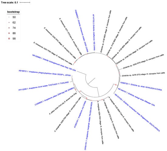
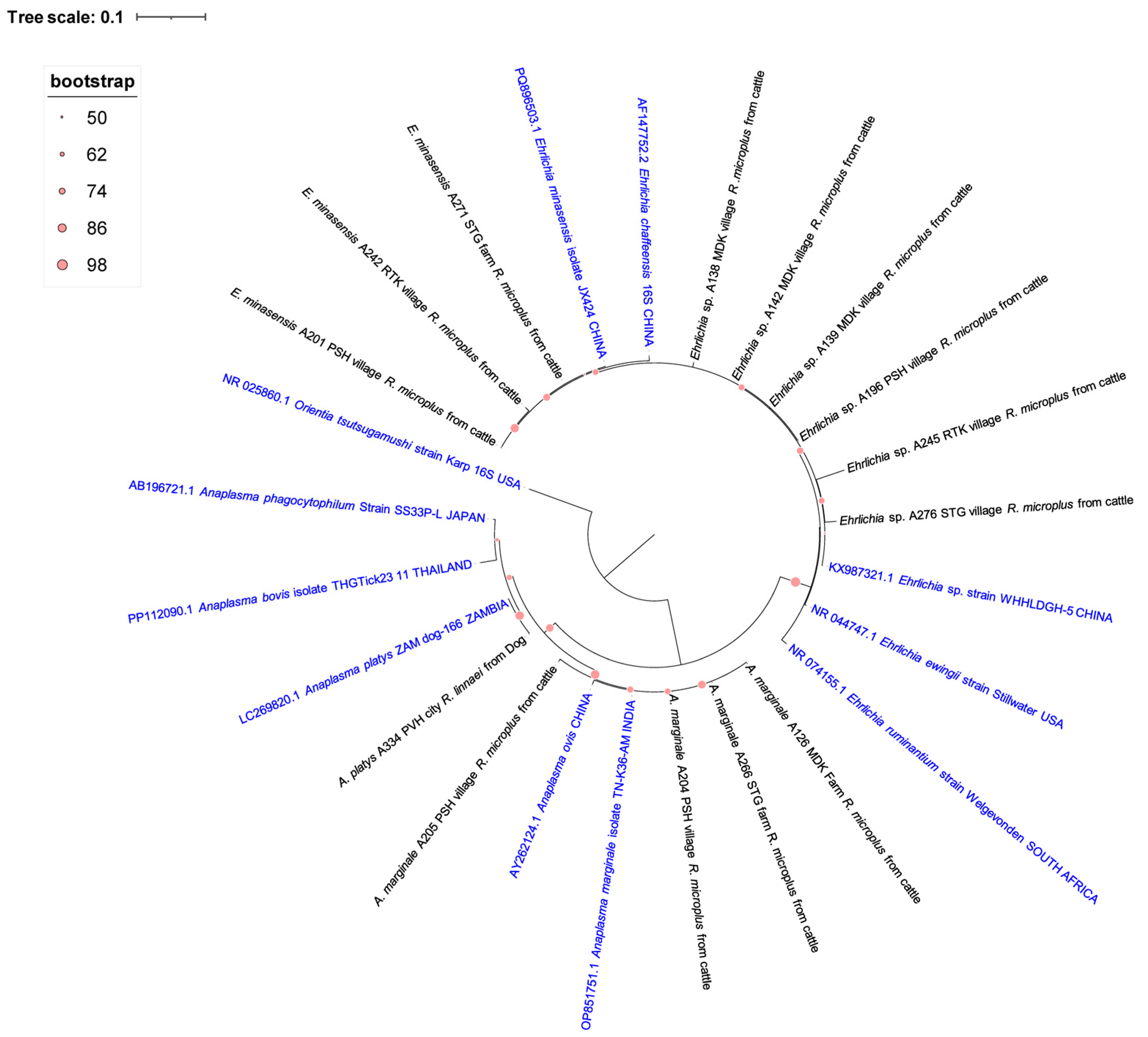
A maximum-likelihood phylogenetic tree based on 16S rRNA gene sequences revealed genetic relatedness among Anaplasma and Ehrlichia isolates from Rhipicephalus microplus and R. linnaei ticks infesting cattle and dogs in Cambodia (Figure 1). Anaplasma marginale strains from Preah Sihanouk (PSH), Mondulkiri (MDK), and Stung Treng (STG) showed >99% similarity to an Indian reference strain (OP851751.1). Anaplasma platys was detected in a pool of R. linnaei collected from a dog in Preah Vihear (PVH) and shared 99.68% similarity with a Zambian strain (LC269820.1). Ehrlichia minasensis isolates from cattle in Preah Sihanouk, Rattanakiri, and Stung Treng exhibited >99% similarity to a Chinese strain (PQ896500.1). An unidentified Ehrlichia sp. displayed 96.11–99.84% similarity to the closest match Ehrlichia sp. in Chinese (KX987321.1) (Figure 2).
Figure 2.
A maximum-likelihood phylogenetic tree was constructed using 16S rRNA gene sequences to explain the relationships between Anaplasma and Ehrlichia species detected in Rhipicephalus microplus and R. linnaei ticks on cattle and dog across different region in Cambodia, alongside reference sequences from GenBank under accession number (PZ515790-PZ515794), (PZ537012-PZ537020). The tree was rooted using Orientia tsutsugamushi as an outgroup. The sequences obtained from this study are highlighted in black.
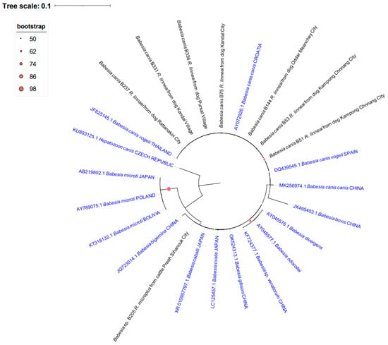
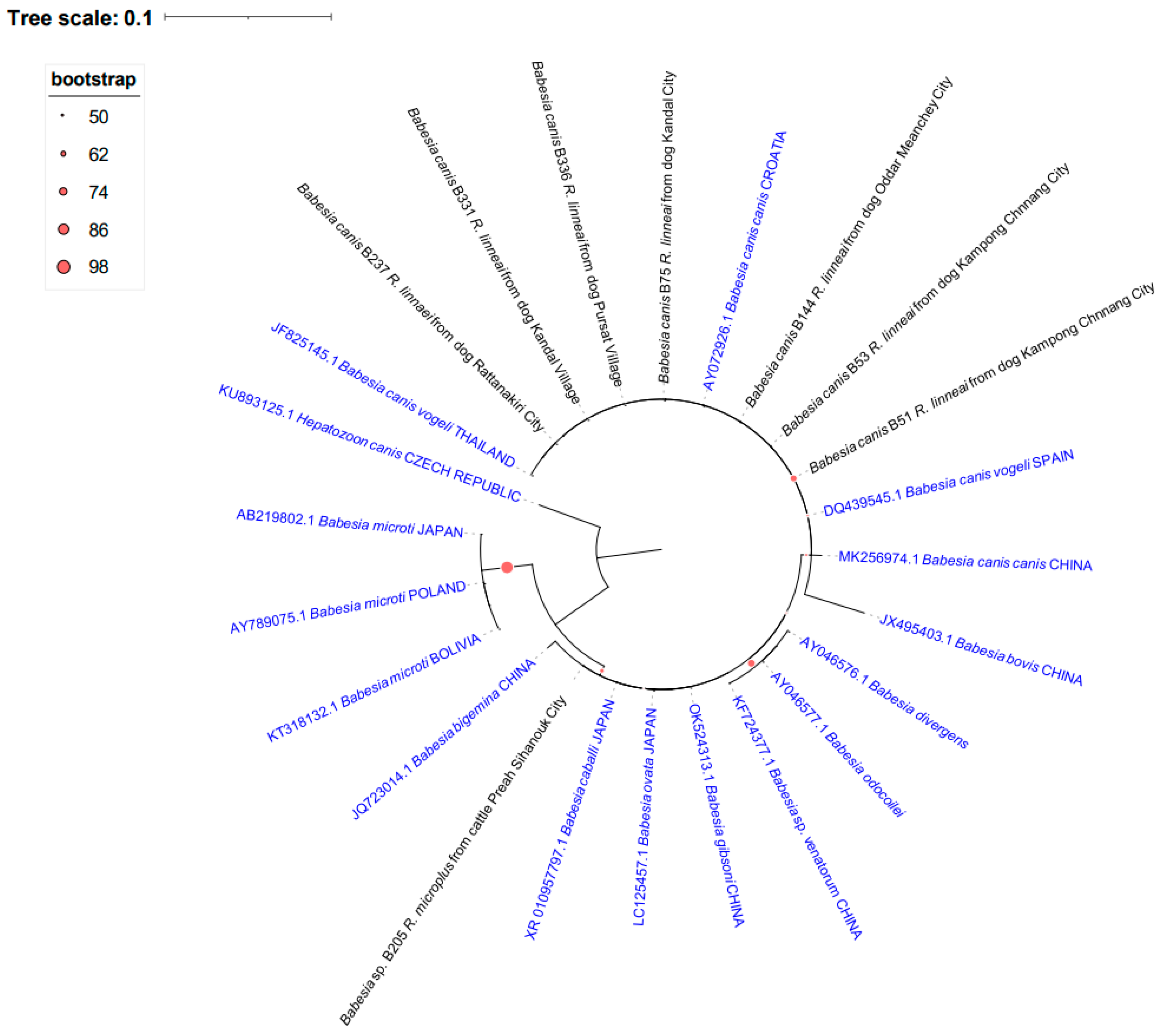
Phylogenetic analysis of 18S rRNA sequences observed that Babesia canis strains from the R. linnaei pool were found on dogs in Ratanakiri, Kandal, Pursat, Odar Meanchey, and Kampong Chhnang. Sequence analysis confirmed that these isolates belonged to the Babesia canis complex. However, the genetic marker used in this study did not provide sufficient resolution to distinguish among the closely related subspecies (B. canis canis, or B. canis vogeli). Given that R. linnaei is the predominant tick species parasitizing dogs in the region and is a recognized vector of B. canis vogeli in Asia, the findings are epidemiologically consistent with this subspecies, although definitive subspecies identification could not be established. These results indicate the circulation of B. canis (sensu lato) among companion animals and associated ticks in the surveyed areas. In contrast, a Babesia sp. detected in R. microplus from cattle in Preah Sihanouk showed no close relationship to any known sequences, indicating a distinct lineage (Figure 3).
Figure 3.
A maximum-likelihood phylogenetic tree was constructed using 18S rRNA gene sequences to visualize the relationships between Babesia canis and Babesia sp. detected in Rhipicephalus linnaei and R. microplus ticks on dogs and cattle across different regions in Cambodia, alongside reference sequences from GenBank under accession number (PZ509201-PZ509194). The tree was rooted using Hepatozoon canis as an appropriate outgroup. The sequences obtained from this study are highlighted in blue.
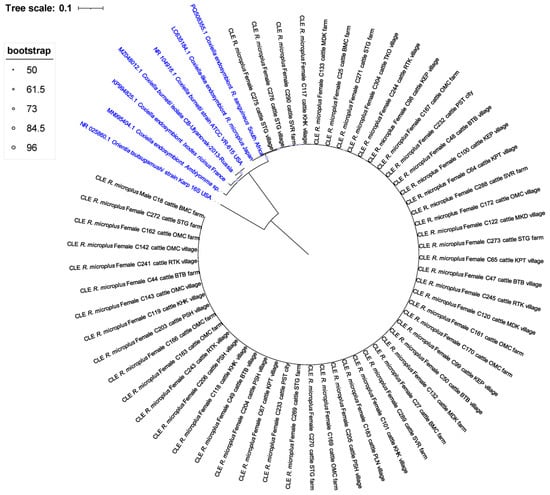
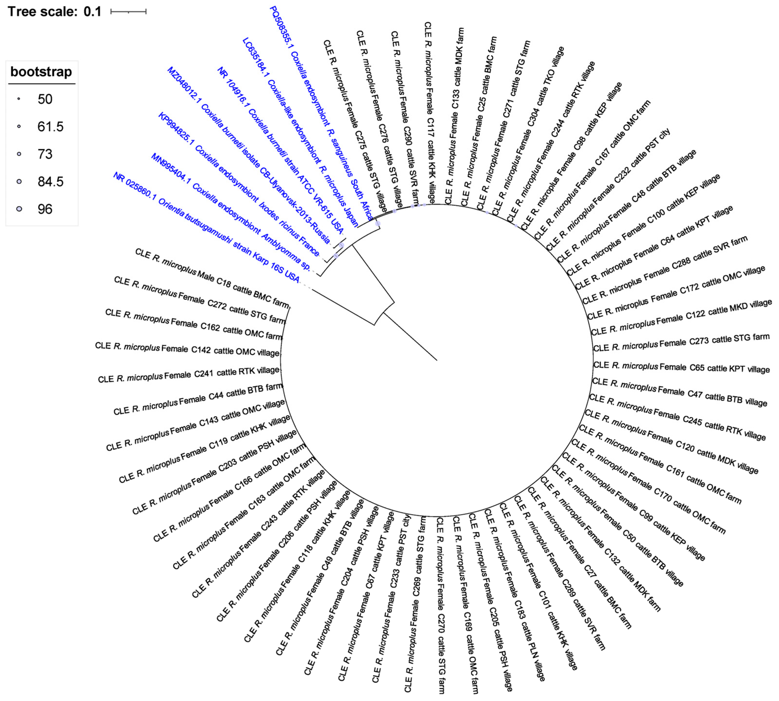
All 55 Coxiella-like endosymbiont sequences obtained from R. microplus ticks on cattle across 14 Cambodian provinces including Banteay Meanchey, Battambang, Kampong Thom, Kep, Koh Kong, Mondulkiri, Oddar Meanchey, Pailin, Preah Sihanouk, Pursat, Ratanakiri, Stung Treng, Svay Rieng, and Takeo exhibited >99% similarity to a Coxiella endosymbiont of Rhipicephalus sanguineus from South Africa (PQ508355.1) (Figure 4).
Figure 4.
A maximum-likelihood phylogenetic tree was constructed using 16S rRNA gene sequences to visualize the relationships between Coxiella-like endosymbionts detected in Rhipicephalus microplus ticks on cattle across different regions in Cambodia, alongside reference sequences from GenBank under accession numbers (PZ510385-PZ510439). The tree was rooted using Orientia tsutsugamushi as an appropriate outgroup. The sequences obtained from this study are highlighted in black.
3.3. Virus Detection in Rhipicephalus microplus
Analysis of 40 Rhipicephalus microplus ticks processed through mNGS revealed the detection of viral RNA in 18 samples (45%), while 22 samples (55%) were negative under the analytical parameters used. Six distinct tick-associated viruses were identified: Guangdong tick manly virus (GTMV), Jingmen tick virus (JMTV), Mogiana tick virus (MGTV), Rhipicephalus-associated rhabdo-like virus, Rhipicephalus-associated flavi-like virus, and Zhangzhou totiv tick virus 1. Single-virus infections were detected in 14 ticks (35%), dual infections in 3 ticks (7.5%), and co-infection with four viruses in 1 tick (2.5%), indicating heterogeneous viral burdens among individual ticks. GTMV was the most prevalent virus, detected in 12 samples (32.5%) from Battambang, Oddar Meanchey, Takeo, and Kampong Speu provinces, predominantly in male ticks (7 males, 2 females, 3 nymphs). JMTV was identified in two samples (5%) from Stung Treng and Mondulkiri (one male, one female). Mogiana tick virus, Rhipicephalus-associated rhabdo-like virus, and Rhipicephalus-associated flavi-like virus were each detected in single samples (2.5%), primarily from Mondulkiri and Kampong Speu provinces. Zhangzhou totiv tick virus 1 was detected in six samples (15%) across five provinces, mostly in male ticks (Table 4).
Table 4.
Summary of viruses detected from Rhipicephalus microplus.
3.4. Viral Abundance and Genome Coverage
Metatranscriptomic sequencing generated 258,910,074 total reads, with an average of 20.8 million reads per sample. GTMV exhibited the highest abundance and genomic completeness. One male tick from Battambang showed a high viral load, with GTMV reads comprising 20.6% of total RNA (~205,800 read per million). Across all GTMV-positive samples, read counts ranged from 12,531 to 360,367, with 99–100% genome coverage and ~96% nucleotide identity to the reference genome (OM264164.1). Zhangzhou totiv tick virus 1 showed read counts ranging from 1880 to 9360 RPM, with 98–100% genome coverage and 94–96% nucleotide identity. JMTV ranged from 1403 to 216,265 reads with genome coverage of 90–97% and ~93% identity, with read abundances up to 217,668 RPM. MGTV and both Rhipicephalus-associated rhabdo-like and flavi-like viruses displayed near-complete to complete genome coverage (97–100%) and nucleotide identities ranging from 93 to 95% relative to reference genomes (MT080097.1, ON812505.1, MH814979.1).
4. Discussion
Our study provides new molecular evidence regarding the circulation of tick-borne pathogens in Rhipicephalus microplus and R. linnaei collected from cattle, goats, and dogs throughout Cambodia. During field sampling, adult ticks were collected in greater numbers than nymphs and larvae. This discrepancy is primarily attributed to the larger size and higher visibility of adult stages, whereas immature stages—particularly larvae—are considerably smaller, more difficult to detect under field conditions, and heavily constrained by collection time limits. Despite these sampling limitations, we detected two bacterial agents: Anaplasma marginale, and Ehrlichia minasensis, as well as non-pathogenic Coxiella-like endosymbionts. Additionally, we found the protozoan pathogen Babesia canis and several tick-associated viruses, including the JMTV, which is known to infect both animals and humans [65,66,67,68,69,70,71].
Anaplasma marginale primarily infects cattle and is not known to cause human infection, in contrast to other members of the genus such as Anaplasma phagocytophilum which are zoonotic [6,14,72,73,74]. The prevalence of A. marginale in Cambodian R. microplus (1.1%) was similar to Vietnam data (1%), but it was slightly lower than reported in the Philippines (6.7%), and Thailand (19.08%) [6,72,75].
The detection of E. minasensis is particularly noteworthy; this emerging Ehrlichia species has close phylogenetic relationships with recognized human pathogens and has recently been identified in cattle ticks globally [14,72,76,77]. In our study, E. minasensis was detected in 0.9% of Cambodian R. microplus. This prevalence is comparable to low levels reported in Thailand (1.97%) [72].
Moreover, Anaplasma platys is a canine pathogen causing cyclic thrombocytopenia; it was detected in 0.3% of R. linnaei. This prevalence is lower than reports from Thailand (22.4%) [78]. We also detected Babesia canis, a protozoan agent of canine babesiosis, confirmed to species level by 18S rRNA sequencing [11]. Reports of B. canis in R. linnaei in Cambodia are limited, with a prevalence of 2.0% observed in our study.
In contrast, the Babesia sp. detected within R. microplus ticks collected from cattle in Preah Sihanouk presents a highly remarkable and unexpected finding. Downstream sequence verification demonstrated that this isolate shared no high-confidence identity or homology with any well-characterized Babesia references currently available in the NCBI GenBank database. This sharp divergence strongly indicates the presence of an unmapped, distinct, or novel Babesia lineage circulating locally in southern Cambodia. However, interpretation of this result should be made cautiously because the sequence quality was limited, likely due to low DNA concentration and faint PCR amplification, which may reduce the accuracy of BLAST identification and phylogenetic inference. In addition, partial 18S rRNA sequences may not always provide sufficient resolution for reliable species-level discrimination among closely related piroplasms [35,48]. Although Coxiella burnetii was not detected in our R. microplus ticks, we observed a high prevalence of CLEs. CLEs are closely related to C. burnetiid, the etiologic agent of Q fever in humans and coxiellosis in animals [79,80]. CLE are increasingly recognized as important components of tick biology, influencing reproduction, survival, and potentially vector competence for other pathogens [19,81]. The absence of C. burnetii may reflect its low prevalence in Cambodia compared to data reported from the Philippines (10.2%) in R. microplus [75]. The non-detection of Coxiella sp. is likely due to the limited sample size, single time-point collection, and the restricted host range sampled (cattle, cats, dogs, goats, and few wildlife animals), which may have reduced the probability of detecting the prevalence of pathogens.
In addition to bacterial and protozoal agents, metagenomic analysis of R. microplus has identified six tick-associated viruses, among which JMTV is notable [69,70,82]. JMTV has been detected in both human febrile cases and animal hosts in various settings across Asia [67]. The presence of JMTV in R. microplus specimens collected from cattle farms in Cambodia indicates a geographic expansion of the virus which has primarily been reported in China, suggesting it may now be circulating more widely throughout Southeast Asia. Because R. microplus commonly infests cattle and will occasionally bite humans, this detection raises important public health and veterinary concerns [83]. In humans, JMTV infection is typically associated with nonspecific febrile illness, including fever, headache, myalgia, malaise, and occasionally gastrointestinal symptoms or rash. In livestock, R. microplus is already a major pest responsible for substantial economic losses; the presence of a viral pathogen further amplifies its significance in veterinary medicine, even though clinical signs of JMTV in animals remain poorly documented. These risks are compounded by the local context in Cambodia, where tick-borne viral infections are likely underdiagnosed and underreported. Due to limited clinical awareness and diagnostic capacity, veterinarians and physicians may not readily recognize tick-borne diseases. Because clinical signs in livestock are often nonspecific such as fever, lethargy, reduced appetite, and decreased productivity, they are easily misdiagnosed or attributed to other endemic conditions, and some infected animals may remain asymptomatic, further obscuring clinical identification. However, the findings of this study must be interpreted cautiously. The detection of JMTV RNA in Cambodian R. microplus confirms its circulation within the tick population, but RNA presence alone does not confirm active viral replication, vector competence, or successful transmission to livestock and humans. While JMTV is linked to human illness in other regions, its clinical significance in Cambodia remains unknown, and no direct evidence of human or animal disease linked to JMTV was obtained in this study. Therefore, conclusions regarding zoonotic risk should remain tentative, and future investigations including virus isolation, transmission dynamics studies, and serological surveys are required to determine its true epidemiological relevance and to sustain vital tick surveillance in the region.
Several limitations should be acknowledged. First, the cross-sectional design does not capture temporal dynamics of tick activity and pathogen circulation. Second, while sampling spanned multiple provinces, localized pathogen diversity may remain underrepresented. Third, the absence of parallel serological or clinical surveillance in animals and humans limits our ability to confirm exposure history or disease associations [84]. Finally, conventional PCR, while specific, can give false positive or false negative results [85], while the use of pooled tick samples restricts the calculation of exact individual prevalence. Furthermore, reliance on reference database–dependent sequence classification introduces potential uncertainty and bias in precise taxonomic assignment, particularly for less-characterized variants.
5. Conclusions
The study provides foundational molecular evidence that tick-borne pathogens, which are significant for veterinary and potential zoonotic concerns, are present in Cambodia. To better understand transmission pathways, assess the economic impact on livestock, and evaluate potential public health risks, it is crucial to expand integrated “One Health” surveillance. This approach should incorporate tick identification, pathogen detection, and host-level diagnostics. Future research that combines longitudinal entomological sampling, host serology, and clinical diagnostics will yield a more comprehensive understanding of the ecology of tick-borne diseases in Cambodia.
Supplementary Materials
The following supporting information can be downloaded at: https://www.mdpi.com/article/10.3390/pathogens15060641/s1, Table S1: BLASTn results of sequenced PCR amplicons obtained from tick samples collected in Cambodia.
Author Contributions
S.Y.: Writing—original draft, writing—review and editing, methodology, validation, formal analysis, investigation. D.B.P.: Writing—original draft, review and editing, investigation, methodology, project administration. L.V., S.C., M.P.: Investigation. S.B. (Sarah Bonnet): supervision. A.B., E.S.-L.: validation, funding acquisition, review and editing. S.B. (Sébastien Boyer): Conceptualization, writing—review and editing, validation, supervision, project administration, and funding acquisition. All authors have read and agreed to the published version of the manuscript.
Funding
This study was supported by the French Embassy in Cambodia through its FSPI—The Solidarity Fund for Innovative Projects. The E.S.-L. laboratory is funded by Institut Pasteur, the INCEPTION program (Investissements d’Avenir grant ANR-16-CONV-0005), the Ixcore foundation for research, the French Government’s Labex IBEID (ANR-10-LABX-62-IBEID), the HERA Project DURABLE (grant no 101102733) and LEAPS (grant no 101094685). The funder had no role in study design, data collection and analysis, decision to publish, or preparation of the manuscript.
Institutional Review Board Statement
The study was approved and supported by the General Directorate of Animal Health and Production from the Ministry of Agriculture, Forestry, and Fisheries of Cambodia under permit No. 2981, signed on 1 October 2021.
Informed Consent Statement
Not applicable. In Cambodia, formal written informed consent from individual owners is not standard for field-level tick sampling on domestic livestock and dogs. However, verbal agreement was obtained from all animal owners or farm managers prior to external parasite collection, and the study was fully covered by the ministerial ethical permit mentioned above.
Data Availability Statement
The newly generated sequences have been submitted to SRA under Bioproject number PRJNA1476874.
Acknowledgments
We extend our gratitude to the medical and veterinary entomology team for their invaluable assistance with tick collection and morphological identification. We also appreciate the support from the Laboratory of Medical Biology (LABM) and the Virology Unit at the Institut Pasteur du Cambodge for providing laboratory facilities for this research.
Conflicts of Interest
The authors declare no conflicts of interest.
References
- Nouhin, D.J.; Maquart, D.P.-O.; Khun, M.L.; Sen, M.S.; Phalla, M.; Heng, M.K.; Ali, S.M.; Leng, S.; Ken, S.; Dziedziech, D.A.; et al. Current knowledge of exposure to tick-borne pathogens among rural population in Cambodia. Int. J. Infect. Dis. 2025, 152, 107419. [Google Scholar] [CrossRef] [Scilit]
- Yean, S.; Prasetyo, D.B.; Marcombe, S.; Hadi, U.K.; Kazim, A.R.; Tiawsirisup, S.; Chinh, V.D.; Matsuno, K.; Low, V.L.; Bonnet, S.; et al. Challenges for ticks and tick-borne diseases research in Southeast Asia: Insight from the first international symposium in Cambodia. PLoS Negl. Trop. Dis. 2024, 18, e0012269. [Google Scholar] [CrossRef] [Scilit] [PubMed]
- Rosario-Cruz, R.; Domínguez-García, D.I.; Almazán, C.; Rosario Dominguez, F. A One Health epidemiological approach for understanding ticks and tick-borne pathogens. Vector Borne Zoonotic Dis. 2024, 24, 473–477. [Google Scholar] [CrossRef] [Scilit] [PubMed]
- Shahzad, S.; Akinsulie, O.C.; Idris, I.; Devnath, P.; Ajagbe, D.; Aliyu, V.A.; Oladoye, M.J.; Ukauwa, C.; Ugwu, C.E.; Ajulo, S.; et al. Ticks and tickborne diseases in Global South countries: Impact and implications of environmental changes. Front. Trop. Dis. 2025, 6, 1597236. [Google Scholar] [CrossRef] [Scilit]
- Sansilapin, C.; Tangwangvivat, R.; Hoffmann, C.S.; Chailek, C.; Lekcharoen, P.; Thippamom, N.; Petcharat, S.; Taweethavonsawat, P.; Wacharapluesadee, S.; Buathong, R.; et al. Severe fever with thrombocytopenia syndrome (SFTS) in Thailand: Using a one health approach to respond to novel zoonosis and its implications in clinical practice. One Health Outlook 2024, 6, 18. [Google Scholar] [CrossRef] [Scilit] [PubMed]
- Huynh, L.N.; Diarra, A.Z.; Pham, Q.L.; Le-Viet, N.; Berenger, J.-M.; Ho, V.H.; Nguyen, X.Q.; Parola, P. Morphological, molecular and MALDI-TOF MS identification of ticks and tick-associated pathogens in Vietnam. PLoS Negl. Trop. Dis. 2021, 15, e0009813. [Google Scholar] [PubMed]
- Madison-Antenucci, S.; Kramer, L.D.; Gebhardt, L.L.; Kauffman, E. Emerging tick-borne diseases. Clin. Microbiol. Rev. 2020, 33, e00083-18. [Google Scholar] [CrossRef] [Scilit] [PubMed]
- Chandra, S.; Ma, G.C.; Burleigh, A.; Brown, G.; Norris, J.M.; Ward, M.P.; Emery, D.; Šlapeta, J. The brown dog tick Rhipicephalus sanguineus sensu Roberts, 1965 across Australia: Morphological and molecular identification of R. sanguineus s.l. tropical lineage. Ticks Tick-Borne Dis. 2020, 11, 101305. [Google Scholar] [CrossRef] [Scilit] [PubMed]
- Almazán, C.; Torres Rodríguez, L.; Alanazi, A.D.; Šlapeta, J. American mitogenome reference for the tropical brown dog tick, Rhipicephalus linnaei (Audouin, 1826). Curr. Res. Parasitol. Vector Borne Dis. 2024, 6, 100194. [Google Scholar] [CrossRef] [Scilit] [PubMed]
- Šlapeta, J.; Chandra, S.; Halliday, B. The “tropical lineage” of the brown dog tick Rhipicephalus sanguineus sensu lato identified as Rhipicephalus linnaei (Audouin, 1826). Int. J. Parasitol. 2021, 51, 431–436. [Google Scholar] [CrossRef] [Scilit] [PubMed]
- Nguyen, V.L.; Colella, V.; Greco, G.; Fang, F.; Nurcahyo, W.; Hadi, U.K.; Venturina, V.; Tong, K.B.Y.; Tsai, Y.-L.; Taweethavonsawat, P.; et al. Molecular detection of pathogens in ticks and fleas collected from companion dogs and cats in East and Southeast Asia. Parasites Vectors 2020, 13, 420. [Google Scholar] [CrossRef] [Scilit] [PubMed]
- Piratae, S.; Senawong, P.; Chalermchat, P.; Harnarsa, W.; Sae-Chue, B. Molecular evidence of Ehrlichia canis and Anaplasma platys and the association of infections with hematological responses in naturally infected dogs in Kalasin, Thailand. Vet. World 2019, 12, 131–135. [Google Scholar] [CrossRef] [Scilit] [PubMed]
- Adao, D.E.V.; Herrera, C.M.T.; Galarion, L.H.; Bolo, N.R.; Carlos, R.S.; Carlos, E.T.; Carlos, S.S.; Rivera, W.L. Detection and molecular characterization of Hepatozoon canis, Babesia vogeli, Ehrlichia canis, and Anaplasma platys in dogs from Metro Manila, Philippines. Korean J. Vet. Res. 2017, 57, 79–88. [Google Scholar] [CrossRef] [Scilit]
- Koh, F.X.; Kho, K.L.; Kisomi, M.G.; Wong, L.P.; Bulgiba, A.; Tan, P.E.; Lim, Y.A.L.; Nizam, Q.N.H.; Panchadcharam, C.; Tay, S.T. Ehrlichia and Anaplasma infections: Serological evidence and tick surveillance in Peninsular Malaysia. J. Med. Entomol. 2018, 55, 269–276. [Google Scholar] [PubMed]
- Marques, R.; Krüger, R.F.; Peterson, A.T.; De Melo, L.F.; Vicenzi, N.; Jiménez-García, D. Climate change implications for the distribution of the babesiosis and anaplasmosis tick vector, Rhipicephalus (Boophilus) microplus. Vet. Res. 2020, 51, 81. [Google Scholar] [CrossRef] [Scilit] [PubMed]
- Srionrod, N.; Nooroong, P.; Poolsawat, N.; Minsakorn, S.; Watthanadirek, A.; Junsiri, W.; Sangchuai, S.; Chawengkirttikul, R.; Anuracpreeda, P. Molecular characterization and genetic diversity of Babesia bovis and Babesia bigemina of cattle in Thailand. Front. Cell Infect. Microbiol. 2022, 12, 1065963. [Google Scholar] [CrossRef] [Scilit] [PubMed]
- Ola-Fadunsin, S.D.; Sharma, R.S.K.; Abdullah, D.A.; Gimba, F.I.; Abdullah, F.F.J.; Sani, R.A. The molecular prevalence, distribution and risk factors associated with Babesia bigemina infection in Peninsular Malaysia. Ticks Tick-Borne Dis. 2021, 12, 101653. [Google Scholar] [CrossRef] [Scilit] [PubMed]
- Li, Y.; Luo, Y.; Cao, S.; Terkawi, M.A.; Lan, D.T.B.; Long, P.T.; Yu, L.; Zhou, M.; Gong, H.; Zhang, H.; et al. Molecular and seroepidemiological survey of Babesia bovis and Babesia bigemina infections in cattle and water buffaloes in the central region of Vietnam. Trop. BioMed 2014, 31, 406–413. [Google Scholar] [PubMed]
- Khoo, J.-J.; Lim, F.-S.; Chen, F.; Phoon, W.-H.; Khor, C.-S.; Pike, B.L.; Chang, L.-Y.; AbuBakar, S. Coxiella detection in ticks from wildlife and livestock in Malaysia. Vector Borne Zoonotic Dis. 2016, 16, 744–751. [Google Scholar] [CrossRef] [Scilit] [PubMed]
- Muramatsu, Y.; Usaki, N.; Thongchai, C.; Kramomtong, I.; Kriengsak, P.; Tamura, Y. Seroepidemiologic survey in Thailand of Coxiella burnetii infection in cattle and chickens and presence in ticks attached to dairy cattle. Southeast Asian J. Trop. Med. Public Health 2014, 45, 1167–1172. [Google Scholar] [PubMed]
- Ybañez, A.P.; Sivakumar, T.; Ybañez, R.H.D.; Ratilla, J.C.; Perez, Z.O.; Gabotero, S.R.; Hakimi, H.; Kawazu, S.-I.; Matsumoto, K.; Yokoyama, N.; et al. First molecular characterization of Anaplasma marginale in cattle and Rhipicephalus (Boophilus) microplus ticks in Cebu, Philippines. J. Vet. Med. Sci. 2013, 75, 27–36. [Google Scholar] [CrossRef] [Scilit] [PubMed]
- Nonthapa, A.; Rodpai, R.; Thanchomnang, T.; Boonroumkaew, P.; Sadaow, L.; Banouvong, V.; Laymanivong, S.; Thinnabut, K.; Tangkawanit, U.; Blair, D.; et al. Molecular detection of tick-borne pathogens in cattle ticks from the Lao People’s Democratic Republic. Parasites Vectors 2025, 19, 21. [Google Scholar] [CrossRef] [Scilit] [PubMed]
- Galon, E.M.; Zafar, I.; Ji, S.; Li, H.; Ma, Z.; Xuan, X. Molecular Reports of Ruminant Babesia in Southeast Asia. Pathogens 2022, 11, 915. [Google Scholar] [CrossRef] [Scilit] [PubMed]
- Shahhosseini, N.; Wong, G.; Babuadze, G.; Camp, J.V.; Ergonul, O.; Kobinger, G.P.; Chinikar, S.; Nowotny, N. Crimean-Congo hemorrhagic fever virus in Asia, Africa and Europe. Microorganisms 2021, 9, 1907. [Google Scholar] [CrossRef] [Scilit] [PubMed]
- Yu, K.-M.; Park, S.-J. Tick-borne viruses: Epidemiology, pathogenesis, and animal models. One Health 2024, 19, 100903. [Google Scholar] [CrossRef] [Scilit] [PubMed]
- Aslam, M.; Abbas, R.Z.; Alsayeqh, A. Distribution pattern of Crimean-Congo Hemorrhagic Fever in Asia and the Middle East. Front. Public Health 2023, 11, 1093817. [Google Scholar] [CrossRef] [Scilit] [PubMed]
- Celina, S.S.; Italiya, J.; Tekkara, A.O.; Černý, J. Crimean-Congo haemorrhagic fever virus in ticks, domestic, and wild animals. Front. Vet. Sci. 2024, 11, 1513123. [Google Scholar] [PubMed]
- Belobo, J.T.E.; Kenmoe, S.; Kengne-Nde, C.; Emoh, C.P.D.; Bowo-Ngandji, A.; Tchatchouang, S.; Wobessi, J.N.S.; Mikangue, C.A.M.; Tazokong, H.R.; Bebey, S.R.K.; et al. Worldwide epidemiology of Crimean-Congo Hemorrhagic Fever Virus in humans, ticks and other animal species, a systematic review and meta-analysis. PLoS Negl. Trop. Dis. 2021, 15, e0009299. [Google Scholar] [CrossRef] [Scilit] [PubMed]
- Papa, A.; Tsergouli, K.; Tsioka, K.; Mirazimi, A. Crimean-Congo hemorrhagic fever: Tick-host-virus interactions. Front. Cell. Infect. Microbiol. 2017, 7, 213. [Google Scholar] [CrossRef] [Scilit] [PubMed]
- Bhowmick, S.; Kasi, K.K.; Gethmann, J.; Fischer, S.; Conraths, F.J.; Sokolov, I.M.; Lentz, H.H.K. Ticks on the run: A mathematical model of Crimean-Congo haemorrhagic fever (CCHF)-key factors for transmission. Epidemiologia 2022, 3, 116–134. [Google Scholar] [CrossRef] [Scilit] [PubMed]
- Damian, D.; Maghembe, R.; Damas, M.; Wensman, J.J.; Berg, M. Application of viral metagenomics for study of emerging and reemerging tick-borne viruses. Vector Borne Zoonotic Dis. 2020, 20, 557–565. [Google Scholar] [CrossRef] [Scilit] [PubMed]
- Sharma, S.N.; Singh, R.; Balakrishnan, N.; Kumawat, R.; Singh, S.K. Vectors of Crimean-Congo hemorrhagic fever (CCHF): Prevention and its control. J. Commun. Dis. 2020, 52, 22–26. [Google Scholar] [CrossRef] [Scilit]
- Garcia, K.; Weakley, M.; Do, T.; Mir, S. Current and future molecular diagnostics of tick-borne diseases in cattle. Vet. Sci. 2022, 9, 241. [Google Scholar] [CrossRef] [Scilit] [PubMed]
- Belova, O.A.; Polienko, A.E.; Averianova, A.D.; Karganova, G.G. Hybrids of Ixodes ricinus and Ixodes persulcatus ticks effectively acquire and transmit tick-borne encephalitis virus. Front. Cell. Infect. Microbiol. 2023, 13, 1104484. [Google Scholar] [CrossRef] [Scilit] [PubMed]
- Gou, H.; Guan, G.; Liu, A.; Ma, M.; Xu, Z.; Liu, Z.; Ren, Q.; Li, Y.; Yang, J.; Chen, Z.; et al. A DNA barcode for piroplasmea. Acta Trop. 2012, 124, 92–97. [Google Scholar] [CrossRef] [Scilit] [PubMed]
- Saba Villarroel, P.M.; Chaiphongpachara, T.; Nurtop, E.; Laojun, S.; Pangpoo-Nga, T.; Songhong, T.; Supungul, D.; Baronti, C.; Thirion, L.; Leaungwutiwong, P.; et al. Seroprevalence study in humans and molecular detection in Rhipicephalus sanguineus ticks of severe fever with thrombocytopenia syndrome virus in Thailand. Sci. Rep. 2024, 14, 13397. [Google Scholar] [CrossRef] [Scilit] [PubMed]
- Jang, H.; Casel, M.A.B.; Jang, S.-G.; Choi, J.H.; Gil, J.; Rollon, R.; Cheun, S.Y.; Kim, Y.-I.; Song, M.S.; Choi, Y.K. Seasonal dynamics of Haemaphysalis tick species as SFTSV vectors in South Korea. Microbiol. Spectr. 2024, 12, e0048924. [Google Scholar] [CrossRef] [Scilit] [PubMed]
- Espada, C.; Cummins, H.; Gonzales, J.A.; Notto, L.; Gaff, H.D. A Comparison of Tick Collection Materials and Methods in Southeastern Virginia. J. Med. Entomol. 2021, 58, 692–698. [Google Scholar] [CrossRef] [Scilit] [PubMed]
- Cooley, R.A. The Genera Boophilus, Rhipicephalus, and Haemaphysalis (Ixodiadae) of the New World; U.S. Government Printing Office: Washington, DC, USA, 1946.
- Estrada-Peña, A.; Venzal, J.M.; Nava, S.; Mangold, A.; Guglielmone, A.A.; Labruna, M.B.; de la Fuente, J. Reinstatement of Rhipicephalus (Boophilus) australis (Acari: Ixodidae) with redescription of the adult and larval stages. J. Med. Entomol. 2012, 49, 794–802. [Google Scholar] [CrossRef] [Scilit] [PubMed]
- Berry, C.M. Resolution of the Taxonomic Status of Rhipicephalus (Boophilus) Microplus. Ph.D. Thesis, University of Glasgow, Glasgow, UK, 2017. [Google Scholar]
- Walker, J.B.; Keirans, J.E.; Horak, I.G. The Genus Rhipicephalus (Acari, Ixodidae): A Guide to the Brown Ticks of the World; Cambridge University Press: Cambridge, UK, 2000. [Google Scholar]
- Kazim, A.R.; Low, V.L.; Tappe, D.; Houssaini, J.; Heo, C.C. Rhipicephalus annulatus, R. australis or R. microplus? Discordance between morphological and genetic data among three cattle tick species. Exp. Appl. Acarol. 2022, 87, 119–131. [Google Scholar] [CrossRef] [Scilit] [PubMed]
- Dao, T.T.H.; Takács, N.; Tran, T.N.; Truong, A.N.; Skinner, K.; Kontschán, J.; Farkas, R.; Hornok, S. Detection of tick-borne pathogens in the pangolin tick, Amblyomma javanense, from Vietnam and Laos, including a novel species of Trypanosoma. Acta Trop. 2024, 260, 107384. [Google Scholar] [CrossRef] [Scilit] [PubMed]
- Geevarghese, G.; Mishra, A.C. Haemaphysalis Ticks of India; Elsevier—Health Sciences Division: Philadelphia, PA, USA, 2011. [Google Scholar]
- Wassef, H.Y.; Hoogstraal, H. Dermacentor (Indocentor) auratus (Acari: Ixodoidea: Ixodidae): Identity of Male and Female. J. Med. Entomol. 1984, 21, 169–173. [Google Scholar] [CrossRef] [Scilit]
- Al-Hosary, A.; Răileanu, C.; Tauchmann, O.; Fischer, S.; Nijhof, A.M.; Silaghi, C. Tick species identification and molecular detection of tick-borne pathogens in blood and ticks collected from cattle in Egypt. Ticks Tick. Borne Dis. 2021, 12, 101676. [Google Scholar] [CrossRef] [Scilit] [PubMed]
- Martínez-García, G.; Santamaría-Espinosa, R.M.; Lira-Amaya, J.J.; Figueroa, J.V. Challenges in tick-borne pathogen detection: The case for Babesia spp. identification in the tick vector. Pathogens 2021, 10, 92. [Google Scholar] [CrossRef] [Scilit] [PubMed]
- Reifenberger, G.C.; Thomas, B.A.; Rhodes, D.V.L. Comparison of DNA extraction and amplification techniques for use with engorged hard-bodied ticks. Microorganisms 2022, 10, 1254. [Google Scholar] [CrossRef] [Scilit] [PubMed]
- Tamura, K.; Stecher, G.; Kumar, S. MEGA11: Molecular Evolutionary Genetics Analysis version 11. Mol. Biol. Evol. 2021, 38, 3022–3027. [Google Scholar] [CrossRef] [Scilit] [PubMed]
- Cicculli, V.; DeCarreaux, D.; Ayhan, N.; Casabianca, F.; de Lamballerie, X.; Charrel, R.; Falchi, A. Molecular screening of Anaplasmataceae in ticks collected from cattle in Corsica, France. Exp. Appl. Acarol. 2020, 81, 561–574. [Google Scholar] [CrossRef] [Scilit] [PubMed]
- Parola, P.; Roux, V.; Camicas, J.L.; Baradji, I.; Brouqui, P.; Raoult, D. Detection of Ehrlichia in African ticks by polymerase chain reaction. Trans. R Soc. Trop. Med. Hyg. 2000, 94, 707–708. [Google Scholar] [CrossRef] [Scilit] [PubMed]
- Koh, F.X.; Panchadcharam, C.; Sitam, F.T.; Tay, S.T. Molecular investigation of Anaplasma spp. in domestic and wildlife animals in Peninsular Malaysia. Vet. Parasitol. 2018, 13, 141–147. [Google Scholar] [CrossRef] [Scilit] [PubMed]
- Qurollo, B.A.; Archer, N.R.; Schreeg, M.E.; Marr, H.S.; Birkenheuer, A.J.; Haney, K.N.; Thomas, B.S.; Breitschwerdt, E.B. Improved molecular detection of Babesia infections in animals using a novel quantitative real-time PCR diagnostic assay targeting mitochondrial DNA. Parasites Vectors 2017, 10, 128. [Google Scholar] [CrossRef] [Scilit] [PubMed]
- Seo, M.-G.; Lee, S.-H.; Ouh, I.-O.; Lee, G.H.; Goo, Y.-K.; Kim, S.; Kwon, O.-D.; Kwak, D. Molecular detection and genotyping of Coxiella-like endosymbionts in ticks that infest horses in South Korea. PLoS ONE 2016, 11, e0165784. [Google Scholar] [CrossRef] [Scilit] [PubMed]
- Minh, B.Q.; Schmidt, H.A.; Chernomor, O.; Schrempf, D.; Woodhams, M.D.; von Haeseler, A.; Lanfear, R. IQ-TREE 2: New models and efficient methods for phylogenetic inference in the genomic era. Mol. Biol. Evol. 2020, 37, 1530–1534. [Google Scholar] [CrossRef] [Scilit] [PubMed]
- Liu, J.; Yu, J.; Yu, X.; Bi, W.; Yang, H.; Xue, F.; Zhang, G.; Zhang, J.; Yi, D.; Ma, R.; et al. Complete mitogenomes of ticks Ixodes acutitarsus and Ixodes ovatus parasitizing giant panda: Deep insights into the comparative mitogenomic and phylogenetic relationship of Ixodidae species. Genes 2022, 13, 2049. [Google Scholar] [CrossRef] [Scilit] [PubMed]
- Tian, J.; Hou, X.; Ge, M.; Xu, H.; Yu, B.; Liu, J.; Shao, R.; Holmes, E.C.; Lei, C.; Shi, M. The diversity and evolutionary relationships of ticks and tick-borne bacteria collected in China. Parasites Vectors 2022, 15, 352. [Google Scholar] [CrossRef] [Scilit] [PubMed]
- Krishnamoorthy, P.; Sudhagar, S.; Goudar, A.L.; Jacob, S.S.; Suresh, K.P. Molecular survey and phylogenetic analysis of tick-borne pathogens in ticks infesting cattle from two South Indian states. Vet. Parasitol. 2021, 25, 100595. [Google Scholar] [CrossRef] [Scilit] [PubMed]
- Cantalupo, P.G.; Pipas, J.M. Detecting viral sequences in NGS data. Curr. Opin. Virol. 2019, 39, 41–48. [Google Scholar] [CrossRef] [Scilit] [PubMed]
- Fracasso, G.; Grillini, M.; Grassi, L.; Gradoni, F.; da Rold, G.; Bertola, M. Effective methods of estimation of pathogen prevalence in pooled ticks. Pathogens 2023, 12, 557. [Google Scholar] [CrossRef] [Scilit] [PubMed]
- Bertola, M.; Montarsi, F.; Obber, F.; Da Rold, G.; Carlin, S.; Toniolo, F.; Porcellato, E.; Falcaro, C.; Mondardini, V.; Ormelli, S.; et al. Occurrence and Identification of Ixodes ricinus Borne Pathogens in Northeastern Italy. Pathogens 2021, 10, 1181. [Google Scholar] [CrossRef] [Scilit] [PubMed]
- Václavík, T.; Balážová, A.; Baláž, V.; Tkadlec, E.; Schichor, M.; Zechmeisterová, K.; Ondruš, J.; Široký, P. Landscape epidemiology of neglected tick-borne pathogens in central Europe. Transbound. Emerg. Dis. 2021, 68, 1685–1696. [Google Scholar] [PubMed]
- Andreassen, A.; Jore, S.; Cuber, P.; Dudman, S.; Tengs, T.; Isaksen, K.; Hygen, H.O.; Viljugrein, H.; Ånestad, G.; Ottesen, P.; et al. Prevalence of tick borne encephalitis virus in tick nymphs in relation to climatic factors on the southern coast of Norway. Parasites Vectors 2012, 5, 177. [Google Scholar] [CrossRef] [Scilit] [PubMed]
- Guo, J.-J.; Lin, X.-D.; Chen, Y.-M.; Hao, Z.-Y.; Wang, Z.-X.; Yu, Z.-M.; Lu, M.; Li, K.; Qin, X.-C.; Wang, W.; et al. Diversity and circulation of Jingmen tick virus in ticks and mammals. Virus Evol. 2020, 6, veaa051. [Google Scholar] [CrossRef] [Scilit] [PubMed]
- Jia, N.; Liu, H.-B.; Ni, X.-B.; Bell-Sakyi, L.; Zheng, Y.-C.; Song, J.-L.; Li, J.; Jiang, B.-G.; Wang, Q.; Sun, Y.; et al. Emergence of human infection with Jingmen tick virus in China: A retrospective study. EBioMedicine 2019, 43, 317–324. [Google Scholar] [CrossRef] [Scilit] [PubMed]
- Wu, Z.; Zhang, M.; Zhang, Y.; Lu, K.; Zhu, W.; Feng, S.; Qi, J.; Niu, G. Jingmen tick virus: An emerging arbovirus with a global threat. MSphere 2023, 8, e0028123. [Google Scholar] [CrossRef] [Scilit] [PubMed]
- Pascoal, J.O.; Siqueira, S.M.; Maia, R.D.C.; Juan Szabó, M.P.; Yokosawa, J. Detection and molecular characterization of Mogiana tick virus (MGTV) in Rhipicephalus microplus collected from cattle in a savannah area, Uberlândia, Brazil. Ticks Tick-Borne Dis. 2019, 10, 162–165. [Google Scholar] [CrossRef] [Scilit] [PubMed]
- Shi, J.; Shen, S.; Wu, H.; Zhang, Y.; Deng, F. Metagenomic profiling of viruses associated with Rhipicephalus microplus ticks in Yunnan Province, China. Virol. Sin. 2021, 36, 623–635. [Google Scholar] [CrossRef] [Scilit] [PubMed]
- Kiwan, P.; Lopez, E.; Gasparine, M.; Piorkowski, G.; Colmant, A.; Paguem, A.; Mvodo, S.; Thirion, L.; de Lamballerie, X.; Charrel, R.; et al. First detection and molecular characterization of Jingmen tick virus with a high occurrence in Rhipicephalus (Boophilus) microplus collected from livestock in Cameroon (2024). Parasites Vectors 2025, 18, 41. [Google Scholar] [CrossRef] [Scilit] [PubMed]
- Yu, Z.-M.; Chen, J.-T.; Qin, J.; Guo, J.-J.; Li, K.; Xu, Q.-Y.; Wang, W.; Lu, M.; Qin, X.-C.; Zhang, Y.-Z. Identification and characterization of Jingmen tick virus in rodents from Xinjiang, China. Infect. Genet Evol. 2020, 84, 104411. [Google Scholar] [CrossRef] [Scilit] [PubMed]
- Thinnabut, K.; Rodpai, R.; Sanpool, O.; Maleewong, W.; Tangkawanit, U. Genetic diversity of tick (Acari: Ixodidae) populations and molecular detection of Anaplasma and Ehrlichia infesting beef cattle from upper-northeastern Thailand. Infect. Genet Evol. 2023, 107, 105394. [Google Scholar] [CrossRef] [Scilit] [PubMed]
- Rehman, A.; Conraths, F.J.; Sauter-Louis, C.; Krücken, J.; Nijhof, A.M. Epidemiology of tick-borne pathogens in the semi-arid and the arid agro-ecological zones of Punjab province, Pakistan. Transbound. Emerg. Dis. 2019, 66, 526–536. [Google Scholar] [PubMed]
- Santos, H.A.; Thomé, S.M.G.; Baldani, C.D.; Silva, C.B.; Peixoto, M.P.; Pires, M.S.; Vitari, G.L.; Costa, R.L.; Santos, T.M.; Angelo, I.C.; et al. Molecular epidemiology of the emerging zoonosis agent Anaplasma phagocytophilum (Foggie, 1949) in dogs and ixodid ticks in Brazil. Parasites Vectors 2013, 6, 348. [Google Scholar] [CrossRef] [Scilit] [PubMed]
- Galay, R.L.; Llaneta, C.R.; Monreal, M.K.F.B.; Armero, A.L., 3rd; Baluyut, A.B.D.; Regino, C.M.F.; Sandalo, K.A.C.; Divina, B.P.; Talactac, M.R.; Tapawan, L.P.; et al. Molecular Prevalence of Anaplasma marginale and Ehrlichia in Domestic Large Ruminants and Rhipicephalus (Boophilus) microplus Ticks From Southern Luzon, Philippines. Front. Vet. Sci. 2021, 8, 746705. [Google Scholar] [CrossRef] [Scilit] [PubMed]
- Xu, J.; Gu, X.L.; Jiang, Z.Z.; Cao, X.Q.; Wang, R.; Peng, Q.M.; Li, Z.-M.; Zhang, L.; Zhou, C.-M.; Qin, X.-R.; et al. Pathogenic Rickettsia, Anaplasma, and Ehrlichia in Rhipicephalus microplus ticks collected from cattle and laboratory hatched tick larvae. PLoS Negl. Trop. Dis. 2023, 17, e0011546. [Google Scholar] [CrossRef] [Scilit] [PubMed]
- Carvalho, I.T.S.; Melo, A.L.T.; Freitas, L.C.; Verçoza, R.V.; Alves, A.S.; Costa, J.S.; Chitarra, C.S.; Nakazato, L.; Dutra, V.; Pacheco, R.C.; et al. Minimum infection rate of Ehrlichia minasensis in Rhipicephalus microplus and Amblyomma sculptum ticks in Brazil. Ticks Tick-Borne Dis. 2016, 7, 849–852. [Google Scholar] [CrossRef] [Scilit] [PubMed]
- Do, T.; Phoosangwalthong, P.; Kamyingkird, K.; Kengradomkij, C.; Chimnoi, W.; Inpankaew, T. Molecular detection of tick-borne pathogens in stray dogs and Rhipicephalus sanguineus sensu lato ticks from Bangkok, Thailand. Pathogens 2021, 10, 561. [Google Scholar] [CrossRef] [Scilit] [PubMed]
- Celina, S.S.; Cerný, J. Coxiella burnetii in ticks, livestock, pets and wildlife: A mini-review. Front. Vet. Sci. 2022, 9, 1068129. [Google Scholar] [CrossRef] [Scilit] [PubMed]
- Nooma, W.; Kaenkan, W.; Trinachartvanit, W.; Baimai, V.; Ahantarig, A. Molecular prevalence of Coxiella like endosymbionts and the first record of Coxiella burnetii in hard ticks from Southern Thailand. Sci. Rep. 2025, 15, 10129. [Google Scholar] [CrossRef] [Scilit] [PubMed]
- Brenner, A.E.; Muñoz-Leal, S.; Sachan, M.; Labruna, M.B.; Raghavan, R. Coxiella burnetii and related tick endosymbionts evolved from pathogenic ancestors. Genome Biol. Evol. 2021, 13, evab108. [Google Scholar] [CrossRef] [Scilit] [PubMed]
- Zhang, L.; Xu, W.; Zhao, Y.; Sui, L.; Song, M.; Liu, Q. Identification and characterization of Jingmen tick virus from Rhipicephalus microplus in Hunan, China. Acta Trop. 2024, 260, 107378. [Google Scholar] [CrossRef] [Scilit] [PubMed]
- Kaur, N.; Prasher, P.; Kumar, K.; Dhingra, S. Rhipicephalus (Boophilus) microplus (Arachnida: Ixodidae) larvae infestation of human eyelids. A rare case. Acarologia 2019, 59, 21–25. [Google Scholar] [CrossRef] [Scilit]
- Yean, S.; Maquart, P.-O.; Delvallez, G.; Boyer, S. First evidence of human borreliosis local transmission in Cambodia. Int. J. Infect. Dis. 2026, 163, 108208. [Google Scholar] [CrossRef] [Scilit] [PubMed]
- Kamau, M.W.; Witte, C.; Goosen, W.; Mutinda, M.; Villinger, J.; Getange, D.; Khogali, R.; von Fricken, M.E.; Fèvre, E.M.; Zimmerman, D.; et al. Comparison of test performance of a conventional PCR and two field-friendly tests to detect Coxiella burnetii DNA in ticks using Bayesian latent class analysis. Front. Vet. Sci. 2024, 11, 1396714. [Google Scholar] [CrossRef] [Scilit] [PubMed]

Disclaimer/Publisher’s Note: The statements, opinions and data contained in all publications are solely those of the individual author(s) and contributor(s) and not of MDPI and/or the editor(s). MDPI and/or the editor(s) disclaim responsibility for any injury to people or property resulting from any ideas, methods, instructions or products referred to in the content. |
© 2026 by the authors. Licensee MDPI, Basel, Switzerland. This article is an open access article distributed under the terms and conditions of the Creative Commons Attribution (CC BY) license.