


Abstract
Enterococci are among the most frequently isolated environmental bacteria that cause mastitis in cows. This study aimed to determine the prevalence of virulence genes, as well as phenotypic and genotypic antibiotic resistance, among eighty enterococcal isolates from cases of bovine mastitis in Polish herds. The presence of virulence and antibiotic resistance genes was determined by PCR. E. faecalis isolates were found to carry more virulence genes than E. faecium isolates, including the efaAfs (100%), ace (98.1%), gelE (86.5%), asa1 (63.5%), esp (57.7%) and cylA (17.3%) genes. The efaAfm gene was the only virulence gene detected in E. faecium isolates. This study revealed that E. faecalis showed a higher virulence gene burden. The ermB gene was present in 90.9% of the Enterococcus spp. that were phenotypically resistant to erythromycin. Almost all tetracycline-resistant Enterococcus isolates carried the tet(M) gene (94.3%), either alone or in combination with the tet(L) and tet(O) genes. Three isolates harboured vanC genes and were susceptible to vancomycin (MIC = 4 μg/mL). The results confirm the high level of antimicrobial resistance of enterococci isolated from cows with mastitis and indicate the genes that may be responsible for this resistance.
1. Introduction
Enterococci are pathogens that contribute to bovine mastitis. It is estimated that they cause approximately 1.5–21% of clinical and subclinical mastitis [1,2,3,4,5]. However, it is possible that some enterococci are misidentified based solely on phenotypic characteristics [6]. Misidentification of the causative agent of mastitis may lead to inappropriate clinical action [7]. Enterococcus spp. and esculin-hydrolysing streptococci isolated from the bovine mammary gland are often treated as a homogeneous group of microorganisms. However, enterococci demonstrate higher minimum inhibitory concentration (MIC) values for some antibiotics, e.g., to ampicillin and penicillin due to intrinsic resistance [8,9]. This highlights the need to identify microorganisms isolated from milk using more accurate methods, such as DNA-based molecular (e.g., 16S rRNA sequencing, PCR, whole genome sequencing; WGS) and proteomic methods (e.g., matrix-assisted laser desorption/ionization time-of-flight mass spectrometry; MALDI-TOF) [7,10,11].
E. faecalis and E. faecium are the enterococcal species most commonly isolated from cows with mastitis [3,4,9,12,13,14]. According to Current Concepts of Bovine Mastitis, both species are important mammary gland pathogens [11]. Enterococci are characterised by the presence of potential virulence factors that favour colonisation, such as an aggregation substance (AS), a collagen-binding protein (Ace), an endocarditis antigen (EfaA) and a surface protein (Esp), and affect host tissues, such as a cytolysin (Cyl), a gelatinase (GelE) and a hyaluronidase (Hyl) [15,16]. Furthermore, Enterococcus spp. can form biofilms [17,18].
Antibiotic therapy is a common method of treating and controlling bovine mastitis; therefore, mastitis is the main reason for the use of antibiotics on dairy farms [19,20,21]. However, there has recently been a greater emphasis on the prudent use of antibiotics when treating food-producing animals, including those with mastitis [21,22]. Inappropriate antibiotic selection when treating bovine mastitis can contribute to the emergence and spread of antibiotic-resistant strains, hence the need for the careful use of antibacterial agents [23]. In vitro antibiotic susceptibility testing of the causative agent of mastitis facilitates the selection of appropriate therapy and enables continuous monitoring of the potential antibiotic resistance of pathogens [24,25]. Treatment of bovine mastitis is often initiated before the in vitro antibiotic susceptibility of the isolate is determined. In such cases, the choice of therapy is based on the available information on the general susceptibility of mastitis pathogens [26]. It is worth emphasising that the sensitivity of mastitis pathogens to antibiotics can vary by country and region [27]. This is due to legal regulations regarding the use of veterinary drugs in different countries and the availability of antibiotics [28].
Contaminated and unpasteurised milk from animals with mastitis, particularly subclinical cases, can be a source of microorganisms, including enterococci. When these enter the human food chain, they can pose a threat to human health due to their potential virulence factors and antibiotic resistance determinants [29,30]. Additionally, the growing consumer demand for natural, unprocessed foods, such as artisan cheeses produced from unpasteurised milk, poses a risk of infection with foodborne pathogens. In addition to the risks associated with consuming raw milk, enterococci from bovine mastitis can pose an occupational hazard to farmers and veterinarians. There is also a potential risk to individuals in close contact with farm animals, particularly those with weakened immune systems [31]. Enterococcus bacteria demonstrate a high rate of intrinsic tolerance/low-level resistance to many classes of antibiotics and can acquire multi-resistance mechanisms [32]. This limits the number of antibiotics available for treating human infections. Another problem is the ease with which virulence and resistance genes can be transmitted to other pathogenic bacteria. These properties make enterococcal infections a serious public health problem [20].
Although some authors have reported on the prevalence of virulence and resistance genes in enterococcal isolates from countries such as the USA, Brazil, South Korea, China and Romania, information on the genotypic characterisation of enterococci as causative agents of bovine mastitis in Europe, including Poland, is limited. Previously published Polish studies on enterococci from bovine milk focused only on the phenotypic resistance of isolates to antibiotics. The assessment of virulence and antimicrobial resistance gene profiles of bovine Enterococcus spp. isolates could fill a knowledge gap on the potential pathogenicity of these pathogens for cows. The aim of this study was therefore to investigate the virulence genes and antibiotic resistance determinants of Enterococcus spp. isolates that caused bovine mastitis in Poland.
2. Materials and Methods
2.1. Bacterial Isolates and Sample Collection
A total of 80 Enterococcus isolates (52 E. faecalis, 11 E. faecium, one E. gallinarum, two E. casseliflavus and 14 isolates of other species) were obtained from the inflammatory secretions of the bovine mammary glands of cows diagnosed with clinical and subclinical mastitis during the previously described study [5]. The samples were collected at different times between 2009 and 2017. The bacteria were isolated from 80 cows kept on 78 farms located in different regions of Poland. To avoid duplication of isolates, only one isolate belonging to a particular species from the same farm was used in the study. Mastitis diagnosis, milk sample collection and milk microbiological examination were performed as previously described [5]. Further phenotypic identification of enterococci was carried out using a serological test (Phadebact Strep D 100 Test; Bactus AB, Huddinge, Sweden) and API biochemical tests (bioMérieux, Marcy-l’Étoile, France). Additionally, enterococcal growth was assessed on a selective medium (Kanamycin esculin azide agar; Merck KGaA, Darmstadt, Germany), which is intended for the isolation of enterococci and group D streptococci, after 24 h incubation at 37 °C in aerobic conditions. Although 16S rDNA sequencing and the MALDI-TOF MS technique are currently considered the standard for bacterial identification [10], these methods are still too expensive for many diagnostic laboratories. Despite excellent sensitivity, rapidity, accuracy and low reagent costs of MALDI-TOF MS technique, the device itself is relatively expensive [33].
2.2. Reference Strains
In addition to the field enterococcal isolates, reference strains (LGC Standards, London, UK) were used in the study (Table 1). Both the tested isolates and the reference strains were stored in cryovials (Microbank; Pro-Lab Diagnostics, Richmond Hill, ON, Canada) at −70 °C.
Table 1.
Reference strains and isolates from the Enterococcus and Staphylococcus genera used to detect virulence and antimicrobial resistance genes.
2.3. DNA Isolation and Gene Detection Using PCR
The DNA of reference strains and enterococcal field isolates was extracted as previously described [34]. The isolates were identified to genus and species level using conventional PCRs (Table 2), after which all enterococci were tested for genes encoding virulence (Table 3) and antibiotic resistance (Table 4) factors. The reaction mixtures, chemicals and thermocycler were the same as those used previously [34], with modifications to the MgCl2 concentration and primer concentration (0.25 µM for vanB). The PCR conditions were optimised based on the original references. Primers for the efaAfs and efaAfm genes were designed based on the GenBank database under accession numbers: U03756 and AF042288, respectively. The primer sequences, PCR conditions and MgCl2 concentrations used in the study are included in Table 2, Table 3 and Table 4. A positive control (1 µL of DNA from a gene-positive strain) and a negative control (1 µL of nuclease-free water) were included in all PCR runs. Electrophoresis and DNA visualisation were carried out as previously described [34].
Table 2.
Sequences of primers and cycling conditions used to identify enterococci isolated from the milk of dairy cows with mastitis by PCR.
Table 3.
Sequences of PCR primers and cycling conditions used to amplify fragments of virulence factor genes of Enterococcus spp. isolated from bovine mastitic milk.
Table 4.
Sequences of the PCR primers and the cycling conditions used to amplify fragments of the antimicrobial resistance genes in the enterococci that were isolated from the milk of cows with mastitis.
2.4. Antimicrobial Susceptibility Testing
The antimicrobial susceptibility of enterococci was tested using the disk diffusion method in Mueller–Hinton II Agar (Graso Biotech, Jabłowo, Poland), in accordance with the guidelines of the Clinical and Laboratory Standards Institute [43]. The following antimicrobial susceptibility discs were used: amoxicillin (AML; 25 µg), ampicillin (AMP; 10 µg), bacitracin (B; 10 u), cephalexin (CL; 30 µg), cefoperazone (CFP; 30 µg), erythromycin (E; 15 µg), cloxacillin (OB; 5 µg), lincomycin (MY; 15 µg), neomycin (N; 30 µg), penicillin G (P; 10 u), tetracycline (TE; 30 µg) (Oxoid Ltd. Basingstoke, Hampshire UK) and cefapirin (CPR; 30 µg) (Mast Diagnostics, Mast Group Ltd., Merseyside, UK). The test results were classified as sensitive (S), intermediate (I) or resistant (R) based on CLSI criteria [43] and information provided by the antibiotic manufacturers.
For enterococcal isolates with vancomycin resistance genes (vanC1, vanC2/3), the minimum inhibitory concentration (MIC) of vancomycin was determined using the broth microdilution method in accordance with the CLSI guidelines [43]. The results were interpreted based on CLSI [43] cut-off values, as well as data from the literature [38,44].
2.5. Statistical Analysis
Statistical analysis was performed with Microsoft Excel 2019 (Microsoft Corporation, Redmond, Washington, DC, USA) and Statistica version 13.3 (Microsoft 2020, Statsoft 2024). To determine whether there are statistically significant differences in the presence of specific virulence genes between clinical and subclinical mastitis, a chi-squared test using a contingency coefficient was performed at a significance level of alpha = 0.05. However, given the small number of subclinical cases (n = 7), the test may have low statistical power when interpreting non-significant associations.
3. Results
3.1. Type of Haemolysis on a Blood Medium
All E. faecium isolates exhibited α-haemolysis on an agar medium supplemented with 5% sheep blood, whereas most E. faecalis isolates exhibited γ-haemolysis (86.5%). Among the remaining Enterococcus bacteria, including E. gallinarum and E. casseliflavus, both α- and γ-haemolysis were observed. No β-haemolysis was demonstrated in any of the Enterococcus isolates tested (Table 5).
Table 5.
Types of haemolysis produced by the 80 enterococcal isolates obtained from cows with mastitis.
3.2. Occurrence of Virulence Genes
The study revealed that E. faecalis isolates carried the ace, asa1, cylA, efaAfs, esp and gelE genes. The efaAfs gene was present in all isolates of E. faecalis tested, while the ace, asa1, cylA, esp and gelE genes occurred at varying frequencies. There was no statistically significant association between the presence of the tested virulence genes and the type of mastitis (Table 6).
Table 6.
Occurrence of virulence determinants in bovine E. faecalis isolates (n = 52) from clinical and subclinical mastitis.
Nine virulence gene profiles were identified in E. faecalis isolates. The most common profile was the ace asa1 efaAfs esp gelE genotype, which was present in 21.2% of isolates (Table 7).
Table 7.
Genotypes of E. faecalis isolated from the milk of dairy cows with mastitis (52 isolates).
All examined E. faecium isolates contained the efaAfm gene. Enterococci belonging to species other than E. faecalis and E. faecium did not harbour any of the tested virulence genes.
Using primers designed to amplify a fragment specific to the E. faecalis EfaA protein gene (efaAfs), amplification products of the expected size (569 bp) were obtained only in E. faecalis isolates and the E. faecalis ATCC 29212 reference strain. In contrast, a second pair of primers designed to amplify a fragment specific to the E. faecium EfaA protein gene (efaAfm) enabled a 361 bp amplicon to be obtained only in E. faecium isolates and the E. faecium ATCC 51559 reference strain. No PCR products were obtained when the above primer pairs were used with samples containing DNA from other Enterococcus species.
3.3. Antimicrobial Resistance
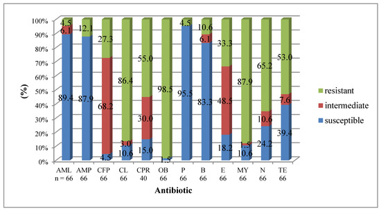
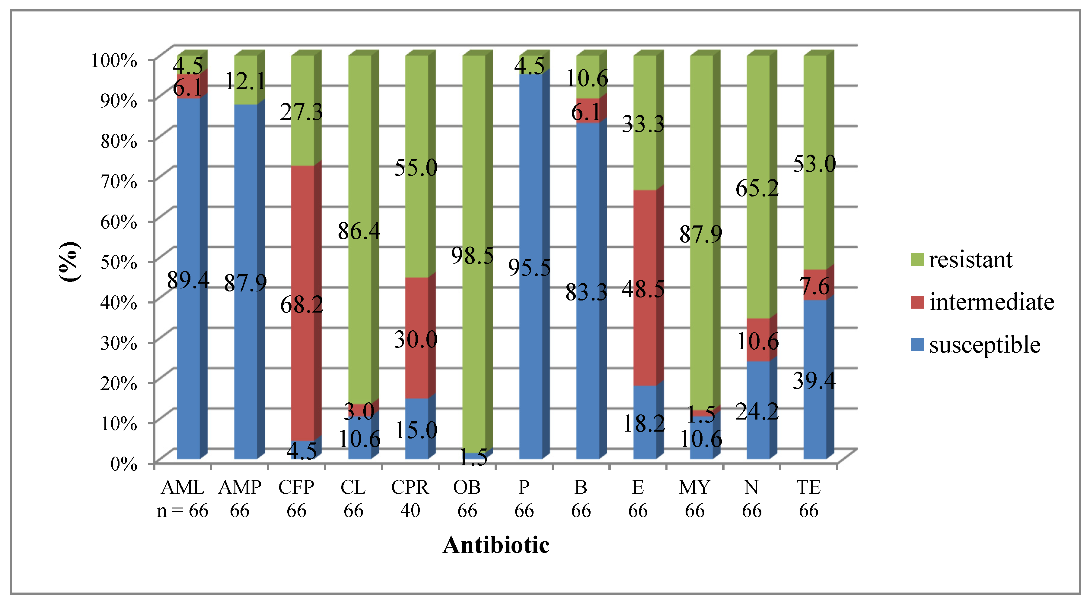
The results of the tests on the susceptibility of enterococcal isolates to antibiotics are presented in Figure 1. The isolates were predominantly resistant to cloxacillin (98.5%), lincomycin (87.9%), cefalexin (86.4%), neomycin (65.2%), cefapirin (55%) and tetracycline (53%). Most Enterococcus isolates were susceptible to penicillin (95.5%), amoxicillin (89.4%), ampicillin (87.9%), and bacitracin (83.3%). Almost 70% of enterococci showed intermediate susceptibility to cefoperazone, approximately 50% to erythromycin, and 30% to cefapirin.
Figure 1.
Antimicrobial susceptibility results for enterococci: AML—amoxicillin (25 µg), AMP—ampicillin (10 µg), CFP—cefoperazone (30 µg), CL—cephalexin (30 µg), CPR—cefapirin (30 µg), OB—cloxacillin (5 µg), P—penicillin G (10 u), B—bacitracin (10 u), E—erythromycin (15 µg), MY—lincomycin (15 µg), N—neomycin (30 µg), TE—tetracycline (30 µg).
Table 8 shows the prevalence of resistance profiles among enterococci.
Table 8.
Antimicrobial resistance (AMR) profiles among Enterococcus spp. isolates (n = 66).
Of the 80 Enterococcus spp. isolates obtained from the mammary glands of cows, 49 (61.3%) were found to carry at least one of the tested tetracycline, erythromycin and vancomycin resistance genes (Table 9). The most prevalent gene was tet(M) (n = 41 isolates), followed by erm(B) (n = 24), tet(L) (n = 20), tet(O) (n = 6), vanC2/3 (n = 2) and vanC1 (n = 1). The majority of isolates carrying the erm(B) gene (87.5%) belonged to the E. faecalis species (Table 10, Table 11 and Table 12). The tet(K), erm(A), erm(C), mef(A), vanA and vanB genes were not detected in the tested isolates. Twenty-eight enterococcal isolates (35%) harboured two or more resistance genes (Table 9).
Table 9.
Occurrence of resistance genes and their combinations in all enterococci examined in the study (n = 80).
Table 10.
Occurrence of resistance genes and their combinations in E. faecalis isolates (n = 52).
Table 11.
Occurrence of resistance genes and their combinations in E. faecium isolates (n = 11).
Table 12.
Occurrence of resistance genes and their combinations in Enterococcus spp. isolates other than E. faecalis and E. faecium (n = 17).
Most Enterococcus isolates carrying the macrolide antibiotic resistance gene erm(B) (95.83%; 23/24) were found to have at least one tetracycline resistance gene (Table 9).
The number of isolates exhibiting phenotypic resistance did not exactly correspond to the number of isolates harbouring the selected resistance genes (Table 13). The erm(B) gene was detected in 90.9% (20/22) of enterococcal isolates that were phenotypically resistant to erythromycin, as well as in the only isolate that showed intermediate resistance to this antibiotic (Table 13). The other macrolide resistance genes tested (erm(A), erm(C) and mef(A)) were not detected. The tet(M) gene was present in 94.3% (33/35) of enterococcal isolates that were phenotypically resistant to tetracycline. In most of these isolates, the tet(M) gene was present either alone (45.7%) or in combination with the tet(L) gene (40.0%) (Table 13).
Table 13.
Prevalence of erythromycin and tetracycline resistance phenotypes and genotypes among Enterococcus spp. isolates (n = 80).
The MIC values for vancomycin were 4 µg/mL for one E. gallinarum isolate carrying the vanC1 gene and two E. casseliflavus isolates carrying the vanC2/3 gene (vancomycin-susceptible isolates according to the CLSI guidelines) [43].
4. Discussion
4.1. The Incidence of Enterococcal Species Causing Mastitis in Cows
In recent years, interest in enterococci has grown. On the one hand, this is due to the widespread involvement of these opportunistic pathogens in causing infections in humans and animals and their increasing resistance to antibiotics used in human and veterinary medicine. On the other hand, it is due to the development of microbial identification techniques. Our previous study showed that enterococci were isolated from 2.8% of clinical and subclinical cases of mastitis in Polish herds [5]. Of the eighty isolates examined in the current study, 78.8% belonged to the two main species of enterococci: E. faecalis (65%) and E. faecium (13.8%). These two species were isolated at a similar frequency from the milk of cows with mastitis in South Korea (86.4% vs. 13.6%) [46]. In this study, 21.2% of the isolates belonged to other species of enterococci. Enterococcal species such as E. durans, E. gallinarum, E. casseliflavus, E. avium and E. hirae have occasionally been isolated from bovine milk and human infections [11,47]. Generally, E. faecalis is more prevalent in infections than E. faecium, which is due to its significantly higher prevalence in the environment and greater number of virulence factors [48]. Recently, an increase in E. faecium-related infections has been observed in human medicine, potentially due to its ability to acquire new resistance or virulence factors [49,50].
4.2. Virulence Factor Genes
The E. faecalis isolates examined in the study harboured multiple virulence genes (from two to six), whereas the only virulence gene detected in the E. faecium isolates was the efaAfm gene. Previous studies have revealed that some virulence genes (ace, asa1, esp and gelE) can occur in E. faecium isolates; however, E. faecalis isolates harboured significantly more virulence factors than E. faecium [46,51,52]. Interestingly, research with Enterococcus spp. isolated from artisanal dairy products has reported that non-faecalis/non-faecium enterococcal isolates carried at least three virulence genes, similar to E. faecalis and E. faecium [53]. In the present study, the absence of tested virulence genes in non-faecalis isolates may be due to the relatively small number of isolates compared to E. faecalis isolates.
The EfaA protein is an adhesin found in the serum of patients with endocarditis [54], which may facilitate the adhesion of bacterial cells to myocardial cells [55]. The presence of genes encoding the EfaA protein has been confirmed in various enterococcal species, including E. faecalis, E. faecium, E. durans and E. solitarius [54]. All E. faecalis and E. faecium isolates examined in our study were found to carry the efaAfs and efaAfm genes, respectively. These genes were also detected in all E. faecalis and E. faecium isolates from human infections, milk and dairy products [51,56] and in 94.6% of E. faecalis isolates from cows with mastitis [57]. These results may indicate that enterococci, which cause mastitis in cows, have the potential to cause disease in humans as well.
Our studies confirmed the observations of other authors regarding the frequent presence of the ace gene, which encodes the Ace adhesion protein, and the gelE gene [52,56,58,59], which is involved in gelatinase and biofilm production in E. faecalis [18]. In our studies, the ace (98.1%) and gelE (86.5%) genes, alongside the efaA gene, were the genes most frequently found in E. faecalis isolates from clinical and subclinical bovine mastitis, similarly to previous studies conducted on Turkish isolates [57].
In our study, neither the gelE nor the ace gene was detected in E. faecium isolates. However, some authors have found one gelE-positive E. faecium isolate in bovine mastitic milk (1/11; 9.1%) [46]. Other authors have reported that the gene was detected more frequently in E. faecalis than in E. faecium [59]. The E. faecalis Ace protein has been shown to be equivalent to the Acm protein (acm gene) in E. faecium, and an additional E. faecium Scm protein (scm gene) has been identified [55]. However, some authors have reported detecting the ace gene in two E. faecium isolates (2/50; 4%) from the milk of cows with mastitis [60].
The aggregating substance found in enterococci is an adhesin encoded by genes located on pheromone-dependent plasmids, including the asa1 gene [16]. In our study, the asa1 gene was present in most of the E. faecalis isolates tested (63.5%), a result consistent with those obtained from raw milk samples (67.1–100%) [46,56,61] and from the milk of cows with mastitis (71.4%) [46]. However, in contrast to our own studies, the asa1 gene was also detected in E. faecium isolates and other species (e.g., E. gallinarum and E. hirae) from bovine milk, including mastitic milk [20,46,60].
In addition to its role in adhesion, Esp is also believed to play a role in evading the host’s immune response, which is an important factor in disease development [62]. Although the esp gene is not essential for biofilm formation [17,18], esp-positive clinical E. faecalis isolates produce a greater quantity of biofilm than isolates lacking this gene [18]. In the present study, the esp gene was detected in 57.7% of E. faecalis isolates from the mammary glands of cows with mastitis. These results are comparable to those of previous studies [46,57,60]. In contrast, the gene was present in 4% of E. faecium isolates from clinical mastitis cases in China [60].
Cytolysin is a pore-forming exotoxin that affects (lyses) both eukaryotic and prokaryotic cells. Produced by haemolytic strains of E. faecalis, it is associated with high virulence and a higher death rate in animal and human infections [63]. Cytolysin production by enterococci (mainly E. faecalis) is a multistep process involving an operon composed of eight genes [16,64]. One of these genes is cylA, which is responsible for expressing protein A, an enzyme activator [15,16]. Some previous studies have demonstrated the presence of the cylA gene in 1.8% and 30% of E. faecalis isolates from clinical and subclinical cases of bovine mastitis [46,57]. In this study, the cylA gene was present in 17.3% of E. faecalis isolates and none of the cylA-positive isolates caused complete lysis of red blood cells (β-haemolysis). Other authors have also observed a discrepancy between phenotypic and genotypic expression of cytolysin [46,56,58]. This may be due to the fact that the genes encoding cytolysin are selectively expressed [65]. Furthermore, in the current study, we used agar supplemented with 5% sheep blood for the examination of haemolysin. However, certain strains of E. faecalis can produce haemolysin/cytolysin, which acts on human, rabbit and horse erythrocytes but not sheep ones [66]. This is associated with different levels of susceptibility of erythrocytes from various species to hemolysin-mediated lysis [65]. For these reasons, to demonstrate the full ability of bovine enterococci to lyse red blood cells, it is justified to determine haemolysis using an agar containing 5% horse blood.
The hylfm gene, which is associated with the production of hyaluronidase, is considered a potential virulence factor in clinical isolates of E. faecium. It is more prevalent in vancomycin-resistant strains than in vancomycin-susceptible strains [67], likely due to its location on large plasmids that may carry glycopeptide resistance genes [68]. In this study, the hylfm gene was not detected in the enterococci, consistent with previous studies of isolates from cow’s milk [20,52,60] and food [69].
4.3. Antimicrobial Resistance of Enterococci Isolated from Bovine Mastitis
The steady increase in bacterial drug resistance observed over the past two decades has prompted various organisations, including the WHO and the European Commission, to take action to reduce and monitor antibiotic use. While most EU countries have experienced a decline in antimicrobial sales, Poland ranks second in terms of total sales of preparations used for food-producing animals and average consumption of antimicrobial substances for these animals (mg/Population Correction Unit; mg/PCU) [70,71]. In recent years, the highest sales were for penicillins, tetracyclines and macrolides [71], which are antibiotics that are often used in preparations to treat bovine mastitis caused by Streptococcus species [23,72]. The resistance to macrolides, lincosamides and tetracyclines observed in our study may be due to the intensive and long-term use of antibiotics for treating mastitis during the lactation and dry periods, which has contributed to the selection of resistant strains [26].
The strains were tested for their susceptibility to the antibiotics commonly used to treat mastitis in cows, which is caused by Gram-positive, catalase-negative cocci. As expected, vast majority of Enterococcus spp. isolates were resistant to certain β-lactam antibiotics, such as cloxacillin and cephalexin. A low percentage of the isolates were resistant to penicillin G, amoxicillin and ampicillin. Previous studies conducted in Poland, Finland, the USA, Brazil and China also reported low percentages of resistance to penicillin (0–5.05%) and ampicillin (0%) [4,20,60,73,74,75]. Other studies have reported higher percentages of penicillin-resistant enterococci, at 62.3% in China [76] and 64.8% in Korea [77] compared with those isolated from Polish dairy cows. In our own studies, a significant proportion of enterococcal isolates were found to be resistant to lincomycin (87.9%), which is consistent with the results of previous research [4,76], and to neomycin (65.2%). However, in a Korean study, the percentage of isolates resistant to neomycin was lower (1.2%) [46]. The very high resistance rates to cloxacillin, cephalexin and lincomycin are due to the intrinsic resistance of enterococci to these antibiotics. Enterococcus spp. exhibit intrinsic resistance to low concentrations of β-lactams (mainly cephalosporins), aminoglycosides and clindamycin (lincosamide), as well as acquired resistance to high concentrations of clindamycin and aminoglycosides [32].
In our study, over half of the enterococcal isolates (53%) were resistant to tetracycline, indicating a lower resistance rate than that reported by other authors in Poland (82%) [4]. Even lower percentages of tetracycline-resistant isolates were reported in the USA (22.5%) and Brazil (27.3%) [20,73].
Many genes conferring resistance to tetracyclines have been identified. In Gram-positive cocci, the most common are tet(M) and tet(O), which encode proteins that protect ribosomes from the action of tetracycline, and tet(K) and tet(L), which encode proteins that pump the antibiotic out of the bacterial cell [78]. Almost all tetracycline-resistant isolates carried the tet(M) gene (33/35; 94.3%). The second most frequently detected gene in tetracycline-resistant isolates was the tet(L) gene (45.7%). The tet(O) gene was detected in 8.6% of isolates. Similar results were obtained in studies of high-level erythromycin-resistant (HLER) E. faecalis isolated from bulk tank milk of dairy companies in Korea [61]. In our own studies, no tetracycline resistance associated with the presence of the tet(K) gene was found. Other authors reported that this gene was rare in enterococci [79]. However, a surprisingly high frequency of the tet(K) gene was reported in other studies, where the tet(K) gene was found in 97.2% and 25% of tetracycline-resistant E. faecalis isolates from subclinical and clinical mastitis cases, respectively [80,81]. The tet(K) gene is generally believed to be primarily found in Gram-positive bacteria belonging to the Staphylococcus genus [82]. The presence of this gene in enterococci could suggest that they have the ability to acquire resistance genes from other species of bacteria.
In our own studies, the prevalence of erythromycin resistance among enterococci was 33.3%, while the percentage of susceptible isolates was 18.2%. Studies conducted in Finland found that 19% of enterococcal isolates were resistant to this antibiotic [74]. In Korea, 57.1% of isolates were resistant and 22.86% were susceptible [77]. In China, resistance to erythromycin was very high, at 83.8% of isolates [60].
There are two mechanisms of erythromycin resistance in bacteria. The first involves ribosome modification by a methylase encoded by erm genes. The second mechanism involves a drug efflux pump, which is a hydrophobic membrane-bound protein encoded by the mef gene [83]. In this study, erm(B) was the only gene conferring erythromycin resistance among the isolates, with 90.9% of the erythromycin-resistant isolates carrying this gene. Two isolates did not contain any of the erm or mef(A) genes tested. Similar results were obtained by Jensen et al. [84], who demonstrated the presence of the erm(B) gene in most erythromycin-resistant enterococcal isolates from cattle, but not the erm(A) or erm(C) genes. In China, the majority of enterococcal isolates from clinical mastitis carried the erm(B) gene (95.5%), although erm(A)-positive (1%) and erm(C)-positive (7.1%) isolates were also detected [60]. Another study found that the erm(A) gene was not present in erythromycin-resistant E. faecalis isolates from the milk of cows with subclinical mastitis, which is consistent with our own findings [80].
Previous studies have shown that enterococcal isolates from cows with mastitis often exhibit greater acquired resistance to tetracycline and erythromycin than to other tested antimicrobials [46,60,80]. These resistance genes are frequently found on the same mobile genetic element, suggesting that tetracycline-resistant strains may play a role in the dissemination of erythromycin resistance [85]. In this study, almost all erm(B)-positive isolates carried at least one tet gene.
Our own study, as well as those of other authors [60,81,82], has revealed an inconsistency between the presence of resistance determinants and phenotypic antibiotic resistance. A variety of mechanisms cause antibiotic resistance, including the presence of specific genes or changes in bacterial metabolism [81]. In this study, several isolates were phenotypically resistant to erythromycin or tetracycline, but genotypically susceptible. This suggests that these isolates may carry antibiotic resistance genes that were not tested for. An example of such a gene is the tetS gene, which confers resistance to tetracycline [60,81]. In this study, two isolates that were phenotypically susceptible carried tetracycline-resistance genes. Previous studies, including those involving WGS, have also detected tet or erm genes in isolates that are phenotypically susceptible; however, the authors demonstrated that it was caused by incorrect phenotypic antibiotic testing results or the presence of silenced antimicrobial resistance genes [86,87]. The absence of antimicrobial resistance gene expression may be due to mutation, a non-functional promoter, integrons or negative transcriptional regulators [88], which were not examined in this study. Another reason for the discrepancy between the presence of resistance genes and phenotypic resistance to antibiotics may be high detection limits (μg/mL). The detection limits determined for most antibiotics used in the treatment of mastitis in cows showed higher values for the disc diffusion method compared to other antibiotic detection methods, such as Delvotest SP and Penzym S 100 (e.g., oxacillin, ampicillin, cefotaxime, cefoperazone, cefquinome and ceftazidime) or Delvotest SP (e.g., chlortetracycline, erythromycin and bacitracin) [89].
Administering sub-therapeutic doses of antibiotics to farm animals through their feed has contributed to the development and spread of multidrug resistance. The glycopeptide antibiotic avoparcin was used in dairy cows in European Union countries until 1997. This resulted in the emergence of Enterococcus spp. strains that were resistant to vancomycin, an important drug used to treat hospital-acquired infections caused by enterococci and a last-resort antibiotic for treating infections caused by methicillin-resistant S. aureus [90]. Ten gene clusters conferring vancomycin resistance have been identified in enterococci. The VanA and VanB clusters are the most prevalent among those isolated from hospital-acquired infections, particularly in E. faecium. These clusters are typically transferred via mobile genetic elements that can integrate into the chromosomes or plasmids [50] of vancomycin-susceptible enterococci, other pathogenic bacteria (e.g., Staphylococcus aureus) or non-pathogenic microorganisms that inhabit the gastrointestinal tract of humans and animals [91]. In our study, the genes responsible for acquired resistance to high vancomycin concentrations (vanA and vanB) were not detected. However, the absence of these genes in the examined isolates does not exclude their presence in other Polish herds or at different times. An E. faecalis isolate carrying the vanB gene was found in cow’s milk in Bangladesh [81]. In our studies, three isolates exhibited the VanC phenotype, characterised by constitutive or induced resistance to low concentrations of vancomycin (MIC = 4–16 µg/mL). This phenotype occurs naturally in most E. gallinarum (vanC1 gene) and E. casseliflavus (vanC2/3) isolates [35,44]. These vanC-positive species remain epidemiologically relevant reservoirs of glycopeptide tolerance.
This study has some limitations. Although efforts were made to avoid duplicating isolates from the same herds, it should be noted that the number of non-faecalis isolates examined in the study was fairly limited. Therefore, the small sample size limits the statistical power available for robust comparisons between species and detailed analysis of less common enterococcal species. The same situation also applies to the relatively small number of Enterococcus spp. isolates from subclinical mastitis cases (n = 7) compared to the number of clinical cases (n = 45). Therefore, conclusions regarding species comparisons and mastitis type associations are exploratory rather than definitive. Furthermore, the identification of the isolates was based on biochemical and serological methods, as well as standard PCR techniques. The use of more advanced methods based on 16S rDNA sequencing, which remains the gold standard in microbial identification, or the MALDI-TOF MS technique, would also enable the identification of species other than E. faecalis and E. faecium. A further limitation of the study is that no investigation was made of the formation of biofilms. The ability of Enterococcus spp. to form biofilms is an important virulence factor. At least two biofilm formation-associated virulence genes (esp and gelE) were found in the majority of E. faecalis isolates tested. Therefore, it would be worthwhile conducting such a study in the future. Phenotypic confirmation of the ability to form biofilms would significantly strengthen the virulence assessment.
5. Conclusions
This study describes the virulence and antibiotic resistance gene profiles of enterococci isolated from clinical and subclinical cases of bovine mastitis in Poland. Our study revealed that E. faecalis showed a higher virulence gene burden. We were unable to find a correlation between the presence of virulence genes and the incidence of subclinical or clinical mastitis in cows. Antibiotic-resistant isolates of enterococci were found in the milk of cows with mastitis in Poland. These isolates may serve as a potential reservoir of resistance genes (erm, tet) for other bacteria colonising the human gastrointestinal tract. However, this theoretical risk is supported by molecular evidence rather than demonstrated transmission. None of the Enterococcus spp. examined in this study exhibited the vanA or vanB genotypes that correspond to acquired resistance to high concentrations of vancomycin. Nevertheless, the absence of these genes in the tested isolates does not exclude their presence in other Polish herds or at different times. Due to the resistance of enterococci, the decision on treatment should be based on the results of a microbiological examination and an antibiogram. Enterococci exhibit natural resistance to bacitracin, cloxacillin, cephalosporins and lincomycin. Antibiotics from the penicillin group (e.g., penicillin, amoxicillin, ampicillin) are often effective, unless confirmed by an antibiogram. Therefore, it is important to accurately identify Gram-positive, catalase-negative cocci isolated from the mammary glands of cows and to monitor their antibiotic susceptibility in different countries, including Poland. Comprehensive monitoring of antimicrobials used in dairy production is required to address the resistance of enterococci to antibiotics.
Author Contributions
Conceptualization, E.Z., M.T., J.G. and H.L.; methodology, E.Z. and H.L.; software, E.Z.; validation, E.Z.; formal analysis, E.Z.; investigation, E.Z. and H.L.; resources, E.Z. and H.L.; data curation, E.Z.; writing—original draft preparation, E.Z.; writing—review and editing, M.T., J.G. and H.L.; visualization, E.Z.; supervision, H.L.; project administration, E.Z.; funding acquisition, E.Z., M.T., J.G. and H.L. All authors have read and agreed to the published version of the manuscript.
Funding
This research was funded by Kazimierz Wielki University. And the APC was funded by Kazimierz Wielki University.
Institutional Review Board Statement
We declare that the milk samples were taken from cows during veterinary care of the herd. The samples were taken as part of routine examination of cows in accordance with the standard procedure for collecting milk samples from animals’ udders.
Informed Consent Statement
Not applicable.
Data Availability Statement
The datasets presented in the current study are included in the article. Further inquiries can be directed to the corresponding author.
Acknowledgments
The collection of bacterial isolates examined in this study was partially investigated at the former Department of Pathophysiology of Reproduction and Mammary Gland at the National Veterinary Research Institute in Pulawy, in the Bydgoszcz Division, where the authors worked until 2014. We would like to thank RADOMIR GRACZYK for his support with statistical analyses.
Conflicts of Interest
Professor HENRYKA LASSA is affiliated with the company ‘Badanie Mleka Henryka Lassa’ (‘Henryka Lassa Milk Testing’). It is a private sole proprietorship. The remaining authors declare that the research was conducted in the absence of any commercial or financial relationships that could be construed as a potential conflict of interest.
Abbreviations
The following abbreviations are used in this manuscript:
| AML | Amoxicillin |
| AMP | Ampicillin |
| AMR | Antimicrobial resistance |
| B | Bacitracin |
| CL | Cephalexin |
| CFP | Cefoperazone |
| E | Erythromycin |
| OB | Cloxacillin |
| MY | Lincomycin |
| N | Neomycin |
| P | Penicillin G |
| TE | Tetracycline |
| CPR | Cefapirin |
References
- Olde Riekerink, R.G.M.; Barkema, H.W.; Kelton, D.F.; Scholl, D.T. Incidence Rate of Clinical Mastitis on Canadian Dairy Farms. J. Dairy Sci. 2008, 91, 1366–1377. [Google Scholar] [CrossRef] [Scilit]
- Petersson-Wolfe, C.S.; Wolf, S.L.; Hogan, J.S. Experimental Challenge of Bovine Mammary Glands with Enterococcus Faecium during Early and Late Lactation. J. Dairy Sci. 2009, 92, 3158–3164. [Google Scholar] [CrossRef] [Scilit]
- Nam, H.M.; Kim, J.M.; Lim, S.K.; Jang, K.C.; Jung, S.C. Infectious Aetiologies of Mastitis on Korean Dairy Farms during 2008. Res. Vet. Sci. 2010, 88, 372–374. [Google Scholar] [CrossRef] [Scilit] [PubMed]
- Różańska, H.; Lewtak-Piłat, A.; Kubajka, M.; Weiner, M. Occurrence of Enterococci in Mastitic Cow’s Milk and Their Antimicrobial Resistance. J. Vet. Res. 2019, 63, 93–97. [Google Scholar] [CrossRef] [Scilit] [PubMed]
- Krukowski, H.; Lassa, H.; Zastempowska, E.; Smulski, S.; Bis-Wencel, H. Etiological Agents of Bovine Mastitis in Poland. Med. Weter. 2020, 76, 221–225. [Google Scholar] [CrossRef] [Scilit]
- Di Domenico, E.G.; Toma, L.; Prignano, G.; Pelagalli, L.; Police, A.; Cavallotti, C.; Torelli, R.; Sanguinetti, M.; Ensoli, F. Misidentification of Streptococcus uberis as a Human Pathogen: A Case Report and Literature Review. Int. J. Infect. Dis. 2015, 33, 79–81. [Google Scholar] [CrossRef] [Scilit]
- Kajdanek, A.; Kluska, M.; Matusiak, R.; Kazimierczak, J.; Dastych, J. A Rapid and Inexpensive PCR Test for Mastitis Diagnosis Based on NGS Data. Pathogens 2024, 13, 423. [Google Scholar] [CrossRef] [Scilit]
- Cameron, M.; Saab, M.; Heider, L.; McClure, J.T.; Rodriguez-Lecompte, J.C.; Sanchez, J. Antimicrobial Susceptibility Patterns of Environmental Streptococci Recovered from Bovine Milk Samples in the Maritime Provinces of Canada. Front. Vet. Sci. 2016, 3, 79. [Google Scholar] [CrossRef] [Scilit]
- Sorge, U.S.; Huber-Schlenstedt, R.; Schierling, K. In vitro antimicrobial resistance profiles of Streptococcus uberis, Lactococcus spp., and Enterococcus spp. from quarter milk samples of cows between 2015 and 2019 in Southern Germany. J. Dairy Sci. 2021, 104, 5998–6012. [Google Scholar] [CrossRef] [Scilit]
- Jahan, N.A.; Godden, S.M.; Royster, E.; Schoenfuss, T.C.; Gebhart, C.; Timmerman, J.; Fink, R.C. Evaluation of the matrix-assisted laser desorption ionization time of flight mass spectrometry (MALDI-TOF MS) system in the detection of mastitis pathogens from bovine milk samples. J. Microbiol. Methods 2021, 182, 106168. [Google Scholar] [CrossRef] [Scilit]
- Kurban, D.; Roy, J.-P.; Kabera, F.; Fréchette, A.; Um, M.M.; Albaaj, A.; Rowe, S.; Godden, S.; Adkins, P.R.F.; Middleton, J.R.; et al. Diagnosing Intramammary Infection: Meta-Analysis and Mapping Review on Frequency and Udder Health Relevance of Microorganism Species Isolated from Bovine Milk Samples. Animals 2022, 12, 3288. [Google Scholar] [CrossRef] [Scilit]
- Simões, J.; Branco, M.; Andrade, J.; Müller, A. Phenotypic antimicrobial susceptibility of environmental bacteria from mastitic milk of pastured dairy cows of S. Miguel (Azores). Trop. Anim. Health Prod. 2020, 52, 407–414. [Google Scholar] [CrossRef] [Scilit]
- de Oliveira, R.P.; Aragão, B.B.; de Melo, R.P.B.; da Silva, D.M.S.; de Carvalho, R.G.; Juliano, M.A.; Farias, M.P.O.; de Lira, N.S.C.; Mota, R.A. Bovine mastitis in northeastern Brazil: Occurrence of emergent bacteria and their phenotypic and genotypic profile of antimicrobial resistance. Comp. Immunol. Microbiol. Infect. Dis. 2022, 85, 101802. [Google Scholar] [CrossRef] [Scilit] [PubMed]
- Santos, P.R.; Kraus, R.B.; Ladeira, S.L.; Pereira, G.M.; Cunha, K.F.; Palhares, K.E.; Silva, A.C.A.; Dors, G.C.; Lima, H.G.; Cereser, N.D.; et al. Resistance profile and biofilm production of Enterococcus spp., Staphylococcus sp., and Streptococcus spp. from dairy farms in southern Brazil. Braz. J. Microbiol. 2023, 54, 1217–1229. [Google Scholar] [CrossRef] [Scilit]
- Vankerckhoven, V.; Van Autgaerden, T.; Vael, C.; Lammens, C.; Chapelle, S.; Rossi, R.; Jabes, D.; Goossens, H. Development of a Multiplex PCR for the Detection of Asa1, gelE, cylA, Esp, and Hyl Genes in Enterococci and Survey for Virulence Determinants among European Hospital Isolates of Enterococcus Faecium. J. Clin. Microbiol. 2004, 42, 4473–4479. [Google Scholar] [CrossRef] [Scilit]
- Piekarska, K. Enterokoki—Czynniki wirulencji i chorobotwórczość. Adv. Microbiol. 2006, 45, 195–207. [Google Scholar]
- Elhadidy, M.; Elsayyad, A. Uncommitted Role of Enterococcal Surface Protein, Esp, and Origin of Isolates on Biofilm Production by Enterococcus Faecalis Isolated from Bovine Mastitis. J. Microbiol. Immunol. Infect. 2013, 46, 80–84. [Google Scholar] [CrossRef] [Scilit] [PubMed]
- Mohamed, J.A.; Huang, D.B. Biofilm formation by enterococci. J. Med. Microbiol. 2007, 56, 1581–1588. [Google Scholar] [CrossRef] [Scilit]
- Sawant, A.A.; Sordillo, L.M.; Jayarao, B.M. A Survey on Antibiotic Usage in Dairy Herds in Pennsylvania. J. Dairy Sci. 2005, 88, 2991–2999. [Google Scholar] [CrossRef] [Scilit]
- Paschoalini, B.R.; Nuñez, K.V.M.; Maffei, J.T.; Langoni, H.; Guimarães, F.F.; Gebara, C.; Freitas, N.E.; Dos Santos, M.V.; Fidelis, C.E.; Kappes, R.; et al. The Emergence of Antimicrobial Resistance and Virulence Characteristics in Enterococcus Species Isolated from Bovine Milk. Antibiotics 2023, 12, 1243. [Google Scholar] [CrossRef] [Scilit]
- Ruegg, P.L. Making Economically Efficient Treatment Decisions for Clinical Mastitis. Vet. Clin. N. Am. Food Anim. Pract. 2025, 41, 181–198. [Google Scholar] [CrossRef] [Scilit]
- Schmerold, I.; van Geijlswijk, I.; Gehring, R. European regulations on the use of antibiotics in veterinary medicine. Eur. J. Pharm. Sci. 2023, 189, 106473. [Google Scholar] [CrossRef] [Scilit]
- Oliver, S.P.; Murinda, S.E. Antimicrobial Resistance of Mastitis Pathogens. Vet. Clin. N. Am. Food Anim. Pract. 2012, 28, 165–185. [Google Scholar] [CrossRef] [Scilit]
- Duarte, R.S.; Miranda, O.P.; Bellei, B.C.; Brito, M.A.V.P.; Teixeira, L.M. Phenotypic and Molecular Characteristics of Streptococcus agalactiae Isolates Recovered from Milk of Dairy Cows in Brazil. J. Clin. Microbiol. 2004, 42, 4214–4222. [Google Scholar] [CrossRef] [Scilit]
- Thomas, V.; De Jong, A.; Moyaert, H.; Simjee, S.; El Garch, F.; Morrissey, I.; Marion, H.; Vallé, M. Antimicrobial Susceptibility Monitoring of Mastitis Pathogens Isolated from Acute Cases of Clinical Mastitis in Dairy Cows across Europe: VetPath Results. Int. J. Antimicrob. Agents 2015, 46, 13–20. [Google Scholar] [CrossRef] [Scilit] [PubMed]
- Guérin-Faublée, V.; Tardy, F.; Bouveron, C.; Carret, G. Antimicrobial Susceptibility of Streptococcus Species Isolated from Clinical Mastitis in Dairy Cows. Int. J. Antimicrob. Agents 2002, 19, 219–226. [Google Scholar] [CrossRef] [Scilit] [PubMed]
- Hendriksen, R.S.; Mevius, D.J.; Schroeter, A.; Teale, C.; Meunier, D.; Butaye, P.; Franco, A.; Utinane, A.; Amado, A.; Moreno, M.; et al. Prevalence of Antimicrobial Resistance among Bacterial Pathogens Isolated from Cattle in Different European Countries: 2002–2004. Acta Vet. Scand. 2008, 50, 28. [Google Scholar] [CrossRef] [Scilit]
- Schmitt-van De Leemput, E.; Zadoks, R.N. Genotypic and Phenotypic Detection of Macrolide and Lincosamide Resistance in Streptococcus uberis. J. Dairy Sci. 2007, 90, 5089–5096. [Google Scholar] [CrossRef] [Scilit]
- White, D.G.; McDermott, P.F. Emergence and Transfer of Antibacterial Resistance. J. Dairy Sci. 2001, 84, E151–E155. [Google Scholar] [CrossRef] [Scilit]
- Tenhagen, B.-A.; Köster, G.; Wallmann, J.; Heuwieser, W. Prevalence of Mastitis Pathogens and Their Resistance Against Antimicrobial Agents in Dairy Cows in Brandenburg, Germany. J. Dairy Sci. 2006, 89, 2542–2551. [Google Scholar] [CrossRef] [Scilit]
- Massella, E.; Russo, S.; Filippi, A.; Garbarino, C.A.; Ricchi, M.; Bassi, P.; Toschi, E.; Torreggiani, C.; Pupillo, G.; Rugna, G.; et al. Does Bovine Raw Milk Represent a Potential Risk for Vancomycin-Resistant Enterococci (VRE) Transmission to Humans? Antibiotics 2025, 14, 814. [Google Scholar] [CrossRef] [Scilit]
- Murray, B.E. The Life and Times of the Enterococcus. Clin. Microbiol. Rev. 1990, 3, 46–65. [Google Scholar] [CrossRef]
- Calderaro, A.; Chezzi, C. MALDI-TOF MS: A Reliable Tool in the Real Life of the Clinical Microbiology Laboratory. Microorganisms 2024, 12, 322. [Google Scholar] [CrossRef] [Scilit]
- Zastempowska, E.; Twarużek, M.; Grajewski, J.; Lassa, H. Virulence Factor Genes and Cytotoxicity of Streptococcus agalactiae Isolated from Bovine Mastitis in Poland. Microbiol. Spectr. 2022, 10, e02224-21. [Google Scholar] [CrossRef] [Scilit]
- Ke, D.; Picard, F.J.; Martineau, F.; Ménard, C.; Roy, P.H.; Ouellette, M.; Bergeron, M.G. Development of a PCR Assay for Rapid Detection of Enterococci. J. Clin. Microbiol. 1999, 37, 3497–3503. [Google Scholar] [CrossRef] [Scilit]
- Dutka-Malen, S.; Evers, S.; Courvalin, P. Detection of Glycopeptide Resistance Genotypes and Identification to the Species Level of Clinically Relevant Enterococci by PCR. J. Clin. Microbiol. 1995, 33, 24–27. [Google Scholar] [CrossRef] [Scilit] [PubMed]
- Yean, C.Y.; Yin, L.S.; Lalitha, P.; Ravichandran, M. A Nanoplex PCR Assay for the Rapid Detection of Vancomycin and Bifunctional Aminoglycoside Resistance Genes in Enterococcus Species. BMC Microbiol. 2007, 7, 112. [Google Scholar] [CrossRef] [Scilit] [PubMed]
- Méndez-Álvarez, S.; Pérez-Hernández, X.; Claverie-Martín, F. Glycopeptide resistance in enterococci. Int. Microbiol. 2000, 3, 71–80. [Google Scholar]
- Billström, H.; Lund, B.; Sullivan, Å.; Nord, C.E. Virulence and Antimicrobial Resistance in Clinical Enterococcus faecium. Int. J. Antimicrob. Agents 2008, 32, 374–377. [Google Scholar] [CrossRef] [Scilit] [PubMed]
- Sutcliffe, J.; Grebe, T.; Tait-Kamradt, A.; Wondrack, L. Detection of Erythromycin-Resistant Determinants by PCR. Antimicrob. Agents Chemother. 1996, 40, 2562–2566. [Google Scholar] [CrossRef] [Scilit]
- Jeric, P.E.; Azpiroz, A.; Lopardo, H.; Centrón, D. Survey of molecular determinants in Gram-positive cocci isolated from hospital settings in Argentina. J. Infect. Dev. Ctries. 2007, 1, 275–283. [Google Scholar] [CrossRef] [Scilit] [PubMed]
- Strommenger, B.; Kettlitz, C.; Werner, G.; Witte, W. Multiplex PCR Assay for Simultaneous Detection of Nine Clinically Relevant Antibiotic Resistance Genes in Staphylococcus aureus. J. Clin. Microbiol. 2003, 41, 4089–4094. [Google Scholar] [CrossRef] [Scilit]
- CLSI (Clinical Laboratory Standards Institute). Performance Standards for Antimicrobial Disc and Dilution Susceptibility Test for Bacteria Isolated from Animals, VET01S, 3rd ed.; Clinical and Laboratory Standards Institute: Wayne, PA, USA, 2015. [Google Scholar]
- Werner, G.; Coque, T.M.; Hammerum, A.M.; Hope, R.; Hryniewicz, W.; Johnson, A.; Klare, I.; Kristinsson, K.G.; Leclercq, R.; Lester, C.H.; et al. Emergence and spread of vancomycin resistance among enterococci in Europe. Euro Surveill. 2008, 13, pii 19046. [Google Scholar] [CrossRef] [Scilit]
- Amuasi, G.R.; Dsani, E.; Owusu-Nyantakyi, C.; Owusu, F.A.; Mohktar, Q.; Nilsson, P.; Adu, B.; Hendriksen, R.S.; Egyir, B. Enterococcus species: Insights into antimicrobial resistance and whole-genome features of isolates recovered from livestock and raw meat in Ghana. Front. Microbiol. 2023, 14, 1254896. [Google Scholar] [CrossRef] [Scilit]
- Kim, H.-J.; Youn, H.-Y.; Kang, H.-J.; Moon, J.-S.; Jang, Y.-S.; Song, K.-Y.; Seo, K.-H. Prevalence and Virulence Characteristics of Enterococcus Faecalis and Enterococcus Faecium in Bovine Mastitis Milk Compared to Bovine Normal Raw Milk in South Korea. Animals 2022, 12, 1407. [Google Scholar] [CrossRef] [Scilit]
- Torres, C.; Alonso, C.A.; Ruiz-Ripa, L.; León-Sampedro, R.; Del Campo, R.; Coque, T.M. Antimicrobial Resistance in Enterococcus spp. of Animal Origin. Microbiol. Spectr. 2018, 6, 1–41. [Google Scholar] [CrossRef] [Scilit]
- Gaspar, F.; Teixeira, N.; Rigottier-Gois, L.; Marujo, P.; Nielsen-LeRoux, C.; Crespo, M.T.B.; Lopes, M.D.F.S.; Serror, P. Virulence of Enterococcus Faecalis Dairy Strains in an Insect Model: The Role of fsrB and gelE. Microbiology 2009, 155, 3564–3571. [Google Scholar] [CrossRef] [Scilit]
- Lester, C.H.; Sandvang, D.; Olsen, S.S.; Schonheyder, H.C.; Jarlov, J.O.; Bangsborg, J.; Hansen, D.S.; Jensen, T.G.; Frimodt-Moller, N.; Hammerum, A.M.; et al. Emergence of Ampicillin-Resistant Enterococcus faecium in Danish Hospitals. J. Antimicrob. Chemother. 2008, 62, 1203–1206. [Google Scholar] [CrossRef] [Scilit]
- McInnes, R.S.; Snaith, A.E.; Dunn, S.J.; Papangeli, M.; Hardy, K.J.; Hussain, A.; van Schaik, W. Integration of vanHAX downstream of a ribosomal RNA operon restores vancomycin resistance in a susceptible Enterococcus faecium strain. npj Antimicrob. Resist. 2024, 2, 2. [Google Scholar] [CrossRef] [Scilit]
- Cariolato, D.; Andrighetto, C.; Lombardi, A. Occurrence of Virulence Factors and Antibiotic Resistances in Enterococcus faecalis and Enterococcus faecium Collected from Dairy and Human Samples in North Italy. Food Control 2008, 19, 886–892. [Google Scholar] [CrossRef] [Scilit]
- Yoon, S.; Lee, Y.J. Molecular Characteristics of Enterococcus Faecalis and Enterococcus faecium from Bulk Tank Milk in Korea. Animals 2021, 11, 661. [Google Scholar] [CrossRef] [Scilit]
- Amidi-Fazli, N.; Hanifian, S. Biodiversity, Antibiotic Resistance and Virulence Traits of Enterococcus Species in Artisanal Dairy Products. Int. Dairy J. 2022, 129, 105287. [Google Scholar] [CrossRef] [Scilit]
- Semedo, T.; Santos, M.A.; Lopes, M.F.; Marques, J.J.F.; Crespo, M.T.; Tenreiro, R. Virulence Factors in Food, Clinical and Reference Enterococci: A Common Trait in the Genus? Syst. Appl. Microbiol. 2003, 26, 13–22. [Google Scholar] [CrossRef] [Scilit]
- Kraszewska, Z.; Skuczyńska, I.; Bogiel, T.; Gospodarek-Komkowska, E. Rola wybranych czynników wirulencji w zakażeniach wywoływanych przez szczepy Enterococcus spp. Adv. Microbiol. 2023, 62, 157–171. [Google Scholar]
- Anderson, A.C.; Jonas, D.; Huber, I.; Karygianni, L.; Wölber, J.; Hellwig, E.; Arweiler, N.; Vach, K.; Wittmer, A.; Al-Ahmad, A. Enterococcus faecalis from Food, Clinical Specimens, and Oral Sites: Prevalence of Virulence Factors in Association with Biofilm Formation. Front. Microbiol. 2016, 6, 1534. [Google Scholar] [CrossRef] [Scilit]
- Yildiz, O.; Turkyilmaz, S. Investigation of virulence genes of Enterococcus faecalis strains isolated from mastitic bovine milk. Isr. J. Vet. Med. 2015, 70, 15–21. [Google Scholar]
- Medeiros, A.W.; Pereira, R.I.; Oliveira, D.V.; Martins, P.D.; d’Azevedo, P.A.; Van Der Sand, S.; Frazzon, J.; Frazzon, A.P.G. Molecular Detection of Virulence Factors among Food and Clinical Enterococcus faecalis Strains in South Brazil. Braz. J. Microbiol. 2014, 45, 327–332. [Google Scholar] [CrossRef] [Scilit]
- Biswas, P.; Dey, S.; Sen, A.; Adhikari, L. Molecular Characterization of Virulence Genes in Vancomycin-Resistant and Vancomycin-Sensitive Enterococci. J. Glob. Infect. Dis. 2016, 8, 16. [Google Scholar] [CrossRef] [Scilit]
- Liu, J.; Liang, Z.; Zhongla, M.; Wang, H.; Sun, X.; Zheng, J.; Ding, X.; Yang, F. Prevalence and Molecular Characteristics of Enterococci Isolated from Clinical Bovine Mastitis Cases in Ningxia. Infect. Drug Resist. 2024, 17, 2121–2129. [Google Scholar] [CrossRef] [Scilit]
- Lee, Y.J.; Kim, K.; Lee, Y.J. Dissemination and characteristics of high-level erythromycin-resistant Enterococcus faecalis from bulk tank milk of dairy companies in Korea. Can. J. Vet. Res. 2023, 87, 51–58. [Google Scholar]
- Tsikrikonis, G.; Maniatis, A.N.; Labrou, M.; Ntokou, E.; Michail, G.; Daponte, A.; Stathopoulos, C.; Tsakris, A.; Pournaras, S. Differences in Biofilm Formation and Virulence Factors between Clinical and Fecal Enterococcal Isolates of Human and Animal Origin. Microb. Pathog. 2012, 52, 336–343. [Google Scholar] [CrossRef] [Scilit]
- Van Tyne, D.; Martin, M.J.; Gilmore, M.S. Structure, function, and biology of the Enterococcus faecalis cytolysin. Toxins 2013, 5, 895–911. [Google Scholar] [CrossRef] [Scilit]
- Gaspar, F.B.; Crespo, M.T.B.; Lopes, M.F.S. Proposal for a Reliable Enterococcal Cytolysin Production Assay Avoiding Apparent Incongruence between Phenotype and Genotype. J. Med. Microbiol. 2009, 58, 1122–1124. [Google Scholar] [CrossRef] [Scilit] [PubMed]
- Semedo, T.; Almeida Santos, M.; Martins, P.; Silva Lopes, M.F.; Figueiredo Marques, J.J.; Tenreiro, R.; Barreto Crespo, M.T. Comparative Study Using Type Strains and Clinical and Food Isolates To Examine Hemolytic Activity and Occurrence of the Cyl Operon in Enterococci. J. Clin. Microbiol. 2003, 41, 2569–2576. [Google Scholar] [CrossRef] [Scilit]
- Izumi, E.; Domingues Pires, P.; Bittencourt de Marques, E.; Suzart, S. Hemagglutinating and hemolytic activities of Enterococcus faecalis strains isolated from different human clinical sources. Res. Microbiol. 2005, 156, 583–587. [Google Scholar] [CrossRef] [Scilit]
- Arshadi, M.; Mahmoudi, M.; Motahar, M.S.; Soltani, S.; Pourmand, M.R. Virulence Determinants and Antimicrobial Resistance Patterns of Vancomycin-resistant Enterococcus faecium Isolated from Different Sources in Southwest Iran. Iran. J. Public Health 2018, 47, 264–272. [Google Scholar]
- Arias, C.A.; Panesso, D.; Singh, K.V.; Rice, L.B.; Murray, B.E. Cotransfer of Antibiotic Resistance Genes and a HylEfm-Containing Virulence Plasmid in Enterococcus faecium. Antimicrob. Agents Chemother. 2009, 53, 4240–4246. [Google Scholar] [CrossRef] [Scilit]
- Elmalı, M.; Can, H.Y. The Prevalence, Vancomycin Resistance and Virulence Gene Profiles of Enterococcus Species Recovered from Different Foods of Animal Origin. Vet. Arh. 2018, 88, 111–124. [Google Scholar] [CrossRef] [Scilit]
- Herman, K.; Nowaczek, A.; Urban-Chmiel, R.; Pyzik, E.; Dec, M. Ograniczanie Zużycia Antybiotyków w Leczeniu Zwierząt Na Terenie UE Na Podstawie Raportów Europejskiego Nadzoru Nad Weterynaryjnym Spożyciem Substancji Przeciwbakteryjnych. Med. Weter. 2024, 80, 477–483. [Google Scholar] [CrossRef] [Scilit]
- European Medicines Agency. Sales of Veterinary Antimicrobial Agents in 31 European Countries in 2022 Thirteenth Esvac Report; Publications Office of the European Union: Luxembourg, 2023. [CrossRef]
- Kromker, V.; Reinecke, F.; Paduch, J.H.; Grabowski, N. Bovine Streptococcus uberis Intramammary Infections and Mastitis. Clin. Microbiol. Open Access 2014, 3, 7. [Google Scholar] [CrossRef]
- Rossitto, P.V.; Ruiz, L.; Kikuchi, Y.; Glenn, K.; Luiz, K.; Watts, J.L.; Cullor, J.S. Antibiotic Susceptibility Patterns for Environmental Streptococci Isolated from Bovine Mastitis in Central California Dairies. J. Dairy Sci. 2002, 85, 132–138. [Google Scholar] [CrossRef] [Scilit]
- Pitkälä, A.; Haveri, M.; Pyörälä, S.; Myllys, V.; Honkanen-Buzalski, T. Bovine Mastitis in Finland 2001—Prevalence, Distribution of Bacteria, and Antimicrobial Resistance. J. Dairy Sci. 2004, 87, 2433–2441. [Google Scholar] [CrossRef] [Scilit]
- Kolar, Q.K.; Goncalves, J.L.; Erskine, R.J.; Ruegg, P.L. Comparison of Minimum Inhibitory Concentrations of Selected Antimicrobials for Non-Aureus Staphylococci, Enterococci, Lactococci, and Streptococci Isolated from Milk Samples of Cows with Clinical Mastitis. Antibiotics 2024, 13, 91. [Google Scholar] [CrossRef] [Scilit]
- Gao, X.; Fan, C.; Zhang, Z.; Li, S.; Xu, C.; Zhao, Y.; Han, L.; Zhang, D.; Liu, M. Enterococcal Isolates from Bovine Subclinical and Clinical Mastitis: Antimicrobial Resistance and Integron-Gene Cassette Distribution. Microb. Pathog. 2019, 129, 82–87. [Google Scholar] [CrossRef] [Scilit] [PubMed]
- Nam, H.M.; Lim, S.K.; Moon, J.S.; Kang, H.M.; Kim, J.M.; Jang, K.C.; Kim, J.M.; Kang, M.I.; Joo, Y.S.; Jung, S.C. Antimicrobial Resistance of Enterococci Isolated from Mastitic Bovine Milk Samples in Korea. Zoonoses Public Health 2010, 57, e59–e64. [Google Scholar] [CrossRef] [Scilit]
- Chopra, I.; Roberts, M. Tetracycline Antibiotics: Mode of Action, Applications, Molecular Biology, and Epidemiology of Bacterial Resistance. Microbiol. Mol. Biol. Rev. 2001, 65, 232–260. [Google Scholar] [CrossRef] [Scilit] [PubMed]
- Hammad, A.M.; Shimamoto, T.; Shimamoto, T. Genetic characterization of antibiotic resistance and virulence factors in Enterococcus spp. from Japanese retail ready-to-eat raw fish. Food Microbiol. 2014, 38, 62–66. [Google Scholar] [CrossRef] [Scilit]
- Yang, F.; Zhang, S.; Shang, X.; Wang, X.; Yan, Z.; Li, H.; Li, J. Short Communication: Antimicrobial Resistance and Virulence Genes of Enterococcus faecalis Isolated from Subclinical Bovine Mastitis Cases in China. J. Dairy Sci. 2019, 102, 140–144. [Google Scholar] [CrossRef] [Scilit]
- Bag, M.A.S.; Arif, M.; Riaz, S.; Khan, M.S.R.; Islam, M.S.; Punom, S.A.; Ali, M.W.; Begum, F.; Islam, M.S.; Rahman, M.T.; et al. Antimicrobial Resistance, Virulence Profiles, and Public Health Significance of Enterococcus faecalis Isolated from Clinical Mastitis of Cattle in Bangladesh. BioMed Res. Int. 2022, 2022, 8101866. [Google Scholar] [CrossRef] [Scilit]
- Ruegg, P.L.; Oliveira, L.; Jin, W.; Okwumabua, O. Phenotypic Antimicrobial Susceptibility and Occurrence of Selected Resistance Genes in Gram-Positive Mastitis Pathogens Isolated from Wisconsin Dairy Cows. J. Dairy Sci. 2015, 98, 4521–4534. [Google Scholar] [CrossRef] [Scilit]
- Domelier, A.-S.; Van Der Mee-Marquet, N.; Arnault, L.; Mereghetti, L.; Lanotte, P.; Rosenau, A.; Lartigue, M.-F.; Quentin, R. Molecular Characterization of Erythromycin-Resistant Streptococcus agalactiae Strains. J. Antimicrob. Chemother. 2008, 62, 1227–1233. [Google Scholar] [CrossRef] [Scilit]
- Jensen, L.B.; Frimodt-Møller, N.; Aarestrup, F.M. Presence of Erm Gene Classes in Gram-Positive Bacteria of Animal and Human Origin in Denmark. FEMS Microbiol. Lett. 1999, 170, 151–158. [Google Scholar] [CrossRef]
- Duarte, R.S.; Bellei, B.C.; Miranda, O.P.; Brito, M.A.V.P.; Teixeira, L.M. Distribution of Antimicrobial Resistance and Virulence-Related Genes among Brazilian Group B Streptococci Recovered from Bovine and Human Sources. Antimicrob. Agents Chemother. 2005, 49, 97–103. [Google Scholar] [CrossRef] [Scilit]
- Tyson, G.H.; Sabo, J.L.; Rice-Trujillo, C.; Hernandez, J.; McDermott, P.F. Whole-genome sequencing based characterization of antimicrobial resistance in Enterococcus. Pathog. Dis. 2018, 76, fty018. [Google Scholar] [CrossRef] [Scilit]
- Coll, F.; Gouliouris, T.; Blane, B.; Yeats, C.A.; Raven, K.E.; Ludden, C.; Khokhar, F.A.; Wilson, H.J.; Roberts, L.W.; Harrison, E.M.; et al. Antibiotic resistance determination using Enterococcus faecium whole-genome sequences: A diagnostic accuracy study using genotypic and phenotypic data. Lancet Microbe 2024, 5, e151–e163. [Google Scholar] [CrossRef] [Scilit]
- Shen, Z.; Zhang, C.Y.; Gull, T.; Zhang, S. Comparison of genotypic and phenotypic antimicrobial resistance profiles of Salmonella enterica isolates from poultry diagnostic specimens. J. Vet. Diagn. Investig. 2024, 36, 529–537. [Google Scholar] [CrossRef] [Scilit]
- Hozová, B.; Kratmüllerová, M. Assay of antibiotic detection limits in cow’s milk model samples and comparison of sensitivity of various detection systems (disk diffusion method, Delvotest SP and Penzym S 100). Czech J. Food Sci. 2001, 19, 125–131. [Google Scholar] [CrossRef] [Scilit]
- Van Tyne, D.; Gilmore, M.S. Friend Turned Foe: Evolution of Enterococcal Virulence and Antibiotic Resistance. Annu. Rev. Microbiol. 2014, 68, 337–356. [Google Scholar] [CrossRef] [Scilit] [PubMed]
- Pesavento, G.; Calonico, C.; Ducci, B.; Magnanini, A.; Lo Nostro, A. Prevalence and Antibiotic Resistance of Enterococcus Spp. Isolated from Retail Cheese, Ready-to-Eat Salads, Ham, and Raw Meat. Food Microbiol. 2014, 41, 1–7. [Google Scholar] [CrossRef] [Scilit]

Disclaimer/Publisher’s Note: The statements, opinions and data contained in all publications are solely those of the individual author(s) and contributor(s) and not of MDPI and/or the editor(s). MDPI and/or the editor(s) disclaim responsibility for any injury to people or property resulting from any ideas, methods, instructions or products referred to in the content. |
© 2026 by the authors. Licensee MDPI, Basel, Switzerland. This article is an open access article distributed under the terms and conditions of the Creative Commons Attribution (CC BY) license.