


Abstract
Johne’s disease is a chronic progressive enteritis in cattle caused by Mycobacterium avium subsp. paratuberculosis (MAP). It causes substantial economic losses and has been suggested to be related to human Crohn’s disease. This study investigated MAP exposure and molecular detection in large-scale dairy herds in Xinjiang, China. A total of 1817 milk and 988 fecal samples were collected from 30 herds located across ten regions. Anti-MAP-specific antibodies were measured in milk samples using a commercially available enzyme-linked immunosorbent assay (ELISA), while MAP DNA was assessed in fecal samples by quantitative PCR (qPCR). The apparent individual seroprevalence was 6.66% (121/1817) based on milk ELISA. At the herd level, 25 of the 30 herds (83.3%) examined by milk ELISA had at least one animal positive for MAP-specific antibodies. MAP DNA was detected in 2.53% (25/988) of fecal samples, and bacterial culture of the qPCR-positive samples yielded 12 MAP isolates, which were confirmed by F57-targeted qPCR and Ziehl–Neelsen staining. Age-related differences were observed according to the diagnostic method, with the highest qPCR positivity recorded in older animals. These findings provide evidence of widespread MAP exposure and molecular detection among large-scale dairy herds in Xinjiang, and support continued monitoring and improved control measures.
1. Introduction
Johne’s disease (JD), also known as paratuberculosis (PTB), is a chronic infectious enteric disease caused by Mycobacterium avium subsp. paratuberculosis (MAP) [1]. As a multifactorial disease influenced by host–pathogen interactions, it affects various domesticated and wild animal species globally [2,3]. JD has substantial economic consequences worldwide [4], largely due to reduced milk production and premature culling [5]. The annual losses in major dairy-producing countries are estimated at hundreds of millions of dollars [6,7].
MAP is transmitted primarily through the fecal–oral route [8,9]. This bacterial species presents a major challenge for dairy herds, not only because of the substantial production losses it causes [10,11], but also due to its potential, albeit debated, zoonotic link to Crohn’s disease in humans [12,13]. It can be directly transmitted from infected animals to susceptible animals through contaminated teats, milk, or colostrum [14,15,16,17]. Other sources of infection include environmental contamination or the introduction of infected animals into naive herds. Although MAP is an intracellular pathogen that replicates mainly within host cells, it can persist in the environment without active replication. Its ability to remain viable for months outside the host contributes to its continuous transmission risk [18,19,20]. Infected animals typically undergo a prolonged latent period, often extending for two years or more before fecal MAP shedding or the onset of clinical symptoms. This slow progression of MAP infection is often exacerbated by management practices that expose susceptible calves to the pathogen, making JD particularly difficult to control. Consequently, control strategies mainly rely on a combination of herd-level diagnostic testing and stringent biosecurity measures [11].
As global concern over JD has grown, numerous studies have assessed its prevalence. A comprehensive review reported that herd prevalence exceeded 20% in approximately half of the surveyed countries, with rates above 40% in some regions [20,21]. In Europe, between-herd seroprevalence has been reported to range from 38% to 68% [22]. A study in 2007 across 17 major dairy states in the U.S. found that 68.1% of herds had at least one animal testing positive for MAP [22,23].
Although China is among the world’s major cattle producers, comprehensive epidemiological data on bovine paratuberculosis remain limited, especially across different farming systems. Regional studies indicate that MAP is widespread but unevenly distributed across the country. For example, 11.7% (121/1038) of dairy cattle and 57.9% (11/19) of dairy herds were tested for seropositivity for MAP [24]. Similarly, 11.8% (433/3674) of cattle from Heilongjiang, Jilin, Liaoning, and Inner Mongolia provinces in northeastern China were serologically positive for MAP [25]. In Xinjiang, Liu et al. previously reported a herd seroprevalence of 50.0% and an individual prevalence of 4.8%, suggesting a substantial MAP burden in this region [26].
The dairy industry in Xinjiang, northwest China, has expanded considerably in recent decades. Nevertheless, farms in the region frequently report cases of intermittent diarrhea in dairy cows that are unresponsive to treatment and often progress to severe emaciation and death. These clinical symptoms highlight the considerable economic burden that JD imposes on regional development. In China, current diagnostic approaches for MAP rely primarily on indirect ELISAs for antibody detection in serum or milk [2], supplemented by bacterial culture and polymerase chain reaction (PCR). However, even with these regional data, integrated epidemiological studies combining indirect serology with direct molecular detection and bacterial culture remain scarce in China, particularly within large-scale dairy production systems with high biosecurity risks. While such systems enhance production efficiency, they may also favor MAP persistence and spread. Factors such as continuous animal contact, shared equipment, and routine management practices can promote fecal–oral transmission without rigorous control. Therefore, understanding MAP infection dynamics in these intensive settings is essential for developing effective and targeted control measures.
This study aimed to investigate MAP epidemiology among dairy herds in Xinjiang, northwest China, and to estimate the apparent prevalence at both the individual and herd levels within large-scale farming systems. An integrated diagnostic approach combining serological assays, qPCR, and bacterial culture was used to meet these objectives.
2. Materials and Methods
2.1. Study Design and Sampling
This cross-sectional study was conducted from July 2024 to October 2025 in large-scale dairy herds (n = 30, each with ≥1000 cows) located across 10 regions in Xinjiang, northwest China. Herds were selected by convenience sampling based on voluntary owner participation. The minimum sample size for each herd was calculated based on the lactating animal size, assuming an expected 10% prevalence, a 95% confidence level, and a 5% margin of error. Within each herd, lactating cows were selected during milking using systematic sampling. A random starting point was first determined within the milking sequence, after which every fifth cow was selected until the target sample size was reached.
A total of 2805 samples were collected. Of these, 988 lactating cows contributed both fecal and milk samples obtained concurrently, whereas an additional 829 cows contributed milk samples only, as fecal sampling was not obtained from these animals. Samples were collected from dairy farms across the following regions: Urumqi (257 milk samples and 201 fecal samples; 4 herds), Kuitun (130 milk samples and 90 fecal samples; 3 herds), Ili (208 milk samples and 78 fecal samples; 4 herds), Aral (89 milk samples and 89 fecal samples; 1 herd), Bayingolin (100 milk samples and 50 fecal samples; 1 herd), Khotan (80 milk samples and 80 fecal samples; 1 herd), Shihezi (94 milk samples and 56 fecal samples; 2 herds), Karamay (129 milk samples and 30 fecal samples; 2 herds), Changji (650 milk samples and 261 fecal samples; 11 herds) and Kashgar (80 milk samples and 53 fecal samples; 1 herd). All animals sampled were apparently clinically healthy and were at least two years old. Milk samples (3 mL) were placed in sterile tubes. Whey was then separated from the milk and stored at −80 °C until ELISA analysis.
Separately, fecal samples (approximately 10 g each) were collected into sterile containers from 988 cows. For serological testing, individual cow milk samples were used rather than serum. Milk was selected because collection is non-invasive and logistically feasible for large-scale screening, enabling cost-effective testing of 1817 animals without animal restraint or venipuncture. All sampled cows were Holsteins.
2.2. Serological Detection
Anti-MAP antibodies were measured in milk samples using a commercially available indirect ELISA kit (Paratuberculosis antibody test kit, IDEXX Laboratories, Westbrook, ME, USA) according to the supplied protocol. Optical density (OD) was measured at 450 nm (reference wavelength: 630 nm), and the sample-to-positive (S/P) ratios were calculated according to the kit guidelines. Results were interpreted according to the recommended cut-off values (S/P ≥ 0.3 for milk/whey), with samples exceeding the threshold classified as positive for MAP-specific antibodies. A herd was defined as antibody-positive if at least one animal in the herd tested positive by milk ELISA.
2.3. DNA Isolation from Fecal Samples and Quantitative PCR
Fecal DNA was extracted individually using the TIANLONG Nucleic Acid Isolation Kit (TIANLONG, Xi’an, China) according to the supplied protocol. qPCR targeting the F57 gene was performed to detect MAP, using primers and probes previously described for MAP identification [27]. An internal amplification control (IAC) was included in every reaction to monitor PCR inhibition. Each 20 μL qPCR mixture contained 2 × TaqMan Fast Advanced Master Mix (10 μL, TianGen, Beijing, China), forward and reverse primers (10 pmol each), the FAM-labeled F57 probe (10 pmol), 500 copies of the IAC plasmid, and DNA template (2 μL). Amplification was carried out with the QuantStudio 5 Real-Time PCR System (Thermo Fisher Scientific, Waltham, MA, USA) under the following conditions: 5 min of initial denaturation at 95 °C; 15 s at 95 °C and 30 s at 60 °C for 40 cycles. Positive (MAP genomic DNA) and no-template control (nuclease-free water) were included in every run. Each sample was analyzed twice, and the mean quantification cycle (Ct) value was recorded. Samples with a Ct value ≤ 35 were considered positive for MAP. A herd was classified as F57 qPCR-positive when at least one animal within that herd tested positive. Herd-level positivity was calculated by dividing the number of qPCR-positive herds by the total number of herds tested.
2.4. MAP Fecal Culture
Fecal samples that tested positive by F57 qPCR were cultured using a modified Herrold’s egg yolk medium (HEYM) [28]. Briefly, 2 g of each sample was suspended in distilled water and mixed thoroughly, then left to stand for 30 min. During decontamination, 0.9% hexadecyl pyridinium chloride (HPC, 30 mL; Thermo Fisher Scientific, Waltham, MA, USA) supplemented with half-strength brain heart infusion (BHI; Qingdao Hope Bio-Technology, Qingdao, China) was added to the supernatant. The mixtures were incubated for 24 h at 37 °C, followed by 20 min of centrifugation (1700× g). Next, the resulting pellet was resuspended in the antibiotic solution (1 mL) containing nalidixic acid (100 µg/mL), polymyxin B (100 µg/mL), amphotericin B (50 µg/mL), and trimethoprim (50 µg/mL), prior to overnight incubation at 37 °C. Finally, 250 μL of the treated suspension was inoculated into each of four HEYM culture bottles and incubated for 3 months at 37 °C. After incubation, F57 qPCR was performed to confirm bacterial growth as described above. A sample was considered culture-positive if the Ct value for the F57 target was ≤35.
2.5. The Ziehl–Neelsen Protocol
Acid-fast staining was performed using a Ziehl–Neelsen staining kit (Qingdao Hope Bio-Technology, Qingdao, China) following the manufacturer’s instructions. Briefly, suspensions from HEYM-positive cultures recovered from culture-positive bottles were used to prepare thin smears on clean glass slides. The smears were air-dried and heat-fixed before staining. Slides were flooded with carbol fuchsin solution and gently heated until steam appeared for approximately 5 min, with additional staining solution added when necessary to prevent drying. After cooling, the slides were rinsed with water and decolorized with acid–alcohol (3% hydrochloric acid in 95% ethanol) until no further red staining was released. The smears were then rinsed and counterstained with methylene blue for approximately 30 s. After final rinsing and air drying, slides were examined under oil immersion microscopy using a 100× objective lens. Organisms appearing red, slightly curved, or beaded bacilli were recorded as acid-fast. Red, slightly curved, or beaded acid-fast bacilli observed were recorded as acid-fast bacilli. A smear was considered positive when at least one typical acid-fast bacillus was detected. Ziehl–Neelsen staining was used as a supportive visualization method and was not regarded as a definitive identification approach for MAP.
2.6. Statistical Analysis
Potential explanatory variables for MAP prevalence were grouped into two categories: individual-level and herd-level variables. Individual-level apparent prevalence was calculated by dividing the number of positive animals by the total number of animals tested, with two-sided 95% confidence intervals (CIs) estimated using the exact binomial Clopper–Pearson method. Differences in rates of MAP positivity across age groups within each sample type were evaluated using Pearson’s Chi-square test based on contingency tables of positive and negative samples. If the expected frequencies within the contingency tables were too low for the Chi-square approximation, Fisher’s exact test was used. The analytical unit was each individual sample, and comparisons were performed separately for each sample type. p < 0.05 indicated statistical significance. R version 4.5.2 was used for statistical analysis.
3. Results
3.1. Prevalence of MAP in Milk and Fecal Samples from Different Regions
The prevalence of MAP infection was assessed by antibody detection in milk samples and molecular detection in fecal samples from large-scale dairy herds across Xinjiang (Table 1). In milk samples, MAP antibodies were detected in 25 of the 30 herds (herd-level positivity: 83.3%, 95% CI: 65.28–94.36%) and in 121 of the 1817 milk samples (apparent individual prevalence: 6.66%, 95% CI: 5.56–7.90%). Regional differences in seropositivity were observed, with animal-level positivity ranging from 2.50% to 16.25%. For fecal samples, 11 of the 30 sampled herds (36.7%, 95% CI: 19.93–56.14%) were MAP-positive (herd-level positivity and 25 of the 988 fecal samples examined (animal-level positivity: 2.53%, 95% CI: 1.64–3.71%). The prevalence of MAP shedding varied substantially across regions. The highest positivity rate was observed in Urumqi (5.97%, 12/201), whereas no fecal samples tested positive in Ili, Bayingolin and Khotan (Table 1). Among the 25 qPCR-positive fecal samples, 14 animals were also positive by milk ELISA, whereas the remaining 11 were milk-ELISA-negative.
Table 1.
MAP prevalence at herd and animal levels in milk and fecal samples from different regions of Xinjiang.
3.2. Animal-Level Milk Seropositivity for MAP at Different Ages
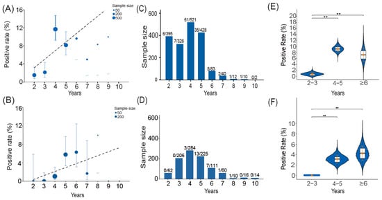
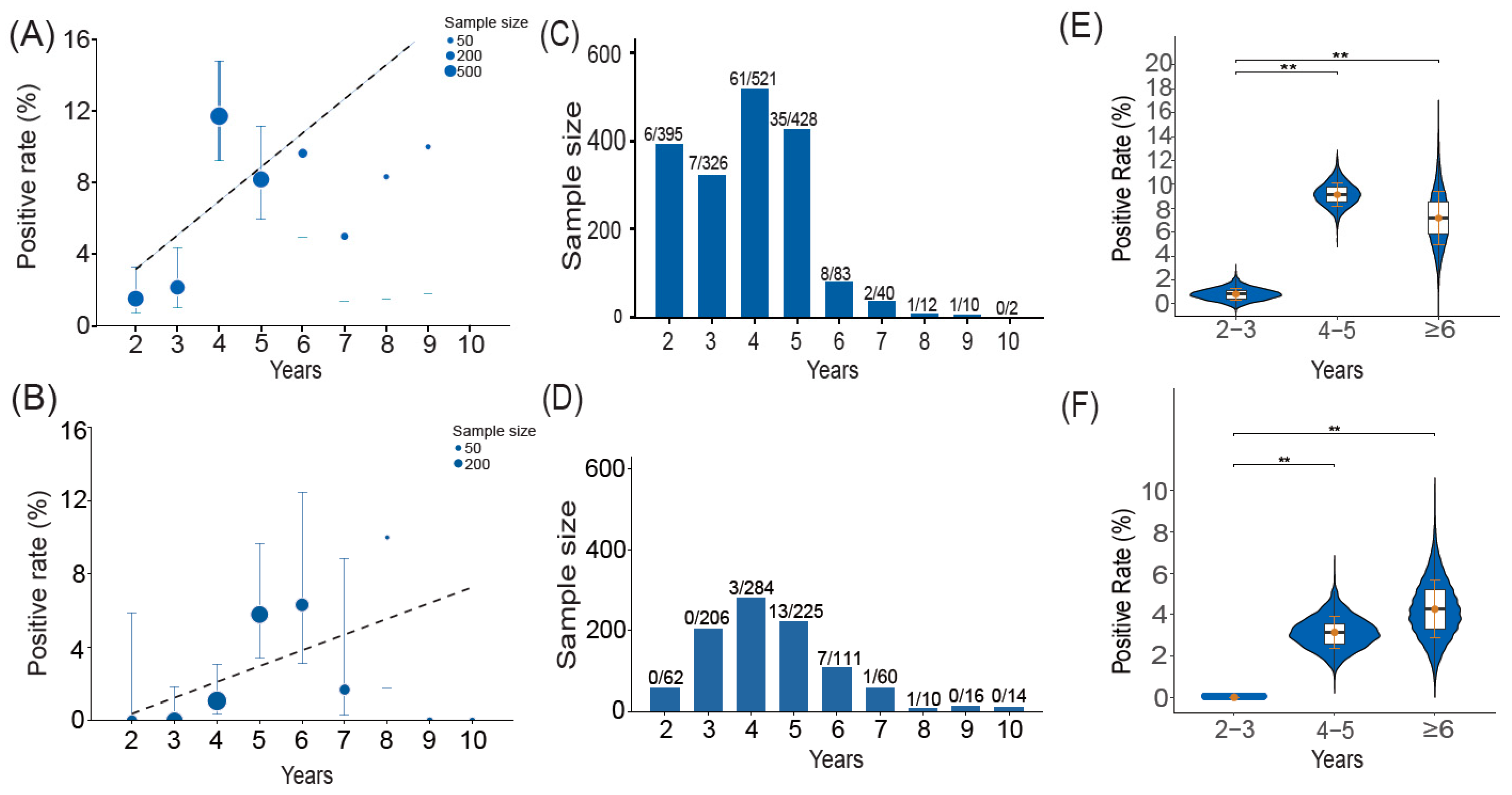
The age-related pattern of MAP milk seropositivity varied among age groups and regions (Figure 1A,C,E). Overall, cows aged 4–5 years had relatively higher milk seropositivity than younger cows (2–3 years). The sample distribution showed that most milk samples were obtained from 4–5-year-old cows, whereas fewer samples were available from older cows (Figure 1C). Statistical comparisons identified significant differences in milk seropositivity among some age groups (Figure 1E). Error bars in Figure 1 denote two-sided 95% confidence intervals calculated using the Clopper–Pearson exact binomial method.
Figure 1.
Animal-level distribution of MAP detection across age groups. (A) Animal-level milk seropositivity rate for MAP by age. (B) Animal-level fecal MAP positivity rate by age. (C) Distribution of milk sample sizes across age groups. (D) Distribution of fecal sample sizes across age groups. Comparison of animal-level milk seropositivity rates (E) and animal-level fecal MAP positivity rates (F) among age groups (2–3, 4–5, and ≥6 years). Statistical significance was assessed using the Chi-square test or Fisher’s exact test. p < 0.01 (**) indicate significance levels. Error bars in panels (A,B,E,F) represent two-sided 95% confidence intervals calculated using the Clopper–Pearson exact binomial method.
3.3. Molecular Detection and Bacterial Culture
All 25 qPCR-positive fecal samples were then cultured on HEYM, and 12 MAP isolates were recovered after three months of incubation. Acid-fast bacilli in all 12 culture-positive isolates were visualized by Ziehl–Neelsen staining, and the isolates were subsequently confirmed to be MAP by F57-targeted qPCR.
3.4. Animal-Level Fecal Positivity for MAP at Different Ages
In fecal samples, the age-related distribution of MAP positivity followed a distinct pattern (Figure 1B,D,F). The fecal MAP positivity rate increased across age groups, with no positive samples detected in the 2–3-year group, whereas positive samples were found in both the 4–5-year and ≥6-year groups, with the highest positivity rate in cows aged ≥6 years (Figure 1B). The distribution of fecal samples was uneven across age categories, with most samples collected from cows aged 4–5 years and fewer samples available from older cows (Figure 1D). Pairwise statistical comparisons showed that the fecal MAP positivity rate was significantly higher in the 4–5-year and ≥6-year groups than in the 2–3-year group (p < 0.01), whereas differences between the 4–5-year and ≥6-year groups were not statistically significant (Figure 1F) (Supplementary Table S1). These results suggest that fecal MAP shedding was detected more often in older cattle, although the small sample sizes in some age categories should be considered in interpreting age-related differences.
4. Discussion
This study indicates that MAP is widely distributed and actively circulating in the large-scale dairy farms of Xinjiang, China. Overall, 25 of the 30 herds tested (83.3%) had at least one seropositive animal based on milk ELISA. This finding is consistent with local reports over the past eight years, which reported higher MAP risk in intensive versus free-range herds [26], suggesting either an increase in herd-level prevalence over time or particularly high infection pressure in the large-scale systems. This widespread distribution suggests that existing biosecurity measures may be insufficient to control Paratuberculosis or curb its spread within these herds. The findings also suggest that the management practices and animal densities typical of large-scale dairy farming can create conditions in which, once introduced, MAP can become endemic and persist despite ongoing control efforts.
The apparent seroprevalence based on milk ELISA was 6.66% at the animal individual level and 83.3% at the herd level, whereas the prevalence of fecal shedding detected by qPCR was 2.53% of animals and 36.7% of herds. Of the 25 qPCR-positive fecal samples, 12 (48%) yielded MAP isolates that were confirmed by repeat F57-targeted qPCR and Ziehl–Neelsen staining, whereas the remaining 13 (52%) yielded no growth and were classified as qPCR-positive but culture-negative. Among these 25 animals, 14 (56%) were also positive by milk ELISA, whereas the remaining 11 (44%) were milk-ELISA-negative. The culture recovery rate of 48% is consistent with the well-documented lower sensitivity of fecal culture relative to molecular detection and should not be attributed to widespread qPCR false-positive results. We therefore interpret these discordant results as a predictable consequence of the limited sensitivity of fecal culture, for both biological and methodological reasons.
Current diagnostic approaches for JD include ELISA and/or fecal qPCR for herd-level assessment based on aggregated individual animal results, as well as testing of individual animals using serological, molecular, and microbiological approaches [29]. Detecting anti-MAP antibodies in serum or milk by ELISA remains the most widely used indirect diagnostic method, and serum is generally considered a more sensitive matrix for serological testing [30]. A critical limitation of these methods is that they detect the host’s immune response rather than the pathogen itself: antibodies appear only after the onset of shedding or clinical signs, so their sensitivity depends strongly on the stage of infection. Pooled estimates indicate that serum ELISA detects only 7–22% of MAP-infected (latent) cattle, rising to 50–87% for clinically affected animals [31]. In a 4-year longitudinal study, the overall sensitivity of an absorbed ELISA was approximately 25%, with stage-specific values of 87% in clinical cases, 75% in high shedders, but only 15% in low shedders [32]. Serological tests are thus well suited to identifying animals in the affected or infectious stages, whereas their low sensitivity in early and subclinical infection may result in underestimation of infection prevalence [31,33,34]. It should also be noted that detection of MAP DNA in feces indicates MAP shedding and infection status but does not by itself establish a diagnosis of clinical JD [31].
Recent studies have demonstrated that serum-based ELISA showed higher inter-kit agreement than milk-based assays in New Zealand dairy herds [30]; the authors also emphasized the practical advantages of milk ELISA, including easier logistics and minimized sampling costs. Milk-based ELISA therefore remains valuable for animal-level monitoring, although antibody prevalence estimates obtained from milk samples should be interpreted cautiously and may represent a conservative estimate compared with serum-based testing.
In addition, MAP has been shown to survive various food-processing steps, including milk pasteurization [20], and has been isolated from raw milk, pasteurized milk, retail cheese (both pasteurized and raw-milk varieties), and powdered infant formula [20]. We combined ELISA, qPCR, and bacterial culture to assess MAP prevalence in large-scale dairy farms in Xinjiang; serological testing, together with fecal pathogen detection, provided complementary evidence for examining paratuberculosis transmission dynamics in farm settings. Understanding MAP distribution within dairy animals therefore remains important for both animal health management and the assessment of potential food-chain exposure risks.
Bacterial culture and qPCR are widely used to detect MAP in feces, and bacterial culture is still considered the gold standard for diagnosing MAP. However, culture has important limitations, including the extremely slow growth rate of MAP and low sensitivity, particularly in animals with subclinical infection or low-level bacterial shedding [8]. A substantial body of evidence indicates that a single fecal culture detects only a minority of infected animals. In a 4-year longitudinal study of 10 dairy herds, only 38% (79/210) of all culture-positive cattle and 42% (67/158) of parturient adults were identified at the first sampling [32]. Consistent with this, a critical review of 102 test-accuracy evaluations estimated the pooled sensitivity of fecal culture for detecting MAP-infected cattle at only 0.23–0.29, reflecting its poor performance in animals during the latent stage of infection [31].
The biological basis for this limitation is well established. During the prolonged subclinical phase, animals shed MAP at low and irregular levels; consequently, a single fecal sample may contain MAP DNA detectable by qPCR but too few viable organisms for isolation in culture [32,33]. Supporting this interpretation, 30% of repeatedly fecal-culture-negative cattle from moderately infected herds were found to have MAP-infected tissues at slaughter [32], demonstrating that culture can fail to detect truly infected animals. Furthermore, a recent systematic review and meta-analysis concluded that qPCR is more sensitive than culture and explicitly acknowledged that, when culture serves as the reference standard, a proportion of true qPCR positives are inevitably misclassified as false positives [6]. Longitudinal field data support this direction of discordance: among known MAP-infectious cows sampled monthly, 78% of fecal samples were positive by qPCR, compared with 46% and 55% by solid and broth culture, respectively [35].
Methodological factors inherent to fecal culture further contribute to the discordance observed in the present study. Chemical decontamination is unavoidable in fecal culture to suppress overgrowth by other microorganisms, but it carries a cost in organism recovery: routine decontamination protocols reduce the number of MAP isolated from feces by approximately 2.7 log10 [36]; the hexadecyl pyridinium chloride concentration itself affects MAP growth and detection on solid media [37]; and direct fecal qPCR avoids decontamination steps that can reduce bacterial numbers below the detection limit of culture [35]. The heterogeneous distribution of MAP within fecal material may further lower the probability of recovery in culture [38]. Accordingly, the 13 discordant samples most likely represent animals with low-level or intermittent shedding, in which MAP DNA was detectable by qPCR but viable organisms fell below the detection limit of culture.
Taken together, 11 of the 30 herds examined had at least one cow with an F57 qPCR-positive fecal sample, corresponding to a herd-level qPCR positivity rate of 36.67% (11/30). Prevalence estimates are intrinsically linked to the molecular target employed for detection. Several molecular targets, including IS900, F57, and ISMav2, have been developed and applied for MAP detection [39,40,41]. Comparative evaluations of IS900-and F57-based assays have demonstrated that IS900 generally provides higher analytical sensitivity owing to its multicopy nature, whereas F57 exhibits higher specificity for MAP identification [40]. Owing to this higher specificity, the F57 target employed here may provide a relatively conservative estimate of fecal MAP shedding, as samples with low bacterial loads could remain undetected. In a comparative evaluation of PCR systems targeting IS900, ISMav2, and F57, the ISMav2 primer system showed inadequate specificity, with false-positive amplification in non-MAP bacteria [39]. In our study, the primary objective was to obtain direct evidence of MAP presence rather than merely to screen for MAP DNA-positive samples; accordingly, all qPCR-positive samples were further examined by bacterial culture, which remains the gold standard for MAP diagnosis. Furthermore, F57 was selected as the molecular target in this study in accordance with the national standard method for real-time PCR detection of MAP (GB/T 27637–2011) [42].
Age-related patterns of MAP detection warrant careful interpretation. In this study, milk ELISA antibody positivity rates were highest in cows aged 4–5 years, which differs from previous reports of increasing MAP seroprevalence with age [25]. This discrepancy may reflect differences in herd characteristics, sample composition, management practices, or infection dynamics. By contrast, fecal qPCR positivity was highest among cows aged ≥6 years. Therefore, the variation observed across age groups should be interpreted as an age-associated distribution pattern rather than as evidence of age-dependent susceptibility or of any particular age group acting as a major transmission reservoir. Because this study did not include longitudinal follow-up, individual production records, or detailed herd management data, further studies are needed to clarify the relationship between age, infection progression, and MAP shedding dynamics.
Marked regional variation in MAP infection indicators was observed among the study regions. At the individual-cow level, milk ELISA positivity ranged from 2.50% (Khotan) to 16.25% (Kashgar), whereas fecal qPCR positivity ranged from 0% (Ili, Bayingolin, and Khotan) to 5.97% (Urumqi). Together, these findings indicate pronounced geographic heterogeneity in MAP infection across Xinjiang. Formal statistical comparison of regional differences and multivariable risk-factor analysis were precluded by the convenience-sampling design, the unequal allocation of herds among regions (1–11 herds per region), heterogeneous regional sample sizes, and the absence of systematically collected herd management, environmental and biosecurity data. Furthermore, although all enrolled herds were large-scale operations (≥1000 cows each), herd size was not recorded as a standardized covariate, which precluded its evaluation as a risk determinant. Accordingly, this study provides baseline descriptive estimates of MAP infection indicators across Xinjiang dairy herds and underscores the need for future analytical epidemiologic investigations employing representative sampling frames and standardized herd-level covariates.
Our findings support a staged, multi-sample approach to regional surveillance. High-throughput screening by milk ELISA, followed by confirmatory qPCR testing, may offer a practical approach for large-scale surveillance programmes enabling initial screening of large numbers of animals or herds and subsequent targeted testing of selected samples.
However, several limitations of our cross-sectional design and diagnostic methods should be acknowledged. First, because the sensitivity of both ELISA and qPCR is suboptimal in the early stages of infection, our estimates reflect apparent prevalence and likely underestimate the actual prevalence of infection. Second, the cross-sectional design and convenience sampling of herds preclude establishing temporal sequences or causal relationships with specific management practices and may limit the generalizability of our prevalence estimates to all large-scale dairy farms in Xinjiang. Third, only lactating cows were sampled; infection dynamics in young stock remain unexplored, even though this cohort is critical for interrupting the transmission cycle. Fourth, because this study was designed to estimate the prevalence of MAP infection rather than to characterize clinical disease, the relationship between MAP shedding and clinical signs such as diarrhea could not be assessed. MAP shedding intensity was not quantitatively evaluated, limiting assessment of how shedding levels contribute to MAP dissemination and transmission dynamics within and between herds.
Beyond these limitations, our study estimated seroprevalence and MAP shedding but did not investigate risk factors associated with elevated MAP prevalence in large-scale farms or the progression from subclinical infection to clinical JD. These gaps highlight priorities for future research. Prospective cohort studies are needed to clarify the management, environmental and host genetic factors that drive MAP transmission and the relationship between MAP infection status, shedding dynamics, and the development of clinical JD. Molecular epidemiological approaches (e.g., whole-genome sequencing) could aid in tracing transmission routes and in distinguishing persistent strains from newly introduced strains. Collectively, these efforts should inform the development of evidence-based and cost-effective control measures to reduce the impact of MAP infection on dairy production systems in Xinjiang, China. Although MAP has been investigated for its possible association with human health outcomes, including Crohn’s disease, the nature and significance of this relationship remain uncertain. The potential public health implications of MAP exposure warrant cautious interpretation and further epidemiological, microbiological and mechanistic investigation.
5. Conclusions
This study documents widespread MAP exposure among large-scale dairy herds sampled in Xinjiang, northwestern China. MAP-specific antibodies were detected in 25 of the 30 herds tested by milk ELISA, corresponding to an overall apparent individual-level prevalence of 6.66%. Among the 988 cows selected for fecal testing, F57-targeted qPCR detected MAP DNA in 25 animals (2.53%), from 11 of the 30 herds examined. In addition, MAP was isolated by culture from 12 of the 25 qPCR-positive fecal samples, and the isolates were confirmed by Ziehl–Neelsen staining and F57-targeted qPCR. Milk ELISA positivity was highest among cows aged 4–5 years, a finding that likely reflects the chronic and progressive course of JD. These findings indicate that MAP represents a substantial concern for animal health and dairy productivity, and may have public health relevance given its suspected zoonotic association. Further research is therefore needed to develop effective control strategies tailored to large-scale dairy farming in Xinjiang, northwest China.
Supplementary Materials
The following supporting information can be downloaded at: https://www.mdpi.com/article/10.3390/pathogens15111190/s1, Table S1: Complete animal-level counts underlying Figure 1.
Author Contributions
Conceptualization, J.-Y.W. and Q.F.; methodology, J.D. and S.-K.M.; software, J.W.; validation, H.-J.S. and J.-Y.W.; formal analysis, Y.S.; investigation, X.-Y.M.; resources, X.-Y.M., J.W., Y.-R.W. and H.-J.S.; data curation, J.D. and Y.S.; writing—original draft preparation, J.D.; writing—review and editing, J.D., Q.F., Y.-R.W., J.-Y.W. and H.-J.S.; supervision, X.-Y.M.; project administration, H.-J.S.; funding acquisition, J.-Y.W. and Y.-R.W. All authors have read and agreed to the published version of the manuscript.
Funding
This research received funding from the National Natural Science Foundation of China (grant number 32260886).
Institutional Review Board Statement
The study protocol received review and approval from the Ethics Committee of the College of Veterinary Medicine at Xinjiang Agricultural University (approval no. 2023051, 20 June 2023), following institutional guidelines for animal welfare and ethical review.
Informed Consent Statement
Verbal informed consent was obtained from the animals’ owners.
Data Availability Statement
The original contributions reported in this study are included in the article and its Supplementary Materials; additional inquiries may be directed to the corresponding author.
Acknowledgments
The authors thank the owners and staff of the participating dairy farms for their cooperation during sample collection.
Conflicts of Interest
The authors declare no conflicts of interest.
Abbreviations
The following abbreviations are used in this manuscript:
| MAP | Mycobacterium avium subsp. paratuberculosis |
| JD | Johne’s disease |
| ELISA | Enzyme-linked immunosorbent assay |
| qPCR | Quantitative PCR |
| HPC | Hexadecyl pyridinium chloride |
| BHI | Brain heart infusion |
| HEYM | Herrold’s egg yolk medium |
| OD | Optical density |
| S/P | Sample-to-positive ratio |
| Ct | Quantification cycle (cycle threshold) |
| CI | Confidence interval |
| IAC | An internal amplification control |
| PCR | polymerase chain reaction |
| PTB | paratuberculosis |
References
- Idris, S.M.; Elmagzoub, W.A.; Isameldin, M.; Arabi, N.; Abdo, A.; Ibrahim, M.; Bakhiet, S.M.; Okuni, J.B.; Ojok, L.; Gameel, A.A.; et al. A Dataset on the Phylogeny of Mycobacterium avium Subsp. paratuberculosis Isolated from Patients in a Referral Gastrointestinal Diseases Centre in the Sudan. Data Brief 2025, 63, 112257. [Google Scholar] [CrossRef] [Scilit] [PubMed]
- Pereira, A.C.; Martins, L.; Biesheuvel, M.M.; Orsel, K.; Buck, J.D.; Barkema, H.W. Mycobacterium avium Subspecies paratuberculosis (MAP) Shedding and Seropositivity in Youngstock in MAP-Infected Dairy Herds in Alberta, Canada. J. Dairy Sci. 2026, 109, 1874–1881. [Google Scholar] [CrossRef] [Scilit] [PubMed]
- Wang, M.; Bissonnette, N.; Dudemaine, P.; Ibeagha-Awemu, E.M. MicroRNA-Mediated Regulatory Mechanisms in Cow’s Ileum and Ileum Lymph Node in Response to Mycobacterium avium Subspecies paratuberculosis Infection. Sci. Rep. 2025, 15, 43030. [Google Scholar] [CrossRef] [Scilit] [PubMed]
- Horan, L.; Mee, J.F.; Field, N.L.; Walsh, S.W.; Valldecabres, A. Farmers’ Knowledge of Johne’s Disease and Opinions of the Irish Johne’s Control Programme: Results of an Online Survey Answered Mostly by Young Farmers. Ir. Vet. J. 2023, 76, 31. [Google Scholar] [CrossRef] [Scilit] [PubMed]
- Dane, H.; Gilbride, B.; Thomas, M.; Grant, I.R. Evidence of Mycobacteriaemias and Mycobacterial Co-infections Uncovered in Cattle at Slaughter Using a Novel Phage-Based Phms-qpcr Assay for Viable Mycobacterium bovis and Mycobacterium avium Subsp. paratuberculosis. Ir. Vet. J. 2025, 78, 28. [Google Scholar] [CrossRef] [Scilit] [PubMed]
- Liu, T.; Lin, H.; Zhu, L.; Yang, D.A.; Yao, H.; Pan, Z. Accuracy of Real-time Pcr for the Detection of Paratuberculosis in Actual Samples: A Systematic Review and Meta-Analysis. Prev. Vet. Med. 2025, 237, 106436. [Google Scholar] [CrossRef] [Scilit] [PubMed]
- Lienhard, J.; Friedel, U.; Paganini, C.; Hilbe, M.; Scherrer, S.; Schmitt, S. Isolation of Mycobacterium avium asp. paratuberculosis and Other Non-tuberculous Mycobacteria from Head Lymph Nodes of Wild Ruminants and Badgers in Switzerland. Front. Vet. Sci. 2024, 10, 1321106. [Google Scholar] [CrossRef] [Scilit] [PubMed]
- Salem, M.; Zaghawa, A.; Housawi, F.; Elsify, A.; Hasan, M.E.; Elrashedy, A.; Salem, K.A.; Amor, N.B.; Naeem, A.A. Comprehensive Genome Analysis of Mycobacterium avium Subsp. paratuberculosis in Camels from Saudi Arabia: Molecular Epidemiology and Antimicrobial Resistance. Vet. World 2025, 18, 859–876. [Google Scholar] [CrossRef] [Scilit] [PubMed]
- Grant, I.R.; Sevilla, I.A.; Molina, E.; Romero Martinez, B.; Lorente-Leal, V.; Thibault-Poisson, V.C.; Cechova, M.; Köhler, H. Evaluation of the Reproducibility and Performance Characteristics of the Phagomagnetic Separation-qpcr Assay for Rapidly Detecting Viable Mycobacterium avium Subsp. paratuberculosis in Bovine Milk and Feces. Front. Vet. Sci. 2025, 12, 1677096. [Google Scholar] [CrossRef] [Scilit] [PubMed]
- Royo, M.; Elguezábal, N.; Arrazuria, R.; Benavides, J.; Fernández, M. Effect of Paratuberculosis Vaccination Before and After Oral Experimental Infection with Mycobacterium avium Subspecies paratuberculosis in Goats. Vet. Q. 2025, 45, 2566363. [Google Scholar] [CrossRef] [Scilit] [PubMed]
- Field, N.L.; McAloon, C.G.; Gavey, L.; Mee, J.F. Mycobacterium avium Subspecies paratuberculosis Infection in Cattle—A Review in the Context of Seasonal Pasture-based Dairy Herds. Ir. Vet. J. 2022, 75, 12. [Google Scholar] [CrossRef] [Scilit] [PubMed]
- Zarei-Kordshouli, F.; Geramizadeh, B.; Khodakaram-Tafti, A. Prevalence of Mycobacterium avium Subspecies paratuberculosis Is 900 Dna in Biopsy Tissues from Patients with Crohn’s Disease: Histopathological and Molecular Comparison with Johne’s Disease in Fars Province of Iran. BMC Infect. Dis. 2019, 19, 23. [Google Scholar] [CrossRef] [Scilit] [PubMed]
- Estevinho, M.M.; Cabeda, J.; Santiago, M.; Machado, E.; Silva, R.; Duro, M.; Pita, I.; Morais, R.; Macedo, G.; Bull, T.J.; et al. Viable Mycobacterium avium Subsp. paratuberculosis Colonizes Peripheral Blood of Inflammatory Bowel Disease Patients. Microorganisms 2023, 11, 1520. [Google Scholar] [CrossRef] [Scilit] [PubMed]
- Gerrard, Z.E.; Swift, B.M.C.; Botsaris, G.; Davidson, R.S.; Hutchings, M.R.; Huxley, J.N.; Rees, C.E.D. Survival of Mycobacterium avium Subspecies paratuberculosis in Retail Pasteurised Milk. Food Microbiol. 2018, 74, 57–63. [Google Scholar] [CrossRef] [Scilit] [PubMed]
- Waddell, L.A.; Rajić, A.; Stärk, K.D.C.; McEwen, S.A. The Zoonotic Potential of Mycobacterium avium ssp. paratuberculosis: A Systematic Review and Meta-Analyses of the Evidence. Epidemiol. Infect. 2015, 143, 3135–3157. [Google Scholar] [CrossRef] [Scilit] [PubMed]
- Waddell, L.A.; Rajić, A.; Stärk, K.D.C.; McEwen, S.A. The Potential Public Health Impact of Mycobacterium avium ssp. paratuberculosis: Global Opinion Survey of Topic Specialists. Zoonoses Public Health 2015, 63, 212–222. [Google Scholar] [CrossRef] [Scilit] [PubMed]
- Lagod, P.P.; Qasem, A.; Naser, S.A. Short Chain Fatty Acids Lower Inflammation and Restore Intestinal Integrity and Function Markers in Mycobacterium paratuberculosis—Infection In Vitro Model. Nutrients 2025, 17, 3663. [Google Scholar] [CrossRef] [Scilit] [PubMed]
- Eppleston, J.; Begg, D.J.; Dhand, N.K.; Watt, B.; Whittington, R.J. Environmental Survival of Mycobacterium avium Subsp. paratuberculosis in Different Climatic Zones of Eastern Australia. Appl. Environ. Microbiol. 2014, 80, 2337–2342. [Google Scholar] [CrossRef] [Scilit] [PubMed]
- Grewal, S.K.; Rajeev, S.; Sreevatsan, S.; Michel, F.C. Persistence of Mycobacterium avium Subsp. paratuberculosis and Other Zoonotic Pathogens During Simulated Composting, Manure Packing, and Liquid Storage of Dairy Manure. Appl. Environ. Microbiol. 2006, 72, 565–574. [Google Scholar] [CrossRef] [Scilit] [PubMed]
- Knific, T.; Ocepek, M.; Kirbiš, A.; Krt, B.; Prezelj, J.; Gethmann, J.M. Quantitative Risk Assessment of Exposure to Mycobacterium avium Subsp. paratuberculosis (MAP) Via Different Types of Milk for the Slovenian Consumer. Foods 2022, 11, 1472. [Google Scholar] [CrossRef] [Scilit] [PubMed]
- Griss, S.; Knific, T.; Buzzell, A.; Carmo, L.P.; Schüpbach-Regula, G.; Meylan, M.; Ocepek, M.; Thomann, B. A Scoping Review on Associations Between Paratuberculosis and Productivity in Cattle. Front. Vet. Sci. 2024, 11, 1352623. [Google Scholar] [CrossRef] [Scilit] [PubMed]
- Ottardi, M.; Lechner, I.; Wang, J.; Schmitt, S.; Schneeberger, M.; Schmid, R.M.; Stephan, R.; Meylan, M. Seroprevalence of Mycobacterium avium Subsp. paratuberculosis in Swiss Dairy Herds and Risk Factors for a Positive Herd Status and Within-Herd Prevalence. Front. Vet. Sci. 2024, 11, 1409694. [Google Scholar] [CrossRef] [Scilit] [PubMed]
- Johnson, P.; McLeod, L.; Qin, Y.; Osgood, N.; Rosengren, L.; Campbell, J.; Larson, K.; Waldner, C. Investigating Effective Testing Strategies for the Control of Johne’s Disease in Western Canadian Cow-Calf Herds Using an Agent-based Simulation Model. Front. Vet. Sci. 2022, 9, 1003143. [Google Scholar] [CrossRef] [Scilit] [PubMed]
- Yue, R.; Liu, C.; Barrow, P.; Liu, F.; Cui, Y.; Yang, L.; Zhao, D.; Zhou, X. The Isolation and Molecular Characterization of Mycobacterium avium Subsp. paratuberculosis in Shandong Province, China. Gut Pathog. 2016, 8, 9. [Google Scholar] [CrossRef] [Scilit] [PubMed]
- Sun, W.; Lv, W.; Cong, W.; Meng, Q.; Wang, C.; Shan, X.; Qian, A. Mycobacterium avium Subspecies paratuberculosis and Bovine Leukemia Virus Seroprevalence and Associated Risk Factors in Commercial Dairy and Beef Cattle in Northern and Northeastern China. BioMed Res. Int. 2015, 2015, 315173. [Google Scholar] [CrossRef] [Scilit] [PubMed]
- Liu, X.; Li, J.; Yang, X.; Wang, D.; Wang, J.; Wu, J. The Seroprevalence of Mycobacterium avium Subspecies paratuberculosis in Dairy Cattle in Xinjiang, Northwest China. Ir. Vet. J. 2017, 70, 1. [Google Scholar] [CrossRef] [Scilit] [PubMed]
- Hosseiniporgham, S.; Biet, F.; Ganneau, C.; Bannantine, J.P.; Bay, S.; Sechi, L.A. A Comparative Study on the Efficiency of Two Mycobacterium avium Subsp. paratuberculosis (MAP)-derived Lipopeptides of L3P and L5P as Capture Antigens in an In-house Milk ELISA Test. Vaccines 2021, 9, 997. [Google Scholar] [CrossRef] [Scilit] [PubMed]
- Hernandez-Reyes, A.; Buck, J.D.; Orsel, K. Risk Factors Associated with Johne’s Disease in a Captive Wood Bison Herd. Can. Vet. J. 2025, 66, 425–434. [Google Scholar] [PubMed]
- Sarkar, S.; Young, B.A.; Daniels, C.S.; Thompson, J.E. Diagnostic and Economic Evaluation of MALDI-TOF MS with Machine Learning for Screening of Johne’s Disease from Dairy Cow Serum. J. Dairy Sci. 2026, 109, 513–526. [Google Scholar] [CrossRef] [Scilit] [PubMed]
- Venkatesh, K.M.; Lopez-Villalobos, N.; Gupta, S.K.; Udy, G.B.; Laven, R.; Chiu, S.; Bugde, P.; Furuya, Y.; Dukkipati, V.S.R. A Comparative Study Between Milk- and Serum-based Antibody Detection Assays for Johne’s Disease in New Zealand Dairy Cattle. Tuberculosis 2025, 155, 102679. [Google Scholar] [CrossRef] [Scilit] [PubMed]
- Nielsen, S.S.; Toft, N. Ante Mortem Diagnosis of Paratuberculosis: A Review of Accuracies of ELISA, Interferon-Gamma Assay and Faecal Culture Techniques. Vet. Microbiol. 2008, 129, 217–235. [Google Scholar] [CrossRef] [Scilit] [PubMed]
- Whitlock, R.H.; Rosenberger, A.E.; Sweeney, R.W.; Spencer, P.A. ELISA and Fecal Culture for Paratuberculosis (Johne’s Disease): Sensitivity and Specificity of Each Method. Vet. Microbiol. 2000, 77, 387–399. [Google Scholar] [CrossRef] [Scilit] [PubMed]
- Sweeney, R.W. Pathogenesis of Paratuberculosis. Vet. Clin. Food Anim. Pract. 2011, 27, 537–546. [Google Scholar] [CrossRef] [Scilit] [PubMed]
- Bissonnette, N.; Ollier, S.; Brousseau, J.; Byrne, A.S.; Savard, C.; Tahlan, K. Systematic Evaluation and Optimization of TaqMan qPCR Assays Targeting F57, ISMAP02, and IS900 for Multiplex Detection of Mycobacterium avium Subsp. paratuberculosis. J. Clin. Microbiol. 2026, 64, e00872-25. [Google Scholar] [CrossRef] [Scilit] [PubMed]
- Laurin, E.L.; Chaffer, M.; McClure, J.T.; McKenna, S.L.; Keefe, G.P. The Association of Detection Method, Season, and Lactation Stage on Identification of Fecal Shedding in Mycobacterium avium ssp. paratuberculosis Infectious Dairy Cows. J. Dairy Sci. 2015, 98, 211–220. [Google Scholar] [CrossRef] [Scilit] [PubMed]
- Reddacliff, L.A.; Vadali, A.; Whittington, R.J. The Effect of Decontamination Protocols on the Numbers of Sheep Strain Mycobacterium avium Subsp. paratuberculosis Isolated from Tissues and Faeces. Vet. Microbiol. 2003, 95, 271–282. [Google Scholar] [CrossRef] [Scilit] [PubMed]
- Johansen, K.A.; Hugen, E.E.; Payeur, J.B. Growth of Mycobacterium avium Subsp. paratuberculosis in the Presence of Hexadecylpyridinium Chloride, Natamycin, and Vancomycin. J. Food Prot. 2006, 69, 878–883. [Google Scholar] [CrossRef] [Scilit] [PubMed]
- Keller, S.M.; Stephan, R.; Kuenzler, R.; Meylan, M.; Wittenbrink, M.M. Comparison of Fecal Culture and F57 Real-Time PCR for the Detection of Mycobacterium avium Subsp. paratuberculosis in Swiss Cattle Herds with a History of Paratuberculosis. Acta Vet. Scand. 2014, 56, 68. [Google Scholar] [CrossRef] [Scilit] [PubMed]
- Möbius, P.; Hotzel, H.; Raßbach, A.; Köhler, H. Comparison of 13 Single-Round and Nested PCR Assays Targeting IS900, ISMav2, f57 and Locus 255 for Detection of Mycobacterium avium Subsp. paratuberculosis. Vet. Microbiol. 2008, 126, 324–333. [Google Scholar] [CrossRef] [Scilit] [PubMed]
- Sidoti, F.; Banche, G.; Astegiano, S.; Allizond, V.; Cuffini, A.M.; Bergallo, M. Validation and Standardization of IS900 and F57 Real-Time Quantitative PCR Assays for the Specific Detection and Quantification of Mycobacterium avium Subsp. paratuberculosis. Can. J. Microbiol. 2011, 57, 347–354. [Google Scholar] [CrossRef] [Scilit] [PubMed]
- Rani, M.; Narang, D.; Kumar, D.; Dhama, K.; Singh, S.V. ISMap02 Element Targeted Nested Polymerase Chain Reaction in the Detection of Mycobacterium avium Subsp. paratuberculosis in Fecal Samples of Cattle and Buffaloes. Vet. World 2018, 11, 397–401. [Google Scholar] [CrossRef] [Scilit] [PubMed]
- GB/T 27637-2011; Real-Time PCR Method for the Detection of Mycobacterium avium Subsp. paratuberculosis. Standards Press of China: Beijing, China, 2011. (In Chinese)

Disclaimer/Publisher’s Note: The statements, opinions and data contained in all publications are solely those of the individual author(s) and contributor(s) and not of MDPI and/or the editor(s). MDPI and/or the editor(s) disclaim responsibility for any injury to people or property resulting from any ideas, methods, instructions or products referred to in the content. |
© 2026 by the authors. Licensee MDPI, Basel, Switzerland. This article is an open access article distributed under the terms and conditions of the Creative Commons Attribution (CC BY) license.