


Abstract
To investigate the temporal dynamics and potential metabolic roles of bacteriophage communities in the marine plastisphere, we characterized phage community dynamics and phage-encoded auxiliary metabolic genes (AMGs) during a 21-day in situ experiment involving four plastic types. Phage communities differed significantly between the early (days 3–12) and late (days 15–21) stages, indicating distinct temporal shifts, whereas no significant differences were observed among polymer types. Changes in phage community composition in the plastisphere might be associated with bacterial host lineages. In particular, Vibrio phages were strongly associated with changes in host-Vibrios. In AMGs, nine metabolic categories comprising a total of nineteen detailed pathways were identified. AMGs associated with cofactor and vitamin metabolism were relatively enriched during the early stages, while sulfur-containing amino acid metabolism became more abundant during the late stages. These findings suggest that plastisphere-associated phages may be linked to microbial community succession and functional potential through dynamic host–phage associations.
1. Introduction
Marine plastic debris continues to accumulate in the ocean, exerting long-term impacts on marine ecosystems [1,2]. When plastic surfaces come into contact with the marine environment, they are rapidly colonized by microorganisms, leading to biofilm formation and the establishment of novel microbial habitats [3]. This colonization process may be facilitated by the hydrophobic nature of polymers, which promotes the adsorption and accumulation of dissolved organic matter, as well as by the release of low-molecular-weight organic compounds from plastic leachates. These microbial habitats, collectively known as the “plastisphere,” are increasingly recognized as distinct microscale environments within marine ecosystems [3]. Furthermore, the high buoyancy of plastic debris and its capacity for long-distance transport can facilitate the dispersal of diverse organisms, including potentially pathogenic microorganisms, across marine regions via the plastisphere [4]. Thus, plastics may act not only as chemical pollutants but also as vectors for the long-distance dispersal of potentially pathogenic microorganisms, with possible implications for disease risks in marine and coastal environments [4,5].
Bacteriophages (phages) are important components of viral communities and exhibit high levels of diversity [6]. Although global phage abundance is estimated at approximately 1031 particles, metagenomic analyses suggest that the full extent of viral diversity remains underexplored [7,8]. However, phages play central roles in shaping microbial community structure and composition and driving organic matter recycling through the viral shunt [9]. Phage diversity and community composition differ between seawater and substrate-associated biofilms [10]. On plastic surfaces, phage communities may be influenced by substrate properties and the succession of biofilm-associated microbial communities [11]. Although plastic polymers differ in physicochemical properties that may influence initial microbial attachment [12], our previous study found no significant differences in bacterial community composition among expanded polystyrene, polyethylene, polypropylene, and polyethylene terephthalate [13]. Because plastic-associated microbial communities originate from the surrounding seawater, their composition and succession may be strongly influenced by environmental factors [14,15]. Accordingly, phage diversity and community composition on plastic surfaces may be shaped by both substrate properties and changes in host bacterial communities during biofilm succession [16]. However, the relative contributions of substrate type and temporal succession to phage community dynamics under natural marine conditions remain poorly understood. Phages can expand the metabolic capabilities of their hosts through auxiliary metabolic genes (AMGs) [17]. Characterizing plastisphere-associated phage communities and their AMGs is therefore important for understanding phage–host interactions and their potential roles in shaping microbial community dynamics and metabolic processes in marine ecosystems affected by plastic pollution. Although Xia et al. [18] suggested that phage-encoded AMGs in the plastisphere may regulate host-associated functions, including nutrient metabolism, quorum sensing, biofilm formation, and antibiotic production, their temporal dynamics and ecological roles on plastic surfaces remain poorly understood. In particular, changes in AMG composition and functional profiles during plastisphere biofilm development have yet to be fully characterized.
Studies on bacteriophage dynamics in the plastisphere remain limited, particularly regarding their associations with microbial communities during biofilm development following plastic immersion in seawater. To address this knowledge gap, we investigated phage community dynamics, phage–bacterium associations, temporal turnover, and phage-encoded auxiliary metabolic genes (AMGs) during a 21-day in situ experiment using four representative plastic polymers: expanded polystyrene, polyethylene, polypropylene, and polyethylene terephthalate. We tested three hypotheses: (1) differences in polymer surface properties would lead to distinct phage community structures among plastic types; (2) temporal changes in phage communities would be associated with shifts in specific potential host bacterial lineages during biofilm succession; and (3) phage community turnover across biofilm developmental stages would involve changes in taxonomic composition and relative abundance, accompanied by shifts in the functional potential of phage-encoded AMGs.
2. Materials and Methods
2.1. Design of In Situ Plastisphere Experiment
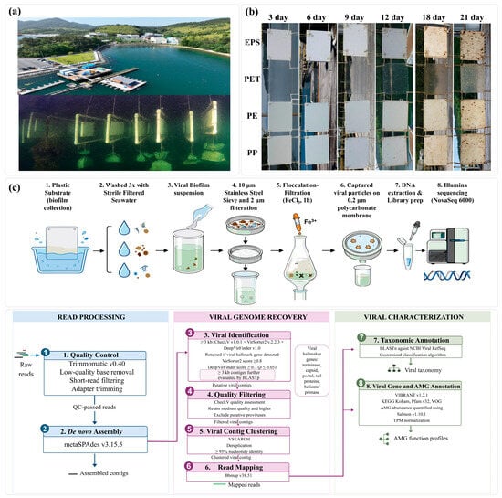
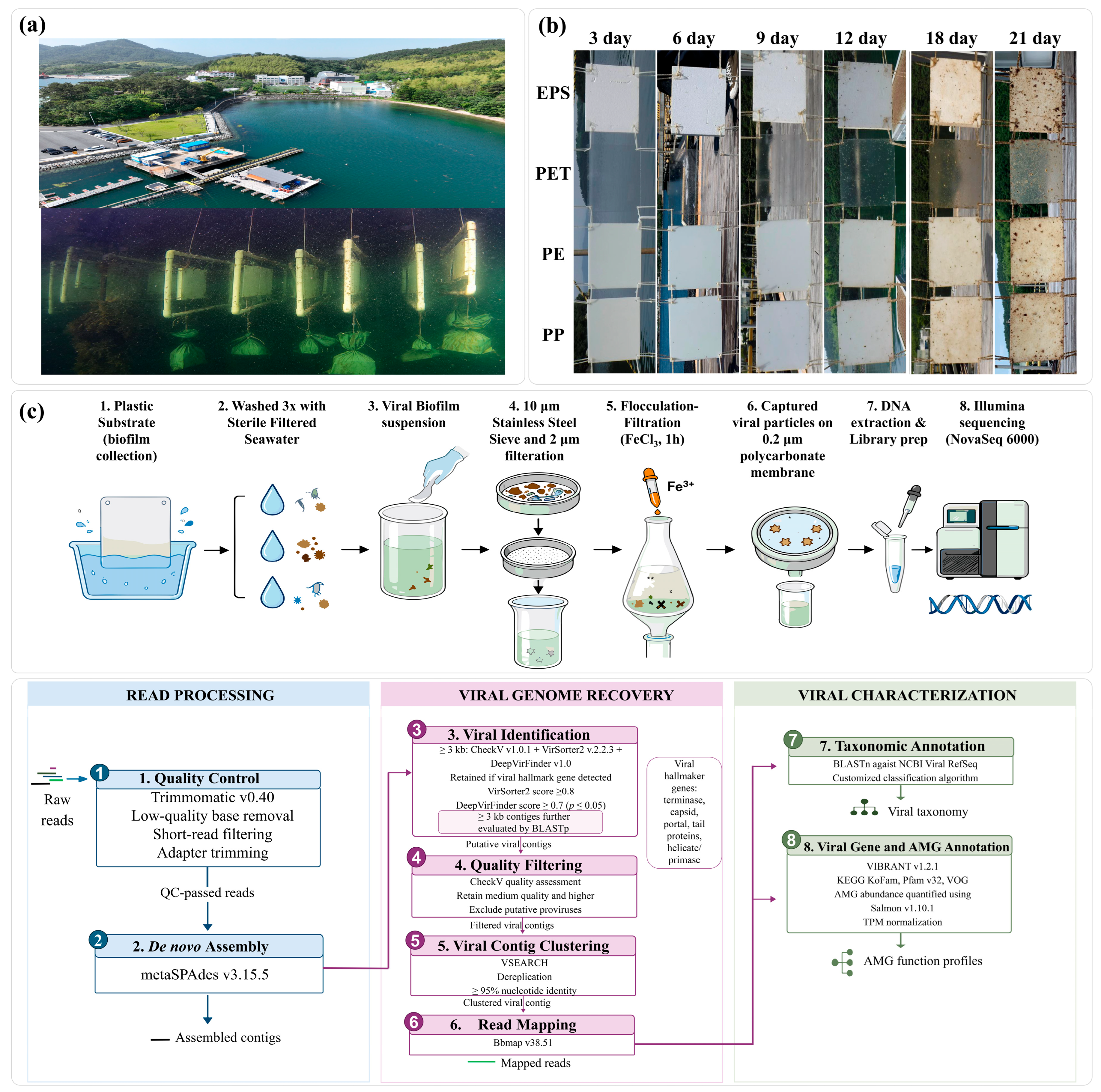
To investigate the microbial communities colonizing plastic surfaces, a 21-day field incubation experiment was conducted from 20 July (Day 0) to 10 August 2021 (investigations initiated on Day 3, 23 July), at the Jangmok Bay Time-Series Monitoring Station (JBTMS; 34°59′37″ N, 128°40′27″ E; Figure 1a) located off the southern coast of South Korea [13]. Four plastic polymers commonly detected in marine environments were used as substrates: polypropylene (PP) sheet manufactured by Goodfellow (density: 0.9 g cm−3, Cat. No. PP303050, Coraopolis, PA, USA), polyethylene (PE; 0.86 g cm−3, Cat. No. ET323280), polyethylene terephthalate (PET; 1.10 g cm−3, Cat. No. ES303030), and expanded polystyrene (EPS) purchased from Zeropeople Co. Ltd. (Sungnam, Republic of Korea, 15 kg m−3). A frame containing four types of plastic substrates was submerged at a depth of 1 m below the water surface (Figure 1a). Each plastic plate was deployed in triplicate at 3-day intervals (days 3, 6, 9, 12, 15, 18, and 21), resulting in a total of 21 samples for each plastic type (Figure 1b). Each frame was stabilized with a 12-kg steel weight, and the in situ experiment was conducted at the mobile outdoor mesocosm laboratory of KIOST (Korea Institute of Ocean Science & Technology) in Geoje, Republic of Korea (Figure 1a). At each sampling time, the plastic substrate was immediately and gently transferred to sterile seawater maintained at 4 °C following retrieval to minimize disturbance and damage associated with air exposure. Biofilms were subsequently collected in the outdoor mesocosm laboratory. A schematic overview of the study workflow is provided in Figure 1c. To analyze phage community associated with the plastic surfaces, one side of each plastic substrate (300 × 300 mm) was gently rinsed three times with sterile filtered seawater to remove loosely attached particles and plankton. Subsequently, the viral communities on biofilms were collected by scraping the surface with a sterile rubber spatula and transferred to a sterile container. Simultaneously, 2 L of ambient seawater was collected from a depth of 1 m at the experimental site for the measurement of environmental variables (inorganic nutrients, dissolved organic carbon (DOC), and chlorophyll-a). Metagenomic analysis was performed within one week of sample collection. This study focused on bacteriophage community dynamics, whereas the bacterial community data were previously reported by Jeong et al. [13].
Figure 1.
Experimental photographs and workflow of phage metagenomics. (a) Photograph of in situ experiment at the Jangmok Bay Time-Series Monitoring Station (JBTMS; 34°59′37″ N, 128°40′27″ E) located off the southern coast of Korea. (b) Photograph of temporal development of biofouling on plastic surfaces: four plastic types of EPS, PET, PE, and PP (triplicate) during the 21-day in situ experiment. The photograph for Day 15 was not taken due to rain. (c) Schematic workflow for sample collection, viral particle recovery, DNA preparation, and metavirome sequencing. (d) Bioinformatic pipeline for analyzing phage community: read processing, viral contig identification and quality filtering, clustering of viral operational taxonomic unit (vOTU), read mapping, taxonomic annotation, and auxiliary metabolic gene (AMG) analysis.
2.2. Analysis of Phage Community
The biofilm suspensions collected from plastic surfaces were pre-filtered sequentially through a 10-µm stainless-steel sieve and a 2-µm polycarbonate membrane filter (TTTP 04700) to remove larger fouling organisms and inorganic particles. The filtrate containing particles smaller than 2 µm, including virus-like particles, was collected, and a 500 mL sample was used for further analysis. To aggregate double strand DNA (dsDNA) viruses, the Fe flocculation–filtration–resuspension (FFR) method [19] was applied. Briefly, for viral precipitation, 50 μL of an iron stock solution (10 g Fe L−1; prepared by dissolving 4.83 g of FeCl3·6H2O in 100 mL of Milli-Q water) was added to each 500 mL aliquot of the filtrate, yielding a final Fe concentration of 1 mg L−1. The sample was continuously mixed by rolling at 20 rpm and 20 °C for 1 h. DNA viruses concentrated from the 500 mL filtrate were collected on a 0.2 µm polycarbonate membrane filter (GTTP04700, 47 mm). To release viral particles from the flocs of Fe ions and the membrane, the filter was cut into eight pieces and transferred to a 50 mL conical tube containing 10 mL of suspension buffer (0.1 M EDTA, 0.2 M MgCl2, and 0.2 M sodium ascorbate). The suspension buffer was adjusted to pH 6.0 with 10 M NaOH. The membrane pieces were gently mixed with the suspension buffer to dissolve the iron-containing flocs and release the concentrated viral particles. Viral genomic DNA was then extracted using the Viral Gene-spin Viral DNA/RNA Extraction Kit (iNtRON Biotechnology, Seoul, Republic of Korea) according to the manufacturer’s instructions.
Genomic DNA libraries for dsDNA viruses were prepared using the NEBNext Ultra II DNA Library Prep Kit (Illumina Inc., San Diego, CA, USA), and paired-end sequencing was performed on the Illumina NovaSeq 6000 platform. Data analyses genomic sequences were performed using a modified workflow (see Figure 1d) based on our previous method [20]. Trimmomatic (v0.40), following standard trimming procedures for Illumina data to discard low-quality bases and short reads prior to downstream analyses [21]. Viral contigs were assembled de novo using metaSPAdes (v3.7.0) [22]. To identify viral sequences, assembled contigs longer than 3000 bp were screened using CheckV v1.0.1 [23] in combination with VirSorter2 (v2.2.3) and DeepVirFinder (v1.0). Contigs were retained as viral if they met the following criteria: detection of viral hallmark genes and prediction scores ≥0.8 in VirSorter2 and ≥0.7 (p ≤ 0.05) in DeepVirFinder [24,25]. In addition, contigs at over 3000 bp were further evaluated using BLASTp, and only those containing viral hallmark genes were retained for downstream analyses. Viral hallmark genes were defined as conserved viral structural and packaging proteins, including terminase large and small subunits, major capsid (head) proteins, DNA polymerase, portal proteins, head–tail adaptor proteins, and tail-associated proteins (e.g., tail fiber, tail sheath, and tape measure proteins), along with selected replication-related proteins such as DNA helicases and primases (Table S1). Contigs identified as putative proviruses were excluded. Sequences sharing ≥95% nucleotide identity were clustered using VSEARCH (v2.31.0) [26], and quality-controlled reads were mapped back to the clustered contigs using BBMap v38.51 [27]. Of the 84 samples initially collected, six failed to meet the metagenomic sequencing quality-control criteria and were excluded from downstream analyses, leaving 78 samples. Of these, 44 samples were retained for the early stage (days 3–12) and 34 for the late stage (days 15–21). An overview of sampling information, sequencing quality metrics, assembly statistics, and viral contig classification based on CheckV quality and size thresholds is provided in Table S2. Taxonomic annotation of the filtered viral contigs was performed using BLASTx implemented in the NCBI BLAST+ suite against the NCBI Viral RefSeq database. Phage community taxonomic profiles were further refined using a customized classification algorithm developed by Kim et al. [28].
All statistical and taxonomic analyses were conducted in R (version 4.5.2; R Core Team) and RStudio (v1.3.959) to ensure analytical reproducibility. The resulting viral operational taxonomic units (vOTUs) were used for downstream analyses. Viral genes and AMGs were predicted and functionally annotated using VIBRANT (v1.2.1), based on the KEGG KoFam, Pfam (v32), and Virus Orthologous Groups (VOG) databases [29]. Phage lifestyles were inferred from the VIBRANT-predicted viral contigs, and only contigs of medium quality or higher were retained for downstream analyses. The relative abundance of AMGs was quantified using Salmon (v1.10.1) by pseudo-aligning metagenomic reads to the predicted AMG-coding sequences, and transcripts per million (TPM) values were used for normalization (Table S3) [30].
2.3. Data Interpretation
Most measurements were performed in triplicate, and data presented in the figures and reported in the Results are expressed as mean values. Relative abundance data of phage communities were square-root-transformed prior to analysis to reduce the influence of highly abundant taxa. Normalized data were subjected to multivariate analyses, including non-metric multidimensional scaling (NMDS), hierarchical clustering, and permutational multivariate analysis of variance (PERMANOVA). Differences in phage community composition among sampling days and plastic polymer types were assessed using PERMANOVA with 999 permutations. To determine whether observed differences were driven by shifts in community centroids rather than dispersion, permutational analysis of multivariate dispersions (PERMDISP) was performed based on Bray–Curtis dissimilarity with 999 permutations. Shannon diversity indices (H′) were calculated to assess differences in phage α-diversity between biofilm developmental stages and among polymer types. A difference between stages was assessed using the Wilcoxon rank-sum test, and differences among polymer types were evaluated using the Kruskal–Wallis test. All multivariate analyses were conducted using PRIMER 7 (PRIMER-e, Auckland, New Zealand) and the vegan package in R (v4.5.2).
Bacterial community data were obtained from a previously published study [13], whereas phage community data were generated in the present study. For comparisons between bacterial and phage communities, the two datasets were matched by plastic polymer type and sampling time point using mean values calculated from the available biological replicates. Community-level associations between bacterial host and phage assemblages were assessed using Procrustes analysis based on Bray–Curtis dissimilarity matrices, with significance evaluated using PROTEST permutation tests [31]. Mantel tests were used to assess correlations between bacterial and phage community dissimilarity matrices [32]. Pearson’s correlation analysis was performed to examine associations between dominant bacterial genera and phage genera, considering genera with a mean relative abundance of >0.5%. Correlations with absolute coefficients |r| > 0.5 were considered strong associations that may indicate potential ecological relationships. The genus-level associations were visualized using Sankey plots generated with the ggplot2 package (v4.0.0) in R. A Venn diagram and heatmap of dominant phage genera (>0.1% relative abundance) were used to summarize shared and stage-specific taxa and to visualize temporal changes in phage composition. To quantify temporal changes (turnover analysis) in phage community structure, abundance-based (AB) Bray–Curtis dissimilarity (AB βbray), abundance-based Bray–Curtis balanced variation (AB βbray.bal), and abundance-based Bray–Curtis abundance-gradient component (AB βbray.gra) were calculated using the betapart package in R [33]. Differences in abundance-based β-diversity between early and late stages were evaluated using permutation tests with 999 randomizations by comparing within-stage and between-stage dissimilarities. All visualizations, including heatmaps and bubble plots of bacterial, phage, and AMG profiles, were generated using ggplot2 in R.
3. Results and Discussion
3.1. Dynamics and Interaction Between Attached Bacteria and Phage Communities on the Plastisphere
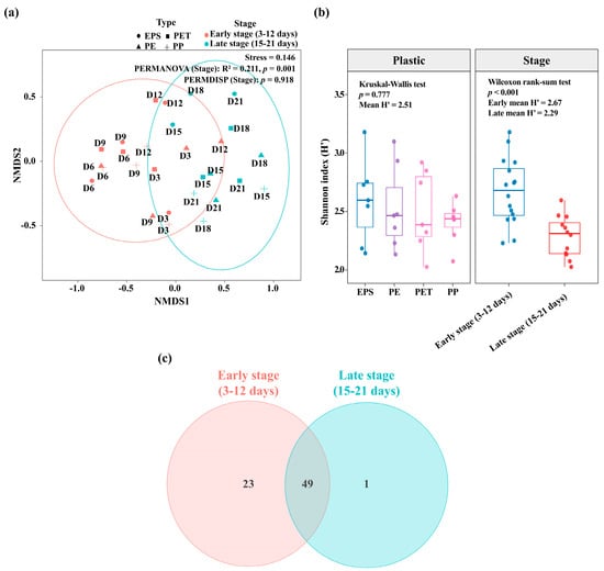
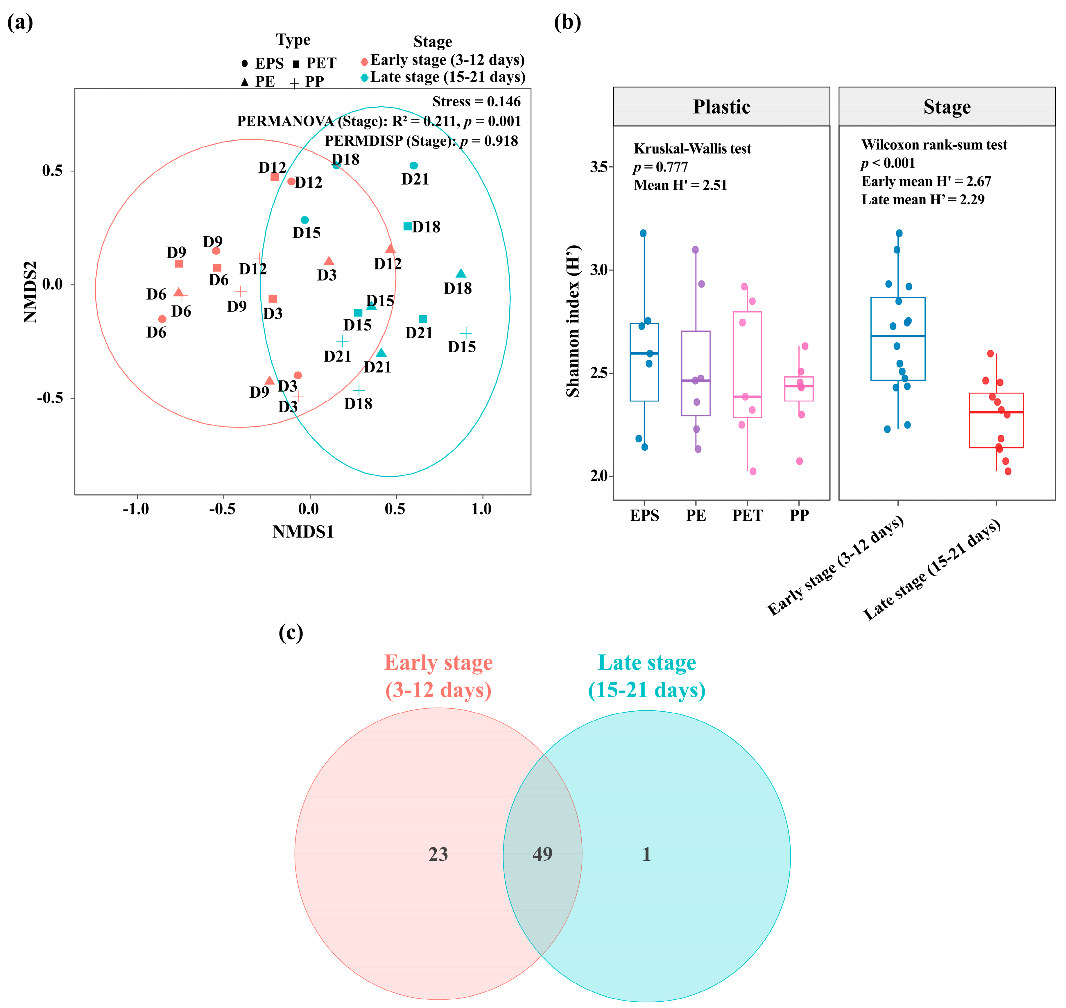
In our previous study, which is closely related to the present work, we examined microbial colonization of plastic surfaces during biofilm development by characterizing the initial attachment and successional dynamics of bacterial assemblages on four polymer types (EPS, PE, PP, and PET) over a 21-day incubation [13]. Consistent with the temporal shifts in bacterial community composition observed in our previous study [13], NMDS analysis revealed a clear separation of phage communities between the early (days 3–12) and late (days 15–21) stages (PERMANOVA, p < 0.001; PERMDISP, p > 0.05; Figure 2a). The separation between stages was unlikely to be attributable to differences in within-group dispersion but instead reflected differences in phage community composition. Polymer type had no significant effect on phage community composition (PERMANOVA, p > 0.05). Nevertheless, polymer surface roughness may have influenced initial microbial attachment. For example, scanning electron micrographs showed that the relatively rough surface of EPS was associated with differences in bacterial community composition during early colonization [13]. Given the absence of significant differences in phage community composition among polymer types, temporal changes in phage communities may be associated with bacterial biofilm succession. Furthermore, shifts in bacterial host lineages during biofilm development may influence phage community composition by altering host availability.
Figure 2.
Dynamics of phage community structure in plastisphere. (a) Non-metric multidimensional scaling (NMDS) ordination of phage community composition based on Bray–Curtis dissimilarity. Samples are colored by developmental stage (early stage: days 3–12; late stage: days 15–21) and shaped by four plastic types (Polypropylene, PP; Polyethylene, PE; Polyethylene terephthalate; PET, and Expanded polystyrene, EPS). Ellipses indicate group clustering, showing clear separation between early- and late-stage community. (b) Boxplot of Shannon diversity (H′) of phage community during developmental plastic types and stages. Boxplots show median and interquartile ranges with individual samples overlaid. (c) Venn diagram showing the unique and shared phage genera between early and late stages.
The Shannon diversity index of the phage community was significantly higher in the early stage than in the late stage (Wilcoxon rank-sum test, p < 0.001; Figure 2b). The diversity peaked on days 6–9 and gradually declined thereafter, resulting in significantly lower diversity during the late stage. The Venn diagram analysis revealed 23 phage taxa unique to the early stage, one unique to the late stage, and 49 shared between stages (Figure 2c). The higher number of early-stage-specific phages, accompanied by higher phage diversity on days 6–9, was consistent with the pattern of increased bacterial diversity. During the initial stage of biofilm formation, some bacterial species become dominant, followed by the rapid colonization of the plastic surface by various bacteria associated with these pioneer species. As the diversity of host bacteria increases, diverse phage lineages are introduced into the plastisphere. In addition, as the biofilm matures over time, specific bacterial taxa often become dominant, leading to the predominance of host-specific phages. These findings suggest that phage community dynamics may be associated with host availability and bacterial attachment to plastic substrates [34]. The substantial overlap between stages, with 49 phage taxa (67.1%) shared across both stages, further suggests that many phage taxa were sustained with biofilm development. The presence of 23 phage taxa unique to the early stage, compared with only one in the late stage, suggests higher phage diversity during early biofilm development, potentially reflecting the coexistence of diverse bacterial hosts and their associated phages. The subsequent enrichment of specific phage taxa may be associated with bacterial host succession. As biofilms developed and bacterial communities changed, phage community composition may have shifted with host availability, potentially contributing to the lower number of stage-specific phage taxa in the late stages [35]. These findings suggest that phage communities in the plastisphere undergo temporal reorganization rather than a simple decline in diversity. Thus, the plastisphere may serve as an ecological habitat and reservoir for phages, potentially facilitating virus–host interactions.
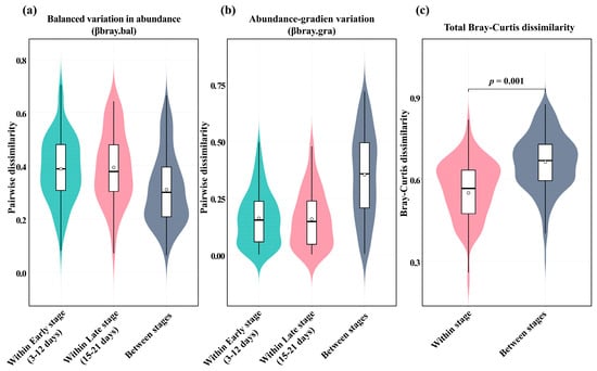
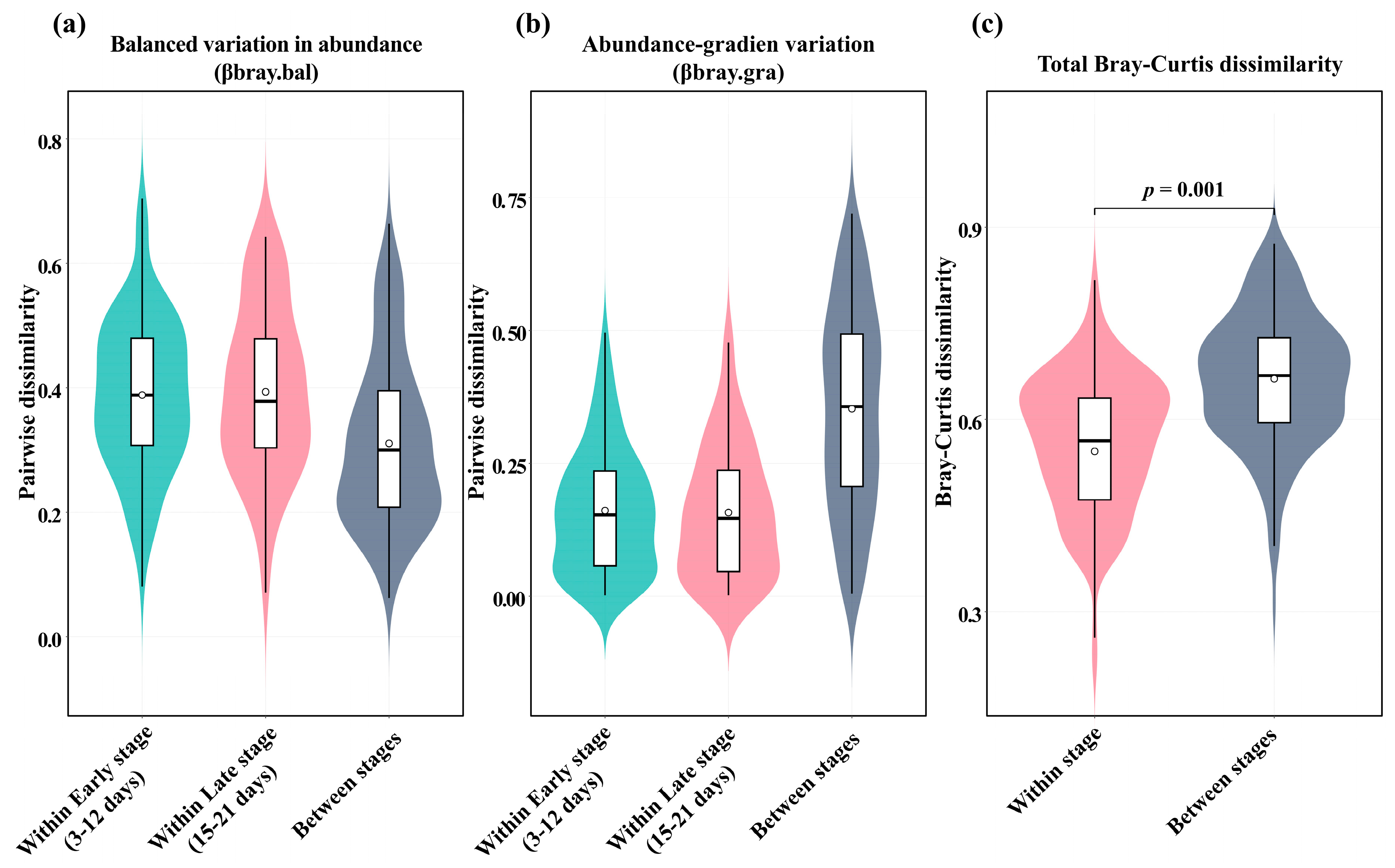
To evaluate the processes underlying temporal changes in the phage community, we performed a turnover analysis. Because the phage community showed no significant differences among plastic materials but differed significantly between the two temporal groups, the analysis was conducted separately for the early (days 3–12) and late (days 15–21) stages. AB βbray.bal was relatively high within each stage but decreased in between-stage comparisons (Figure 3a). In contrast, AB βbray.gra remained relatively low within each stage but increased between stages (Figure 3b). Abundance-based turnover was significantly higher between stages than within stages (p < 0.001; Figure 3c), indicating different phage community compositions between the early and late stages. Thus, these results indicate temporal shifts in phage community composition between the early and late stages, accompanied by increased abundance-gradient variation. In particular, the temporal dynamics of Vibrio populations and their associated phages are consistent with the possibility that phage-mediated top-down pressure contributed to changes in Vibrio abundance and subsequent bacterial community turnover (Figure 4a). Similar turnover-dominated dynamics have been reported in other virus–host systems [36], where phage communities may reorganize in response to changes in host availability and infection dynamics. This interpretation is also consistent with Tan et al. [35], who demonstrated that Vibrio phages reduced Vibrio populations within biofilms and altered bacterial community composition through the selection of phage-resistant strains. Therefore, these observations suggest that phage–host interactions may contribute to bacterial population dynamics and community turnover in biofilm environments. The observed decline in Vibrio abundance may have been partly associated with host-specific phage predation. More broadly, the temporal turnover of phage communities in the plastisphere may reflect changes in host bacterial community composition rather than simply the appearance or disappearance of individual taxa.
Figure 3.
Temporal changes (turnover analysis) in phage community structure of abundance-based (AB) Bray–Curtis dissimilarity (AB βbray). (a) Abundance-based Bray–Curtis balanced variation (AB βbray.bal) for within- and between-stage comparisons (early: days 3–12; late: days 15–21). (b) Abundance-based Bray–Curtis abundance-gradient component (AB βbray.gra) for within- and between-stage comparisons (early: days 3–12; late: days 15–21). (c) Comparison of balanced variation in abundance among comparison groups using a permutation test (p < 0.001). Violin plots with embedded boxplots show the distribution of pairwise dissimilarities, with medians and interquartile ranges indicated.
Figure 4.
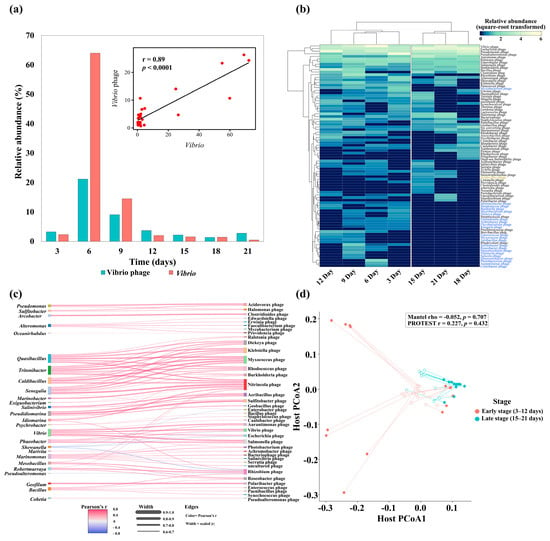
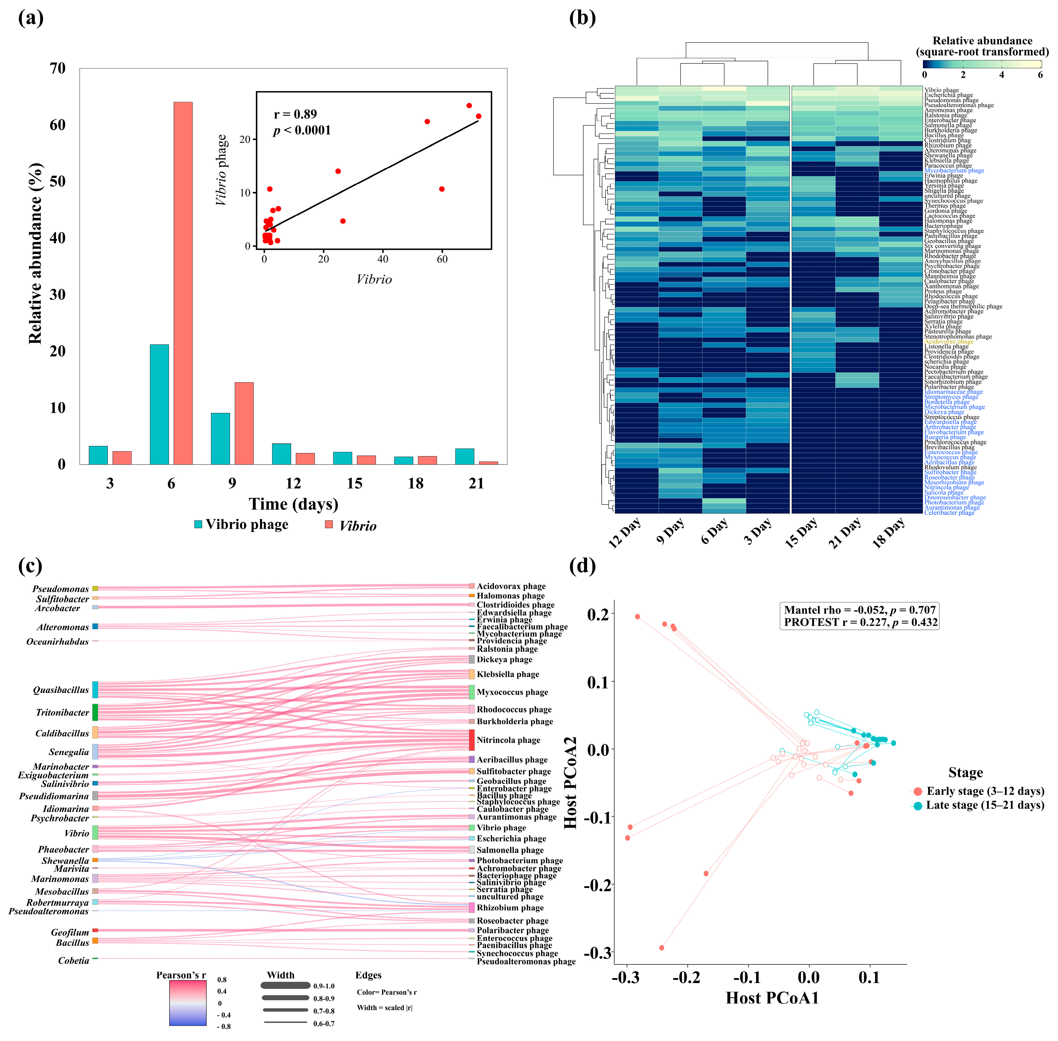
Host–phage associations within plastisphere. (a) Bar plot showing a relationship between Vibrio spp. and Vibrio phages. The inset graph shows significant positive correlation between the bacteria and the phage. (b) Heatmap showing the relative abundance (square-root-transformed) of dominant phage genera. Rows represent phage genera and columns represent sampling time points, with hierarchical clustering highlighting temporal patterns. (c) Sankey diagram illustrating correlation between common bacteria and phages. Link width represents the strength of host–phage associations based on correlation analysis. The information on abbreviation of species is shown in Table S4. (d) Procrustes analysis based on PCoA ordinations comparing host and phage communities. Points represent samples colored by stage (early: days 3–12; late: days 15–21), and lines connect paired host–phage configurations.
A total of 73 phage genera were identified in this study, and their temporal distributions at the genus level are presented in the heatmap (Figure 4b). In the early stage, the community was strongly dominated by specific lineages, including Vibrio phage, Escherichia phage, Pseudoalteromonas phage, and Pseudomonas phage. Similarly, early-stage biofilms were characterized by pioneer bacterial hosts, such as Vibrio, Pseudoalteromonas, and Escherichia [13]. The phage communities identified in this study exhibited host-associated patterns. A Sankey network analysis suggested a potential host-associated interaction pattern (Figure 4c, Table S4). In seawater, interactions between free-living bacteria and phages may be relatively diffuse [37], whereas plastic-associated biofilms may favor more specific phage–host associations [18]. In particular, the phages detected in this study were predicted to be associated primarily with Gammaproteobacteria, a group frequently reported on artificial substrates, including plastics and antifouling-coated hulls [13,14,15]. Moreover, some taxa within Gammaproteobacteria have been reported to be prevalent in organically enriched or contaminated environments [14]. These predicted associations suggest potential ecological links between phages and bacterial taxa commonly found on artificial surfaces, although direct interactions between the phages and their putative hosts remain to be confirmed. Nitrincola phage, Rhizobium phage, and Sulfitobacter phage showed correlations with various hosts, while most phages exhibited specific correlations with particular bacteria. Phages have been reported to be adsorbed onto plastic surfaces through non-specific electrostatic or hydrophobic interactions [38]. Mantel and Procrustes analyses indicated not significant correspondence between bacterial and phage communities (Mantel’s p = 0.707; Procrustes’s p = 0.432; Figure 4d), suggesting that host–phage interactions were localized to specific genus-level associations rather than parallel changes in the entire community. Our previous studies of planktonic marine microbial communities have reported broad and temporally variable virus–host associations [39]. In contrast, the composition of phage communities in plastispheres appears to be closely linked to the formation and maturation of bacterial communities within biofilm matrices on substrates. Although direct infection relationships were not established for most bacterial and phage taxa, these patterns may be interpreted as potential ecological associations rather than evidence of direct causal interactions. The host specificity of phages may allow multiple phage taxa to coexist within bacterial communities dominated by a limited number of host taxa. During biofilm maturation, changes in bacterial diversity may be associated with shifts in phage community composition, potentially reflecting interactions between host availability and phage community dynamics [40].
Vibrio phages increased rapidly in the early stage, paralleling the persistently high relative abundance of Vibrios over the same period. Moreover, host–phage abundances were strongly and positively correlated within the Vibrios lineage (Spearman’s r = 0.89; Figure 4a), supporting tight genus-level co-dynamics and closely coupled population trajectories during early succession. Many Vibrios adopt a “feast-or-famine” strategy, characterized by rapid growth when labile organic substrates become available [41]. Such localized enrichment of labile substrates may create transient nutrient hotspots that favor fast-growing heterotrophs such as Vibrio, thereby promoting their rapid colonization during the early stages of biofilm formation [41,42]. Such density-dependent amplification is consistent with predictions of the “kill-the-winner” framework, in which dominant bacterial taxa support corresponding viral proliferation [43]. Despite the absence of significant whole-community concordance, the strong relationship between Vibrios and Vibrio phages suggests that host–phage coupling occurred primarily within specific bacterial lineages rather than uniformly across the entire community. In addition, structural maturation and biomass accumulation of biofilms increase host density and contact frequency, thereby promoting sustained host–phage interactions and strengthening the persistence of dominant phage lineages.
3.2. Auxiliary Metabolic Genes in Phages on the Plastisphere
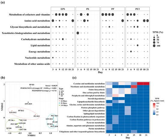
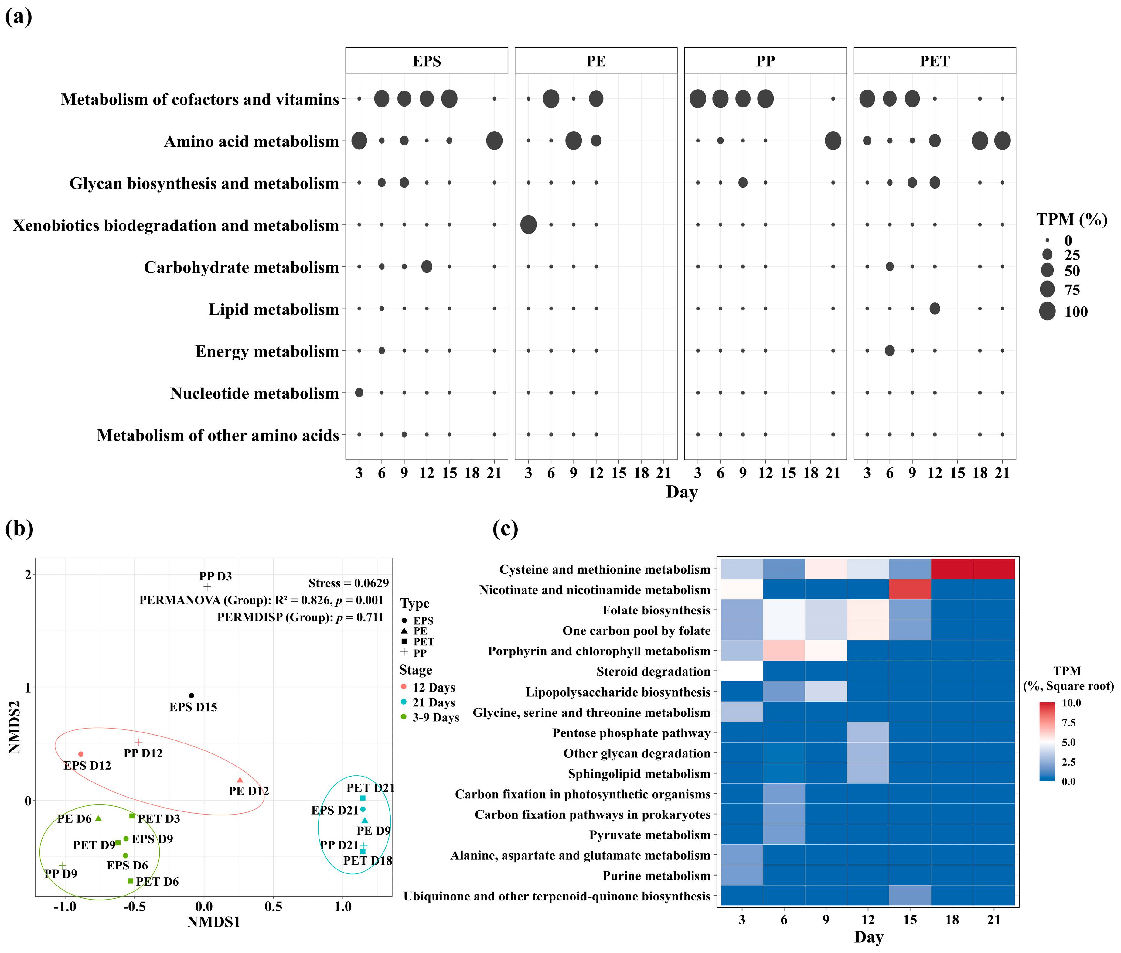
Phages can acquire AMGs from their hosts and transfer these genes back to host cells. These genes may enhance host metabolic activity and facilitate the production of new phage particles [17,44,45]. The AMGs identified in approximately 2700 phage vOTUs were classified into nine metabolic categories encompassing 19 pathways (Figure 5a). Among these, cofactor and vitamin, amino acid, and carbohydrate metabolism were consistently represented in all plastic types. Although metabolic functional differences among plastic types were generally limited, xenobiotic biodegradation-related AMGs were particularly enriched in the PE plastics during early biofilm formation. The dynamics of phage AMGs indicated changes in the environmental conditions of the plastisphere. Niu et al. [40] reported that phages play an important role in regulating key microbial metabolic processes in the environment. NMDS analysis based on AMG profiles revealed distinct clustering patterns in functional composition, with samples classified into three groups based on their AMG pathway profiles rather than sampling dates alone (Figure 5b). The AMG composition varied between temporal stages (Figure 5c). Cysteine and methionine metabolism was the most abundant pathway (30.84%), followed by porphyrin and chlorophyll metabolism (14.36%), folate biosynthesis (13.83%), and one-carbon pool by folate (13.83%).
Figure 5.
Temporal dynamics of phage-associated auxiliary metabolic genes (AMGs) in plastisphere. (a) Bubble plots showing the rate of transcripts per million mapped reads (TPM) of AMG functional categories during sampling times for each plastic type (Polypropylene, PP; Polyethylene, PE; Polyethylene terephthalate, PET; and Expanded polystyrene, EPS). (b) Non-metric multidimensional scaling (NMDS) ordination based on the results of phage AMGs. Samples are colored by biofilm developmental stage and shaped by plastic type. Ellipses indicate sample grouping. The stress value and PERMANOVA results are shown. (c) Heatmap showing the relative abundance of dominant AMG pathways across sampling days (square-root-transformed TPM), highlighting temporal shifts in functional composition. Samples were divided into three groups according to similarities in their AMG pathway composition, as determined by cluster analysis.
Vitamin- and porphyrin-related metabolic pathways predominated during the early stage, whereas pathways associated with sulfur-containing amino acid metabolism, including cysteine and methionine metabolism, were more abundant during the later stage. These temporal shifts suggest that the metabolic potential associated with phage AMGs may change throughout biofilm development. The enrichment of folate biosynthesis, one-carbon metabolism, and porphyrin-related pathways during the early stage may support nucleotide synthesis, redox balance, and cofactor supply during rapid microbial colonization [46]. In particular, tetrapyrrole compounds, including chlorophyll, heme, and the corrin ring of cobalamin (vitamin B12), are involved in diverse cellular processes and are synthesized through pathways [47,48]. Genes involved in tetrapyrrole biosynthesis have also been identified in the genomes of some phages isolated from marine environments, supporting the possibility that phages influence host cofactor metabolism [49]. The tetrapyrrole-related AMGs identified in this study suggest that metabolic interactions between phages and their hosts may be particularly relevant during the early stages of biofilm development. The representation of cysteine and methionine metabolism during the later stage suggests that sulfur-containing amino acid metabolism may become increasingly important as biofilms mature. Sulfur-containing amino acids are essential for protein synthesis, oxidative stress responses, and the maintenance of redox homeostasis [50,51]. This pattern is consistent with the findings of Niu et al. [40], who reported the enrichment of sulfur metabolism-related genes, including those involved in assimilatory sulfate reduction, in phage communities associated with plastic biofilms. These observations suggest that redox gradients and nutrient limitation within biofilms may be associated with changes in microbial sulfur metabolism [52]. However, further investigation is required to determine whether these environmental factors contribute to the observed patterns. Thus, the temporal shifts in AMG composition suggest that metabolic interactions between phages and their hosts may change during plastisphere development. Previous studies have proposed that phage-mediated metabolic modulation in plastisphere biofilms may enhance key host metabolic pathways under environmental constraints [18]. Accordingly, phage-derived AMGs may contribute to host metabolic adaptation, potentially supporting cofactor-related processes during early biofilm formation and amino acid and sulfur metabolism during later stages. Nevertheless, the AMG profiles identified in this study do not directly demonstrate gene expression or the functional contributions of these genes to host metabolism. Further studies are therefore needed to validate their expression and functional roles during biofilm development.
4. Conclusions
Our findings indicate that biofilm succession was more strongly associated with phage community dynamics and predicted functional potential than polymer identity in the plastisphere. Phage communities exhibited pronounced temporal turnover, characterized by shifts in taxon abundance and changes in community composition, alongside changes in bacterial community composition. Concurrent stage-dependent shifts in AMG composition, including the greater representation of sulfur metabolism-related genes during later biofilm development, suggest temporal variation in the predicted metabolic potential of plastisphere-associated phages. These findings highlight the plastisphere as a dynamic microbial habitat in which biofilm development is associated with changes in phage community composition and the distribution of phage-encoded metabolic genes. However, as this study was based on DNA genome, the presence and relative abundance of AMGs do not directly demonstrate their expression or functional contributions to host metabolism. Further studies incorporating gene expression analyses and direct measurements of metabolic activity are needed to determine whether these genetic patterns translate into functional changes in phage–host interactions during biofilm development.
Supplementary Materials
The following supporting information can be downloaded at: https://www.mdpi.com/article/10.3390/pathogens15111144/s1. Table S1, BLAST-based annotation of viral sequences with functional categorization. Viral proteins were classified into major functional groups, including DNA packaging, structural components, tail-associated proteins, host recognition, and replication-related proteins; Table S2, Sampling information, sequencing quality, assembly statistics, and viral contig classification based on CheckV; Table S3, Functional annotation and TPM-normalized abundance of auxiliary metabolic genes (AMGs) based on KEGG and Pfam classification. Each AMG was annotated with KEGG Orthology (KO), metabolic pathway, and Pfam domain information. TPM values represent normalized abundance estimated using Salmon; Table S4, Information of Bacterial hosts and bacteriophages used in Sankey plot. Bacterial hosts (H) and bacteriophages (P) are abbreviated in Figure 4c.
Author Contributions
Conceptualization, S.W.J.; Methodology, K.E.K.; G.E.J. and H.-J.K.; investigation, G.E.J. and H.-J.K.; data curation, K.E.K.; S.W.J.; writing—original draft preparation, K.E.K. and S.W.J.; visualization, K.E.K.; supervision, S.W.J.; project administration, S.W.J.; funding acquisition, S.W.J. All authors have read and agreed to the published version of the manuscript.
Funding
This work was financially supported by the National Research Foundation of Korea (NRF), funded by the Ministry of Science and ICT (Grant Number: RS-2026-25549102).
Institutional Review Board Statement
Not applicable.
Informed Consent Statement
Not applicable.
Data Availability Statement
The prokaryotic community data used for the host–bacteriophage comparative analyses were derived from Jeong et al. (2023), for which the raw 16S rDNA sequencing data are publicly available in the NCBI Sequence Read Archive (SRA) under BioProject accession number PRJNA928105. The bacteriophage community data have been deposited in the NCBI Sequence Read Archive (SRA) under BioProject accession PRJNA1551614.
Acknowledgments
The genomic DNA samples were stored the Library of Marine Samples of Korea Institute of Ocean Science & Technology (KIOST), Republic of Korea. During the preparation of this manuscript, the authors used ChatGPT (GPT-6) for language polishing and grammar correction. In addition, Figurelabs (www.figurelabs.ai) was used to design Figure 1c and the graphical abstract. The authors have reviewed and edited the output and take full responsibility for the content of this publication.
Conflicts of Interest
The authors declare no conflicts of interest.
References
- Auta, H.S.; Emenike, C.U.; Fauziah, S.H. Distribution and importance of microplastics in the marine environment: A review of the sources, fate, effects, and potential solutions. Environ. Int. 2017, 102, 165–176. [Google Scholar] [CrossRef] [Scilit] [PubMed]
- Jambeck, J.R.; Geyer, R.; Wilcox, C.; Siegler, T.R.; Perryman, M.; Andrady, A.; Narayan, R.; Law, K.L. Plastic waste inputs from land into the ocean. Science 2015, 347, 768–771. [Google Scholar] [CrossRef] [Scilit] [PubMed]
- Zettler, E.R.; Mincer, T.J.; Amaral-Zettler, L.A. Life in the “plastisphere”: Microbial communities on plastic marine debris. Environ. Sci. Technol. 2013, 47, 7137–7146. [Google Scholar] [CrossRef] [Scilit] [PubMed]
- Keswani, A.; Oliver, D.M.; Gutierrez, T.; Quilliam, R.S. Microbial hitchhikers on marine plastic debris: Human exposure risks at bathing waters and beach environments. Mar. Environ. Res. 2016, 118, 10–19. [Google Scholar] [CrossRef] [Scilit] [PubMed]
- Kirstein, I.V.; Kirmizi, S.; Wichels, A.; Garin-Fernandez, A.; Erler, R.; Löder, M.; Gerdts, G. Dangerous hitchhikers? Evidence for potentially pathogenic Vibrio spp. on microplastic particles. Mar. Environ. Res. 2016, 120, 1–8. [Google Scholar] [CrossRef] [Scilit] [PubMed]
- Suttle, C.A. Viruses in the sea. Nature 2005, 437, 356–361. [Google Scholar] [CrossRef] [Scilit] [PubMed]
- Cobián Güemes, A.G.; Youle, M.; Cantú, V.A.; Felts, B.; Nulton, J.; Rohwer, F. Viruses as winners in the game of life. Annu. Rev. Virol. 2016, 3, 197–214. [Google Scholar] [CrossRef] [Scilit] [PubMed]
- Martinez-Hernandez, F.; Fornas, O.; Lluesma Gomez, M.; Bolduc, B.; de la Cruz Peña, M.J.; Martínez Martínez, J.; Anton, J.; Gasol, J.M.; Rosselli, R.; Rodriguez-Valera, F.; et al. Single-virus genomics reveals hidden cosmopolitan and abundant viruses. Nat. Commun. 2017, 8, 15892. [Google Scholar] [CrossRef] [Scilit] [PubMed]
- Wilhelm, S.W.; Suttle, C.A. Viruses and Nutrient Cycles in the Sea: Viruses play critical roles in the structure and function of aquatic food webs. BioScience 1999, 49, 781–788. [Google Scholar]
- Ding, W.; Wang, R.; Liang, Z.; Zhang, R.; Qian, P.-Y.; Zhang, W. Expanding our understanding of marine viral diversity through metagenomic analyses of biofilms. Mar. Life Sci. Technol. 2021, 3, 395–404. [Google Scholar] [CrossRef] [Scilit] [PubMed]
- Yamada, Y.; Guillemette, R.; Baudoux, A.C.; Patel, N.; Azam, F. Viral attachment to biotic and abiotic surfaces in seawater. Appl. Environ. Microbiol. 2020, 86, e01687-19. [Google Scholar] [CrossRef] [Scilit] [PubMed]
- Amaral-Zettler, L.A.; Zettler, E.R.; Mincer, T.J. Ecology of the plastisphere. Nat. Rev. Microbiol. 2020, 18, 139–151. [Google Scholar] [CrossRef] [Scilit] [PubMed]
- Jeong, G.E.; Kim, H.-J.; Kim, K.E.; Kim, Y.J.; Lee, T.K.; Shim, W.J.; Jung, S.W. Selective attachment of prokaryotes and emergence of potentially pathogenic prokaryotes on four plastic surfaces: Adhesion study in a natural marine environment. Mar. Pollut. Bull. 2023, 193, 115149. [Google Scholar] [CrossRef] [Scilit] [PubMed]
- Kim, H.-J.; Park, J.S.; Kim, S.M.; Kim, K.E.; Kim, Y.J.; Kim, M.-J.; Cha, H.-G.; Hyun, B.; Shim, W.J.; Lee, S.H.; et al. Effects of environmental factors on the population dynamics of potential pathogenic bacterial community attached to marine plastic debris. J. Hazard. Mater. 2025, 495, 138755. [Google Scholar] [CrossRef] [Scilit] [PubMed]
- Kim, H.-J.; Park, J.S.; Lee, T.-K.; Kang, D.; Kang, J.-H.; Shin, K.; Jung, S.W. Dynamics of marine bacterial biofouling communities after initial Alteromonas genovensis biofilm attachment to anti-fouling paint substrates. Mar. Pollut. Bull. 2021, 172, 112895. [Google Scholar] [CrossRef] [Scilit] [PubMed]
- Yu, Y.; Miao, L.; Adyel, T.M.; Waldschläger, K.; Wu, J.; Hou, J. Aquatic plastisphere: Interactions between plastics and biofilms. Environ. Pollut. 2023, 322, 121196. [Google Scholar] [CrossRef] [Scilit] [PubMed]
- Thompson, L.R.; Zeng, Q.; Kelly, L.; Huang, K.H.; Singer, A.U.; Stubbe, J.; Chisholm, S.W. Phage auxiliary metabolic genes and the redirection of cyanobacterial host carbon metabolism. Proc. Natl. Acad. Sci. USA 2011, 108, E757–E764. [Google Scholar] [CrossRef] [Scilit] [PubMed]
- Xia, R.; Yin, X.; Balcazar, J.L.; Huang, D.; Liao, J.; Wang, D.; Alvarez, P.J.J.; Yu, P. Bacterium-Phage Symbiosis Facilitates the Enrichment of Bacterial Pathogens and Antibiotic-Resistant Bacteria in the Plastisphere. Environ. Sci. Technol. 2025, 59, 2948–2960. [Google Scholar] [CrossRef] [Scilit] [PubMed]
- John, S.G.; Mendez, C.B.; Deng, L.; Poulos, B.; Kauffman, A.K.; Kern, S.; Brum, J.; Polz, M.F.; Boyle, E.A.; Sullivan, M.B. A simple and efficient method for concentration of ocean viruses by chemical flocculation. Environ. Microbiol. Rep. 2011, 3, 195–202. [Google Scholar] [CrossRef] [Scilit] [PubMed]
- Kim, Y.J.; Kim, K.E.; Kim, H.-J.; Park, J.S.; Kim, M.-J.; Kim, S.M.; Lee, T.; Jung, S.W. Dynamics of the DNA Viral Community in Korean Coastal Waters. Sci. Data 2025, 12, 1782. [Google Scholar] [CrossRef] [Scilit] [PubMed]
- Bolger, A.M.; Lohse, M.; Usadel, B. Trimmomatic: A flexible trimmer for Illumina sequence data. Bioinformatics 2014, 30, 2114–2120. [Google Scholar] [CrossRef] [Scilit] [PubMed]
- Nurk, S.; Meleshko, D.; Korobeynikov, A.; Pevzner, P.A. metaSPAdes: A new versatile metagenomic assembler. Genome Res. 2017, 27, 824–834. [Google Scholar] [CrossRef] [Scilit] [PubMed]
- Nayfach, S.; Camargo, A.P.; Schulz, F.; Eloe-Fadrosh, E.; Roux, S.; Kyrpides, N.C. CheckV assesses the quality and completeness of metagenome-assembled viral genomes. Nat. Biotechnol. 2021, 39, 578–585. [Google Scholar] [CrossRef] [Scilit] [PubMed]
- Guo, J.; Bolduc, B.; Zayed, A.A.; Varsani, A.; Dominguez-Huerta, G.; Delmont, T.O.; Pratama, A.A.; Gazitúa, M.C.; Vik, D.; Sullivan, M.B.; et al. VirSorter2: A multi-classifier, expert-guided approach to detect diverse DNA and RNA viruses. Microbiome 2021, 9, 37. [Google Scholar] [CrossRef] [Scilit] [PubMed]
- Ren, J.; Song, K.; Deng, C.; Ahlgren, N.A.; Fuhrman, J.A.; Li, Y.; Xie, X.; Poplin, R.; Sun, F. Identifying viruses from metagenomic data using deep learning. Quant. Biol. 2020, 8, 64–77. [Google Scholar] [CrossRef] [Scilit] [PubMed]
- Rognes, T.; Flouri, T.; Nichols, B.; Quince, C.; Mahé, F. VSEARCH: A versatile open source tool for metagenomics. PeerJ 2016, 4, e2584. [Google Scholar] [CrossRef] [Scilit] [PubMed]
- Bushnell, B. BBMap: A Fast, Accurate, Splice-Aware Aligner; Report LBNL-7065E; Lawrence Berkeley National Laboratory: Berkeley, CA, USA, 2014.
- Kim, K.E.; Jung, S.W.; Park, J.S.; Kim, H.-J.; Lee, C.; Ha, S.-Y.; Lee, T.-K. Optimized Metavirome Analysis of Marine DNA Virus Communities for Taxonomic Profiling. Ocean Sci. J. 2022, 57, 259–268. [Google Scholar] [CrossRef] [Scilit]
- Kieft, K.; Zhou, Z.; Anantharaman, K. VIBRANT: Automated recovery, annotation and curation of microbial viruses, and evaluation of viral community function from genomic sequences. Microbiome 2020, 8, 90. [Google Scholar] [CrossRef] [Scilit] [PubMed]
- Patro, R.; Duggal, G.; Love, M.I.; Irizarry, R.A.; Kingsford, C. Salmon provides fast and bias-aware quantification of transcript expression. Nat. Methods 2017, 14, 417–419. [Google Scholar] [CrossRef] [Scilit] [PubMed]
- Jackson, D.A. PROTEST: A PROcrustean Randomization TEST of community environment concordance. Écoscience 1995, 2, 297–303. [Google Scholar] [CrossRef] [Scilit]
- Peres-Neto, P.R.; Jackson, D.A. How well do multivariate data sets match? The advantages of a Procrustean superimposition approach over the Mantel test. Oecologia 2001, 129, 169–178. [Google Scholar] [CrossRef] [Scilit] [PubMed]
- Baselga, A. Separating the two components of abundance-based dissimilarity: Balanced changes in abundance vs. abundance gradients. Methods Ecol. Evol. 2013, 4, 552–557. [Google Scholar] [CrossRef] [Scilit]
- Kutralam-Muniasamy, G.; Shruti, V.C.; Pérez-Guevara, F. Plastisphere-hosted viruses: A review of interactions, behavior, and effects. J. Hazard. Mater. 2024, 472, 134533. [Google Scholar] [CrossRef] [Scilit]
- Tan, D.; Dahl, A.; Middelboe, M. Vibriophages Differentially Influence Biofilm Formation by Vibrio anguillarum Strains. Appl. Environ. Microbiol. 2015, 81, 4489–4497. [Google Scholar] [CrossRef] [Scilit] [PubMed]
- Needham, D.M.; Chow, C.-E.T.; Cram, J.A.; Sachdeva, R.; Parada, A.; Fuhrman, J.A. Short-term observations of marine bacterial and viral communities: Patterns, connections and resilience. ISME J. 2013, 7, 1274–1285. [Google Scholar] [CrossRef] [Scilit] [PubMed]
- Kim, K.E.; Joo, H.M.; Kim, Y.J.; Kang, D.; Lee, T.-K.; Jung, S.W.; Ha, S.-Y. Ecological Interaction between Bacteriophages and Bacteria in Sub-Arctic Kongsfjorden Bay, Svalbard, Norway. Microorganisms 2024, 12, 276. [Google Scholar] [CrossRef] [Scilit] [PubMed]
- Dika, C.; Duval, J.F.L.; Francius, G.; Perrin, A.; Gantzer, C. Isoelectric point is an inadequate descriptor of MS2, Phi X 174 and PRD1 phages adhesion on abiotic surfaces. J. Colloid Interface Sci. 2015, 446, 327–334. [Google Scholar] [CrossRef] [Scilit] [PubMed]
- Kim, M.-J.; Kim, K.E.; Kim, H.-J.; Kim, Y.J.; Lee, T.-K.; Kim, S.M.; Cha, H.-G.; Jung, S.W. Co-occurrence patterns between Chlorophyta and nucleocytoplasmic large DNA virus in coastal ecosystem, South Korea. Mar. Environ. Res. 2025, 204, 106944. [Google Scholar] [CrossRef] [Scilit] [PubMed]
- Niu, L.; Zhao, S.; Chen, Y.; Li, Y.; Zou, G.; Tao, Y.; Zhang, W.; Wang, L.; Zhang, H. Diversity and potential functional characteristics of phage communities colonizing microplastic biofilms. Environ. Res. 2023, 219, 115103. [Google Scholar] [CrossRef] [Scilit] [PubMed]
- Eilers, H.; Pernthaler, J.; Amann, R. Succession of pelagic marine bacteria during enrichment: A close look at cultivation-induced shifts. Appl. Environ. Microbiol. 2000, 66, 4634–4640. [Google Scholar] [CrossRef] [Scilit] [PubMed]
- Yang, J.W.; Chang, F.-H.; Yeh, Y.-C.; Tsai, A.-Y.; Chiang, K.-P.; Shiah, F.-K.; Gong, G.C.; Hsieh, C.H. Trade-Offs between Competitive Ability and Resistance to Top-Down Control in Marine Microbes. mSystems 2023, 8, e01017-22. [Google Scholar] [CrossRef] [Scilit] [PubMed]
- Winter, C.; Bouvier, T.; Weinbauer, M.G.; Thingstad, T.F. Trade-offs between competition and defense specialists among unicellular planktonic organisms: The “killing the winner” hypothesis revisited. Microbiol. Mol. Biol. Rev. 2010, 74, 42–57. [Google Scholar] [CrossRef] [Scilit] [PubMed]
- Mahmoudabadi, G.; Milo, R.; Phillips, R. Energetic cost of building a virus. Proc. Natl. Acad. Sci. USA 2017, 114, E4324–E4333. [Google Scholar] [CrossRef] [Scilit] [PubMed]
- Hurwitz, B.L.; U’Ren, J.M. Viral metabolic reprogramming in marine ecosystems. Curr. Opin. Microbiol. 2016, 31, 161–168. [Google Scholar] [CrossRef] [Scilit] [PubMed]
- Zhou, K.; Wong, T.Y.; Long, L.; Anantharaman, K.; Zhang, W.; Wong, W.C.; Zhang, R.; Qian, P.-Y. Genomic and transcriptomic insights into complex virus–prokaryote interactions in marine biofilms. ISME J. 2023, 17, 2303–2312. [Google Scholar] [CrossRef] [Scilit] [PubMed]
- Zappa, S.; Li, K.; Bauer, C.E. The Tetrapyrrole Biosynthetic Pathway and Its Regulation in Rhodobacter capsulatus. Adv. Exp. Med. Biol. 2010, 675, 229–250. [Google Scholar] [CrossRef] [Scilit] [PubMed]
- Czarnecki, O.; Grimm, B. Post-translational control of tetrapyrrole biosynthesis in plants, algae, and cyanobacteria. J. Exp. Bot. 2012, 63, 1675–1687. [Google Scholar] [CrossRef] [Scilit] [PubMed]
- Wegner, H.; Roitman, S.; Kupczok, A.; Braun, V.; Woodhouse, J.N.; Grossart, H.-P.; Zehner, S.; Béjà, O.; Frankenberg-Dinkel, N. Identification of Shemin pathway genes for tetrapyrrole biosynthesis in bacteriophage sequences from aquatic environments. Nat. Commun. 2024, 15, 8783. [Google Scholar] [CrossRef] [Scilit] [PubMed]
- Turnbull, A.L.; Surette, M.G. Cysteine biosynthesis, oxidative stress and antibiotic resistance in Salmonella typhimurium. Res. Microbiol. 2010, 161, 643–650. [Google Scholar] [CrossRef] [Scilit] [PubMed]
- Levine, R.L.; Mosoni, L.; Berlett, B.S.; Stadtman, E.R. Methionine residues as endogenous antioxidants in proteins. Proc. Natl. Acad. Sci. USA 1996, 93, 15036–15040. [Google Scholar] [CrossRef] [Scilit] [PubMed]
- Stewart, P.S.; Franklin, M.J. Physiological heterogeneity in biofilms. Nat. Rev. Microbiol. 2008, 6, 199–210. [Google Scholar] [CrossRef] [Scilit] [PubMed]
Disclaimer/Publisher’s Note: The statements, opinions and data contained in all publications are solely those of the individual author(s) and contributor(s) and not of MDPI and/or the editor(s). MDPI and/or the editor(s) disclaim responsibility for any injury to people or property resulting from any ideas, methods, instructions or products referred to in the content. |
© 2026 by the authors. Licensee MDPI, Basel, Switzerland. This article is an open access article distributed under the terms and conditions of the Creative Commons Attribution (CC BY) license.