


Abstract
Schlafen proteins are interferon-responsive regulators of cellular and antiviral pathways that can profoundly influence viral replication and host defense. Diverse viral infections are known to induce expression of Schlafen family members in human cells, while infection studies in mice have been primarily limited to mSlfn2, a group I Schlafen protein without a clear ortholog in humans. RNA-seq analysis of three distinct infection models reveals that mouse Schlafens are induced in response to murine gammaherpesvirus 68 (MHV68) in fibroblasts, B cells, and primary peritoneal macrophages. Notably, mSlfn8 expression is induced under multistep low-MOI infection conditions, whereas infections initiated with higher viral inoculum fail to induce mSlfn8. To assess whether mSlfn8 or related Slfns impact MHV68 replication, we performed siRNA-mediated knockdown and CRISPR/Cas9 knockout of mSlfn2, mSlfn8, and mSlfn9 in NIH 3T3 cells. Two independent clonal mSlfn2 and mSlfn8 knockout cell lines supported higher viral titer, indicating that these mSlfns are restriction factors for MHV68 replication in murine fibroblasts. Collectively, these results demonstrate that mSlfn2 and mSlfn8 function as antiviral effectors during MHV68 infection and support a model in which MHV68 counteracts interferon-induced mouse Schlafen expression to promote efficient replication.
1. Introduction
The Schlafen protein family are a group of vertebrate-specific regulatory proteins involved in modulating several cellular responses, including viral infection, cell cycle arrest, and DNA damage [1]. Several of the Schlafen genes are type I interferon (IFN)-stimulated with direct immunological and antiviral roles, which has led to the classification of many SLFNs as broad antiviral restriction factors effective against numerous RNA and DNA virus families [1,2]. Humans encode six different Schlafen proteins (hSLFNs), while mice encode nine different Schlafen proteins (mSlfns). All Schlafen proteins encode an N-terminal core SLFN domain defined by a conserved motif called the SLFN box [3,4,5]. Additionally, the SLFN domain contains three endonuclease active site residues found in the majority of Schlafen proteins. The Schlafen protein family is further classified into groups I, II, or III based on overall domain structure and size [3]. While group I Schlafen proteins comprise a Schlafen core domain only, groups II and III contain a SWAVDL linker domain following their N-terminal Schlafen core domain. Group III proteins have a third helicase domain at their C-terminus with implications in ssDNA binding and DNA damage [4,5,6,7]. At present, the functions of these additional domains in Schlafen proteins are still being defined.
Schlafen proteins are known to bind and, in some cases, cleave transfer RNAs (tRNAs) through their N-terminal domain and endonuclease active site residues; moreover, tRNA binding and cleavage has been implicated in Schlafen antiviral activity [3,8,9,10,11,12]. Human SLFN11 (hSLFN11) and hSLFN13 can restrict HIV-1 infection by selectively inhibiting the production of viral proteins based on codon usage, suggesting that SLFN tRNA binding activity plays a role in its antiviral function [8,10]. Further, in vitro studies clearly demonstrate mouse Slfn8 (mSlfn8) and hSLFN11-14 can cleave mature tRNAs at D and T-loop regions, generating various tRNA fragments (tRFs) [10,12,13]. This endonuclease activity affords SLFN proteins the potential to manipulate viral translation by inactivating mature tRNAs, as well as to produce effector RNAs that can independently modulate gene expression. In support of this hypothesis, N-terminal domain mutants of hSLFN11, 12, 13, and 14 exhibit reduced antiviral activity against several RNA viruses, an activity that is attributed to their tRNA/rRNA endoribonuclease activity [2,10,14,15,16]. Finally, mature and premature tRFs induced by viral infection have been shown to silence gene expression, but whether these are generated by Schlafen proteins is unknown [17,18,19]. Overall, it appears that Schlafens effectively restrict cellular tRNA functionality, which in turn inhibits viral translation.
To date, hSLFN5, hSLFN11, hSLFN12, and hSLFN14 have been shown to have antiviral activity against herpesviruses [20,21,22,23]. hSLFN5 suppresses HSV-1 transcription by directly binding to the viral genome and limiting RNA polymerase accessibility [20,24]. hSLFN11 and hSLFN12 have both been shown to restrict beta- and gammaherpesvirus infection, respectively, while hSLFN14 has an antiviral effect against the alphaherpesvirus, varicella zoster virus [21,22,23,25,26]. However, the specific functions of hSLFN11, hSLFN12, and hSLFN that drive herpesvirus restriction remain to be characterized. Additionally, which mouse Slfn proteins participate in herpesvirus restriction is unknown, though mSlfn2 mutant mice (also known as elektra-mutant mice) are more susceptible to murine cytomegalovirus infection [27]. The only clear orthologs of hSLFNs are mSlfn5 (59% sequence identity with hSLFN5), mSlfn14 (70% sequence identity with hSLFN14), and mSlfn9 (63% sequence identity with hSLFN13) [2]. Although the murine and human Schlafen families differ substantially in their composition and many family members lack direct orthologous relationships, studies in mice have been instrumental in defining fundamental roles for Schlafen proteins in immune regulation and host–pathogen interactions [27,28,29,30].
To further clarify whether mouse Slfns are induced by and respond to herpesvirus infection, we became interested in exploring mouse Slfn proteins as potential restriction factors for murine gammaherpesvirus 68 (MHV68), a model murine virus related to human Kaposi sarcoma-associated herpesvirus and Epstein–Barr virus. We report that mSlfn8 is upregulated in response to MHV68 infection in both mouse fibroblasts, B cells, and primary peritoneal macrophages. We find that blocking MHV68 viral DNA replication enhances mSlfn8 expression, demonstrating viral antagonism of mSlfn8 in a manner similar for other interferon-stimulated genes. Finally, we find evidence of control of MHV68 by mSlfn8, as well as family member mSlfn2, suggesting broad antiviral activity of gammaherpesvirus infection by mouse Slfn proteins.
2. Materials and Methods
2.1. Cell Lines and Culturing Conditions
NIH 3T3 (NIH/Swiss-derived) murine fibroblasts, NIH 3T12 murine fibroblasts, and HEK293T human embryonic kidney cells (all obtained from ATCC, Manassas, VA, USA) were maintained in high-glucose Dulbecco’s modified Eagle’s medium (DMEM; Gibco 11965-092, Brooklyn, NY, USA) supplemented with 10% fetal bovine serum (FBS; Gibco A5670701). Cultures were screened regularly for mycoplasma (Invivogen, San Diego, CA, USA) and cultured in the absence of antibiotics. NIH 3T12s were used for virus propagation. Peritoneal cells from C57BL/6 mice were isolated following euthanasia by lavage with 10 mL of ice cold RPMI1640 (Gibco 11875-093) + 10% FBS + 1% pen/strep and cultured in the presence of 50 ng/mL murine M-CSF. Forty-eight hours after plating, non-adherent cells were removed by washing with PBS to obtain enriched peritoneal macrophage cultures.
2.2. siRNA Transfection
For siRNA transfection, pooled siRNA (Table 1) purchased from Dharmacon (Lafayette, CO, USA) was nucleofected into 5 × 10⁵ NIH 3T3 cells at 200 nM with the Neon™ Transfection System (Thermo Fisher, Waltham, MA, USA) using buffer E2, 100 uL tips and the settings: 1400 volts/ 20 ms/ 2 pulses. Cells were ejected into 2 mL DMEM + 10% FBS and seeded in 6-well dishes. Cells were infected 24 h post-transfection or post-nucleofection. No media was changed until time of inoculation with virus as detailed in Section 2.3.
Table 1.
Oligonucleotide sequences.
2.3. Virus Infections
MHV68-GFP was amplified in NIH 3T12s and titered by TCID50 on NIH 3T3s as described previously [31]. For infection, MHV68 virus was added to ½ culture volume of media, placed on NIH 3T3 cells as indicated for 60–90 min at a multiplicity of infection (MOI) of 5 or 0.05 (as calculated by TCID50) to allow viral entry. Cells were washed with PBS to remove unabsorbed virus, then replenished with fresh DMEM + 10% FBS medium. The same protocol was used for infection of peritoneal macrophages, except an MOI = 10 was used. For phosphonoacetic acid (PAA) treatments, PAA (Sigma, St. Louis, MO, USA) was added at a final concentration of 200 μg/mL into the media after viral absorption. Supernatants were titered by TCID50 on NIH 3T3.
2.4. RNA-Seq Datasets
Datasets from MHV68-infected NIH 3T3s and A20-HE-RITs were downloaded from the SRA database: SRR19792326, SRR19792325, SRR19792324, SRR19792321, SRR25823338, SRR25823339 [32]. MHV68-infected peritoneal macrophages were suspended in Trizol and RNA was purified and sent to Genewiz. Whole transcriptome sequencing was performed via rRNA-depletion and analysis performed by Genewiz’s basic bulk RNA-sequencing package. Sequence reads were trimmed using Trimmomatic v.0.36 and mapped to a custom combined mouse + MHV68 genome file using the STAR aligner v.2.5.2b. Differential expression analysis was performed with DESeq2. The percent-of-family calculation was done by taking ERCC normalized read counts or transcripts per million (TPM) for each of the eight mSlfn genes analyzed here (Slfn1, 2, 3, 4, 5, 8, 9, 14), then calculating the percent for each gene over the total for the family. Raw data is deposited under BioProject accession PRJNA1520822 (SRX35015971, SRX35015972, SRX35015973, SRX35015974, SRX35015975, SRX35015976).
2.5. Schlafen Gene Knockout
Lentiviruses expressing sgRNA were generated by cloning gRNA oligos (sequences in Table 1) into the pMCB320 lentiviral vector carrying mCherry (Addgene #89359). Lentiviruses were packaged with 3rd generation packaging plasmids in HEK293Ts as follows. HEK293T cells were seeded 24 h before transfection in. 6-well dish at a density of 1.5 × 10⁶ cells/well. HEK293T cells were co-transfected with pMD2.G (0.31 µg), pMDL.G (0.63 µg), pRSV (0.31 µg), and pMCB320 (1.25 µg) using PolyJet (SignaGen). Lentivirus-containing supernatant was collected 48h post-transfection, 0.45 µM syringe-filtered, and frozen at −80 °C. Cas9-expressing NIH3T3s (Dharmacon; HD Cas9-010) were plated in a 6-well at 1 × 10⁶ cell/well and transduced with sgRNA lentivirus in the presence of polybrene (4 µg/mL). Plates were spinfected for 2 h at 800× g. The medium was changed at 48 h and antibiotic selection for pMCB320 (puromycin (5 µg/mL)) and Cas9 (blastocidin (5 µg/mL)) was added. Single cell clones positive for mCherry expression were FACS-sorted using a Cytek Aurora 2 Flow (Fremont, CA, USA) into a 96-well dish with the UI Flow Cytometry Facility.
2.6. Knockout Validation
Genomic DNA was isolated from Slfn knockout and non-targeting control cells using the DNeasy Blood and Tissue Kit (Zymo Research, Irvine, CA, USA). The sgRNA target site was amplified by PCR and fragments were resolved on a 2% agarose gel (primer sequences in Table 1). Bands were excised and purified using the Zymoclean Gel DNA Recovery Kit (Zymo Research), followed by the Zymo Clean and Concentrate Kit (Zymo Research) to further purify the DNA. Purified fragments were submitted to Plasmidsaurus for Premium PCR service. The chromatograms were analyzed using the ICE analysis program for the presence of indels (insertion/deletion) [33]. The KO score was >95% for all clones except mSlfn2-KO1, which was manually verified to carry a 170 bp deletion at the cut site, which generated a premature stop codon and truncates mSlfn2 after amino acid K40 (UniProt: Q9Z0I6).
2.7. Cell Growth and Viability Assays
To measure cellular proliferation, control and mSLFN-KO cells were seeded in triplicate at 3.5 × 10³ cells per well in a 96-well plate. Cells were then imaged at 24, 48, and 72 h post-seeding. Images of the plate were taken with the Agilent Biotek Cytation 5 with the high contrast brightfield accessory kit. Measurements of cell counts were analyzed with Agilent BioTek Gen5 image analysis software. Cell counts were converted into fold change over time and plotted using GraphPad Prism 11. LDH release was measured using an LDH-Glo kit (Promega, Madison, WI, USA) and a Tecan plate reader.
2.8. RT-qPCR
Total RNA was isolated from cells using TRIzol (Invitrogen, Carlsbad, CA, USA) and was treated with TURBO DNase (Ambion, Austin, TX, USA) to remove contaminating DNA. TURBO-treated RNA was then reverse transcribed with AMV RT (Promega) primed with random 9-mers. Gene expression was measured by amplifying cDNA using iTaq SYBR Green Supermix (Biorad, Hercules, CA, USA) on a QuantStudio 3 (Applied Biosystems, Foster City, CA, USA), and all primers are listed in Table 1. RT-qPCR was analyzed using the delta delta Ct (ΔΔCt) method. Average fold changes were calculated relative to 18s internal controls.
2.9. Statistical Analysis
All experiments were performed in at least biological duplicate or triplicate, meaning that experiments were performed on different days with different cell populations in terms of stock vial or passage number. All RT-qPCR statistical analysis was performed using raw ΔCt values. The statistical test used is indicated in the figure legends.
3. Results
3.1. MHV68 Infection Induces mSlfn Expression
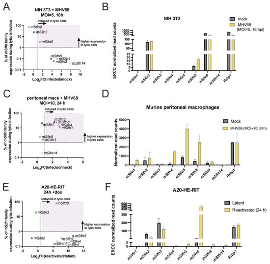
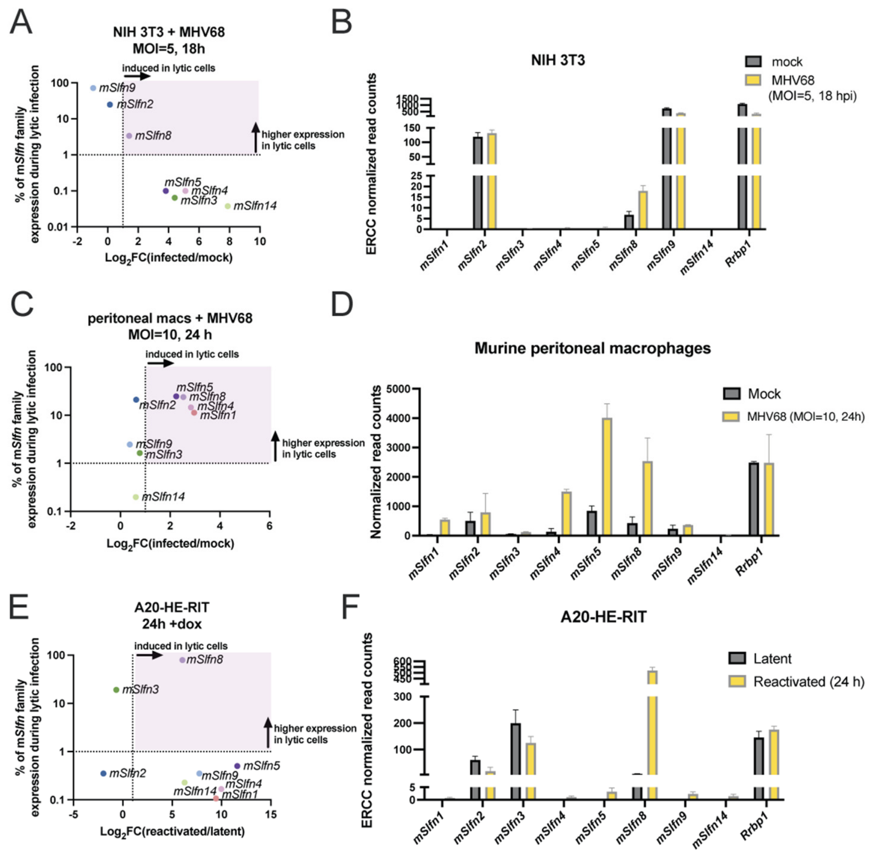
Human SLFN proteins have been shown to play a key role in the response to human herpesvirus infection, including HSV-1 and HCMV [20,21]. Mice encode at least nine Schlafen genes (mSlfn1, 2, 3, 4, 5, 8, 9, 10, and 14), but which of these might be antiviral apart from mSlfn2 is not known [27]. To explore commonly expressed Schlafen mRNAs across multiple MHV68 infection/reactivation models, we analyzed RNA-seq data from three different cell culture models of MHV68 infection: previously published de novo MHV68 infection (MOI = 5, 18 h) in NIH 3T3 fibroblasts [32] (Figure 1A,B), de novo MHV68 infection (MOI = 10, 24 h) in primary murine peritoneal macrophages (generated in our lab; Figure 1B,C), and published MHV68 lytic reactivation (24h doxycycline treatment) in A20-HE-RIT B cells [32,34] (Figure 1E,F). mSlfn10 was not included in our analysis, as it is classified as a non-coding pseudogene in C57BL/6, and whether it has coding capacity is not well defined for all cell lines and mouse strains. The first parameter we examined was percent of family expression (i.e., the relative expression of each mSlfn as a percentage of the eight measured mSlfn genes, specifically during lytic replication) (Figure 1A,C,E; Y-axis). Percent of family expression was chosen as a metric to cross-compare the different sequencing datasets, as these datasets were generated by different groups using different methodology. Differences in methodology used for RNA-seq can make direct comparison of metrics like transcript-per-million (TPM) inaccurate. Instead, the percent of family expression metric highlights the key mSlfn genes expressed during infection in each model. The second parameter we measured was log2(fold change) of each lytic condition vs. control (i.e., which mSlfns are induced by MHV68 de novo infection or reactivation) (Figure 1A,C,E; X-axis). Following infection in NIH 3T3 fibroblasts, mSlfn3, mSlfn4, mSlfn5, mSlfn8, and mSlfn14 were induced 2- to 200-fold by MHV68 (Figure 1A; X-axis). However, with the exception of mSlfn8, we also found these genes to be poorly expressed relative to other mSlfns in infected NIH 3T3s (Figure 1A; Y-axis; Figure 1B). Following infection of primary murine peritoneal macrophages, mSlfns were more evenly expressed across the family, with mSlfn1, mSlfn4, mSlfn5, mSlfn8 showing the strongest induction with MHV68 infection (Figure 1C,D). Reactivation of MHV68 in A20-HE-RITs led to induction of a slightly different profile, including enhanced expression of mSlfn1, mSlfn4, mSlfn5, mSlfn8, mSlfn9, and mSlfn14 (Figure 1E,F). mSlfn8 expression made up the majority of mSlfn family expression in A20-HE-RITs (Figure 1E). Overall, this analysis suggests that a subset of mSlfn genes is induced by active MHV68 infection, with three family members, mSlfn4, mSlfn5, and mSlfn8, induced in all analyzed cell culture models of infection, though expression level varied across the cell types and/or infection models.
Figure 1.
Mouse Schlafen genes are induced following MHV68 infection in three different cell types and infection models. Data was pulled from RNA-seq experiments performed in: (A,B) NIH 3T3 infected with MHV68 at an MOI = 5 for 18 h [32], (C,D) C57BJ/6 primary peritoneal macrophages infected with MHV68 at an MOI = 10 for 24 h, or (E,F) A20-HE-RITs reactivated from latency using doxycycline for 24 h [32,34]. For the dot plots (A,C,E), the log2(fold change of lytic/control) is presented on the X-axis. The percent of mSlfn family expression (the relative expression of each mSlfn as a percentage of the total eight measured mSlfn family transcripts) specifically from the lytic infected cells is presented on the Y-axis. The purple shaded area represents genes that are induced (>2-fold= dotted vertical line) and expressed at >1% of total mSlfn family levels (dotted horizontal line). For the bar graphs (B,D,F), normalized read counts for mouse Slfn genes in mock vs. infected or latent vs. reactivated are shown. Error bars represent the standard deviation across two independent experiments. Rrbp1 is shown as a representative host gene that is not interferon-stimulated. As the datasets were produced by different groups and with different methodologies, it is only appropriate to quantitatively compare within each infection model, and not across models.
To appreciate the difference in mSlfn expression following MHV68 infection specifically in NIH 3T3 fibroblasts, we noted that reads were detectable (defined as >10 reads) for mSlfn2, mSlfn8, and mSlfn9 transcripts, while read counts for other members of the mSlfn gene family were extremely low (<2 reads) (Figure 2A). mSlfn9 appeared to be constitutively expressed in NIH 3T3s, on par with another host mRNA Rrbp1, and exhibited decreased expression (down 2-fold) in response to MHV68. This trend is consistent with other host mRNAs (see Rrbp1 in Figure 1A; down 3.3-fold with infection) during infection in NIH 3T3s, likely a result of gammaherpesvirus-induced host shutoff [32,35]. In contrast, mSlfn2 expression was stable before and after infection, while mSlfn8 was induced 2.6-fold in response to MHV68 infection. Interestingly, mSlfn8 was induced in all three of the different cell culture models. Moreover, mSlfn8 shares approximately 60% amino acid identity with hSLFN11 and 64% with hSLFN13 [2,10]. Due to their high sequence similarity, and because hSLFN11 and hSLFN13 are known to have antiviral activity, we initially investigated a potential role for mSlfn8 during MHV68 infection.
Figure 2.
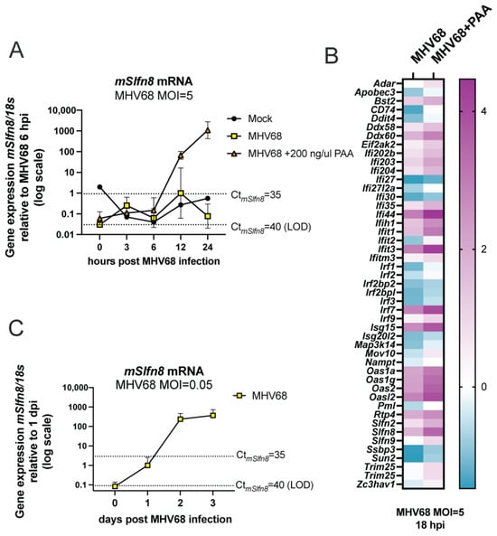
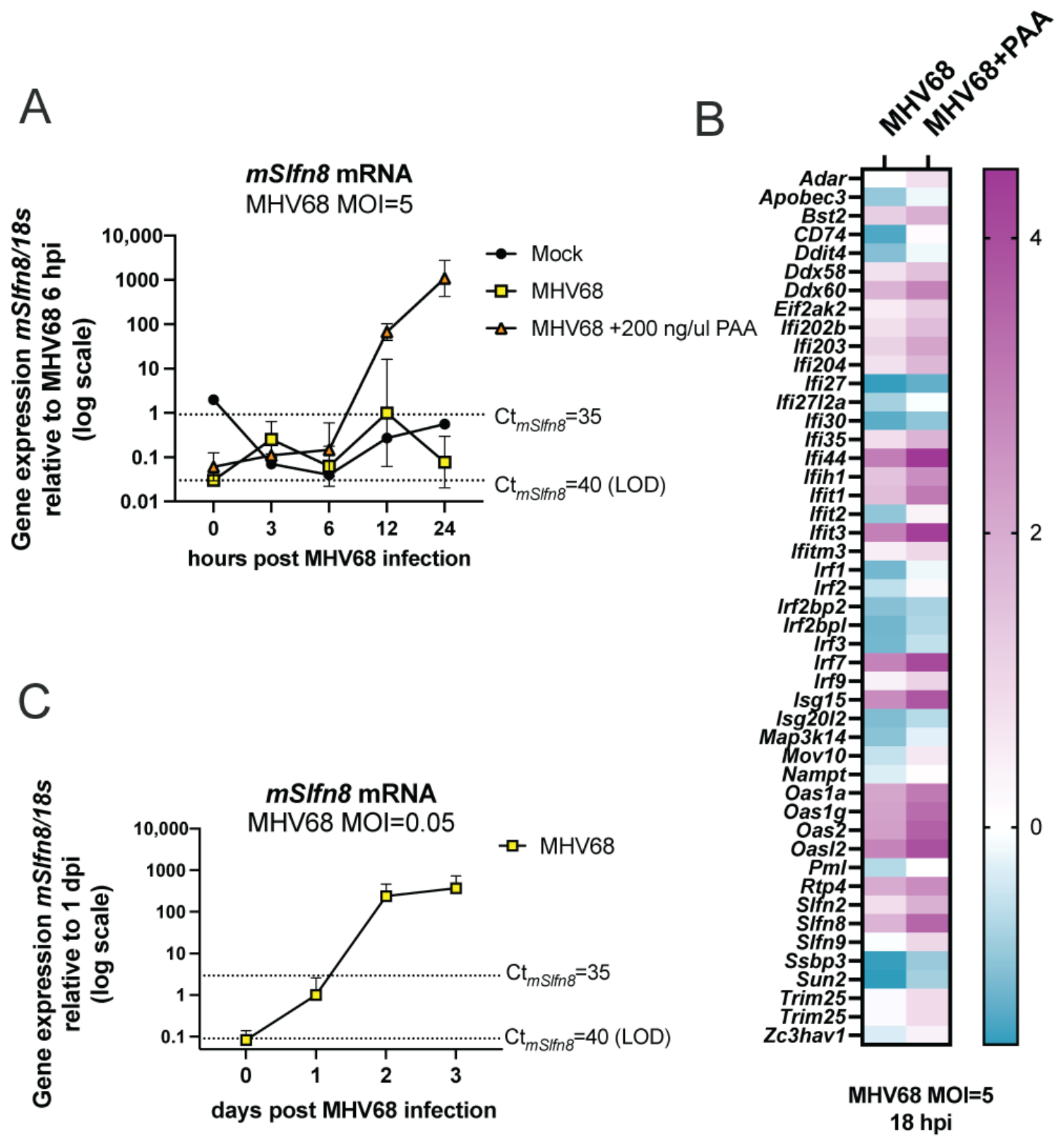
mSlfn8 is induced during MHV68 infection in NIH 3T3 fibroblasts. (A) NIH 3T3 cells were infected with MHV68 at an MOI of 5 and collected at the indicated times post-infection. RNA was isolated and analyzed by RT-qPCR using primers for mSlfn8 and 18s. Fold change was calculated relative to values at 12 hpi (+ MHV68/− PAA). Each data point represents the average of two independent experiments for mock and MHV68 and three for MHV68 + PAA conditions. Horizontal dotted lines indicate estimated Ct value limits for mSlfn8 at 35 and 40, with 40 representing the limit of detection (LOD). mSlfn8 is not abundantly expressed in mock or infected cells (Ct values between 35 and 40). (B) Log2(fold change of MHV68/mock) for a representative set of interferon-stimulated genes from published experiments performed in the presence or absence of PAA [32]. (C) NIH 3T3 cells were infected with MHV68 at an MOI of 0.05 and collected at the indicated times post-infection. RNA was isolated and analyzed by RT-qPCR using primers for mSlfn8 and 18s. Fold change was calculated relative to values at 1 dpi (+MHV68) and represents the average of three independent experiments.
3.2. PAA Treatment and Low-MOI Infection Enhance mSlfn8 Expression
Of the three Schlafen genes expressed in NIH 3T3s, mSlfn8 exhibited the strongest induction following MHV68 infection (Figure 1). To determine the parameters that dictate mSlfn8 induction, we explored RNA-seq data from NIH 3T3s infected with MHV68 in the presence of phosphonoacetic acid (PAA), which inhibits the viral DNA polymerase, blocking viral DNA replication and subsequent late gene expression [36]. Surprisingly, mSlfn8 expression appeared to be enhanced when viral DNA replication and late gene expression were blocked with PAA [32]. While mSlfn8 was shown previously to be stimulated by IFNα treatment [22,37,38], to our knowledge, PAA has not been formally reported to enhance the IFN response. We then decided to determine the kinetics of mSlfn8 expression upon MHV68 infection across a synchronized cycle of infection (MOI = 5, 24 h), both in the absence and presence of 200 µg/ml PAA (Figure 2B). Although mSlfn8 was detected in the previous RNA-seq data (Figure 1A,B), mSlfn8 was difficult to detect by RT-qPCR (Ct > 35) in both mock and MHV68-infected cells (Figure 2A). In contrast, mSlfn8 was strongly induced in the presence of MHV68 and PAA, unlike with MHV68 alone (Figure 2A), supporting published RNA-seq data [32]. Next, we reasoned that PAA treatment might broadly enhance ISG expression by permitting accumulation of immunostimulatory viral DNA replication intermediates or by reducing expression of replication-dependent viral immune antagonists [39,40,41]. To examine this, we plotted the expression of host ISGs following MHV68 infection (MOI = 5 for 18 h) in the presence or absence of PAA [32] (Figure 2B). As expected, a subset of the selected ISGs show enhanced expression upon MHV68 infection in fibroblasts. PAA addition further enhances the expression level of these ISGs above that seen with infection alone, suggesting that PAA addition during MHV68 infection does indeed enhance the expression of ISGs. Based on this information, we predicted that mSlfn8 induction might be enhanced in a low-MOI infection setting (MOI = 0.05), where MHV68 does not initially infect every cell in the culture, allowing enhanced paracrine signaling and propagation of antiviral pathways [42]. We measured the expression of mSlfn8 in NIH3T3 cells infected with MHV68 at an MOI of 0.05. Expression of mSlfn8 was evident by 2 days post-infection and peaked 3 days post-infection (Figure 2C). Induction of mSlfn8 following a low-MOI infection was much stronger (~1000 fold) than that observed with a high MOI. These results demonstrate that mSlfn8 expression increases in response to MHV68 infection when viral DNA replication is inhibited with PAA and is more robust during a low-MOI infection compared to a high-MOI infection.
3.3. siRNAs Targeting mSlfn8/9 Minimally Impact MHV68 Titer
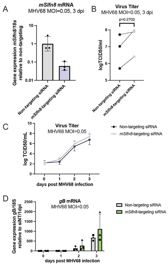
Next, we sought to determine whether mSlfns can restrict an MHV68 infection. We first performed a pilot experiment to assess the performance of siRNA knockdown for mSlfn8. NIH 3T3 cells were treated with ON-TARGETplus SMARTpool siRNA (mSlfn8-targeting siRNA) or nontargeting control siRNA (Non-targeting siRNA) as a negative control. NIH 3T3 cells were infected 24 h post-siRNA treatment with MHV68 at an MOI of 0.05. Though mSlfn8 antibodies have been made recently, this production was small-scale and not commercially available [38]. We thus validated mSlfn8 knockdown by RT-qPCR, specifically at 3 dpi when mSlfn8 Ct values are reliably measured (see Figure 2C) and found that mSlfn8 expression decreased by approximately 20-fold in mSlfn8-targeting siRNA-treated cells compared to non-targeting siRNA treatment (Figure 3A). Upon checking the siRNA sequences in our SMARTpool, we discovered the siRNA pool could potentially target mSlfn9, which shares 93% mRNA identity with mSlfn8. Accordingly, we found our siRNA pool led to reduced expression of mSlfn9 in addition to reduced expression of mSlfn8. Knockdown of mSlfn8 and mSlfn9 together resulted in 2- to 9-fold higher viral titers by 3 dpi, but this did not reach statistical significance (Figure 3B,C). We also measured gB mRNA, a MHV68 late transcript, but there was no difference between infected cells treated with non-targeting versus mSlfn8/9-targeting siRNAs (Figure 3D). Together, this data suggests that the combined knockdown of mSlfn8 and mSlfn9 did not significantly affect MHV68 replication. Although these pooled siRNA experiments were not pursued further, our findings emphasize the importance of accounting for off-target effects when targeting highly conserved gene families, such as the Schlafens.
Figure 3.
mSlfn8 knockdown minimally impacts MHV68 titer. (A) NIH 3T3 cells were nucleofected with non-targeting or mSlfn8-targeting siRNAs and infected with MHV68 at an MOI of 0.05 post-nucleofection. Knockdown was confirmed by RT-qPCR using primers for mSlfn8 and 18s. (B,C) Supernatants from (A) were titered by a TCID50 assay. The results of a paired t-test of the values from 3 days post-infection (dpi) are shown. (D) MHV68 late gene expression was assessed by RT-qPCR using primers for gB and 18s. Symbols refer to different biological replicates.
3.4. Validation and Growth Analysis of mSlfn-KO Cells
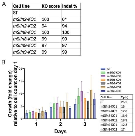
Our previous results underscore the importance of rigorously validating siRNA-mediated knockdowns. Off-target effects, particularly when using pooled siRNAs, can confound interpretation of phenotypes. Thus, we used CRISPR/Cas9 to make a clean knockout of mSlfn8. We also made knockout cell lines of the other two Schlafen genes expressed in NIH 3T3, mSlfn2 and mSlfn9, though we note that these were not induced across all infection models (Figure 1A,B). We used the Broad Institute CRISPick library to design two gene-specific guide RNAs per mSlfn gene (Table 1). Upon transduction and puromycin selection, we selected and amplified single cell clones and verified loss of coding capacity by amplicon sequencing and ICE analysis (Figure 4A; see details in Materials and Methods). Several groups have reported enhanced growth phenotypes following mSlfn knockout [7,37]. To evaluate the effect of mSlfn2, mSlfn8, and mSlfn9 knockout on cellular growth, we performed a growth curve using an imaging plate reader, including a single clone per guide alongside cells expressing safe-targeting guides (ST) to assess the growth of the cells following mSlfn knockout (Figure 4B). Over a 3-day growth period, we measured an average doubling time (Td) of 25.2 h for safe-targeting control cells. We observed enhanced growth for mSlfn-KO clones, with Td ranging from 12 to 19 h. Enhanced growth was consistently robust with both mSlfn2-KO cell lines, consistent with other reports [37].
Figure 4.
mSlfn2,8,9-KO confirmation and growth characteristics. (A) NIH 3T3 Cas9-expressing cells were transduced with a safe-targeting or mSlfn-targeting sgRNA lentiviruses, with mSlfn2, mSlfn8, and mSlfn9 targeted with two separate sgRNA sequences (1 or 2). Each locus was amplified for PCR, subjected to long read amplicon sequencing, and ICE analysis to calculate knockout score. * mSlfn2-KO2 lacks simple indels detected by ICE, but contains a 170 bp deletion disrupting the mSlfn2 ORF. (B) Cell count was measured using a Cytation 5 instrument and was normalized to the respective cell count at day 0. Doubling time (Td) in hours (h) was measured by averaging growth over three days.
3.5. mSlfn-KO Cells Support Higher Titer Replication of MHV68
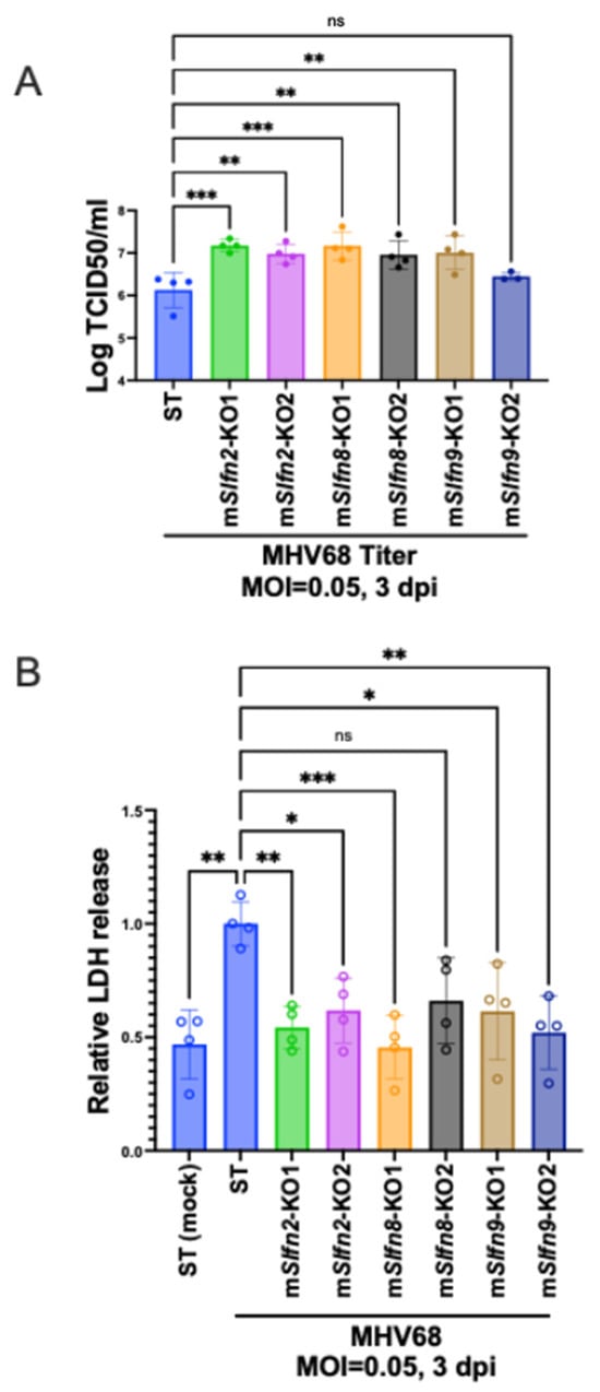
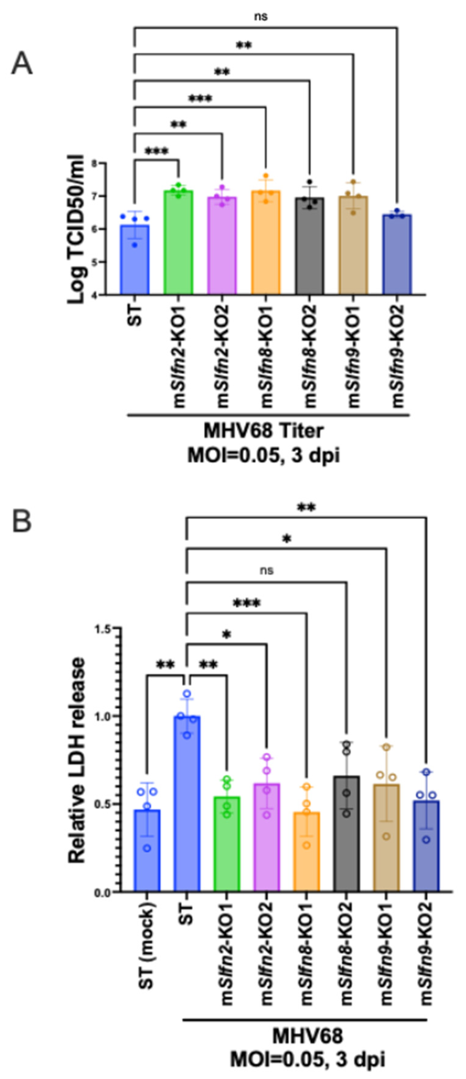
Using these validated mSlfn-KO clones, we next investigated whether mSlfn2, mSlfn8, or mSlfn9 restrict MHV68 replication. Because mSlfn8 expression was most strongly induced during low-MOI infection (Figure 2C), we infected safe-targeting cells and the corresponding mSlfn-KO cell lines with MHV68 at an MOI of 0.05 and quantified infectious virus production at 3 dpi (Figure 5A). Because of the observed difference in growth (Figure 4), we seeded safe-targeting and mSlfn-KO cells at 100% confluency prior to infection to limit differential cellular growth during the experiment. Disruption of either mSlfn2 or mSlfn8 consistently resulted in substantially higher viral titers compared with safe-targeting cells, with increases ranging from approximately 6- to 10-fold. A similar increase was observed in one mSlfn9 knockout clone (mSlfn9-KO1), whereas a second clone using a separate guide RNA (mSlfn9-KO2) did not reproducibly enhance virus production (Figure 5A). Importantly, both independent mSlfn2-KO clones and both mSlfn8-KO clones, each made with a unique guide RNA, exhibited elevated MHV68 replication, providing strong evidence that loss of either gene enhances viral growth. Next, we analyzed cell viability of our cultures at the endpoint of infection by measuring LDH release into the media (Figure 5B). With our safe-targeting cell line, we observed a 2-fold increase in LDH release upon infection, consistent with cell death following MHV68 lytic replication. However, LDH release for the infected mSlfn-KO cell lines remained similar to uninfected safe-targeting cells. This assay supports other studies demonstrating that Schlafens play important roles in cell death in response to DNA damage or interferon stimulation [7,11,37]. These findings identify mSlfn2 and mSlfn8 as host restriction factors that limit productive MHV68 infection, perhaps by promoting cell death.
Figure 5.
mSlfn2, 8, 9-KO cells support higher MHV68 titer and less cell death. mSlfn2, 8, 9-KO NIH 3T3 cells were seeded at 100% confluency and infected with MHV68 at an MOI of 0.05. Data is from four technical replicates collected from two independent experiments. (A) Supernatants were collected at 3 dpi and viral titers were measured by TCID50. (B) Supernatants were assayed for LDH using an LDH-Glo cytotoxicity assay. Supernatant from mock-infected safe-targeting control cells (ST) is included as an uninfected control. Data are presented as mean + SD. Statistical analysis was performed by one-way ANOVA. ns, not significant; * p < 0.05; ** p < 0.01; *** p < 0.001.
4. Discussion
Our findings demonstrate that mSlfn8 expression is induced during MHV68 infection, and that knockout of mSlfn8, along with mSlfn2, enhances viral titer in mouse NIH 3T3 fibroblasts. This observation emphasizes the role of mouse Schlafen family members in antiviral defense within the broader landscape of interferon-stimulated genes (ISGs) that restrict gammaherpesvirus replication. MHV68, like other herpesviruses, encodes multiple strategies to counteract interferon signaling and ISG activity. Consistent with this model, mSlfn8 induction was low except when we infected under low-MOI conditions, where multiple rounds of replication are expected to amplify innate immune signaling in bystander cells. We also observed high levels of mSlfn8 expression when cells were infected in the presence of PAA, which we hypothesize leads to enhanced ISG expression due to incomplete viral DNA intermediates or by blocking expression of viral immune antagonists. The induction of mSlfn8 suggests that it is part of the host antiviral response activated during infection. However, the observation that mSlfn8 expression is minimal following a high dose of MHV68 (MOI = 5) is suggestive of a viral mechanism that suppresses the IFN response, though we did not define this mechanism in this work. If viral antagonism is indeed at play, mSlfn8 may represent a biologically relevant barrier to efficient MHV68 replication.
The increased viral titers observed in independent mSlfn8 knockout clones provide direct functional evidence that mSlfn8 restricts productive MHV68 infection. Notably, a similar phenotype was observed in mSlfn2-KO cells. Because the effects were reproducible across independent knockout clones, these findings strongly support a bona fide antiviral role for both proteins. In contrast, the inconsistent phenotype observed between the two mSlfn9 knockout clones prevents definitive conclusions regarding a role for mSlfn9 in restricting MHV68 infection. We predict that the mSlfn9-KO2 cell line may have accumulated suppressor mutations or acquired off-target genetic perturbations but have not yet tested for these possibilities. Additional studies using complementation approaches will be required to determine whether mSlfn9 contributes to antiviral defense. Finally, it is possible that there is functional redundancy among mSlfns and that knocking out multiple Schlafens is required to enhance the antiviral phenotype observed.
Although the molecular mechanisms underlying mSlfn2- and mSlfn8-mediated restriction remain unclear, several possibilities can be considered. Schlafen family proteins have been implicated in diverse biological processes, including regulation of cellular proliferation, differentiation, translation, RNA metabolism, and innate immune signaling [1]. Mouse Schlafen proteins are less well characterized than their human SLFN counterparts, and many of their biological functions remain poorly understood. mSlfn2 is a group I Schlafen protein carrying a single Schlafen core domain and shown to regulate immune cell quiescence and protect tRNAs from stress-induced cleavage [27,29]. In contrast, mSlfn8 is a group III Schlafen protein consisting of a Schlafen core domain, SWADL linker, and an RNA/DNA helicase domain, and has similarities to human SLFN11/13 that serve antiviral roles and are important in the DNA damage response [7]. Not only do mSlfn2 and mSlfn8 differ by number of protein domains, but they also differ by the presence of functional endonuclease activity within their shared Schlafen core domain. While mSlfn8 retains an endonuclease active site and can cleave tRNAs in vitro, mSlfn2 lacks this activity and instead binds and protects tRNAs from cleavage [10,29]. Indeed, we found distinct roles of mouse Schlafens regarding cellular growth. Knockout of mSlfn2, 8, and 9 all had positive effects on cell growth, with mSlfn2-KO cells showing the most robust increase in growth. Due to these findings, we performed MHV68 infection on fully confluent monolayers to avoid growth effects on viral titer. Finally, future studies dissecting the stage of MHV68 replication that is enhanced in each mSlfn-KO cell line will be informative toward understanding the antiviral mechanisms of mSlfn proteins. Taken together, we posit that the differences in mSlfn2 and mSlfn8 domain structure and function suggest that these proteins likely restrict MHV68 through distinct mechanisms, which we are currently investigating.
Several limitations of the present study should be acknowledged. First, mSlfn knockouts were made and tested in NIH 3T3 fibroblasts, and the contribution of mSlfn8 to antiviral defense may differ in cell types that are naturally infected by MHV68 in vivo, including B cells, macrophages, and dendritic cells [43]. Second, although our knockout studies demonstrate that mSlfn2 and mSlfn8 limit virus replication, complementation and overexpression experiments restoring gene expression would further strengthen the causal relationship between Schlafen loss and enhanced viral growth. Thus, we cannot rule out the possibility that enhanced replication observed in mSlfn-KO cells arises from off-target gene editing or adaptation during selection of these cell lines. Third, given that mSlfn8 and mSlfn9 display functional redundancy, at least with regard to DNA damage response, MHV68 infection should also be characterized in mSlfn8/9 double knockout cells [7]. Finally, the viral factor or factors responsible for mSlfn8 (and other ISG) antagonism remain unknown and represent an important topic for future investigation.
Though more work is required to rigorously define the role of mouse Schlafens during viral infection, this study suggests that mSlfn8 and mSlfn2 contribute to antiviral defense against the gammaherpesvirus MHV68. The induction of mSlfn8 during infection, coupled with possible antagonism by MHV68 and the enhanced viral replication observed in knockout cells, supports a model in which mSlfn8 functions as part of the infection-induced antiviral response. These findings expand the understanding of Schlafen protein function during herpesvirus infection and establish a foundation for future studies aimed at defining the molecular mechanisms by which Schlafen family members restrict gammaherpesvirus replication.
Author Contributions
Conceptualization, S.G. and J.M.T.; methodology, S.G., A.N.L. and J.M.T.; validation, S.G., A.N.L. and J.M.T.; formal analysis, S.G., A.N.L. and J.M.T.; investigation, S.G., A.N.L. and J.M.T.; writing—original draft preparation, S.G. and J.M.T.; writing—review and editing, S.G. and J.M.T.; visualization, S.G. and J.M.T.; supervision, J.M.T.; project administration, J.M.T.; funding acquisition, S.G. and J.M.T. All authors have read and agreed to the published version of the manuscript.
Funding
This research was funded by the National Institutes of Health, grant R21AI173703 to J.M.T.; grant T32AI007533 to S.G.; the Office of Undergraduate Research at the University of Iowa, ICRU fellowship to A.N.L.
Institutional Review Board Statement
Not applicable.
Informed Consent Statement
Not applicable.
Data Availability Statement
RNA-seq data used in this manuscript can be found at SRA: SRR19792326, SRR19792325, SRR19792324, SRR19792321, SRR25823338, SRR25823339 [32]. RNA-seq data generated for this work can be found under BioProject accession PRJNA1520822 (SRX35015971, SRX35015972, SRX35015973, SRX35015974, SRX35015975, SRX35015976).
Acknowledgments
We thank Sarah Dremel for help in analyzing RNA-seq data, Paige Donovan and Wendy Maury for isolating the peritoneal macrophages, and Aimee Potter for assistance and training on the Cytation 5. We also thank members of the Tucker lab and Rich Roller for critical reading of the manuscript. Knockout cells were sorted at the Flow Cytometry Facility, which is a Carver College of Medicine/Holden Comprehensive Cancer Center core research facility at the University of Iowa. The facility is funded through user fees and the generous financial support of the Carver College of Medicine, Holden Comprehensive Cancer Center, and Iowa City Veteran’s Administration Medical Center.
Conflicts of Interest
The authors declare no conflicts of interest.
Abbreviations
The following abbreviations are used in this manuscript:
| SLFN | Schlafen |
| hSLFN | Human Schlafen |
| mSlfn | Mouse Schlafen |
| MHV68 | Murine gammaherpesvirus 68 |
| IFN | Interferon |
| ISG | Inteferon-stimulated genes |
| tRNA | Transfer RNAs |
| tRF | Transfer RNA fragment |
| PAA | Phosphonoacetic acid |
| HU | hydroxyurea |
| LDH | Lactate dehydrogenase |
References
- Mavrommatis, E.; Fish, E.N.; Platanias, L.C. The schlafen family of proteins and their regulation by interferons. J. Interferon Cytokine Res. 2013, 33, 206–210. [Google Scholar] [CrossRef] [Scilit] [PubMed]
- Kim, E.T.; Weitzman, M.D. Schlafens Can Put Viruses to Sleep. Viruses 2022, 14, 442. [Google Scholar] [CrossRef] [Scilit] [PubMed]
- Valdez, F.; Salvador, J.; Palermo, P.M.; Mohl, J.E.; Hanley, K.A.; Watts, D.; Llano, M. Schlafen 11 Restricts Flavivirus Replication. J. Virol. 2019, 93, 10-1128. [Google Scholar] [CrossRef] [Scilit] [PubMed]
- Murai, J.; Tang, S.W.; Leo, E.; Baechler, S.A.; Redon, C.E.; Zhang, H.; Al Abo, M.; Rajapakse, V.N.; Nakamura, E.; Jenkins, L.M.M.; et al. SLFN11 Blocks Stressed Replication Forks Independently of ATR. Mol. Cell 2018, 69, 371–384 e376. [Google Scholar] [CrossRef] [Scilit] [PubMed]
- Murai, J.; Thomas, A.; Miettinen, M.; Pommier, Y. Schlafen 11 (SLFN11), a restriction factor for replicative stress induced by DNA-targeting anti-cancer therapies. Pharmacol. Ther. 2019, 201, 94–102. [Google Scholar] [CrossRef] [Scilit] [PubMed]
- Xiao, H.; Han, Z.; Tang, Y.; Gao, X.; Xu, M.; Qiu, S.; Ren, N.; Yi, Y.; Zhou, C. SLFN11, far from being limited to responding to cancer DNA damage. Clin. Exp. Med. 2025, 25, 304. [Google Scholar] [CrossRef] [Scilit] [PubMed]
- Alvi, E.; Mochizuki, A.L.; Katsuki, Y.; Ogawa, M.; Qi, F.; Okamoto, Y.; Takata, M.; Mu, A. Mouse Slfn8 and Slfn9 genes complement human cells lacking SLFN11 during the replication stress response. Commun. Biol. 2023, 6, 1038. [Google Scholar] [CrossRef] [Scilit] [PubMed]
- Li, M.; Kao, E.; Gao, X.; Sandig, H.; Limmer, K.; Pavon-Eternod, M.; Jones, T.E.; Landry, S.; Pan, T.; Weitzman, M.D.; et al. Codon-usage-based inhibition of HIV protein synthesis by human schlafen 11. Nature 2012, 491, 125–128. [Google Scholar] [CrossRef] [Scilit] [PubMed]
- Stabell, A.C.; Hawkins, J.; Li, M.; Gao, X.; David, M.; Press, W.H.; Sawyer, S.L. Non-human Primate Schlafen11 Inhibits Production of Both Host and Viral Proteins. PLoS Pathog. 2016, 12, e1006066. [Google Scholar] [CrossRef] [Scilit] [PubMed]
- Yang, J.Y.; Deng, X.Y.; Li, Y.S.; Ma, X.C.; Feng, J.X.; Yu, B.; Chen, Y.; Luo, Y.L.; Wang, X.; Chen, M.L.; et al. Structure of Schlafen13 reveals a new class of tRNA/rRNA- targeting RNase engaged in translational control. Nat. Commun. 2018, 9, 1165. [Google Scholar] [CrossRef] [Scilit] [PubMed]
- Li, M.; Kao, E.; Malone, D.; Gao, X.; Wang, J.Y.J.; David, M. DNA damage-induced cell death relies on SLFN11-dependent cleavage of distinct type II tRNAs. Nat. Struct. Mol. Biol. 2018, 25, 1047–1058. [Google Scholar] [CrossRef] [Scilit] [PubMed]
- Metzner, F.J.; Wenzl, S.J.; Kugler, M.; Krebs, S.; Hopfner, K.P.; Lammens, K. Mechanistic understanding of human SLFN11. Nat. Commun. 2022, 13, 5464. [Google Scholar] [CrossRef] [Scilit] [PubMed]
- Lee, S.; Hoyt, S.; Wu, X.; Garvie, C.; McGaunn, J.; Shekhar, M.; Totzl, M.; Rees, M.G.; Cherniack, A.D.; Meyerson, M.; et al. Velcrin-induced selective cleavage of tRNA(Leu)(TAA) by SLFN12 causes cancer cell death. Nat. Chem. Biol. 2023, 19, 301–310. [Google Scholar] [CrossRef] [Scilit] [PubMed]
- Luo, M.; Jia, X.; Wang, Z.W.; Yang, J.Y.; Wang, W.; Chen, J.; Ou, J.Y.; Feng, J.X.; Yu, B.; Wang, S.; et al. Structural and functional characterization of human SLFN14. Nucleic Acids Res. 2025, 53, gkaf484. [Google Scholar] [CrossRef] [Scilit] [PubMed]
- Valenzuela, C.; Saucedo, S.; Llano, M. Schlafen14 Impairs HIV-1 Expression in a Codon Usage-Dependent Manner. Viruses 2024, 16, 502. [Google Scholar] [CrossRef] [Scilit] [PubMed]
- Kobayashi-Ishihara, M.; Frazao Smutna, K.; Alonso, F.E.; Argilaguet, J.; Esteve-Codina, A.; Geiger, K.; Genesca, M.; Grau-Exposito, J.; Duran-Castells, C.; Rogenmoser, S.; et al. Schlafen 12 restricts HIV-1 latency reversal by a codon-usage dependent post-transcriptional block in CD4+ T cells. Commun. Biol. 2023, 6, 487. [Google Scholar] [CrossRef] [Scilit] [PubMed]
- Cho, H.; Lee, W.; Kim, G.W.; Lee, S.H.; Moon, J.S.; Kim, M.; Kim, H.S.; Oh, J.W. Regulation of La/SSB-dependent viral gene expression by pre-tRNA 3′ trailer-derived tRNA fragments. Nucleic Acids Res. 2019, 47, 9888–9901. [Google Scholar] [CrossRef] [Scilit] [PubMed]
- Liu, J.; Shao, L.; Trang, P.; Yang, Z.; Reeves, M.; Sun, X.; Vu, G.P.; Wang, Y.; Li, H.; Zheng, C.; et al. Inhibition of herpes simplex virus 1 gene expression and replication by RNase P-associated external guide sequences. Sci. Rep. 2016, 6, 27068. [Google Scholar] [CrossRef] [Scilit] [PubMed]
- Wang, Q.; Lee, I.; Ren, J.; Ajay, S.S.; Lee, Y.S.; Bao, X. Identification and functional characterization of tRNA-derived RNA fragments (tRFs) in respiratory syncytial virus infection. Mol. Ther. 2013, 21, 368–379. [Google Scholar] [CrossRef] [Scilit] [PubMed]
- Kim, E.T.; Dybas, J.M.; Kulej, K.; Reyes, E.D.; Price, A.M.; Akhtar, L.N.; Orr, A.; Garcia, B.A.; Boutell, C.; Weitzman, M.D. Comparative proteomics identifies Schlafen 5 (SLFN5) as a herpes simplex virus restriction factor that suppresses viral transcription. Nat. Microbiol. 2021, 6, 234–245. [Google Scholar] [CrossRef] [Scilit] [PubMed]
- Nightingale, K.; Potts, M.; Hunter, L.M.; Fielding, C.A.; Zerbe, C.M.; Fletcher-Etherington, A.; Nobre, L.; Wang, E.C.Y.; Strang, B.L.; Houghton, J.W.; et al. Human cytomegalovirus protein RL1 degrades the antiviral factor SLFN11 via recruitment of the CRL4 E3 ubiquitin ligase complex. Proc. Natl. Acad. Sci. USA 2022, 119, e2108173119. [Google Scholar] [CrossRef] [Scilit] [PubMed]
- Liu, S.Y.; Sanchez, D.J.; Aliyari, R.; Lu, S.; Cheng, G. Systematic identification of type I and type II interferon-induced antiviral factors. Proc. Natl. Acad. Sci. USA 2012, 109, 4239–4244. [Google Scholar] [CrossRef] [Scilit] [PubMed]
- Seong, R.K.; Seo, S.W.; Kim, J.A.; Fletcher, S.J.; Morgan, N.V.; Kumar, M.; Choi, Y.K.; Shin, O.S. Schlafen 14 (SLFN14) is a novel antiviral factor involved in the control of viral replication. Immunobiology 2017, 222, 979–988. [Google Scholar] [CrossRef] [Scilit] [PubMed]
- Metzner, F.J.; Huber, E.; Hopfner, K.P.; Lammens, K. Structural and biochemical characterization of human Schlafen 5. Nucleic Acids Res. 2022, 50, 1147–1161. [Google Scholar] [CrossRef] [Scilit] [PubMed]
- Pisareva, V.P.; Muslimov, I.A.; Tcherepanov, A.; Pisarev, A.V. Characterization of Novel Ribosome-Associated Endoribonuclease SLFN14 from Rabbit Reticulocytes. Biochemistry 2015, 54, 3286–3301. [Google Scholar] [CrossRef] [Scilit] [PubMed]
- Fletcher, S.J.; Pisareva, V.P.; Khan, A.O.; Tcherepanov, A.; Morgan, N.V.; Pisarev, A.V. Role of the novel endoribonuclease SLFN14 and its disease-causing mutations in ribosomal degradation. RNA 2018, 24, 939–949. [Google Scholar] [CrossRef] [Scilit] [PubMed]
- Berger, M.; Krebs, P.; Crozat, K.; Li, X.; Croker, B.A.; Siggs, O.M.; Popkin, D.; Du, X.; Lawson, B.R.; Theofilopoulos, A.N.; et al. An Slfn2 mutation causes lymphoid and myeloid immunodeficiency due to loss of immune cell quiescence. Nat. Immunol. 2010, 11, 335–343. [Google Scholar] [CrossRef] [Scilit] [PubMed]
- Geserick, P.; Kaiser, F.; Klemm, U.; Kaufmann, S.H.; Zerrahn, J. Modulation of T cell development and activation by novel members of the Schlafen (slfn) gene family harbouring an RNA helicase-like motif. Int. Immunol. 2004, 16, 1535–1548. [Google Scholar] [CrossRef] [Scilit] [PubMed]
- Yue, T.; Zhan, X.; Zhang, D.; Jain, R.; Wang, K.W.; Choi, J.H.; Misawa, T.; Su, L.; Quan, J.; Hildebrand, S.; et al. SLFN2 protection of tRNAs from stress-induced cleavage is essential for T cell-mediated immunity. Science 2021, 372, eaba4220. [Google Scholar] [CrossRef] [Scilit] [PubMed]
- Schwarz, D.A.; Katayama, C.D.; Hedrick, S.M. Schlafen, a new family of growth regulatory genes that affect thymocyte development. Immunity 1998, 9, 657–668. [Google Scholar] [CrossRef] [Scilit] [PubMed]
- Manning, A.C.; Bashir, M.M.; Rapchak, K.; Huff, C.J.; Jimenez, A.R.; Gonzalez, S.; Woodruff, C.L.; Upton, H.E.; Collins, K.; Lowe, T.M.; et al. Gammaherpesvirus infection triggers the formation of tRNA fragments from premature tRNAs. mBio 2025, 16, e0087525. [Google Scholar] [CrossRef] [Scilit] [PubMed]
- Dremel, S.E.; Tagawa, T.; Koparde, V.N.; Hernandez-Perez, C.; Arbuckle, J.H.; Kristie, T.M.; Krug, L.T.; Ziegelbauer, J.M. Interferon induced circRNAs escape herpesvirus host shutoff and suppress lytic infection. EMBO Rep. 2024, 25, 1541–1569. [Google Scholar] [CrossRef] [Scilit] [PubMed]
- Conant, D.; Hsiau, T.; Rossi, N.; Oki, J.; Maures, T.; Waite, K.; Yang, J.; Joshi, S.; Kelso, R.; Holden, K.; et al. Inference of CRISPR Edits from Sanger Trace Data. CRISPR J. 2022, 5, 123–130. [Google Scholar] [CrossRef] [Scilit] [PubMed]
- Santana, A.L.; Oldenburg, D.G.; Kirillov, V.; Malik, L.; Dong, Q.; Sinayev, R.; Marcu, K.B.; White, D.W.; Krug, L.T. RTA Occupancy of the Origin of Lytic Replication during Murine Gammaherpesvirus 68 Reactivation from B Cell Latency. Pathogens 2017, 6, 9. [Google Scholar] [CrossRef] [Scilit] [PubMed]
- Covarrubias, S.; Richner, J.M.; Clyde, K.; Lee, Y.J.; Glaunsinger, B.A. Host shutoff is a conserved phenotype of gammaherpesvirus infection and is orchestrated exclusively from the cytoplasm. J. Virol. 2009, 83, 9554–9566. [Google Scholar] [CrossRef] [Scilit] [PubMed]
- Rochford, R.; Lutzke, M.L.; Alfinito, R.S.; Clavo, A.; Cardin, R.D. Kinetics of murine gammaherpesvirus 68 gene expression following infection of murine cells in culture and in mice. J. Virol. 2001, 75, 4955–4963. [Google Scholar] [CrossRef] [Scilit] [PubMed]
- Katsoulidis, E.; Carayol, N.; Woodard, J.; Konieczna, I.; Majchrzak-Kita, B.; Jordan, A.; Sassano, A.; Eklund, E.A.; Fish, E.N.; Platanias, L.C. Role of Schlafen 2 (SLFN2) in the generation of interferon alpha-induced growth inhibitory responses. J. Biol. Chem. 2009, 284, 25051–25064. [Google Scholar] [CrossRef] [Scilit] [PubMed]
- Silva-Espinoza, J.C.; Rodriguez Rodriguez, M.I.; Perucho, C.E.; Terrazas, B.A.; Valenzuela, C.; Palos Vargas, S.; Carlin, A.; Prospero, D.L.; Francia, G.; Llano, M. Generation of Schlafen 8-Specific Antibodies. Antibodies 2026, 15, 16. [Google Scholar] [CrossRef] [Scilit] [PubMed]
- Honess, R.W.; Watson, D.H. Herpes simplex virus resistance and sensitivity to phosphonoacetic acid. J. Virol. 1977, 21, 584–600. [Google Scholar] [CrossRef] [Scilit] [PubMed]
- Ma, Z.; Damania, B. The cGAS-STING Defense Pathway and Its Counteraction by Viruses. Cell Host Microbe 2016, 19, 150–158. [Google Scholar] [CrossRef] [Scilit] [PubMed]
- Stempel, M.; Chan, B.; Brinkmann, M.M. Coevolution pays off: Herpesviruses have the license to escape the DNA sensing pathway. Med. Microbiol. Immunol. 2019, 208, 495–512. [Google Scholar] [CrossRef] [Scilit] [PubMed]
- Zaritsky, L.A.; Bedsaul, J.R.; Zoon, K.C. Virus Multiplicity of Infection Affects Type I Interferon Subtype Induction Profiles and Interferon-Stimulated Genes. J. Virol. 2015, 89, 11534–11548. [Google Scholar] [CrossRef] [Scilit] [PubMed]
- Wang, Y.; Tibbetts, S.A.; Krug, L.T. Conquering the Host: Determinants of Pathogenesis Learned from Murine Gammaherpesvirus 68. Annu. Rev. Virol. 2021, 8, 349–371. [Google Scholar] [CrossRef] [Scilit] [PubMed]

Disclaimer/Publisher’s Note: The statements, opinions and data contained in all publications are solely those of the individual author(s) and contributor(s) and not of MDPI and/or the editor(s). MDPI and/or the editor(s) disclaim responsibility for any injury to people or property resulting from any ideas, methods, instructions or products referred to in the content. |
© 2026 by the authors. Licensee MDPI, Basel, Switzerland. This article is an open access article distributed under the terms and conditions of the Creative Commons Attribution (CC BY) license.