


Abstract
The aim of this study was to compare the virulence-associated characteristics and metabolic activity of fluoroquinolone-resistant (FR) Escherichia coli O25 ST131 isolates recovered from urine samples of outpatients in Zagreb, Croatia, during two study periods, 2011/2012 and 2017/2018. The O25 ST131 clonal lineage was identified by rapid multiplex PCR targeting the O25pabB and trpA genes. PCR was used to detect blaCTX-M, blaCTX-M-15, blaTEM, and blaSHV β-lactamase genes, as well as the virulence-associated genes fimH, papC, hlyA, cnf1, and traT. Metabolic activity was evaluated using the VITEK 2 GN card, and serum resistance was assessed using a serum susceptibility assay. Differences between study periods were analyzed using linear regression and the chi-square test. A total of 4354 and 7717 E. coli isolates were recovered during the first and second study periods, respectively, and 170 FR E. coli O25 isolates were collected. Among the O25 ST131 isolates, 30/45 (66.7%) from the first study period and 75/114 (65.8%) from the second study period were ESBL-positive, with CTX-M-15 predominating. The fimH gene was detected in all 159 O25 ST131 isolates (100%), whereas hlyA, papC, cnf1, and traT were detected in 35 (22%), 43 (27%), 35 (22%), and 123 (77%) isolates, respectively. Compared with the first study period, isolates from the second period showed significantly higher frequency of papC, hlyA, and cnf1, as well as higher serum resistance and metabolic potential (p < 0.01). These findings demonstrate temporal differences in selected virulence-associated characteristics, serum resistance, and metabolic activity among FR E. coli O25 ST131 isolates and support the need for continued surveillance of this clinically important clonal lineage in the community.
1. Introduction
Antimicrobial resistance (AMR) is a major global public health challenge, with substantial consequences for the prevention and treatment of infectious diseases in humans and animals. Antibiotics remain essential for modern medicine and veterinary practice, and the increasing prevalence of multidrug-resistant (MDR) bacteria threatens the effectiveness of established therapeutic strategies. MDR bacteria are now widespread globally, with resistant organisms capable of spreading locally, nationally, and internationally through human and animal movement and travel. This contributes to increased morbidity and mortality and imposes a substantial economic burden on healthcare systems. Coordinated efforts are therefore required to preserve the effectiveness of antimicrobial agents for future generations [1].
Escherichia coli O25 ST131 is a globally disseminated MDR clonal lineage that is predominant among extraintestinal pathogenic E. coli (ExPEC). The ST131 lineage comprises several genetic clades (A, B, and C), with clade C being the most prevalent worldwide [2,3]. The sequential acquisition of antimicrobial resistance and virulence-associated determinants, including genes carried on mobile genetic elements, contributed to the emergence and global dissemination of clade C and its subclades C1 and C2 during the 1990s and 2000s [2]. Although ST131 has substantially influenced the population structure of ExPEC, the specific combination of characteristics underlying its successful global dissemination remains incompletely understood [2]. Reports of ST131 in humans, wild and companion animals, food-producing animals, and the environment have increased worldwide [4,5,6,7,8,9]. Although the potential transmission of MDR E. coli between animals and humans has received increasing attention, the relationships among humans, animals, the environment, and the transmission of antimicrobial-resistant E. coli remain incompletely understood.
Urinary tract infections (UTIs) are among the most common bacterial infections in humans and are a major indication for antimicrobial treatment. The global dissemination of MDR E. coli ST131 has created additional challenges for the treatment of community-acquired infections, including UTIs. The ability of ST131 to persistently colonize the intestinal tract, including in the absence of antimicrobial exposure, may contribute to its persistence and dissemination and indicates that antimicrobial stewardship alone may not be sufficient to control its spread [10]. Understanding the bacterial and host factors contributing to the successful dissemination of ST131 is therefore important for developing strategies to limit its expansion [10]. AMR of uropathogenic E. coli (UPEC) to first-line antimicrobial agents, including co-trimoxazole, ciprofloxacin, and third-generation cephalosporins, as well as to last-line agents such as carbapenems, is of particular concern [11]. The convergence of antimicrobial resistance and virulence-associated characteristics in successful E. coli lineages highlights the need for continued investigation of the determinants underlying their persistence, dissemination, and pathogenic potential in animals, humans and the environment.
Bacterial metabolic activity may contribute to fitness by influencing the ability of bacteria to obtain energy, synthesize cellular components, and adapt to specific ecological niches. Characterizing metabolic and virulence-associated properties of successful antimicrobial-resistant clones may therefore provide insight into their persistence and dissemination.
The aim of this study was to compare selected virulence-associated characteristics and metabolic activity of fluoroquinolone-resistant (FR) E. coli O25 ST131 isolates recovered from midstream urine samples collected during two study periods, 2011/2012 and 2017/2018, from outpatients with community-acquired urinary tract infections (CA-UTIs) in Zagreb, Croatia. In this study, CA-UTI refers to acute urinary tract infection in outpatients without a documented history of recent hospitalization who underwent microbiological investigation because of symptoms suggestive of acute UTI.
Data on the dissemination of the ST131 clone in Croatia remain limited. The clone was first detected in Croatia in 2009, although the corresponding study investigated hospital isolates collected in 2002/2003. In a previous study of MDR E. coli O25 ST131 in the Croatian outpatient population, antimicrobial resistance patterns were compared between isolates recovered from urine samples during 2011/2012 and 2017/2018 [12,13]. In the present study, we further investigated virulence-associated characteristics and metabolic activity of isolates from these two periods in relation to their antimicrobial resistance phenotype, with the aim of assessing temporal differences in characteristics potentially contributing to the persistence and dissemination of this clonal lineage.
2. Materials and Methods
2.1. Urine Culture and Bacterial Identification
Midstream urine samples were inoculated onto non-selective blood agar and differential selective MacConkey agar (Oxoid Ltd., Basingstoke, Hampshire, UK). E. coli was identified using standard biochemical tests, including glucose and lactose fermentation, indole production, citrate and phenylalanine utilization, and lysine and/or ornithine decarboxylation.
2.2. Bacterial Isolates
This retrospective cross-sectional study investigated non-duplicate, consecutive fluoroquinolone-resistant (FR) E. coli O25 ST131 isolates obtained from midstream urine samples received at the Dr. Andrija Štampar Teaching Institute of Public Health, Zagreb, Croatia, during two study periods: March 2011 to January 2012 and October 2017 to August 2018. During the first study period, 4354 E. coli isolates were recovered from 29,544 urine samples, whereas during the second period, 7717 E. coli isolates were recovered from 52,193 urine samples.
Inclusion criteria were fluoroquinolone resistance, a positive leukocyte esterase test, and a bacterial count of ≥105 CFU/mL of urine in outpatients diagnosed with acute urinary tract infection.
A total of 170 O25 isolates were selected from 519 FR E. coli isolates identified during the two study periods. Isolates were stored at −80 °C in commercial cryopreservation medium (Microbank, Pro-Lab Diagnostics, Richmond Hill, ON, Canada) and were used for further analysis.
2.3. Detection of Clonal Lineage E. coli O25 ST131
All 519 collected FR E. coli isolates were initially tested by agglutination with E. coli O25 antiserum (Denka Seiken Co., Ltd., Chuo-ku, Tokyo, Japan) according to the manufacturer’s instructions. The ST131 clonal group was subsequently detected among the 159/170 O25-positive FR E. coli isolates using a rapid PCR method with the primers O25pabB.F (5′-TCCAGCAGGTGCTGGATCGT-3′), O25pabB.R (5′-GCGAAATTTTTCGCCGTACTGT-3′), trpA.F (5′-GCTACGAATCTCTGTTTGCC-3′), and trpA.R (5′-GCAACGCGGCCTGGCGGAAG-3′), as previously described by Clermont et al. [14].
Briefly, PCR was performed in a total volume of 20 μL containing 2 μL of 10× buffer (Invitrogen, Thermo Fisher Scientific, Waltham, MA, USA), 2 μL of O25pabB.F primer (20 pmol, Microsynth AG, Balgach, Switzerland), 2 μL of O25pabB.R primer (20 pmol, Microsynth AG), 1.2 μL of trpA.F primer (12 pmol, Microsynth AG), 1.2 μL of trpA.R primer (12 pmol, Microsynth AG), 8 μL of dNTPs (20 μM, Invitrogen, Thermo Fisher Scientific), 0.2 μL of Taq polymerase (1 U, Invitrogen, Carlsbad, CA, USA), 0.6 μL of MgCl2 (5 mM), and 2.8 μL of bacterial lysate.
The PCR conditions were as follows: initial denaturation at 94 °C for 4 min, followed by 30 cycles of denaturation at 94 °C for 5 s and annealing at 65 °C for 10 s, with a final extension at 72 °C for 5 min. PCR products were analyzed using the MCE-202 MultiNA microchip electrophoresis system for DNA/RNA analysis (Shimadzu Co., Kyoto, Japan). An E. coli O25 ST131 isolate previously confirmed by multilocus sequence typing (MLST) was used as the positive control, and E. coli ATCC 25922 was used as the negative control.
2.4. Metabolic Profiling of FR E. coli O25ST131
Metabolic profiling was performed using the VITEK 2 Compact automated system (bioMérieux, Marcy-l’Étoile, France). The GN identification card provided very good or excellent identification results for all FR E. coli O25 ST131 isolates.
The metabolic activity of the 159 detected FR E. coli O25 ST131 isolates was calculated as the ratio of the number of positive biochemical reactions to the total number of 47 biochemical reactions tested, as previously described by Gibreel et al. [15].
2.5. Detection of FR and ESBL Phenotype
The FR and ESBL phenotypes were detected according to the methods recommended by EUCAST [16]. All isolated E. coli strains were routinely screened with a 5 μg ciprofloxacin disk and fluoroquinolone resistance in 159 O25 ST131 isolates was confirmed by using the microdilution method with ciprofloxacin concentrations ranging from 0.06 to 128 μg/mL.
The presence of the ESBL phenotype in the 170 E. coli O25 isolates was detected by using a combined disk test and a double-disk synergy test. For the combination disk test, disks containing cefotaxime, ceftazidime, ceftriaxone, and cefepime, alone and in combination with clavulanic acid, were used. An increase in the inhibition zone diameter of ≥5 mm in the presence of clavulanic acid was interpreted as evidence of ESBL production. For the double-disk synergy test, disks containing cefotaxime, ceftazidime, and cefepime were placed in proximity to a disk containing amoxicillin–clavulanic acid. Distortion or enhancement of the cephalosporin inhibition zone toward the clavulanic acid-containing disk was interpreted as a confirmatory result for ESBL production.
2.6. Detection of CTX-M, CTX-M15, TEM and SHV β-Lactamases
PCR was performed to detect the blaCTX-M, blaCTX-M-15, blaTEM, and blaSHV β-lactamase genes in 105 ESBL-positive E. coli O25 ST131 isolates. The primers used for detection of CTX-M, CTX-M-15, TEM, and SHV β-lactamases are listed in Appendix A, Table A1.
The PCR targeting blaCTX-M-15 amplified the entire coding sequence of the blaCTX-M-15 gene, enabling identification of this specific allelic variant without sequencing.
For CTX-M detection, the PCR mixture had a final volume of 20 μL and contained 10 μL of Takara EmeraldAmp PCR Master Mix, 1 μL of each primer (20 pmol; Microsynth AG), 7 μL of PCR-grade water, and 1 μL of bacterial lysate. The PCR conditions were as follows: initial denaturation at 95 °C for 5 min, followed by 35 cycles of 95 °C for 1 min, 53 °C for 30 s, and 72 °C for 50 s, with a final extension at 72 °C for 10 min [17].
The presence of the mobile insertion sequence IS26 upstream of blaCTX-M was determined by PCR mapping using a forward primer targeting the insertion element in combination with the universal reverse primer MA3 targeting the blaCTX-M gene as previously described [18].
For CTX-M-15 detection, the PCR mixture had a final volume of 20 μL and contained 10 μL of Takara EmeraldAmp PCR Master Mix, 1 μL of each primer (20 pmol; Microsynth AG), 7 μL of PCR-grade water, and 1 μL of bacterial lysate. The PCR conditions were as follows: initial denaturation at 94 °C for 3 min, followed by 35 cycles of 95 °C for 30 s, 55 °C for 30 s, and 72 °C for 45 s, with a final extension at 72 °C for 5 min [19].
For blaTEM detection, the PCR mixture had a final volume of 50 μL and contained 25 μL of Takara EmeraldAmp PCR Master Mix, 1 μL of each primer (20 pmol; Microsynth AG), 20 μL of PCR-grade water, and 3 μL of bacterial lysate. The PCR conditions were as follows: initial denaturation at 95 °C for 5 min, followed by 35 cycles of 95 °C for 1 min, 55 °C for 30 s, and 72 °C for 50 s, with a final extension at 72 °C for 10 min [20].
For blaSHV detection, the PCR mixture had a final volume of 50 μL and contained 25 μL of Takara EmeraldAmp PCR Master Mix, 1 μL of each primer (20 pmol; Microsynth AG), 21 μL of PCR-grade water, and 2 μL of bacterial lysate. The PCR conditions were as follows: initial denaturation at 94 °C for 5 min, followed by 30 cycles of 94 °C for 30 s, 68 °C for 1 min, and 72 °C for 1 min, with a final extension at 72 °C for 10 min [21].
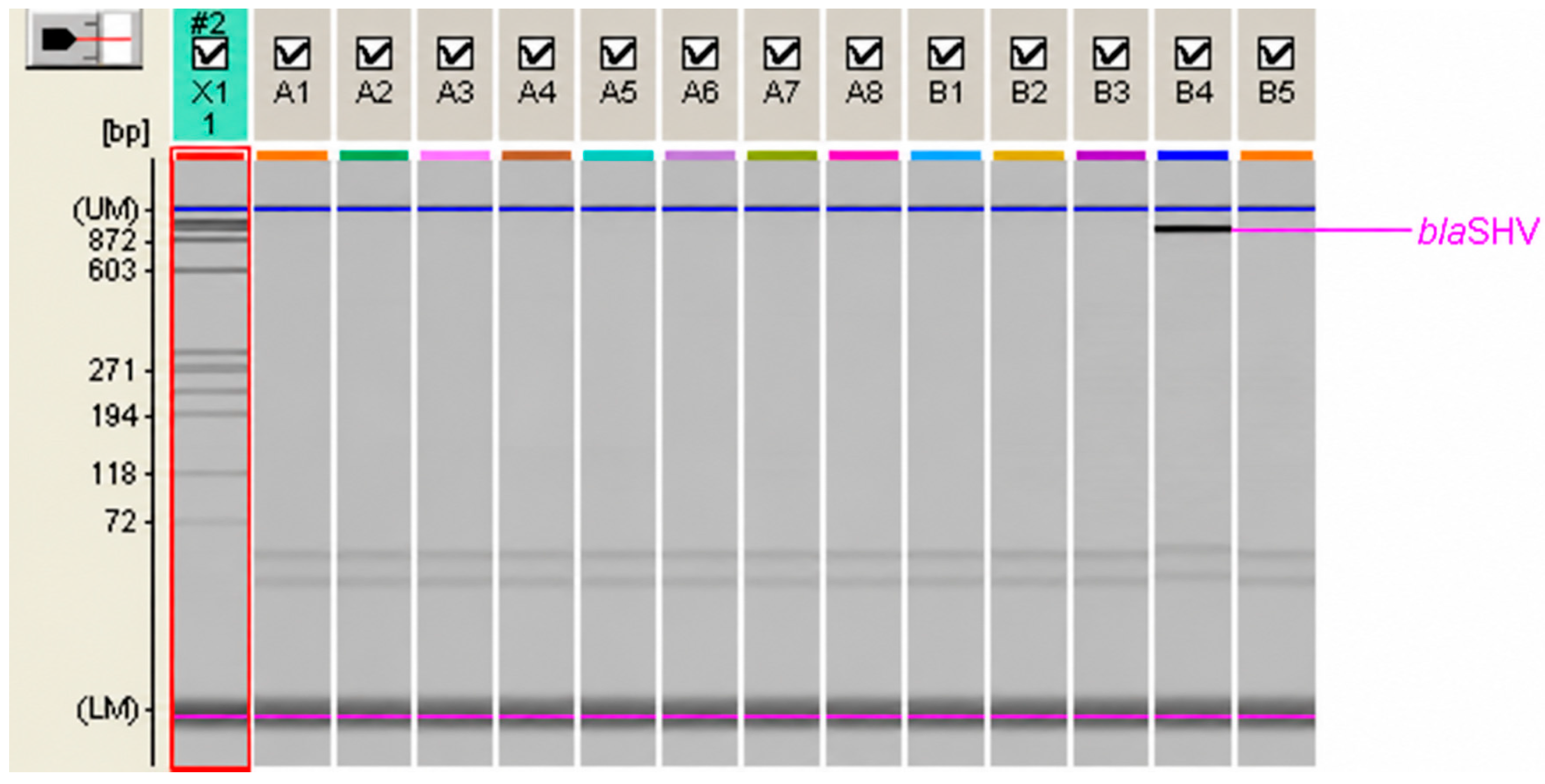
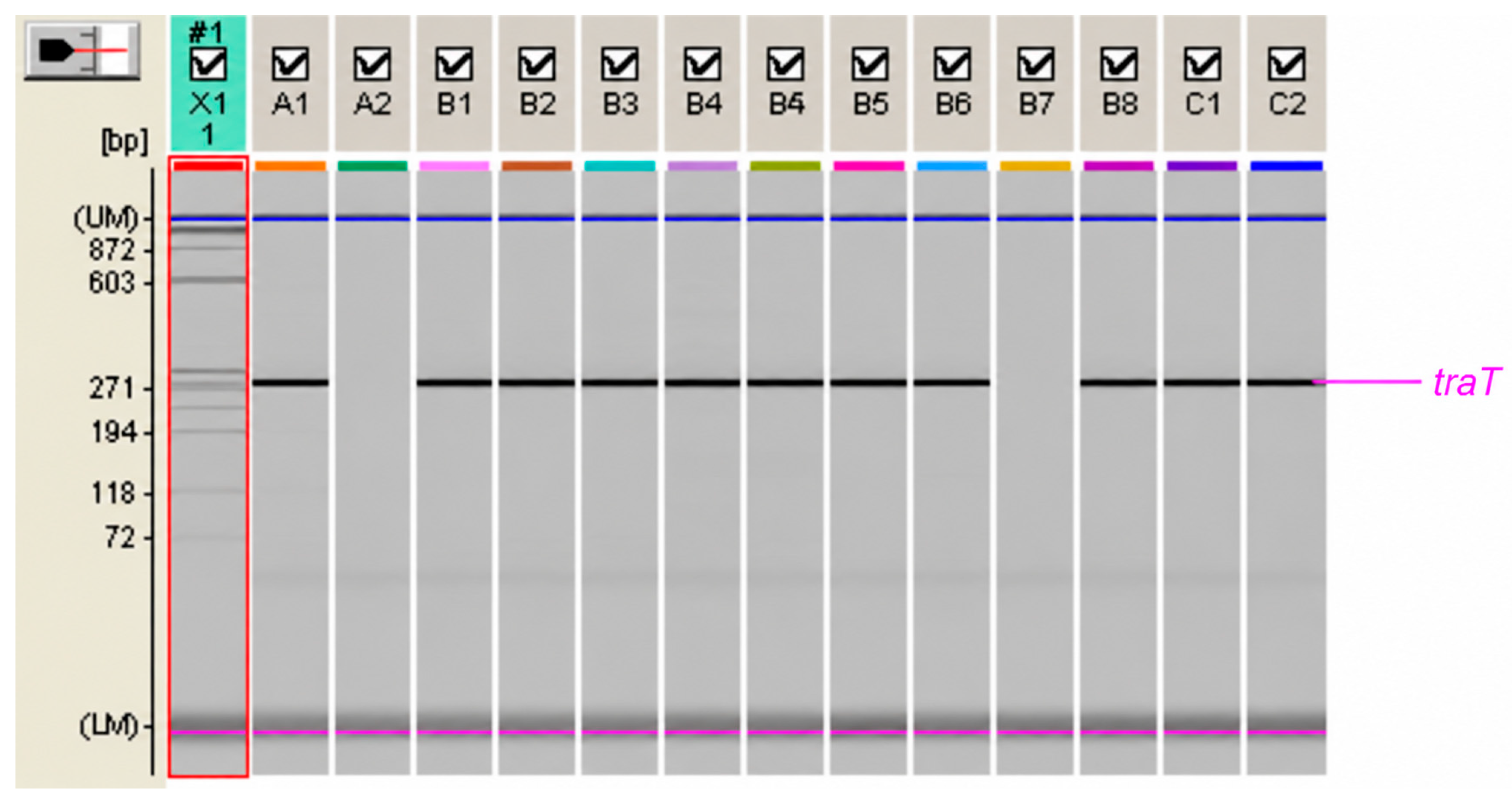
All PCR amplification products were analyzed using the MCE-202 MultiNA microchip electrophoresis system for DNA/RNA analysis (Shimadzu Co., Kyoto, Japan), as shown in Appendix A, Figure A1, Figure A2, Figure A3 and Figure A4.
2.7. Detection of fimH, papC, hlylA, cnf1 and traT Virulence Genes
PCR amplification was performed to detect the fimH, papC, hlyA, cnf1, and traT virulence genes in 159 FR E. coli O25 ST131 isolates, as previously described [22]. The primers used for gene detection are listed in Appendix B, Table A2.
Briefly, the PCR mixture had a final volume of 25 μL and contained 20 μL of Takara EmeraldAmp PCR Master Mix (Takara Bio, San Jose, CA, USA), 1 μL of each primer (20 pmol; Microsynth AG, Balgach, Switzerland), and 3 μL of bacterial lysate. The PCR conditions were as follows: initial denaturation at 95 °C for 5 min, followed by 25 cycles of 94 °C for 30 s, 63 °C for 30 s, and 68 °C for 3 min, with a final extension at 72 °C for 10 min.
2.8. Serum and Serum Susceptibility Assay
Blood samples were obtained from three healthy volunteers and allowed to clot at room temperature for 30 min. Serum was separated by centrifugation at 1000× g for 15 min at 4 °C and subsequently pooled. An aliquot of the pooled serum was heat-inactivated at 56 °C for 30 min and used as a control.
Phenotypic serum susceptibility was assessed using the assay described by Schiller and Hatch [23]. Serum susceptibility was evaluated based on the reduction in colony-forming units (CFUs) following exposure to fresh human serum, reflecting complement-mediated bacterial killing.
Briefly, the bacterial suspension was standardized to approximately 104 CFU/mL. An aliquot of 100 μL of the bacterial suspension was mixed with 100 μL of fresh, undiluted pooled human serum and incubated at 37 °C for 2 h. As a negative control, 100 μL of heat-inactivated serum was mixed with 100 μL of the bacterial suspension. Following incubation, samples were plated for CFU enumeration after overnight incubation at 37 °C. The survival percentage was calculated relative to the heat-inactivated serum control.
Serum resistance was defined as a reduction in CFU counts of <90%, indicating limited complement-mediated killing. Serum susceptibility was defined as a reduction in CFU counts of ≥90% relative to the heat-inactivated serum control, indicating effective serum bactericidal activity [23].
2.9. Statistical Analysis
Statistically significant differences in the research results were tested with the χ2-square test with the help of 2 × 2 contingency tables with the application of Yates correction. The value p < 0.05 was considered significant.
In the descriptive statistics for metabolic potential results, absolute numbers and percentages were used to present categorical variables, while for numerical variables, median and range were used due to deviation from normal distribution. In the bivariate analysis of metabolic potential data, the χ2-square test or Fisher’s exact test (adjusted according to the frequency of expected frequencies in the contingency table) for categorical variables and the Mann–Whitney U test for numerical variables were used. Linear regression was also used for data analysis, with the dependent variable being the metabolic average while the independent variables were year of isolation and ESBL status (yes/no). The regression model showed a good fit with a Durbin–Watson test of 1.911 and an adjusted R2 of 7.1%. Statistical analysis was performed using the IBM SPSS Statistics program (v21.0; IBM, Armonk, NY, USA) and results with p < 0.05 were considered statistically significant.
3. Results
3.1. ESBL Phenotype and Detection of CTX-M, CTX-M-15, and TEM β-Lactamases Genes
Among the uropathogenic E. coli isolates collected during the first and second study periods, 4354 and 7717 isolates, respectively, the proportion of ESBL-positive isolates ranged from 4.8% to 7.0%.
Among the 519 fluoroquinolone-resistant (FR) uropathogenic E. coli isolates collected during the two study periods, 48/151 (31.8%) isolates from the first study period and 122/368 (33.1%) from the second study period were ESBL-positive, with no statistically significant difference between the periods (p > 0.05). Among the O25 ST131 isolates (n = 159), ESBL-positivity rates were 66.7% in the first and 65.8% in the second study period. Similarly, among the 170 O25 isolates, ESBL positivity was detected in 30/48 (62.5%) and 75/122 (61.5%) of isolates, respectively, during the same periods (p > 0.05).
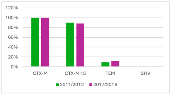
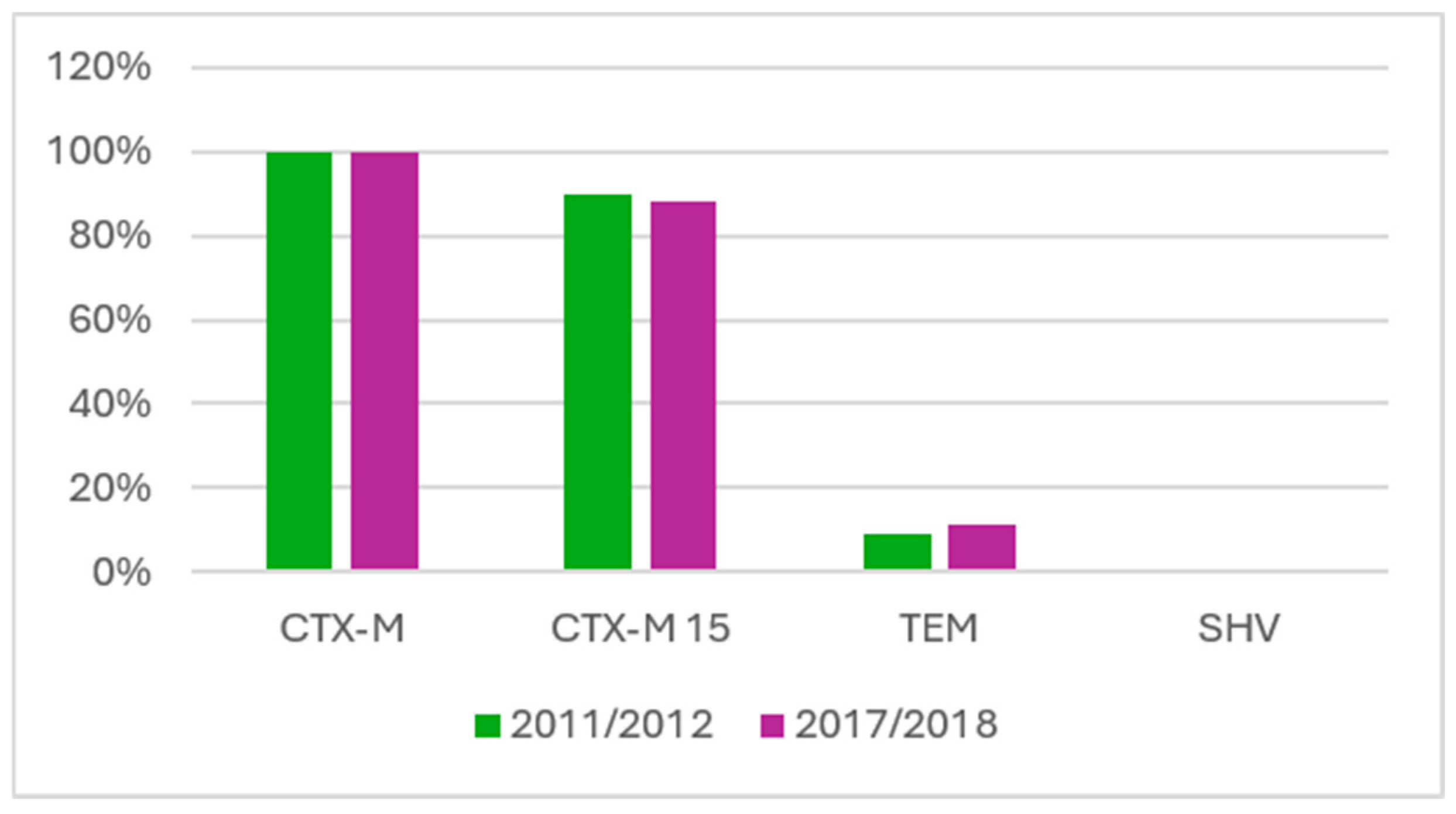
The frequencies of blaCTX-M, blaCTX-M-15, and blaTEM genes detected among the 105 ESBL-positive FR E. coli O25 ST131 isolates collected during both study periods are shown in Figure 1.
Figure 1.
Detection of CTX-M, CTX-M-15 and TEM β-lactamases in 105 ESBL-positive FR O25 ST131 E. coli isolates.
All investigated ESBL-positive O25 ST131 isolates carried the blaCTX-M gene. The blaCTX-M-15 gene was detected in 27 (90%) isolates from the first study period; among these, 12 (44%) also carried the IS26 insertion sequence upstream of blaCTX-M. In the second study period, blaCTX-M-15 was detected in 66 (88%) ESBL-positive O25 ST131 isolates, while IS26 was detected upstream of blaCTX-M in 12 (18%) isolates. No statistically significant difference was observed in the frequency of blaCTX-M-15 detection between the two study periods (χ2 = 0.08, p > 0.05).
The blaTEM gene was detected in four FR O25 ST131 ESBL-positive E. coli isolates from the first study period and in 13 isolates from the second study period. The simultaneous presence of blaTEM and blaCTX-M-15 was observed in two isolates from the first study period and in 12 isolates from the second study period. None of the ESBL-positive isolates carried the blaSHV gene during either study period.
3.2. Virulence Genes fimH, papC, hlylA, cnf1 and traT in O25 ST131 Isolates
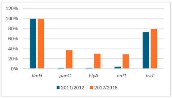
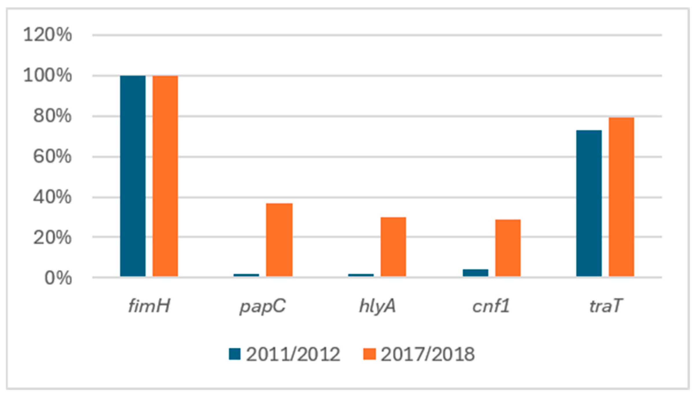
The fimH gene was detected in all 159 isolates (100%), whereas papC, hlyA, cnf1, and traT were detected in 43 (27%), 35 (22%), 35 (22%), and 123 (77%) isolates, respectively.
During the first study period, papC and hlyA were detected in one isolate each, cnf1 in two isolates, and traT in 33 isolates. During the second study period, papC was detected in 42 isolates, hlyA in 34, cnf1 in 33, and traT in 90 isolates.
A statistically significant difference was observed between the two study periods in the detection of papC, hlyA, and cnf1 (χ2 = 13.71, p < 0.01), with these virulence-associated genes detected more frequently during the second study period (Figure 2). In the first study period, only one of 45 isolates was papC-positive, compared with 42 of 114 isolates in the second study period. A similar pattern was observed for hlyA and cnf1. Only one hlyA-positive and two cnf1-positive isolates were detected among the isolates from the first study period.
Figure 2.
Detection of virulence genes in 159 FR O25 ST131 isolates during the two study periods.
A statistically significant difference was also observed in the distribution of papC, hlyA, and cnf1 between ESBL-positive and ESBL-negative E. coli O25 ST131 isolates (p < 0.01) during both study periods. The simultaneous presence of these virulence genes was detected in 34 E. coli O25 ST131 isolates with an ESBL phenotype, including one isolate from the first study period and 33 from the second study period. In the second study period, one ESBL-negative E. coli O25 ST131 isolate also carried fimH, papC, hlyA, and cnf1 simultaneously.
Overall, 35 E. coli O25 ST131 isolates carried fimH, papC, hlyA, and cnf1 simultaneously, and traT was detected in 33 of these 35 isolates. The distribution of virulence-associated genes differed between the two study periods, with a higher frequency of papC, hlyA, and cnf1 among isolates from the second study period and an association between these virulence profiles and the ESBL phenotype (p < 0.01).
3.3. Metabolic Activity of FR O25 ST131 Isolates During the Two Study Periods
Metabolic potential was calculated as the proportion of positive biochemical reactions among the 47 biochemical reactions assessed using the VITEK 2 GN card and was expressed both as the number of positive reactions and as the corresponding proportion (%).
During the first study period, the mean number of positive biochemical reactions was 17 (range, 16–19) among ESBL-negative E. coli O25 ST131 isolates. Among ESBL-positive isolates, the mean number of positive reactions was 18 (range, 14–21) out of 47 tested reactions.
During the second study period, the mean number of positive biochemical reactions was 19 (range, 14–21) among ESBL-negative E. coli O25 ST131 isolates and 19 (range, 14–23) among ESBL-positive isolates. The distribution of metabolic potential values is presented in Table 1.
Table 1.
Metabolic potential of FR O25 ST131 E. coli isolates.
The biochemical profiles of E. coli O25 ST131 isolates were compared between the two study periods based on the proportion of positive reactions for each individual biochemical substrate. Statistical significance was assessed using the chi-square test and Fisher’s exact test. The p-values for individual substrates comparing the two study periods are presented in Appendix C, Table A3.
Regression analysis demonstrated a statistically significant difference in metabolic potential between the two study periods (p < 0.01), whereas ESBL status (negative vs. positive) was not a statistically significant predictor of metabolic potential (p = 0.093; Table 2).
Table 2.
Linear regression analysis results, with the metabolic average as the dependent variable.
No statistically significant differences in metabolic potential according to ESBL status were observed within either study period or when comparing ESBL-positive and ESBL-negative isolates across the two study periods (p > 0.05).
3.4. Serum Susceptibility of Isolates During the Two Study Periods
A total of 89/159 (56.0%) E. coli O25 ST131 isolates demonstrated resistance to the bactericidal activity of serum. The proportion of serum-resistant (SR) isolates was 18/45 (40.0%) during the first study period and 71/114 (62.3%) during the second study period.
A statistically significant difference in the frequency of serum-resistant isolates was observed between the two study periods, with a higher proportion of SR isolates in the second period (χ2 = 6.50, p = 0.01).
No statistically significant association was observed between serum resistance and ESBL phenotype among E. coli O25 ST131 isolates (p > 0.05).
The presence of the traT gene, which has been associated with resistance to serum bactericidal activity, did not consistently correspond to the phenotypic serum susceptibility results. Overall, traT was detected in 123/159 (77.4%) isolates. The gene was detected in 33/45 (73.3%) isolates from the first study period and 90/114 (78.9%) isolates from the second study period, with no statistically significant difference between the periods (p > 0.05).
Among the 123 traT-positive isolates, 49 (39.8%) were serum-susceptible and 74 (60.2%) were serum-resistant. In the first study period, 20 serum-susceptible and 13 serum-resistant isolates carried traT, whereas in the second study period, 29 serum-susceptible and 61 serum-resistant isolates carried traT. The presence of traT was significantly associated with the serum susceptibility phenotype (χ2 = 3.87, p = 0.049).
The distribution of serum-susceptible and serum-resistant E. coli O25 ST131 isolates is presented in Appendix C, Table A4.
3.5. Patients’ Demographic Characteristics
FR E. coli isolates were recovered from 124 female and 27 male patients (male-to-female ratio, 1:4.6) aged 11–95 years during the first study period and from 288 female and 80 male patients (male-to-female ratio, 1:3.6) aged 9–98 years during the second study period.
The O25 ST131 clone was detected in 39 female and six male patients aged 14–95 years during the first study period and in 83 female and 31 male patients aged 9–93 years during the second study period. The mean age was 69 years among ESBL-positive patients in the first study period and 72 years in the second study period. No statistically significant difference was observed between the two study periods (χ2 = 0.07, p > 0.05).
A limitation of this retrospective study was the lack of clinical information beyond patient demographic characteristics.
Preliminary results were presented in a poster session at the 35th European Congress of Clinical Microbiology and Infectious Diseases (ECCMID), held in Vienna, Austria, from 27 to 30 April 2025 (Anušić M, Vujnović T, Vlahek L, Tičić V, Kolčić I, Marijan T, Bedenić B, Vraneš J. Virulence and fitness of CTX-M-producing Escherichia coli O25 ST131 in community-acquired urinary tract infections in Croatia).
4. Discussion
The pandemic clone E. coli O25 ST131 is divided into three major genetic clades (A, B, and C). Most contemporary E. coli O25 ST131 isolates belong to clade C, which is characterized by the fimH30 allele encoding the type 1 fimbrial adhesin and is further divided into the H30-R (fluoroquinolone-resistant) and H30-Rx (fluoroquinolone-resistant and ESBL-producing) subclades, corresponding to C1 and C2, respectively. Type 1 fimbriae are recognized as an important virulence factor in uropathogenic E. coli (UPEC).
In our study, all E. coli O25 ST131 isolates carried the fimH gene, whereas papC, hlyA, and cnf1 were detected significantly more frequently during the second study period. Virulence-associated genes present in the E. coli O25 ST131 clonal lineage have been used to define several virotypes (A–G). Virotype A is characterized by the presence of afa/draBC and an afa operon gene, with the sat gene being variably present. Virotype B is characterized by the presence of iroN, with sat being variably present, whereas virotype C is characterized by the presence of sat. Virotype D is characterized by variable presence of afa/draBC, afa operon, iroN, sat, cnf1, hlyA, papGIII, cdtB, and neuC-K1, together with the presence of ibeA. Virotype E is characterized by the presence of sat, papGII, cnf1, and hlyA, whereas virotypes F and G are characterized by the presence of iroN, papGII, and cnf1, and sat and papGII, respectively [24,25].
Calhau et al. demonstrated the presence of pathogenicity islands PAI I CFTO73, PAI II CFTO73, and PAI IV 536 in E. coli ST131 isolates, containing virulence determinants associated with P fimbriae, siderophores, and α-hemolysin [26]. In our study, the fimH/papC/hlyA/cnf1/traT virulence-associated gene profile predominated among FR E. coli O25 ST131 ESBL-positive isolates during the second study period. The simultaneous presence of papC, hlyA, and cnf1 was detected in 33 of 75 ESBL-positive isolates during the second study period, compared with one of 39 ESBL-negative isolates. Further studies are required to determine whether these isolates correspond to virotype E and to assess the presence of the relevant pathogenicity islands, as well as their potential contribution to UTI development in outpatients without known predisposing factors.
ESBL production has frequently been associated with reduced virulence compared with ESBL-negative E. coli isolates. However, Banerjee et al. reported that the ESBL-positive E. coli ST131 clonal lineage carried a greater number of characterized virulence genes than non-ST131 E. coli isolates [27]. Similarly, our findings are consistent with those reported by Hojabri et al., who found that ESBL-positive E. coli ST131 H30Rx isolates were more frequently associated with papA, papC, papEF, hlyA, cnf1, k5, and kpsMT II, and with the findings of Gibreel et al., who reported a higher number of virulence genes among CTX-M-15-producing UPEC ST131 isolates than among non-ST131 isolates [28,29].
In our study, ESBL-positive E. coli O25 ST131 isolates carried a higher number of the selected virulence-associated genes than ESBL-negative isolates. These findings suggest an association between ESBL production and the presence of the selected virulence-associated genes in E. coli O25 ST131. However, the presence of virulence-associated genes does not necessarily demonstrate increased phenotypic virulence, and functional and clinical studies are required to establish whether these genetic differences translate into differences in pathogenic potential.
The O25 ST131 E. coli clonal lineage requires metabolic adaptation to transition from the gastrointestinal tract to the urinary tract and to respond to changes in pH and nutrient availability. The metabolic findings from our study can be compared with those of two studies conducted in England. Gibreel et al. reported significantly higher metabolic potential among O25 ST131 isolates than among other sequence types, including ST10, ST14, ST69, ST73, ST88, ST95, ST127, and ST405. In that study, O25 ST131 was significantly associated with six positive biochemical reactions—AGAL (α-galactosidase), BGUR (β-glucuronidase), ODC (ornithine decarboxylase), ProA (L-proline arylamidase), SAC (sucrose/sucrose), and ILATk (L-lactate alkalinization)—and with two negative biochemical reactions, ILATa (L-lactate assimilation) and 5KG (5-keto-D-gluconate), using the VITEK 2 ID GNB card [15].
In the same study, comparisons of susceptible and multidrug-resistant E. coli O25 ST131 isolates showed that CTX-M-15-producing isolates had significantly higher metabolic potential. The biochemical characteristics associated with CTX-M-15-producing E. coli O25 ST131 included positive reactions for tyrosine arylamidase, L-lactate alkalinization, phosphatase, and succinate alkalinization. Cluster analysis of biochemical profiles identified a large cluster containing multidrug-resistant E. coli O25 ST131 isolates [15].
In contrast, Alqasim et al., using four API kits and the Biolog automated system, did not demonstrate differences in metabolic potential between E. coli ST131 and other UPEC sequence types, including ST69, ST73, ST95, and ST10. In that study, ST131 isolates showed low utilization of five substrates—5KG (8%), D-arabinose (20%), esculin (24%), cellobiose (12%), and dulcitol (36%)—compared with other sequence types. Genome sequence analysis also showed little difference in the presence or absence of metabolic operons between ST131 and other sequence types. The authors therefore concluded that E. coli ST131 was not associated with a distinct biochemical profile and that observed differences were more likely related to individual ST131 strains than to the clonal lineage as a whole [30].
In our study, the metabolic potential of UPEC E. coli O25 ST131 isolates was evaluated during two study periods without comparison with other fluoroquinolone-resistant UPEC isolates. A total of 112 biochemical profiles were observed; therefore, a characteristic biochemical profile specific to E. coli O25 ST131 could not be established. Nevertheless, several biochemical reactions were comparable with those reported in previous studies. The negative reactions ILATa (L-lactate assimilation) and 5KG (5-keto-D-gluconate) reported by Gibreel et al. were also predominantly negative in our isolates [15]. In our study, ILATa was positive in 1.75% (2 isolates) during the first study period and 7.89% (9 isolates) during the second period, whereas 5KG was positive in only one isolate (0.87%) during the second period. D-cellobiosis was negative during both study periods, consistent with the findings of Alqasim et al. [30].
Of the six positive biochemical reactions associated with O25 ST131 by Gibreel et al.—AGAL, BGUR, ILATk, ODC, ProA, and SAC—all were also positive among the E. coli O25 ST131 isolates investigated in our study. Metabolic activity was significantly higher among isolates collected during the second study period than among those collected during the first period, whereas ESBL status did not significantly affect metabolic potential. Further studies are required to determine whether the observed temporal differences in metabolic activity are associated with bacterial fitness, including the ability to colonize the gastrointestinal tract and cause urinary tract infections.
Resistance to the bactericidal activity of serum is also considered a virulence-associated trait and may be particularly relevant to bacteria capable of invasive infection and hematogenous dissemination. The bactericidal activity of human and animal serum is mediated primarily by the complement system. Phan et al. investigated genes associated with serum resistance in EC958, a representative UPEC ST131 isolate, and identified strong associations with genes involved in the synthesis of the murein lipoprotein Lpp and A-lipids, as well as genes regulating O-antigen chain length [31].
In our study, the presence of the traT gene and the phenotypic susceptibility of FR E. coli O25 ST131 isolates to the bactericidal activity of human serum were assessed. The traT gene was detected in 83.1% (74/89) of serum-resistant isolates and in 70.0% (49/70) of serum-susceptible isolates (p < 0.05). In contrast, Kanukollu et al. reported no statistically significant difference in the prevalence of traT between serum-resistant and serum-susceptible E. coli isolates [32].
The precise mechanism by which TraT contributes to serum resistance remains incompletely characterized. It has been proposed that the TraT outer membrane lipoprotein may interfere with complement-mediated membrane damage [33]. Although we observed an association between the presence of traT and serum resistance, complete concordance was not observed, likely reflecting the contribution of additional mechanisms to resistance against the bactericidal activity of serum [34,35,36].
In our study, comparison of the phenotypic susceptibility of FR E. coli O25 ST131 isolates to serum bactericidal activity between the two study periods demonstrated a significantly higher frequency of serum-resistant isolates during the second period. The simultaneous presence of fimH, papC, hlyA, cnf1, and traT in serum-resistant isolates, particularly among ESBL-positive isolates during the second study period, suggests the occurrence of isolates carrying multiple virulence-associated traits. Further clinical studies are required to determine whether this combination of characteristics is associated with complicated urinary tract infections, bacteremia, or urosepsis.
Recent studies have reported that E. coli ST131 isolates carrying blaCTX-M genes, including blaCTX-M-15, may exhibit enhanced in vitro fitness and prolonged intestinal persistence, challenging the traditional assumption that antimicrobial resistance necessarily imposes a substantial fitness cost [37,38,39,40,41]. Type 1 fimbriae and other virulence-associated factors may contribute to efficient intestinal colonization. The evolution of E. coli O25 ST131 has therefore been characterized by the acquisition and maintenance of antimicrobial resistance and virulence-associated determinants, including fluoroquinolone resistance and ESBL genes [42,43,44]. These characteristics may contribute to the successful persistence and dissemination of this clonal lineage and its involvement in urinary tract and bloodstream infections.
The present study has several limitations. The analysis was restricted to two selected time periods, and the study population consisted of isolates recovered from outpatients with acute urinary tract infections. In addition, the analysis of virulence was based on the detection of selected genes rather than on functional assays of virulence. Therefore, the observed differences in gene prevalence should not be interpreted as direct evidence of differences in pathogenicity. Prospective studies incorporating broader genomic characterization, functional assays, and clinical outcome data are warranted to further investigate temporal changes in this high-risk clonal lineage.
Author Contributions
Conceptualization, J.V. and M.A.; methodology, J.V., M.A. and V.T.; software, I.K.; validation, J.V. and M.A.; formal analysis, I.K. and S.Š.; investigation, M.A., V.T. and T.M.; resources, J.V.; data curation, M.A.; writing—original draft preparation, J.V.; writing—review and editing, B.B.; visualization, S.Š.; supervision, B.B.; project administration, S.Š.; funding acquisition, J.V. All authors have read and agreed to the published version of the manuscript.
Funding
This research was funded by the “Molecular epidemiology of multidrug-resistant uropathogens and their dissemination potential in the community” grant, number IP-2025-02-6757, by the Croatian Science Foundation.
Institutional Review Board Statement
The study was conducted in accordance with the Declaration of Helsinki and approved by the Ethics Committee of Dr. Andrija Štampar Teaching Institute of Public Health, Zagreb, Croatia, protocol code-class number 641-01/25-01/1 and date of 20 February 2025.
Informed Consent Statement
Informed consent was waived because this was a retrospective study. The isolates were collected in the framework of routine microbiology diagnostics. The patients were not subject to any additional procedures for the purpose of the study. Informed consent for collecting serum from three healthy volunteers was waived because the serum was collected from the authors of the study (M.A., V.T. and T.M.).
Data Availability Statement
The original contributions presented in this study are included in the article. Further inquiries can be directed to the corresponding author.
Acknowledgments
We thank Matea Kvaternik Celjak for providing technical assistance with the PCR and MultiNA procedures, and Ivan Barišić for providing control strains for the microchip electrophoresis.
Conflicts of Interest
The authors declare no conflicts of interest.
Abbreviations
The following abbreviations are used in this manuscript:
| AMR | Antimicrobial resistance |
| MDR | Multidrug resistant |
| ExPEC | Extraintestinal pathogenic Escherichia coli |
| UTI | Urinary tract infection |
| UPEC | Uropathogenic Escherichia coli |
| CA-UTI | Community-acquired urinary tract infection |
| ESBL | Extended-spectrum beta-lactamases |
| EUCAST | European Committee on Antimicrobial Susceptibility Testing |
| PCR | Polymerase chain reaction |
| SS | Serum-susceptible |
| SR | Serum-resistant |
| CFU | Colony-forming units |
| TEM | Temoneira beta-lactamase |
| SHV | Sulfhydryl reagent variable beta-lactamase |
| CTX-M | CefoTaXimase first isolated in Munich |
| FR | Fluoroquinolone resistant |
| MLST | MultiLocus Sequence Typing |
| ST | Sequence type |
| APPA | Ala-Phe-Pro-arylamidase |
| H2S | H2S production |
| BGLU | Beta-glucosidase |
| PROA | L-Proline Arylamidase |
| SAC | Saccharose/sucrose |
| ILATK | L-lactate alkalinization |
| GLYA | Glycine arylamidase |
| O129R | O/129 resistance (comp.vibrio.) |
| ADO | Adonitol |
| BNAG | Beta-N-acetyl-glucosaminidase |
| DMAL | D-maltose |
| LIP | Lipase |
| IARL | L-arbitol |
| DGLU | D-glucose |
| DMNE | D-mannose |
| TYRA | Tyrosine arylamidase |
| CIT | Citrate |
| NAGA | Beta-N-acetyl-galactosaminidase |
| IHISA | L-histidine assimilation |
| ELLM | Ellman |
| DCEL | D-cellobiose |
| GGT | Gamma-glutamyl-transferase |
| BXYL | Beta-xylosidase |
| URE | Urease |
| DTAG | D-tagatose |
| AGLU | Alpha-glucosidase |
| ODC | Ornithine decarboxylase |
| GGAA | Glu-Gly-Arg-arylamidase |
| PYRA | L-pyrrolydonyl-arylamidase |
| AGLTP | Glutamyl arylamidase pNA |
| DMAN | D-mannitol |
| PLE | Palatinose |
| DTRE | D-trehalose |
| SUCT | Succinate alkalinization |
| LDC | Lysine decarboxylase |
| IMLTA | L-malate assimilation |
| MNT | Malonate |
| AGAL | Alpha-galactosidase |
| CMT | Coumarate |
| ILATA | L-lactate assimilation |
| BGAL | Beta-galactosidase |
| OFF | Fermentation/glucose |
| BAIAP | Beta-alanine-arylamidase pNA |
| DSOR | D-sorbitol |
| 5KG | 5-keto-D-gluconate |
| PHOS | Phosphatase |
| BGUR | Beta-glucuronidase |
Appendix A
Table A1.
Selected primers used in the study.
Figure A1.
Microchip electrophoresis for the CTX-M gene (542 bp). DNA range bp—X1; positive samples—A1, A2, A3, A4, A5, A6, A7, A8, B1, B2; positive control K. pneumoniae CTX-M—C3; negative control E. coli ATCC 25922—C4.
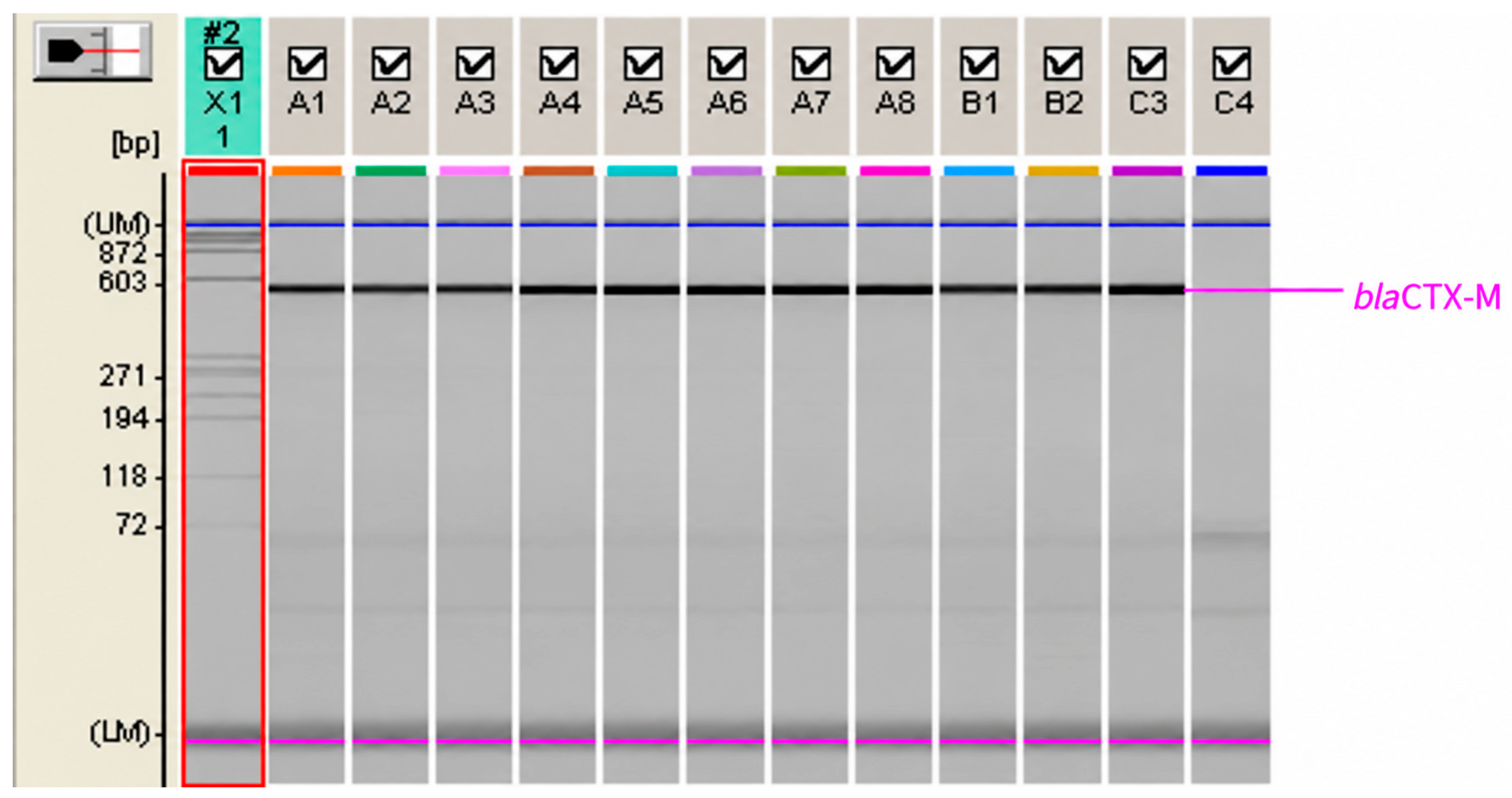
Figure A2.
Microchip electrophoresis for the CTX-M-15 gene (1020 bp). DNA range bp—X1; positive samples—B1, B2, B3, B4, B7, B8, C6; negative samples—A2, B5, B6; positive control E. coli CTX-M-15—A1; negative control—E. coli ATCC 25922—C8.
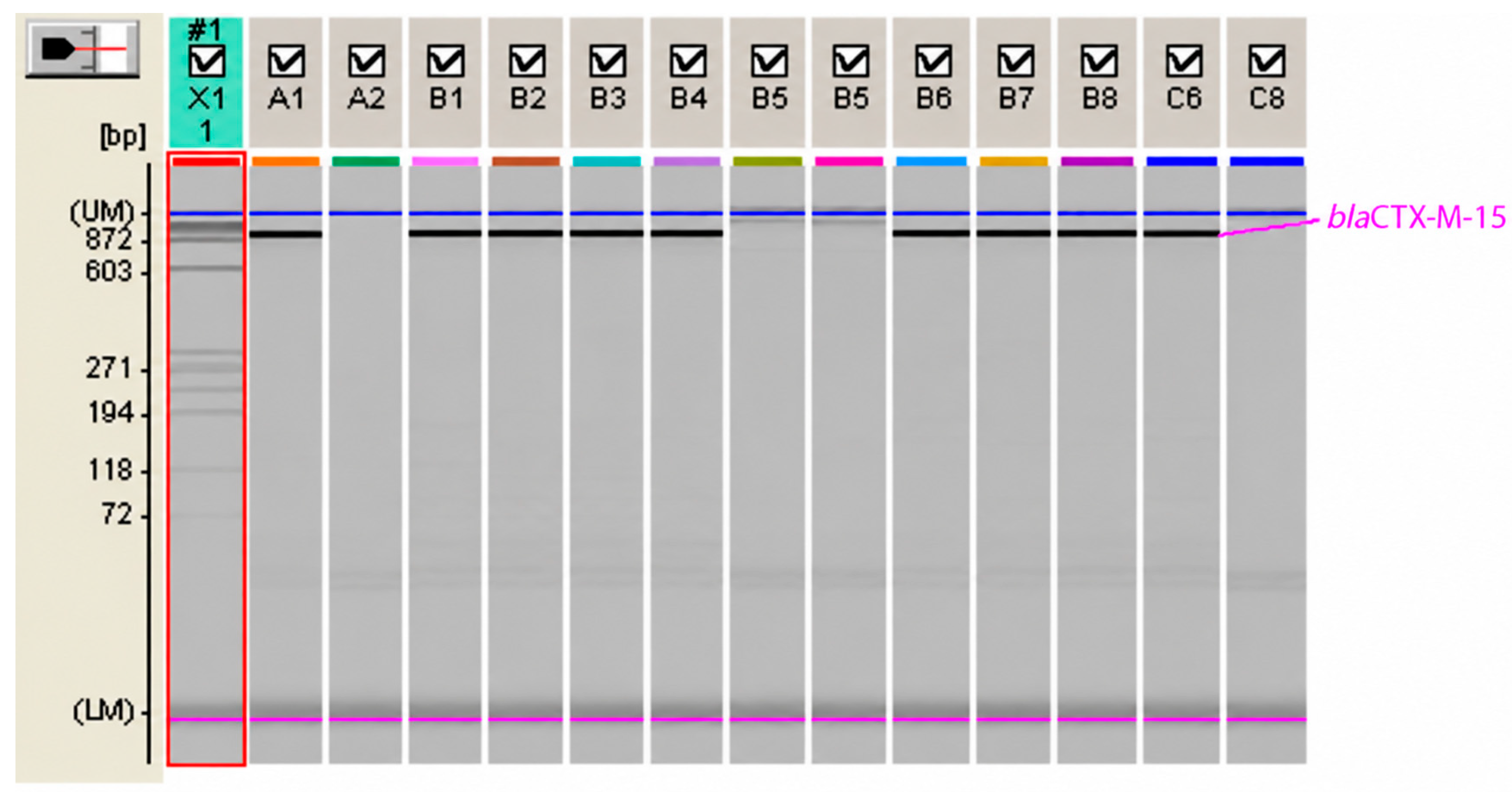
Figure A3.
Microchip electrophoresis for the TEM gene (868 bp). DNA range bp—X1; positive samples—A1, A7, B1, B4; negative samples—A2, A3, A4, A5, A8, B3, C4; positive control TEM2 producing E. coli—C2, negative control E. coli ATCC 25922—C3.
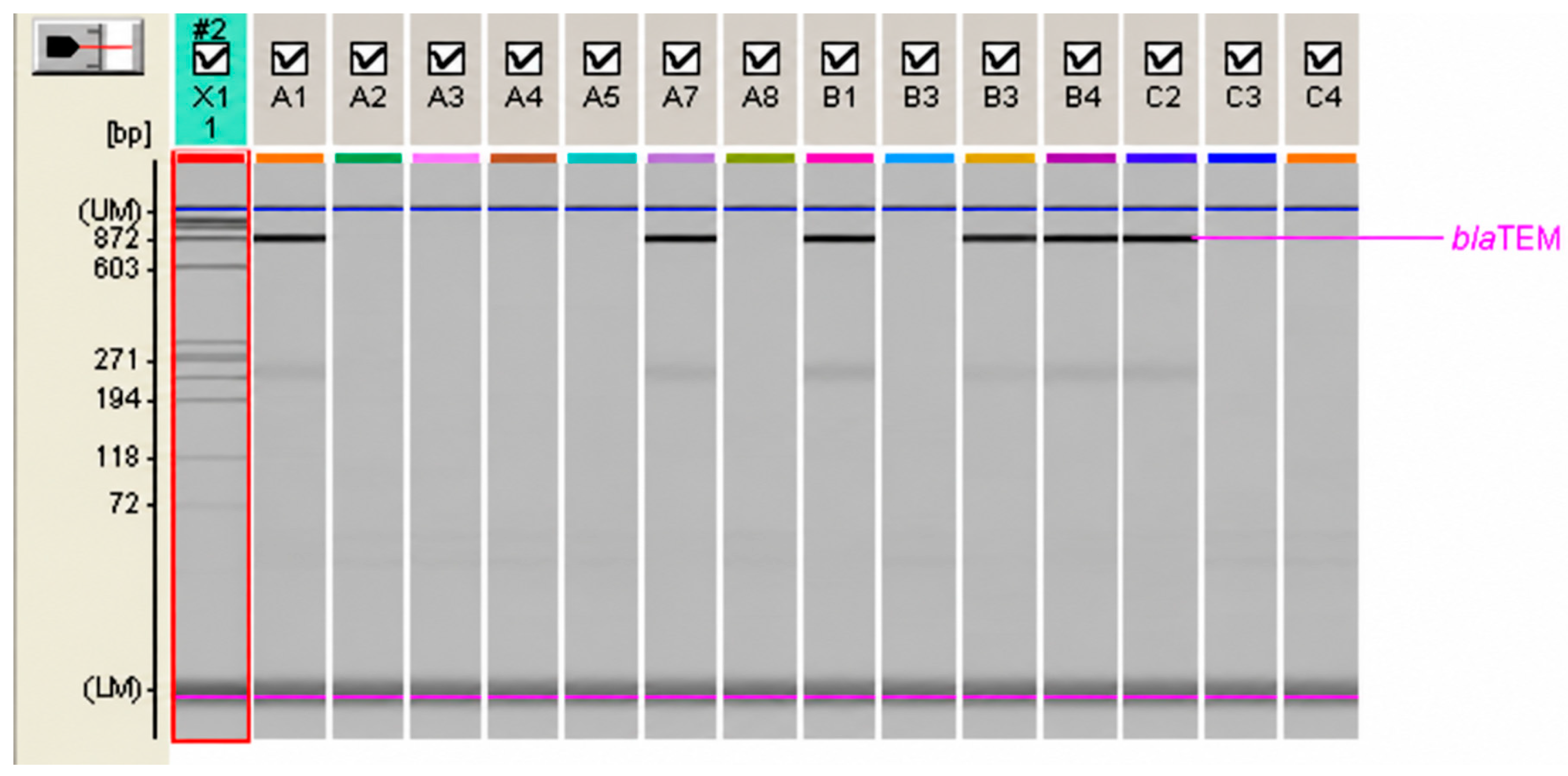
Figure A4.
Microchip electrophoresis for the SHV gene (1127 bp). DNA range bp—X1; negative samples—A1, A2, A3, A4, A5, A6, A7, A8, B1, B; positive control SHV2a producing K. pneumoniae—B4; negative control E. coli ATCC 25922—B3, B5.
Appendix B
Table A2.
Primers used for the detection of fimH, papC, hlyA, cnf1 and traT genes.
Figure A5.
Microchip electrophoresis for the virulence gene fimH (508 bp). DNA range bp—X1; positive samples—A1, A2, A3, A4, A5, A6, A7, A8, B, B2; positive control—C4; negative control-C5.
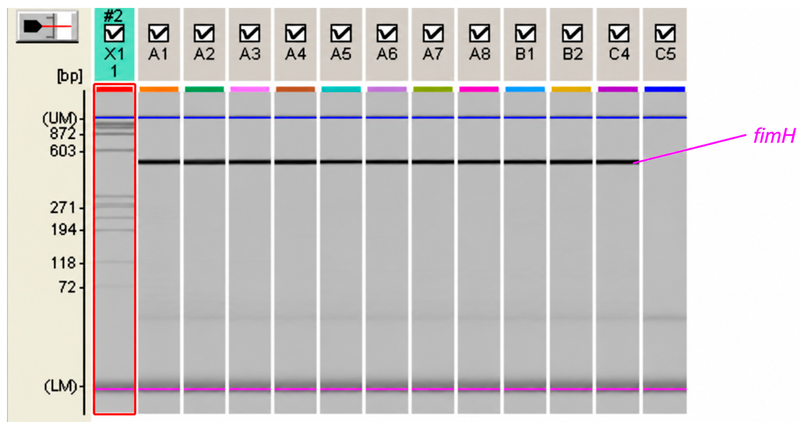
Figure A6.
Microchip electrophoresis for the virulence gene papC (205 bp). DNA range bp—X1; positive samples—A6, A8, B2, B6; negative samples—A1, A2, A3, A7, B1, B5; positive control—C7; negative control—C8.
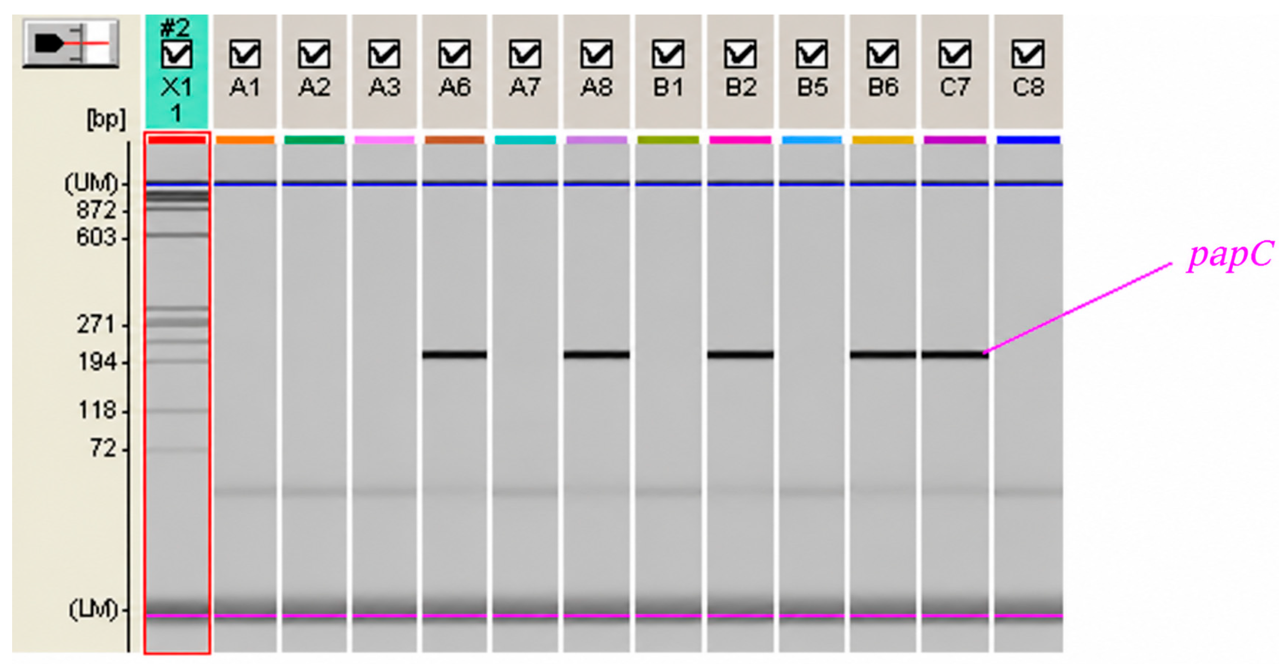
Figure A7.
Microchip electrophoresis for the virulence gene hlyA (1257 bp). DNA range bp—X1; positive samples—B5, B6, B7, C1, C2, C6; negative samples—B1, B2, B3, B8; positive control—A1, negative control—A2.
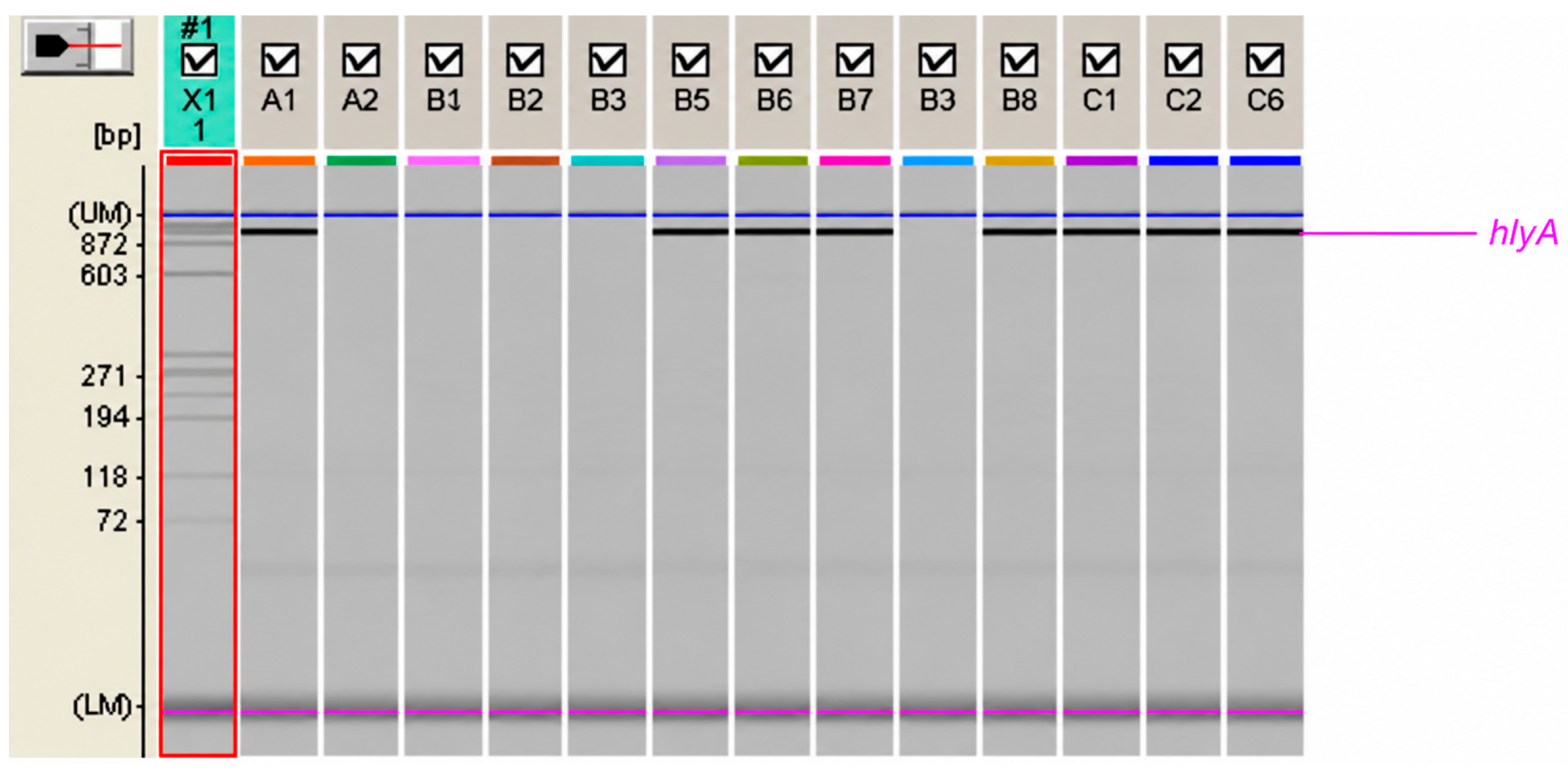
Figure A8.
Microchip electrophoresis for the virulence gene cnf1 (498 bp). DNA range bp—X1; positive samples—B3, C2, C3, C7, C8; negative samples—B1, B2, B4, C4, C6; positive control—A1; negative control—A2.
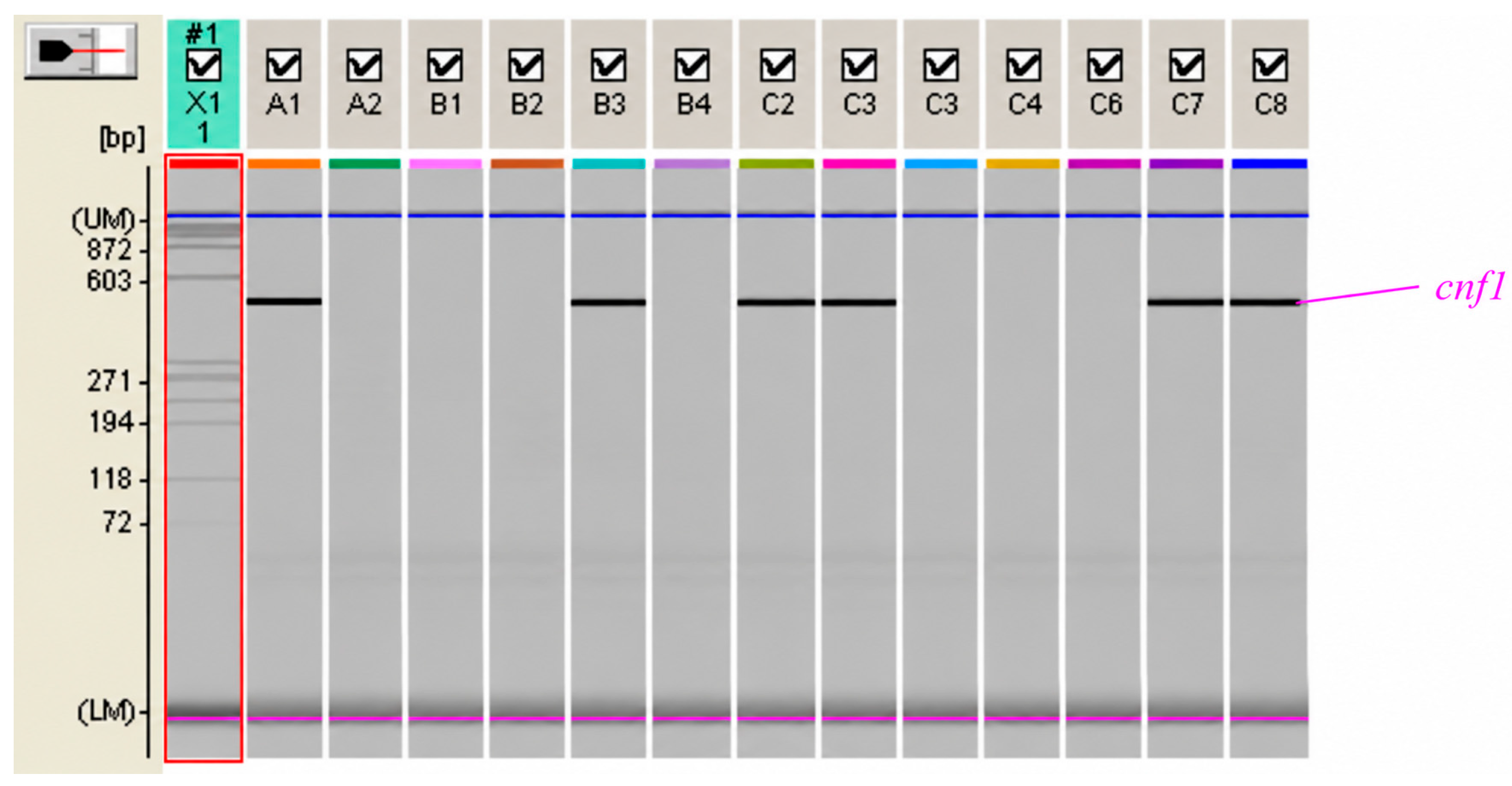
Figure A9.
Microchip electrophoresis for the virulence gene traT (290 bp). DNA range bp—X1; positive samples—B1, B2, B3, B4, B5, B6, B7, C1, C2; negative samples—B8; positive control—A1; negative control—A2.
Appendix C
Table A3.
Comparison of biochemical reactions of FR E. coli O25 ST131 isolates in two study periods.
Table A4.
Detection of serum-susceptible (SS) and serum-resistant (SR) E. coli O25 ST131 isolates.
References
- Izydorczyzk, C.; Church, D.L. Evolution and rise of antimicrobial resistance in key public health bacteria within and across the One Health sectors. Future Microbiol. 2026, 21, 181–215. [Google Scholar] [CrossRef] [Scilit] [PubMed]
- Pitout, J.D.D.; DeVinney, R. Escherichia coli ST131: A multidrug resistant clone primed for global domination. F1000Research 2017, 6, 195. [Google Scholar] [CrossRef] [Scilit] [PubMed]
- Nicolas-Chanoine, M.-H.; Bertrand, X.; Madec, J.-Y. Escherichia coli ST131, an intriguing clonal group. Clin. Microbiol. Rev. 2014, 27, 543–574. [Google Scholar] [CrossRef] [Scilit] [PubMed]
- Eger, E.; Heiden, S.E.; Korolew, K.; Bayingana, C.; Ndoli, J.M.; Sendegeya, A.; Gahutu, J.B.; Kurz, M.S.E.; Mockenhaupt, F.P.; Müller, J.; et al. Circulation of extended-spectrum beta-lactamase producing Escherichia coli of pandemic sequence types 131, 648 and 410 among hospitalized patients, caregivers, and the community in Rwanda. Front. Microbiol. 2021, 12, 662575. [Google Scholar] [CrossRef] [PubMed]
- Lübcke, P.; Heiden, S.E.; Homeier-Bachmann, T.; Bohnert, J.A.; Schulze, C.; Eger, E.; Schwabe, M.; Guenther, S.; Schaufer, K. Multidrug-resistant high-risk clonal Escherichia coli lineages occur along an antibiotic residue gradient in the Baltic Sea. npj Clean Water 2024, 7, 94. [Google Scholar] [CrossRef] [Scilit]
- Elankumaran, P.; Cummins, M.L.; Browning, G.F.; Marenda, M.S.; Reid, C.J.; Djordjevic, S.P. Genomic and temporal trends in canine ExPEC reflect those of human ExPEC. Microbiol. Spectr. 2022, 10, e0129122. [Google Scholar] [CrossRef] [Scilit] [PubMed]
- Ewers, C.; Grobbel, M.; Stamm, I.; Kopp, P.A.; Diehl, I.; Semmler, T.; Fruth, A.; Beutlich, J.; Guerra, B.; Wieler, L.H.; et al. Emergence of human pandemic O25:H4-ST131 CTX-M-15 extended-spectrum-beta-lactamase-producing Escherichia coli among companion animals. J. Antimicrob. Chemother. 2010, 65, 651–660. [Google Scholar] [CrossRef] [Scilit] [PubMed]
- Soncini, J.G.M.; Cerdeira, L.; Sano, E.; Koga, V.L.; Tizura, A.T.; Tano, Z.N.; Nakazato, G.; Kobayashi, R.K.T.; Aires, C.A.M.; Lincopan, N.; et al. Genomic insights of high-risk clones of ESBL-producing Escherichia coli isolated from community infections and commercial meat in southern Brazil. Sci. Rep. 2022, 12, 9354. [Google Scholar] [CrossRef] [Scilit] [PubMed]
- Brendecke, J.; Homeier-Bachmann, T.; Schmitz Ornẻs, A.; Guenther, S.; Heiden, S.E.; Schwabe, M.; Eger, E.; Schaufler, K. Multidrug-resistant high-risk Escherichia coli and Klebsiella pneumoniae clonal lineages occur in black-headed gulls from two conservation islands in Germany. Antibiotics 2022, 11, 1357. [Google Scholar] [CrossRef] [Scilit]
- Whitmer, G.R.; Moorthy, G.; Arshad, M. The pandemic Escherichia coli sequence type 131 strain is acquired even in the absence of antibiotic exposure. PLoS Pathog. 2019, 15, e1008162. [Google Scholar] [CrossRef] [Scilit] [PubMed]
- Walker, L.L.; Phan, M.-D.; Permana, B.; Lian, Z.J.; Nhu, N.T.K.; Cuddihy, T.; Peters, K.M.; Ramsay, K.A.; Stewart, C.; Pfennigwerth, N.; et al. Emergence of a carbapenem-resistant atypical uropathogenic Escherichia coli clone as an increasing cause of urinary tract infection. Nat. Commun. 2025, 16, 8200. [Google Scholar] [CrossRef] [Scilit] [PubMed]
- Literacka, E.M.; Bedenic, B.; Baraniak, A.; Fiett, J.; Tonkic, M.; Jajic-Bencic, I.; Gniadkowski, M. blaCTM-X genes in Escherichia coli strains from Croatian hospitals are located in new (blaCTX-M-3a) and widely spread (blaCTX-M3a and blaCTX-M-15) genetic structures. Antimicrob. Agents Chemother. 2009, 53, 1630–2635. [Google Scholar] [CrossRef] [Scilit] [PubMed]
- Anušić, M.; Marijan, T.; Mlinarić Džepina, A.; Tičić, V.; Gršković, L.; Vraneš, J. A first report on multidrug-resistant Escherichia coli O25 ST131 dissemination in an outpatient population in Zagreb, Croatia. Antibiotics 2025, 14, 109. [Google Scholar] [CrossRef] [Scilit]
- Clermont, O.; Dhanji, H.; Upton, M.; Gibreel, T.; Fox, A.; Boyd, D.; Mulvey, M.R.; Nordmann, P.; Ruppé, E.; Sarthou, J.L.; et al. Rapid detection of the O25b-ST131 clone of Escherichia coli encompassing the CTX-M-15-producing strains. J. Antimicrob. Chemother. 2009, 64, 74–77. [Google Scholar] [CrossRef] [Scilit] [PubMed]
- Gibreel, T.M.; Dodgson, A.R.; Cheesbrough, J.; Bolton, F.J.; Fox, A.J.; Upton, M. High metabolic potential may contribute to the success of ST131 uropathogenic Escherichia coli. J. Clin. Microbiol. 2012, 50, 3202–3207. [Google Scholar] [CrossRef] [Scilit] [PubMed][Green Version]
- The EUCAST Guidelines in Detection of Resistance Mechanisms v 2.0 (2017-07-11) 2019. Available online: http://www.eucast.org/resistance_mechanisms/ (accessed on 24 September 2026).
- Leflon-Guibout, V.; Jurand, C.; Bonacorsi, S.; Espinasse, F.; Guelfi, M.C.; Duportail, F.; Heym, B.; Bingen, E.; Nicolas-Chanoine, M.-H. Emergence and spread of three clonally related virulent isolates of CTX-M-15-producing Escherichia coli with variable resistance to aminoglycosides and tetracycline in a French geriatric hospital. Antimicrob. Agents Chemother. 2004, 48, 3736–3742. [Google Scholar] [CrossRef] [Scilit] [PubMed]
- Saladin, M.; Cao, V.T.B.; Lambert, T.; Donay, J.L.; Herrmann, J.L.; Ould-Hocine, Z.; Verdet, C.; Delisle, F.; Philippon, A.; Arlet, G. Diversity of CTX-M β-lactamases and their promoter regions from Enterobacteriaceae isolated in three Parisian hospitals. FEMS Microbiol. Lett. 2002, 209, 161–168. [Google Scholar] [CrossRef] [Scilit] [PubMed]
- Muzaheed; Doi, Y.; Adams-Haduch, J.M.; Endimiani, A.; Sidjabat, H.E.; Gaddad, S.M.; Paterson, D.L. High prevalence of CTX-M-15-producing Klebsiella pneumoniae among inpatients and outpatients with urinary tract infection in Southern India. J. Antimicrob. Chemother. 2008, 61, 1393–1394. [Google Scholar] [CrossRef] [Scilit] [PubMed]
- Arlet, G.; Brami, G.; Décrè, D.; Flippo, A.; Gaillot, O.; Lagrange, P.H.; Philippon, A. Molecular characterisation by PCR-restriction fragment length polymorphism of TEM beta-lactamases. FEMS Microbiol. Lett. 1995, 134, 203–208. [Google Scholar] [CrossRef] [Scilit]
- Nüesch-Inderbinen, M.T.; Hächler, H.; Kayser, F.H. Detection of genes coding for extended- spectrum SHV beta-lactamases in clinical isolates by a molecular genetic method, and comparison with the E test. Eur. J. Clin. Microbiol. Infect. Dis. 1996, 15, 398–402. [Google Scholar] [CrossRef] [Scilit] [PubMed]
- Johnson, J.R.; Stell, A.L. Extended virulence genotypes of Escherichia coli strains from patients with urosepsis in relation to phylogeny and host compromise. J. Infect. Dis. 2000, 181, 261–272. [Google Scholar] [CrossRef] [Scilit] [PubMed]
- Schiller, N.L.; Hatch, R.A. The serum sensitivity, colonial morphology, and outer membrane protein of Pseudomonas aeruginosa strains isolated from several clinical sites. Diagn. Microbiol. Infect. Dis. 1983, 1, 145–157. [Google Scholar] [CrossRef] [Scilit] [PubMed]
- Barrios-Villa, E.; Cortés-Cortés, G.; Lozano-Zaraín, P.; Arenas-Hernández, M.M.P.; Martínez de la Peña, C.F.; Martínez-Laguna, Y.; Torres, C.; Rocha-Gracia, R.D.C. Adherent/invasive Escherichia coli (AIEC) isolates from asymptomatic people: New E. coli ST131 O25:H4/H30-Rx virotypes. Ann. Clin. Microbiol. Antimicrob. 2018, 17, 42. [Google Scholar] [CrossRef] [Scilit] [PubMed]
- Blanco, J.; Mora, A.; Mamani, R.; López, C.; Blanco, M.; Dahbi, G.; Herrera, A.; Marzoa, J.; Fernandez, V.; de la Cruz, F.; et al. Four main virotypes among extended-spectrum-β-lactamase-producing isolates of Escherichia coli O25b:H4-B2-ST131: Bacterial, epidemiological, and clinical characteristics. J. Clin. Microbiol. 2013, 51, 3358–3367. [Google Scholar] [CrossRef] [Scilit] [PubMed]
- Calhau, V.; Ribeiro, G.; Mendonça, N.; Da Silva, G.J. Prevalent combination of virulence and plasmidic-encoded resistance in ST 131 Escherichia coli strains. Virulence 2013, 4, 726–729. [Google Scholar] [CrossRef] [Scilit] [PubMed]
- Banerjee, R.; Johnston, B.; Lohse, C.; Porter, S.B.; Clabots, C.; Johnson, J.R. Escherichia coli sequence type 131 is a dominant, antimicrobial-resistant clonal group associated with healthcare and elderly hosts. Infect. Control Hosp. Epidemiol. 2013, 34, 361–369. [Google Scholar] [CrossRef] [Scilit] [PubMed]
- Hojabri, Z.; Darabi, N.; Arab, M.; Saffari, F.; Pajand, O. Clonal diversity, virulence genes content and subclone status of Escherichia coli sequence type 131: Comparative analysis of E. coli ST131 and non-ST131 isolates from Iran. BMC Microbiol. 2019, 19, 117. [Google Scholar] [CrossRef] [Scilit] [PubMed]
- Gibreel, T.M.; Dodgson, A.R.; Cheesbrough, J.; Fox, A.J.; Bolton, F.J.; Upton, M. Population structure, virulence potential and antibiotic susceptibility of uropathogenic Escherichia coli from Northwest England. J. Antimicrob. Chemother. 2012, 67, 346–356. [Google Scholar] [CrossRef] [Scilit] [PubMed]
- Alqasim, A.; Emes, R.; Clark, G.; Newcombe, J.; La Ragione, R.; McNally, A. Phenotypic microarrays suggest Escherichia coli ST131 is not metabolically distinct lineage of extra-intestinal pathogenic E. coli. PLoS ONE 2014, 9, e88374. [Google Scholar] [CrossRef] [Scilit] [PubMed]
- Phan, M.D.; Peters, K.M.; Sarkar, S.; Lukowski, S.W.; Allsopp, L.P.; Gomes Moriel, D.; Achard, M.E.S.; Totsika, M.; Marshall, V.M.; Upton, M.; et al. The serum resistome of a globally disseminated multidrug resistant uropathogenic Escherichia coli clone. PLoS Genet. 2013, 9, e1003834. [Google Scholar] [CrossRef] [Scilit] [PubMed]
- Kanukollu, U.; Bieler, S.; Hull, S.; Hull, R. Contribution of the traT gene to serum resistance among clinical isolates of Enterobacteriaceae. J. Med. Microbiol. 1985, 19, 61–67. [Google Scholar] [CrossRef] [Scilit] [PubMed]
- Sukupolvi, S.; O’Connor, C.D. TraT lipoprotein, a plasmid-specified mediator of interactions between gram-negative bacteria and their environment. Microbiol. Rev. 1990, 54, 331–341. [Google Scholar] [CrossRef] [PubMed]
- Montenegro, M.A.; Bitter-Suermann, D.; Timmis, J.K.; Agüero, M.E.; Cabello, F.C.; Sanyal, S.C.; Timmis, K.N. traT gene sequences, serum resistance and pathogenicity-related factors in clinical isolates of Escherichia coli and other Gram-negative bacteria. J. Gen. Microbiol. 1985, 131, 1511–1521. [Google Scholar] [CrossRef] [Scilit] [PubMed]
- Miajlovic, H.; Smith, S.G. Bacterial self-defence: How Escherichia coli evades serum killing. FEMS Microbiol. Let. 2014, 354, 1–9. [Google Scholar] [CrossRef] [Scilit] [PubMed]
- Freire, C.A.; Silva, R.M.; Ruiz, R.C.; Pimenta, D.C.; Bryant, J.A.; Henderson, I.R.; Barbosa, A.S.; Elias, W.P. Secreter autotransporter toxin (Sat) mediates innate immune system evasion. Front. Immunol. 2022, 13, 844878. [Google Scholar] [CrossRef] [Scilit] [PubMed]
- Palkovicova, J.; Sukkar, I.; Delafuente, J.; Valcek, A.; Medvecky, M.; Jamborova, I.; Bitar, I.; Phan, M.-D.; San Millan, A.; Dolejska, M. Fitness effects of blaCTX-M-15-harbouring F2:A1:B-plasmids on their native Escherichia coli ST131 H30Rx hosts. J. Antimicrob. Chemother. 2022, 77, 2960–2963. [Google Scholar] [CrossRef] [Scilit] [PubMed]
- Hertz, F.B.; Marving, R.L.; Frimodt-Møller, N.; Nielsen, K.L. In vitro relative fitness, in vivo intestinal colonization and genomic differences of Escherichia coli of ST131 carrying blaCTX-M-15. Front. Microbiol. 2022, 12, 798473. [Google Scholar] [CrossRef] [Scilit] [PubMed]
- Nkene, I.; Alapati, S.; Ribeiro, A.; Okoliegbe, L.; Unnikrishnan, S.; Ironside, C.; Wilson, B.; Hijazi, K. Genomic and growth fitness study of extended-spectrum β-lactamase-producing Escherichia coli from bloodstream infections after introduction of a national 4C antimicrobial stewardship policy in Scotland. Int. J. Antimicrob. Agents 2024, 64, 107380. [Google Scholar] [CrossRef] [Scilit] [PubMed]
- Sarkar, S.; Hutton, M.L.; Vagenas, D.; Ruter, R.; Schüller, S.; Lyras, D.; Schembri, M.A.; Totsika, M. Intestinal colonization traits of pandemic multidrug-resistant Escherichia coli ST131. J. Infect. Dis. 2018, 218, 979–990. [Google Scholar] [CrossRef] [Scilit] [PubMed]
- Johnson, J.R.; Clabots, C.; Porter, S.B.; Bender, T.; Johnston, B.D.; Thuras, P. Intestinal persistence of colonizing Escherichia coli strains, especially ST131-H30, in relation to bacterial and host factors. J. Infect. Dis. 2022, 225, 2197–2207. [Google Scholar] [CrossRef] [Scilit] [PubMed]
- Decano, A.G.; Downing, T. An Escherichia coli ST131 pangenome atlas reveals population structure and evolution across 4071 isolates. Sci. Rep. 2019, 9, 17394. [Google Scholar] [CrossRef] [Scilit]
- Ben Zakour, N.L.; Alsheikh-Hussain, A.S.; Ashcroft, M.M.; Khanh Nhu, N.T.; Roberts, L.W.; Stanton-Cook, M.; Schembri, M.A.; Beatsona, S.A. Sequential acquisition of virulence and fluoroquinolone resistance has shaped the evolution of Escherichia coli ST131. mBio 2016, 7, e00347-16, Erratum in mBio 2016, 7, e00958-16. [Google Scholar] [CrossRef] [Scilit] [PubMed]
- Biggel, M.; Moons, P.; Nguyen, M.N.; Goossens, H.; Van Puyvelde, S. Convergence of virulence and antimicrobial resistance in increasingly prevalent Escherichia coli ST131 papGII+ sublineages. Commun. Biol. 2022, 5, 752. [Google Scholar] [CrossRef] [Scilit] [PubMed]

Disclaimer/Publisher’s Note: The statements, opinions and data contained in all publications are solely those of the individual author(s) and contributor(s) and not of MDPI and/or the editor(s). MDPI and/or the editor(s) disclaim responsibility for any injury to people or property resulting from any ideas, methods, instructions or products referred to in the content. |
© 2026 by the authors. Licensee MDPI, Basel, Switzerland. This article is an open access article distributed under the terms and conditions of the Creative Commons Attribution (CC BY) license.