


Abstract
Austenitic stainless steels serve as load-bearing structural materials in the divertor and vacuum vessel components of fusion devices, where they are shielded from the plasma by beryllium or tungsten. Local loss of that armour layer during off-normal events exposes the underlying steel to direct plasma contact, a condition for which surface degradation data remain limited. In this work, 08Cr16Ni11Mo3Ti (analog of 316 Ti) austenitic stainless steel was irradiated with helium plasma at 2 keV ion energy and specimen temperatures of 250, 400, and 550 °C for exposures of 500 and 5000 s. Surface degradation depended strongly on the irradiation conditions; temperature appeared to be the dominant factor, although it co-varied with the ion flux: exposure at 250 °C produced no resolvable change in surface relief, whereas grain-boundary etching appeared at 400 °C and gave way at 550 °C to pronounced preferential sputtering that exposed grains, twin boundaries, and subgrain cellular structure. Mass-loss measurements indicate removal of approximately 3.4 µm after 5000 s at 550 °C, corresponding to a mean erosion rate of ~0.7 nm/s. Microhardness increased by 30–40 HV 0.05 after 500 s at all three temperatures and remained at a similar level after 5000 s at 400 and 550 °C, whereas an unexplained decrease below the unirradiated value was found at 250 °C. The results indicate that under sustained low-energy plasma exposure, the structural integrity of the section is retained while degradation is confined to a surface layer whose properties are tailored by the balance between implantation and erosion.
1. Introduction
The relevance of transitioning to clean and highly efficient energy sources is driven by the growing global demand for energy resources and the urgent need to reduce greenhouse gas emissions [1]. The number of potential non-fossil energy sources capable of replacing the current large-scale use of fossil fuels is very limited: renewable energy, nuclear fission, and thermonuclear fusion. Controlled thermonuclear fusion offers the prospect of a safe, inexhaustible, and waste-free energy source for future generations [2,3]. The experience accumulated at existing experimental facilities in confining high-temperature plasma [4,5,6,7] has enabled progress toward a decisive stage—the construction of the International Thermonuclear Experimental Reactor (ITER). The ITER facility serves as a bridge between existing research tokamaks and the future industrial demonstration power plant DEMO (DEMOnstration Power Plant), forming the scientific and technological foundation for the entire fusion energy sector [8,9]. A key objective of ITER is to demonstrate the feasibility of controlled fusion and long-pulse plasma confinement. The subsequent success of the transition to the DEMO power plant depends directly on resolving materials science challenges. The radiation and thermal resistance of the structural materials used in the first wall and blanket currently defines the engineering limits and operational lifetime of future thermonuclear reactors [10,11,12].
The extreme environment of nuclear fusion reactors necessitates complex, multi-layered in-vessel components capable of isolating the plasma and protecting the underlying structural materials from degradation. The interaction between plasma that can reach temperatures of 1.5 × 10⁸ °C and plasma-facing components is one of the most critical challenges [13,14]. The primary in-vessel components form the reactor core immediately surrounding the plasma within the vacuum vessel [15]. In ITER, for example, the main structures include the divertor and the blanket with an integrated first wall. This first wall is a multi-layered structure featuring a tungsten armor layer [15] (originally beryllium [16]) to withstand intense plasma heat fluxes, a copper alloy heat sink for thermal dissipation, and an ITER-grade 316 austenitic stainless steel backbone for structural support and water cooling [17].
A primary degradation factor for structural materials in thermonuclear reactors is the generation of highly energetic 14.1 MeV fast neutrons from the deuterium-tritium (D-T) fusion reaction [18]. While the extreme surface heat flux is primarily absorbed by the plasma-facing armor (such as tungsten or beryllium), these highly penetrating neutrons cause severe microstructural damage in the underlying austenitic steels, such as ITER-grade 316. Under extended neutron irradiation, these steels accumulate a high density of radiation-induced defects [19], undergo nuclear transmutation of alloying elements [20,21], and experience radiation-induced segregation (RIS) at the grain boundaries [22,23]. Consequently, the steel suffers critical mechanical and chemical degradation, including a decrease in corrosion resistance (particularly intergranular corrosion) [24,25,26,27], a significant loss of uniform ductility coupled with radiation hardening (an increase in yield strength) [28,29,30,31,32], and overall radiation-induced embrittlement [33,34,35,36,37].
While thermonuclear fusion offers profound inherent safety advantages, the structural consequences of extreme off-normal events must be anticipated and mitigated. One of the possible critical emergency scenarios involves the failure or destruction of plasma-facing shielding during unmitigated plasma disruptions. Such an event would bypass the protective armor, directly exposing the underlying structural austenitic steels to extreme thermal loads. During these transients, the incident power density can reach ~50 MW/m2·s1/2 at the melt threshold for tungsten [38]. It is established that direct exposure to such intense heat fluxes induces severe and rapid macroscopic degradation of the steel’s surface layers, leading to melting, vaporization, and ablation [39]. The specific microstructural consequences and erosion mechanisms of such impacts on austenitic steels remain insufficiently understood. Existing studies report varying degrees of resistance to such damage. Pimenov et al. [40] demonstrated that microsecond-scale irradiation of low-activation austenitic steels with deuterium ions and plasma induces toroidal rings and flat-bottomed craters on the metal surface. Notably, the erosion resistance of the examined austenitic steels was found to be inferior to that of the ferritic-martensitic steel 10Cr9WV. Conversely, Moldabekov et al. [41], employing the PF-30 Plasma Focus facility at heat loads of 10–230 J/cm2, found stainless steel to erode more slowly than carbon steel: at the highest tested load (230 J/cm2), the erosion rate was ~0.4 μm/pulse for stainless steel versus 0.46 μm/pulse for carbon steel, approaching the erosion resistance of titanium alloys (~0.37 μm/pulse). However, data on the response of austenitic steel to continuous low-energy helium plasma at moderate temperatures, i.e., in the absence of melting, remain limited. Therefore, this paper investigates the microstructural, crystallographic, and mechanical evolution of annealed 08Cr16Ni11Mo3Ti austenitic stainless steel subjected to helium plasma irradiation.
2. Materials and Methods
2.1. Material
The primary object of the present materials science investigation was the austenitic corrosion-resistant steel 08Cr16Ni11Mo3Ti (Russian analog of AISI 316Ti). The exact chemical composition of the steel was determined by energy-dispersive X-ray spectroscopy (EDS, Japan) on a Hitachi TM-4000 scanning electron microscope(Hitachi High-Tech Corporation, Tokyo, Japan) (Table 1).
Table 1.
Chemical composition of 08Cr16Ni11Mo3Ti, wt.%.
2.2. Preparation
Specimens were subjected to 20% cold rolling followed by annealing for 30 min at 1050 °C in an evacuated tube (residual pressure no worse than 1 Pa) in a Nabertherm B-130 muffle furnace ( Nabertherm GmbH, Lilienthal, Germany) and cooled together with the evacuated tube in flowing water. Specimens were sectioned using a Buehler IsoMet precision cutting saw (Buehler Ltd., Lake Bluff, USA) and a DGT EP-03 electrical discharge machining (EDM) unit. Surface preparation for microstructural examination and irradiation comprised sequential mechanical grinding on silicon carbide (SiC) paper with grit sizes from 220 to 2500, followed by mechanical polishing. The final surface-treatment step was electrolytic polishing in a specially prepared electrolyte (H3PO4—98 mL, H2SO4—2 mL, CrO3—20 g) at a voltage of 30 V. For transmission electron microscopy (TEM, Japan), 3-mm-diameter discs with a thickness not exceeding 0.2 mm were die-punched from the specimens. Electron-transparent regions were obtained by twin-jet electrolytic polishing in a perchloric acid–ethanol electrolyte (20% HClO4 + 80% C2H5OH).
2.3. Helium Plasma Irradiation Parameters
Specimens were irradiated with helium plasma using the Plasma-beam installation (PBI) at the National Nuclear Center of the Republic of Kazakhstan [42,43] at a residual pressure no worse than 1.33 × 10⁻⁴ Pa. The ion energy was 2 keV. Helium ions are produced by a deuterium and tritium fusion reaction and impinge on wall materials together with fuel ions [44]. The energy of ions is comparable with that used in previous papers [45]. Vacuum was produced by Varian SH-110 and ESVP 75 forevacuum pumps together with TMU-262 and TMU-521 turbomolecular pumps, and monitored using Pfeiffer IMR-265, PBR-260, and PKR 261 gauges installed in the electron-gun, discharge, and interaction chambers, respectively, with pumping controlled via a Combi-Gauge controller. Temperature was monitored using a WRe5/20 tungsten–rhenium thermocouple placed on the reverse side of the specimens and read out with a TRM-201 single-channel controller; the collector cooling-water flow rate was 0.06 kg/s. The plasma composition in the facility’s discharge chamber was analyzed online using an Ocean Optics HR2000+ES optical spectrometer (OceanView software, 500 ms integration time); the recorded spectrum was dominated by peaks at 388.4, 491.5, 501.1, 587.0, 667.0, 706.0, and 727.7 nm, corresponding to characteristic helium spectral lines. The specimen temperature was measured by a pyrometer from the front side and by a tungsten–rhenium thermocouple on the reverse side. The electron-beam power was varied by the operator as the main control parameter for heating and maintaining the specified specimen temperatures. A change in beam power alters the degree of ionization of the working gas in the volume and near the specimen, and hence the plasma density and the extracted ion current. The irradiation temperature, beam power and ion current were therefore coupled and could not be varied independently: the higher temperatures required higher beam power and were accompanied by a higher ion current (Table 2), which increased by a factor of 1.6–1.8 between 250 and 550 °C. Local plasma parameters of the discharge were measured with a cylindrical Langmuir probe. The fluence was calculated from the ion current and the exposure time; it was not measured directly. Ion current is used here as a measure of the ion flux. Specimens were irradiated for 500 and 5000 s at each irradiation temperature.
Table 2.
Irradiation parameters for 08Cr16Ni11Mo3Ti steel specimens irradiated for 500 and 5000 s.
2.4. Methods of Analysis of Microstructure and Physical and Mechanical Properties
The microstructure of the initial and irradiated steels was examined by optical metallography (Neophot-2 microscope, VEB Carl Zeiss JENA, Jena, Germany), scanning electron microscopy (SEM, Hitachi TM-4000, Tokyo, Japan), and transmission electron microscopy (TEM, JEOL JEM-2100, Tokyo, Japan). X-ray diffraction (XRD) analysis was performed on a D8 ADVANCE diffractometer (Bruker, Karlsruhe, Germany). Diffraction patterns were recorded over a 2θ range of 20–90°, with a counting time of 1 s per point and a scanning step size of 0.02°. Microhardness was measured by the Vickers method on a PMT-3 tester (Russia) using a diamond pyramid indenter with a 136° apex angle, before and after plasma irradiation. The indenter load was 50 g (0.49 N), with a dwell time of 7–10 s. The reported microhardness value was obtained by averaging at least 10–15 measurements; the measurement error was 3%. A total of 22 specimens were cut from an unirradiated wrapper tube made of 08Cr16Ni11Mo3Ti steel. 18 of them were irradiated, with 3 specimens used for each of the six irradiation conditions (250, 400 and 550 °C; 500 and 5000 s). Material density and specimen mass were determined using a KERN analytical balance (KERN & SOHN GmbH, Balingen, Germany) with a resolution of 0.1 mg. Density measurements were performed by hydrostatic weighing in ethanol at room temperature (20–22 °C). After plasma exposure, the specimens were weighed without additional cleaning to preserve the as-irradiated surface for subsequent microscopy and EDS characterization. Density measurements were carried out at room temperature (20–22 °C), with the final value obtained by averaging the results of at least three measurements.
3. Results
3.1. Initial Microstructure
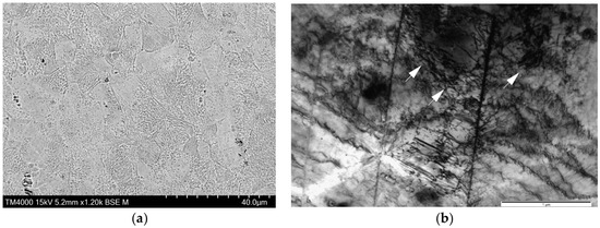
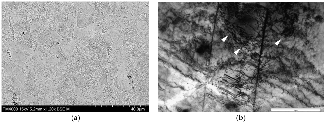
The microstructure of the unirradiated, as-received 08Cr16Ni11Mo3Ti steel samples consists of polyhedral austenite grains with an average grain size of 20–30 μm (Figure 1). As expected from the 20% cold-worked condition detailed in the methodology, the matrix exhibits an elevated dislocation density 1010–1011 cm−2 characterized by mesh and mesh-cellular structures, alongside both mechanical and growth twins. Various secondary phase inclusions are distributed both within the matrix and along the grain boundaries. EDS analysis confirmed that the majority of these precipitates are globular titanium carbide (TiC) particles. Additionally, highly etchable stringer-type precipitates, which do not conform to the austenite grain boundaries, are observed. Molybdenum is present in the matrix in low solid-solution concentrations and appears to be preferentially partitioned into molybdenum-enriched secondary phases.
Figure 1.
Microstructure of steel 08Cr16Ni11Mo3Ti after 20% cold work, obtained by SEM (a) and TEM (b). Arrows in (b) indicate representative dense dislocation networks.
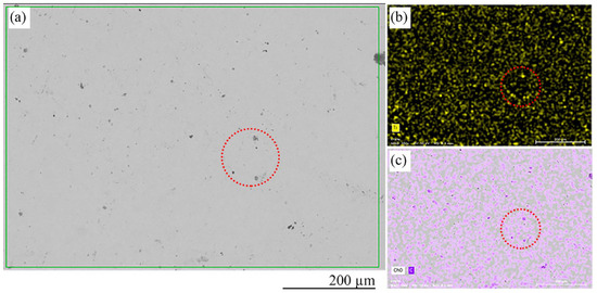
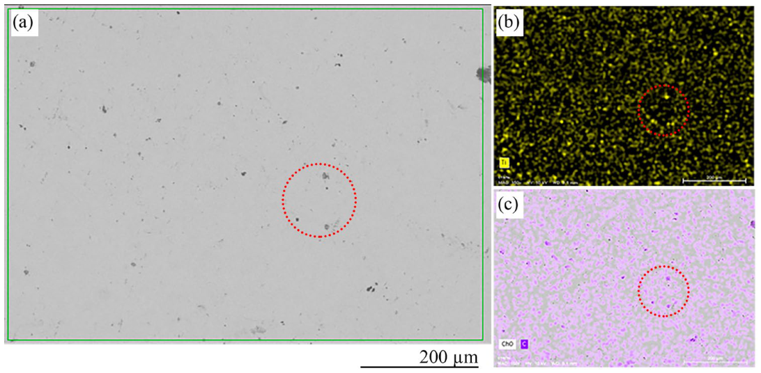
Following a solution annealing treatment at 1050 °C for 30 min, the 20% cold-worked microstructure underwent complete primary recrystallization. The dense mesh-cellular dislocation networks were eliminated, replaced by a strain-free matrix with equiaxed austenite grains containing characteristic straight-edged annealing twins. However, isolated inclusions, primarily titanium carbides, remain in the material (Figure 2).
Figure 2.
Micrograph of the surface of an unirradiated 08Cr16Ni11Mo3Ti steel specimen (a). Elemental distribution maps of titanium (b) and carbon (c) over the surface. Red circles indicate clusters of titanium carbides.
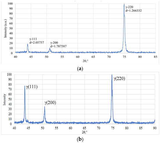
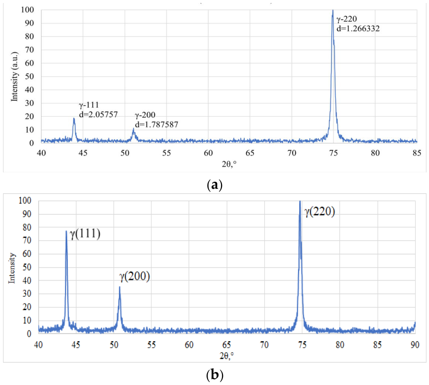
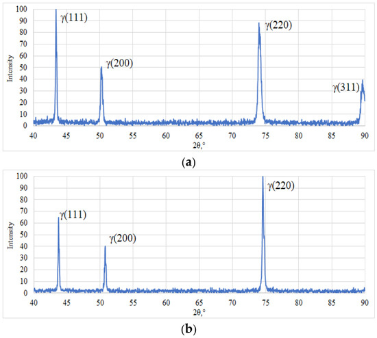
X-ray diffraction (XRD) analysis was conducted to evaluate the crystallographic evolution of the 08Cr16Ni11Mo3Ti steel before and after thermal treatment (Figure 3). In the as-received, 20% cold-worked condition, the diffractogram exhibits pronounced peak broadening, most notably in the dominant γ(220) reflection. This broadening confirms the presence of high residual microstrains and the elevated dislocation density characteristic of the plastically deformed matrix. Additionally, the heavily suppressed intensities of the γ(111) and γ(200) peaks relative to the γ(220) peak indicate a strong preferred crystallographic orientation (texture) induced by the cold deformation process. Following solution annealing at 1050 °C for 30 min, the diffraction peaks become significantly sharper and narrower, reflecting the relief of internal lattice strains and a substantial reduction in dislocation density driven by complete primary recrystallization. Furthermore, the relative intensities of the γ(111) and γ(200) reflections increase markedly, signaling a transition toward a more random crystallographic orientation consistent with the nucleation and growth of new, strain-free equiaxed austenite grains.
Figure 3.
X-ray diffractograms illustrating the crystallographic evolution of 08Cr16Ni11Mo3Ti steel: (a) the as-received 20% cold-worked state, exhibiting characteristic peak broadening and strong deformation texture, and (b) the fully recrystallized state following solution annealing at 1050 °C for 30 min, showing stress relief and randomized crystallographic orientation.
3.2. Microstructure After Helium Plasma Irradiation
Plasma exposure may generate porosity and microcracking within austenitic steels, and mass and density measurements were therefore combined to distinguish material removed from the surface from discontinuities formed inside the material. The specimen irradiated at 550 °C for 5000 s lost 2.1 mg. Referred to the exposed area of 78.5 mm2 and assuming uniform removal, this corresponds to an eroded layer of approximately 3.4 µm, i.e., a mean erosion rate of ~0.7 nm/s. The bulk density of the irradiated specimens, determined by hydrostatic weighing, did not differ significantly from that of the unirradiated material (7.893 g/cm3). Since the eroded layer represents well below one percent of the specimen volume, this measurement is not sensitive to porosity confined to the near-surface region; it does, however, exclude the formation of closed voids distributed throughout the bulk. The mass loss is thus attributed to progressive removal of material from the plasma-facing surface rather than to internal void formation.
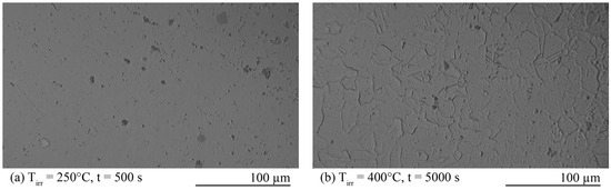
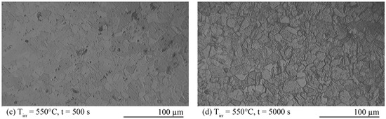
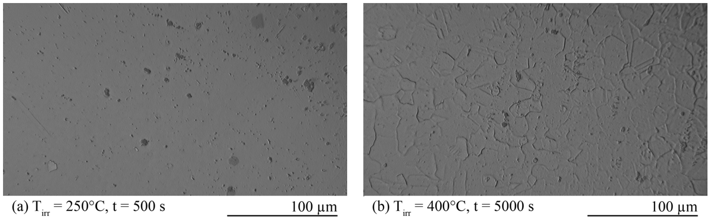
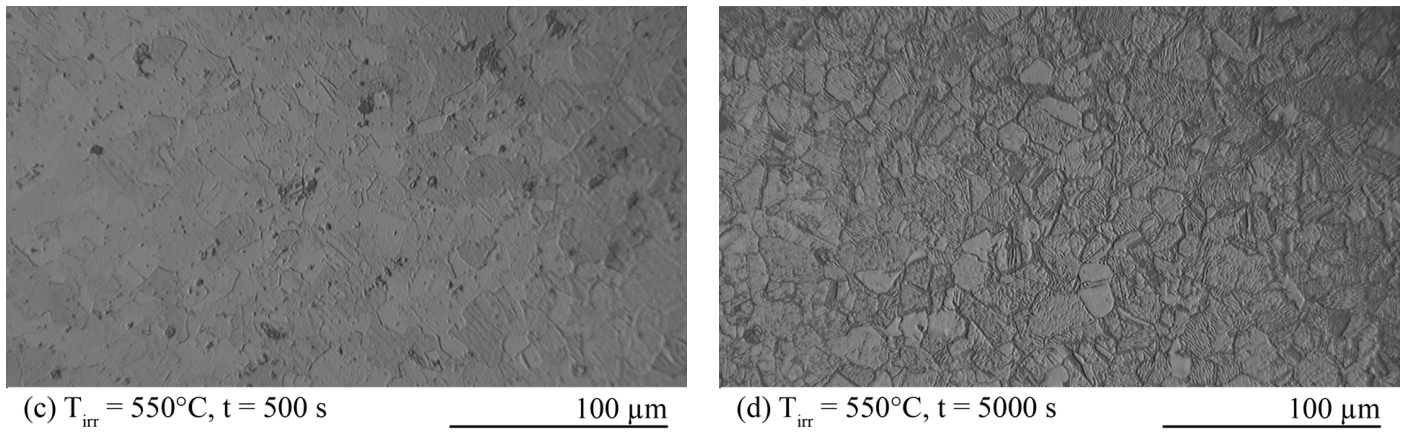
Figure 4 presents the surface microstructures of the 08Cr16Ni11Mo3Ti specimens following plasma irradiation, captured via optical metallography. At a relatively low irradiation temperature of 250 °C (Figure 4a), the plasma exposure induced no macroscopic changes to the surface relief; the matrix remained largely featureless, with only isolated 1–5 μm molybdenum and titanium carbide inclusions visible. As the irradiation temperature increased to 400 °C (Figure 4b), the onset of plasma-induced ion and thermal etching became apparent, selectively revealing the underlying grain boundaries and causing the macroscopic surface to appear matte. This selective etching effect is drastically accelerated at 550 °C. After just 500 s at 550 °C (Figure 4c), the grain structure is heavily etched and distinctly visible, showing a high density of fine grains and characteristic twins. Extending the plasma exposure to 5000 s at 550 °C (Figure 4d) resulted in pronounced preferential sputtering along grain boundaries, exposing a highly developed surface relief characterized by distinct, varying crystallographic orientations, prominent twin boundaries, and internal subgrain cellular structures.
Figure 4.
Microstructure of 08Cr16Ni11Mo3Ti steel samples after plasma irradiation. Irradiation conditions are shown in the photographs.
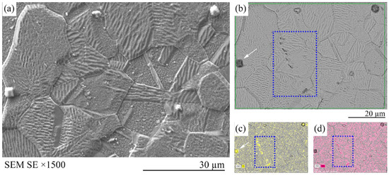
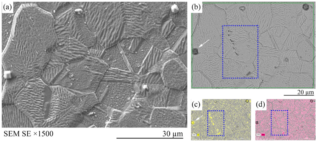
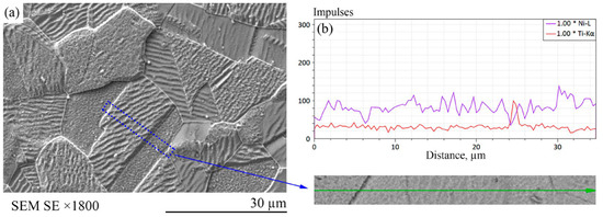
Helium plasma irradiation at 400 °C induced a noticeable increase in surface precipitation (Figure 5). Fine titanium-rich particles (~1 μm) precipitated predominantly along the grain boundaries, while larger, primary titanium and molybdenum inclusions (~10 μm) remained distributed throughout both the intragranular matrix and the boundaries. Increasing the irradiation temperature to 550 °C (for a 5000 s exposure) resulted in a highly developed surface relief driven by intense preferential sputtering of the matrix (Figure 5a). This plasma etching exposed a distinct polycrystalline structure with grains of varying sizes and no apparent crystallographic texturing. Notably, a pronounced sub-cellular and lamellar-like microstructure was revealed within the larger crystallites. Energy-dispersive X-ray spectroscopy (EDS) mapping (Figure 5b–d) highlights a highly heterogeneous elemental distribution across this irradiated surface. Complex Mo–Ti–C precipitates formed in distinct linear arrays (stringers) not exceeding 2–3 μm in size, alongside the larger isolated titanium inclusions. Furthermore, EDS line scanning across the microstructural interfaces (Figure 6) confirmed significant elemental redistribution driven by the irradiation. Specifically, the specimen irradiated at 550 °C for 5000 s exhibited localized depletion of nickel and iron at the grain boundaries, accompanied by a concomitant enrichment of titanium, indicating the onset of radiation-induced segregation (RIS).
Figure 5.
Surface characterization of the specimen irradiated with helium plasma for 5000 s at 550 °C: (a) Secondary electron (SE) image of the surface topography; (b) reference area selected for EDS mapping; and corresponding surface distribution maps of (c) titanium and (d) molybdenum. The blue rectangle marks a region with a high concentration of Mo–Ti–C particles, while the white arrow indicates an isolated titanium inclusion.
Figure 6.
Non-uniform distribution of chemical elements in the specimen irradiated with helium plasma for 5000 s at 550 °C. In the SEM image (SE) of the surface (a), the blue rectangle marks the region in which the linear distribution of titanium and nickel was examined (b).
After helium plasma irradiation at 400 °C and 550 °C (Figure 7), the diffraction patterns remain closely similar to those of the material in the pre-irradiation solution-annealed condition (Figure 3b). The γ(220) reflection remains dominant; the relative intensities of the γ(111) and γ(200) peaks are essentially unchanged, and the peak profiles remain sharp and narrow. No additional reflections attributable to secondary phases were detected, and no measurable peak broadening indicative of accumulated microstrain was observed. Since the specimens were solution-annealed prior to irradiation, this indicates that helium plasma exposure introduced no lattice strain, texture change, or phase transformation resolvable by X-ray diffraction.
Figure 7.
X-ray diffraction patterns of 08Cr16Ni11Mo3Ti steel specimens after helium plasma irradiation for 5000 s at 400 °C (a) and 550 °C (b).
3.3. The Effect of Helium Plasma Irradiation on Microhardness of the Steel
The surface microhardness of the specimens was evaluated using a PMT-3 microhardness tester under a 50 g load (Table 3). The baseline microhardness of the unirradiated 08Cr16Ni11Mo3Ti steel was measured at 126 HV 0.05. Following helium plasma irradiation, the surface layer exhibited distinct radiation-induced hardening; with the exception of the specimen exposed at 250 °C for 5000 s, the plasma treatments increased the microhardness by an average of 30–40 HV 0.05. The hardness distribution was also highly dependent on the localized microstructure. For instance, in the specimen irradiated at 400 °C for 5000 s, regions comprising fine-grained clusters exhibited a substantially higher microhardness (239 HV 0.05) compared to the larger recrystallized grains (162 HV 0.05), a variance consistent with the Hall-Petch strengthening mechanism.
Table 3.
Microhardness * of 08Cr16Ni11Mo3Ti steel samples after plasma irradiation.
Overall, at a constant exposure time of 500 s, the measured microhardness increased moderately from 158 to 173 HV 0.05 as the irradiation temperature increased from 250 to 550 °C. After 5000 s, the hardness values at 400 and 550 °C, 162/239 and 164 HV 0.05, respectively, remained close to those measured after 500 s. In contrast, the hardness at 250 °C decreased to 101 HV 0.05, below the unirradiated value of 126 HV 0.05. The prolonged exposure did not produce a systematic decrease in hardness, and the low value obtained at 250 °C cannot be attributed conclusively to thermal recovery. The origin of this behavior remains unclear and warrants systematic investigation using additional materials and a broader range of irradiation conditions.
4. Discussion
This study investigates the microstructural, crystallographic, and mechanical evolution of annealed 08Cr16Ni11Mo3Ti austenitic stainless steel subjected to helium plasma irradiation. Surface degradation changed markedly with irradiation temperature, which in these experiments co-varied with ion flux and beam power: morphological effects were negligible at 250 °C, grain-boundary etching initiated at 400 °C, and aggressive preferential sputtering at 550 °C exposed a highly developed relief of grains, twins, and sub-cellular structures. This evolution coincided with intense grain-boundary precipitation of titanium carbides and complex Mo–Ti–C arrays. The surface microhardness demonstrated a competing dependence on plasma parameters; while short exposures (500 s) showed radiation hardening that increased with temperature (and the accompanying ion flux), extending the exposure time (5000 s) induced localized structural softening across all temperatures due to thermal recovery. It should be noted that, owing to the operating principle of the facility, the specimen temperature, electron-beam power and ion current (and hence the ion flux) could not be varied independently: the beam power is the main parameter for heating and also controls the ionization of the working gas and the extracted ion current. The effects of temperature, flux and fluence therefore cannot be fully separated on the basis of the present data.
The exposure conditions examined here differ in several respects from ion-beam irradiation to comparable energy, and these differences bear directly on how the present results should be interpreted. In a beam experiment the incident flux is focused, and the implanted layer is retained, so damage accumulates monotonically with fluence in a fixed volume beneath the surface. Under plasma exposure, the surface is simultaneously implanted and eroded. The mass-loss measurements reported above correspond to removal of approximately 3.4 µm during 5000 s at 550 °C, a mean erosion rate of ~0.7 nm/s. Since the projected range of 2 keV helium ions in austenitic steel is of the order of dozens of nanometers, any given layer of material remains within the ballistic damage zone for only a few tens of seconds before the receding surface reaches it. The accumulated displacement dose retained in the surviving material therefore saturates within the first minute of exposure and is essentially the same after 500 s and after 5000 s. Ballistic damage accumulation consequently accounts for the observed low dependence of microhardness on exposure time at 550 °C.
Comparing the results obtained for austenitic steel 08Cr16Ni11Mo3Ti with the data of Pimenov et al. [39], who investigated the effect of deuterium plasma on reduced-activation austenitic steels (10Cr12Mn20W, 25Cr12Mn20W), makes it possible to distinguish the physical degradation mechanisms operating under steady-state (prolonged) and pulsed irradiation regimes. In the work of Pimenov et al., at power densities of q ~ 107–108 W/cm2, the dominant factor was intense thermal shock. It induced cascading melting and evaporation of the surface layer, the formation of resolidified melt beads, toroidal craters, and microcracks, and masked the material’s own diffusion processes through redeposition of chamber structural materials (in particular, Cu). In contrast, under continuous low-energy helium irradiation (2 keV), the surface layer of 08Cr16Ni11Mo3Ti steel does not undergo melting. Surface degradation instead proceeds through selective ion and thermal etching. Ti-rich and Mo–Ti–C particles became more clearly visible on the plasma-exposed surface. However, similar Ti-rich particles were already present in the unirradiated material, and morphology and EDS data alone cannot establish whether the observed particles precipitated during irradiation or were pre-existing particles exposed by preferential removal of the surrounding matrix. Therefore, these features are described as particles revealed on the irradiated surface rather than as irradiation-induced precipitates.
The absence of melting and droplet-type defects, together with the development of grain boundaries and sub-cellular structure at 400–550 °C, indicates that the change in surface relief is governed by ion sputtering and temperature-stimulated diffusion of elements rather than by thermal melting of the material. This interpretation is consistent with the results of [45], who investigated 304 stainless steel, a chromium-nickel austenitic steel of similar composition and structure, under comparable low-energy helium ion exposure. They showed that, in the absence of melting, the dominant degradation mechanism shifts toward nanostructural reorganization: a high density of helium nanobubbles 1–2 nm in diameter accumulates in the subsurface layer, and helium trapping saturates upon reaching a critical dose of about 1021 m−2, followed by low-temperature desorption due to detrapping from the crystal lattice surrounding the bubbles [45]. Since the fluences in the present work (2 × 1025–4 × 1026 m−2, Table 2) exceed this saturation dose by several orders of magnitude, the formation of similar subsurface helium bubbles can be expected here. We emphasize, however, that bubbles, defects and damage depth were not examined directly in this study, so this mechanism should be regarded as a hypothesis based on the literature rather than an observed result.
The experimental conditions examined in the paper simulate a scenario where the protective armor has been locally compromised, and the exposed structural steel continues to operate. Over a sustained plasma campaign—particularly when accounting for the higher particle fluxes expected at a divertor surface—this cumulative material loss becomes substantial. In such operational regimes, the degraded state of the surface layer, rather than the bulk material properties, will ultimately dictate the component’s remaining service life. While the erosion resistance (0.7 nm/s) of the austenitic steel under these isolated plasma conditions is relatively favorable, it is imperative to note that this study does not account for synergistic reactor conditions. Specifically, these results isolate plasma effects from prolonged 14.1 MeV fast neutron irradiation, a primary degradation factor against which protective plasma-facing armor such as beryllium or tungsten offers no defense.
5. Conclusions
In this study, the microstructural, crystallographic, and mechanical evolution of annealed 08Cr16Ni11Mo3Ti austenitic stainless steel under helium plasma irradiation was investigated.
- Surface degradation under helium plasma is highly temperature-dependent. While low-temperature exposure (250 °C) causes minimal morphological change, elevated temperatures (400–550 °C) trigger aggressive preferential sputtering and thermal etching.
- This extreme localized environment accelerates the precipitation of fine titanium-rich and Mo–Ti–C complexes, and prolonged exposure initiates localized Radiation-Induced Segregation (RIS), characterized by nickel and iron depletion alongside titanium enrichment at the grain boundaries.
- Generally, the microhardness of the specimens increased after irradiation. Nevertheless, the mechanical integrity of the irradiated surface is governed by a competing balance between radiation damage and thermal recovery. Short-duration plasma transients induce radiation hardening influenced locally by grain size according to the Hall-Petch relationship. Conversely, prolonged exposure drives insignificant internal stress relaxation and localized structural softening in the specimens irradiated above 400 °C.
The findings provide critical insights into the surface degradation mechanisms of structural materials subjected to extreme off-normal fusion reactor transients.
Author Contributions
Conceptualization, A.T., G.I. and B.C.; Methodology, L.D. and S.K.; Formal analysis, A.T. and Y.Z.; Investigation, A.T., L.D., Y.Z., I.S., G.Z., T.T. and A.M.; Resources, S.K., I.S., G.Z., T.T., A.M. and B.C.; Writing—original draft, A.T. and B.C.; Writing—review and editing, A.T., G.I. and B.C.; Visualization, A.T.; Supervision, S.K. and B.C.; Project administration, B.C. All authors have read and agreed to the published version of the manuscript.
Funding
The work was carried out within the framework of the scientific and technical program IRN BR23891779, “Scientific and Technical Support of Experimental Research at the Kazakhstan Materials Science KTM Tokamak” with program-targeted funding from the Agency of the Republic of Kazakhstan for Atomic Energy.
Data Availability Statement
The raw data supporting the conclusions of this article will be made available by the authors on request.
Acknowledgments
The authors would like to thank M.S. Merezhko, K.V. Tsay, and L.G. Turubarova for their assistance in this work. During the preparation of this work, the authors used Google Translate, Google Gemini (3.6 Flash, 3.1 Pro), Anthropic Claude (Sonnet 5, Opus 5) in order to improve the translation from Russian to English and readability of the manuscript, with caution. After using these services, all authors reviewed and edited the content as needed and take full responsibility for the content of the publication.
Conflicts of Interest
The authors declare no conflicts of interest.
Abbreviations
The following abbreviations are used in this manuscript:
| ITER | International Thermonuclear Experimental Reactor |
| DEMO | Demonstration Power Plant |
| SEM | Scanning Electron Microscopy |
| TEM | Transmission Electron Microscopy |
| XRD | X-ray Diffraction |
| EDS | Energy-Dispersive X-ray Spectroscopy |
| EDM | Electrical Discharge Machining |
| RIS | Radiation-Induced Segregation |
| HV | Vickers Hardness |
| AISI | American Iron and Steel Institute |
References
- Alba, R.; Iglesias, R.; Cerdeira, M.Á. Materials to Be Used in Future Magnetic Confinement Fusion Reactors: A Review. Materials 2022, 15, 6591. [Google Scholar] [CrossRef] [Scilit]
- Ongena, J.; Ogawa, Y. Nuclear Fusion: Status Report and Future Prospects. Energy Policy 2016, 96, 770–778. [Google Scholar] [CrossRef] [Scilit]
- Donné, A.J.H. The European Roadmap towards Fusion Electricity. Phil. Trans. R. Soc. A 2019, 377, 20170432. [Google Scholar] [CrossRef] [Scilit]
- Joffrin, E.; Abduallev, S.; Abhangi, M.; Abreu, P.; Afanasev, V.; Afzal, M.; Aggarwal, K.M.; Ahlgren, T.; Aho-Mantila, L.; Aiba, N.; et al. Overview of the JET Preparation for Deuterium–Tritium Operation with the ITER like-wall. Nucl. Fusion 2019, 59, 112021. [Google Scholar] [CrossRef] [Scilit]
- Hawryluk, R.J. Results from Deuterium-Tritium Tokamak Confinement Experiments. Rev. Mod. Phys. 1998, 70, 537–587. [Google Scholar] [CrossRef] [Scilit]
- Kamada, Y.; Barabaschi, P.; Ishida, S.; Ide, S.; Lackner, K.; Fujita, T.; Bolzonella, T.; Suzuki, T.; Matsunaga, G.; Yoshida, M.; et al. Plasma Regimes and Research Goals of JT-60SA towards ITER and DEMO. Nucl. Fusion 2011, 51, 073011. [Google Scholar] [CrossRef] [Scilit]
- Li, J.; Guo, H.Y.; Wan, B.N.; Gong, X.Z.; Liang, Y.F.; Xu, G.S.; Gan, K.F.; Hu, J.S.; Wang, H.Q.; Wang, L.; et al. A Long-Pulse High-Confinement Plasma Regime in the Experimental Advanced Superconducting Tokamak. Nat. Phys. 2013, 9, 817–821. [Google Scholar] [CrossRef] [Scilit]
- The ITER Organization; Domestic Agencies and ITER Collaborators; Campbell, D.J.; Akiyama, T.; Barnsley, R.; Bassan, M.; Baylor, L.R.; Bertalot, L.; Escourbiac, F.; Giancarli, L.M.; et al. Innovations in Technology and Science R&D for ITER. J. Fusion Energ. 2019, 38, 11–71. [Google Scholar] [CrossRef] [Scilit]
- Federici, G.; Bachmann, C.; Barucca, L.; Baylard, C.; Biel, W.; Boccaccini, L.V.; Bustreo, C.; Ciattaglia, S.; Cismondi, F.; Corato, V.; et al. Overview of the DEMO Staged Design Approach in Europe. Nucl. Fusion 2019, 59, 066013. [Google Scholar] [CrossRef] [Scilit]
- Knaster, J.; Moeslang, A.; Muroga, T. Materials Research for Fusion. Nat. Phys. 2016, 12, 424–434. [Google Scholar] [CrossRef] [Scilit]
- Zinkle, S.J.; Busby, J.T. Structural Materials for Fission & Fusion Energy. Mater. Today 2009, 12, 12–19. [Google Scholar] [CrossRef] [Scilit]
- Huang, Q.; Baluc, N.; Dai, Y.; Jitsukawa, S.; Kimura, A.; Konys, J.; Kurtz, R.J.; Lindau, R.; Muroga, T.; Odette, G.R.; et al. Recent Progress of R&D Activities on Reduced Activation Ferritic/Martensitic Steels. J. Nucl. Mater. 2013, 442, S2–S8. [Google Scholar] [CrossRef] [Scilit]
- Pacchioni, G. The Road to Fusion. Nat. Rev. Phys. 2019, 1, 360–362. [Google Scholar] [CrossRef] [Scilit]
- Ongena, J.; Koch, R.; Wolf, R.; Zohm, H. Magnetic-Confinement Fusion. Nat. Phys. 2016, 12, 398–410. [Google Scholar] [CrossRef] [Scilit]
- Abdou, M.; Morley, N.B.; Smolentsev, S.; Ying, A.; Malang, S.; Rowcliffe, A.; Ulrickson, M. Blanket/First Wall Challenges and Required R&D on the Pathway to DEMO. Fusion Eng. Des. 2015, 100, 2–43. [Google Scholar] [CrossRef] [Scilit]
- Ioki, K.; Barabaschi, P.; Bruno, L.; Cardella, A.; Danner, W.; Elio, F.; Hechler, M.; Kodama, T.; Lodato, A.; Loesser, D.; et al. ITER First Wall/Shield Blanket. Fusion Eng. Des. 1998, 39–40, 585–592. [Google Scholar] [CrossRef] [Scilit]
- Shim, H.-J.; Kim, S.; Park, B.; Kim, D.-H.; Johansson, V. Fabrication and Characteristic of the 316L(N)-IG Forged Block and Rolled Plate for Application to ITER Blanket Shield Block. Fusion Eng. Des. 2020, 156, 111738. [Google Scholar] [CrossRef] [Scilit]
- Gietl, H.; Worrall, M.J.; Chapman, D.B.; Dromey, J.M.E.; Frazer, D.; Gonderman, S.; Brookman, J.V.; Woolstenhulme, N.E. Neutron Irradiation & Thermomechanical Experiment (NITE)—Design. Fusion Eng. Des. 2026, 222, 115539. [Google Scholar] [CrossRef] [Scilit]
- Nordlund, K.; Zinkle, S.J.; Sand, A.E.; Granberg, F.; Averback, R.S.; Stoller, R.E.; Suzudo, T.; Malerba, L.; Banhart, F.; Weber, W.J.; et al. Primary Radiation Damage: A Review of Current Understanding and Models. J. Nucl. Mater. 2018, 512, 450–479. [Google Scholar] [CrossRef] [Scilit]
- Garner, F.A. 3.02—Radiation-Induced Damage in Austenitic Structural Steels Used in Nuclear Reactors. In Comprehensive Nuclear Materials, 2nd ed.; Konings, R.J.M., Stoller, R.E., Eds.; Elsevier: Oxford, UK, 2020; Volume 3, pp. 57–168. ISBN 978-0-08-102866-7. [Google Scholar]
- Akayev, S.O.; Ganeev, G.Z.; Dikov, A.S.; Kislitsin, S.B.; Khromushin, I.V.; Partyka, Y. Helium accumulation in Cr18Ni10Ti austenitic steel under different saturation methods. J. Phys. Conf. Ser. 2022, 2155, 012007. [Google Scholar] [CrossRef] [Scilit]
- Nastar, M.; Soisson, F. 1.08—Radiation-Induced Segregation. In Comprehensive Nuclear Materials, 2nd ed.; Konings, R.J.M., Stoller, R.E., Eds.; Elsevier: Oxford, UK, 2020; Volume 1, pp. 235–264. ISBN 978-0-08-102866-7. [Google Scholar]
- Etienne, A.; Radiguet, B.; Cunningham, N.J.; Odette, G.R.; Pareige, P. Atomic Scale Investigation of Radiation-Induced Segregation in Austenitic Stainless Steels. J. Nucl. Mater. 2010, 406, 244–250. [Google Scholar] [CrossRef] [Scilit]
- Merezhko, D.A.; Merezhko, M.S.; Gusev, M.N.; Busby, J.T.; Maksimkin, O.P.; Short, M.P.; Garner, F.A. Investigation of Pitting Corrosion in Sensitized Modified High-Nitrogen 316LN Steel after Neutron Irradiation. In Minerals, Metals and Materials Series; Springer: Berlin/Heidelberg, Germany, 2019; pp. 1125–1140. [Google Scholar]
- Andresen, P.L.; Was, G.S. 4.07—Irradiation Assisted Stress Corrosion Cracking. In Comprehensive Nuclear Materials, 2nd ed.; Konings, R.J.M., Stoller, R.E., Eds.; Elsevier: Oxford, UK, 2019; pp. 190–217. ISBN 978-0-08-102866-7. [Google Scholar]
- Andresen, P.L.; Was, G.S. A Historical Perspective on Understanding IASCC. J. Nucl. Mater. 2019, 517, 380–392. [Google Scholar] [CrossRef] [Scilit]
- Was, G.S.; Andresen, P.L. Mechanisms behind Irradiation-Assisted Stress Corrosion Cracking. In Nuclear Corrosion; Elsevier: Oxford, UK, 2020; pp. 47–88. ISBN 9780128237199. [Google Scholar]
- Merezhko, M.S.; Merezhko, D.A.; Tarapeyeva, A.Y.; Kapanov, A.S.; Short, M.P.; Burtebayev, N.T.; Sakhiyev, S.K. Strain Induced Martensitic Transformations Reduce Embrittlement in Neutron Irradiated Austenitic Stainless Steels. J. Nucl. Mater. 2026, 633, 156996. [Google Scholar] [CrossRef] [Scilit]
- Merezhko, M.S.; Merezhko, D.A. Localization of Plastic Deformation in the Copper and Stainless Steels Samples, Irradiated with Neutrons. J. Phys. Conf. Ser. 2022, 2155, 012009. [Google Scholar] [CrossRef] [Scilit]
- Tsay, K.V.; Rofman, O.V.; Merezhko, M.S.; Short, M.P. Simultaneous Strength and Toughness Increase in Low Dose Neutron Irradiated Austenitic Stainless Steel at Sub-Zero Temperatures Due to Changes in Strain-Induced Martensitic Transformation. Acta Mater. 2025, 288, 120786. [Google Scholar] [CrossRef] [Scilit]
- Akaev, S.O.; Dikov, A.S.; Dikova, L.A.; Kislitsin, S.B.; Firsova, V.V.; Larionov, A.S.; Chernov, I.I.; Staltsov, M.S. The effect of neutron irradiation in a helium atmosphere on the mechanical properties of 18Cr10NiTi austenitic steel. IOP Conf. Ser. Mater. Sci. Eng. 2020, 1005, 012007. [Google Scholar] [CrossRef] [Scilit]
- Dikov, A.; Kislitsin, S.; Ivanov, B.; Kiryanov, R.; Maksimkin, E. Changes in the Structure and Mechanical Properties of the SAV-1 Alloy and Structural Fe-Cr-Ni Steels After Long-Term Service as Core Materials in Nuclear Reactors. Materials 2025, 18, 3391. [Google Scholar] [CrossRef] [Scilit]
- Margolin, B.; Sorokin, A.; Shvetsova, V.; Minkin, A.; Potapova, V.; Smirnov, V. The Radiation Swelling Effect on Fracture Properties and Fracture Mechanisms of Irradiated Austenitic Steels. Part I. Ductility and Fracture Toughness. J. Nucl. Mater. 2016, 480, 52–68. [Google Scholar] [CrossRef] [Scilit]
- Margolin, B.; Minkin, A.; Fomenko, V.; Smirnov, V.; Pirogova, N.; Sorokin, A.; Shvetsova, V. Features of the Test Temperature and Damage Dose Effects on Fracture Toughness and Fatigue Crack Growth Rate for Austenitic Chromium-Nickel Steel Irradiated during 45 Years in WWER-440. J. Nucl. Mater. 2026, 627, 156600. [Google Scholar] [CrossRef] [Scilit]
- Margolin, B.; Sorokin, A.; Belyaeva, L. A Link between Neutron and Ion Irradiation Hardening for Stainless Austenitic and Ferritic-Martensitic Steels. Metals 2024, 14, 99. [Google Scholar] [CrossRef] [Scilit]
- Hojna, A. Overview of Intergranular Fracture of Neutron Irradiated Austenitic Stainless Steels. Metals 2017, 7, 392. [Google Scholar] [CrossRef] [Scilit]
- Griffiths, M.; Xu, S.; Ramos Nervi, J.E. Swelling and He-Embrittlement of Austenitic Stainless Steels and Ni-Alloys in Nuclear Reactors. Metals 2022, 12, 1692. [Google Scholar] [CrossRef] [Scilit]
- Riccardo, V.; Arnoux, G.; Cahyna, P.; Hender, T.C.; Huber, A.; Jachmich, S.; Kiptily, V.; Koslowski, R.; Krlin, L.; Lehnen, M.; et al. JET Disruption Studies in Support of ITER. Plasma Phys. Control. Fusion 2010, 52, 124018. [Google Scholar] [CrossRef] [Scilit]
- Polskii, V.I. Erosion of First-Wall Materials of a Thermonuclear Reactor under Conditions Simulating Plasma Disruptions. Ph.D. Thesis, National Research Nuclear University MEPhI, Moscow, Russia, 2005. [Google Scholar]
- Pimenov, V.N.; Gribkov, V.A.; Ivanov, L.I.; Demina, E.V.; Masliyaev, S.A.; Dubrovskiy, A.V.; Scholz, M.; Miklaszewski, R.; Ugaste, Y.E.; Kolman, B. Extreme pulsed effect of energy on low-activated austenitic steels. Vopr. At. Nauk. Tekh. Ser. Fiz. Radiats. Povrezhdeniy Radiats. Materialoved. 2005, 3, 144–150. [Google Scholar]
- Moldabekov, Z.M.; Zhukeshov, A.M.; Gabdullina, A.T.; Amrenova, A.U.; Serik, K. The influence of pulsed plasmas on erosion of the surface of metal materials. NNC RK Bull. 2018, 1, 97–101. [Google Scholar] [CrossRef] [Scilit]
- Skakov, M.; Miniyazov, A.; Batyrbekov, E.; Baklanov, V.; Koyanbayev, Y.; Gradoboev, A.; Kozhakhmetov, Y.; Sokolov, I.; Tulenbergenov, T.; Zhanbolatova, G. Influence of the Carbidized Tungsten Surface on the Processes of Interaction with Helium Plasma. Materials 2022, 15, 7821. [Google Scholar] [CrossRef] [Scilit]
- Skakov, M.; Batyrbekov, E.; Sokolov, I.; Miniyazov, A.; Tulenbergenov, T.; Sapataev, Y.; Orazgaliyev, N.; Bukina, O.; Zhanbolatova, G.; Kozhakhmetov, Y. Influence of Hydrogen Plasma on the Surface Structure of Beryllium. Materials 2022, 15, 6340. [Google Scholar] [CrossRef] [Scilit]
- Ueda, Y.; Peng, H.Y.; Lee, H.T.; Ohno, N.; Kajita, S.; Yoshida, N.; Doerner, R.; De Temmerman, G.; Alimov, V.; Wright, G. Helium Effects on Tungsten Surface Morphology and Deuterium Retention. J. Nucl. Mater. 2013, 442, S267–S272. [Google Scholar] [CrossRef] [Scilit]
- Tokitani, M.; Miyamoto, M.; Tokunaga, K.; Iwakiri, H.; Fujiwara, T.; Yoshida, N. Desorption of Helium from Austenitic Stainless Steel Heavily Bombarded by Low Energy He Ions. J. Nucl. Mater. 2004, 329–333, 761–765. [Google Scholar] [CrossRef] [Scilit]

Disclaimer/Publisher’s Note: The statements, opinions and data contained in all publications are solely those of the individual author(s) and contributor(s) and not of MDPI and/or the editor(s). MDPI and/or the editor(s) disclaim responsibility for any injury to people or property resulting from any ideas, methods, instructions or products referred to in the content. |
© 2026 by the authors. Licensee MDPI, Basel, Switzerland. This article is an open access article distributed under the terms and conditions of the Creative Commons Attribution (CC BY) license.