


Simple Summary
Insecticide resistance makes the brown planthopper, Nilaparvata lugens, increasingly difficult to control. Resistance is often linked to enzymes that break down insecticides, but these enzymes also depend on energy and other metabolites produced by the insect. In this study, we compared insecticide-resistant and susceptible brown planthoppers to investigate how metabolism contributes to resistance. Resistant insects showed coordinated changes in pathways related to energy production, protective metabolites, and insecticide detoxification. Two genes, mitochondrial NADP-dependent isocitrate dehydrogenase (NADP) and UDP-glucosyltransferase 2 (UGT2), were consistently more active in two independently selected resistant strains. Silencing either gene increased the insects’ sensitivity to the neonicotinoid insecticides nitenpyram and clothianidin. These results suggest that neonicotinoid resistance is supported by interconnected changes in both central metabolism and detoxification. NADP and UGT2 may therefore represent useful molecular targets for monitoring resistance and developing improved pest-management strategies.
Abstract
Metabolic resistance is commonly attributed to the overexpression of detoxification enzymes, whereas the metabolic systems that sustain detoxification remain less well resolved. Here, we integrated widely targeted metabolomics and transcriptomics to compare the clothianidin-resistant brown planthopper strain CLR with the susceptible strain CLS and validated candidate genes in an independent nitenpyram-resistant background. CLR and CLS exhibited distinct metabolomic profiles. Differential genes and metabolites converged on carbon metabolism, glycolysis/gluconeogenesis, the tricarboxylic acid cycle, glutathione metabolism, pentose and glucuronate interconversions, cytochrome P450-mediated xenobiotic metabolism, and ABC transporters. These changes were summarized into four interconnected modules involving glycolytic energy supply, protective sugar-derived metabolites, pyruvate–TCA–malate metabolism, and UGT-mediated glycosylation. Expression analysis identified a mitochondrial NADP-dependent isocitrate dehydrogenase gene and UDP-glucosyltransferase 2 (UGT2) as consistently upregulated in both resistant backgrounds. Silencing NADP reduced the LC50 of nitenpyram and clothianidin by 1.84- and approximately 1.81-fold, respectively, whereas UGT2 silencing produced corresponding reductions of 1.89- and 1.83-fold. These findings indicate that neonicotinoid resistance in Nilaparvata lugens is supported by coordinated remodelling of central metabolism and detoxification and identify two candidate metabolic nodes for further resistance management research.
1. Introduction
The brown planthopper, Nilaparvata lugens Stål, is one of the most destructive migratory pests of rice in Asia, where it damages plants through sustained phloem feeding, causes hopperburn during outbreaks and transmits rice viruses [1]. Its high fecundity, long-distance migration and rapid population growth make chemical control an important component of emergency management in many rice-growing regions [1]. Neonicotinoid insecticides have been widely used against N. lugens and other sap-feeding pests because they act mainly on insect nicotinic acetylcholine receptors and show strong systemic activity in plants [2,3,4,5]. Nevertheless, intensive and repeated use of neonicotinoids has accelerated resistance evolution in many insect pests, reducing field efficacy and increasing the need for resistance management strategies [6,7,8]. Understanding the mechanisms underlying neonicotinoid resistance in N. lugens is therefore important for improving insecticide stewardship and delaying resistance development.
Insecticide resistance is usually a multifactorial phenotype involving altered target-site sensitivity, reduced penetration, behavioural avoidance and enhanced metabolic detoxification [6,7,9,10]. Among these mechanisms, metabolic resistance has received particular attention because it can reduce the internal dose of insecticides before they reach their molecular targets [9,10]. In N. lugens, overexpression of the cytochrome P450 gene CYP6ER1 has been associated with imidacloprid resistance, providing direct evidence that P450-mediated detoxification contributes to neonicotinoid resistance in this species [11]. Additional studies and reviews have emphasized that P450s, glutathione S-transferases (GSTs), carboxylesterases, UDP-glycosyltransferases and ATP-binding cassette (ABC) transporters may contribute to insecticide resistance through altered expression, substrate metabolism, conjugation or transport [9,11,12,13,14]. For example, GSTs participate in glutathione-dependent conjugation and oxidative-stress defence, whereas ABC transporters can export xenobiotics or their metabolites across membranes [12,13,15]. These studies have greatly advanced our understanding of detoxification-based resistance, but they have generally focused on detoxification genes themselves rather than on the metabolic conditions that allow these reactions to be sustained.
From a biochemical perspective, detoxification is not an isolated enzymatic process. P450-catalysed oxidation requires electron transfer from NADPH through NADPH-cytochrome P450 reductase, GST-mediated conjugation depends on reduced glutathione, and UGT-mediated glycosylation requires activated UDP-sugars as donor substrates [13,14,16]. Detoxification products may also need to be transported across membranes through ATP-dependent transport systems [12,15]. Thus, enhanced detoxification capacity is likely to impose demands on central metabolism, especially pathways that generate ATP, NADPH, glutathione and sugar donors [16,17,18,19]. This requirement is consistent with the classical framework of xenobiotic metabolism, in which phase I oxidation, phase II conjugation and phase III transport are functionally connected to cellular energy, redox balance and precursor supply [9,12,16,20]. Therefore, insecticide resistance may involve not only the up-regulation of detoxification enzymes, but also the reallocation of metabolic fluxes that supply cofactors and substrates for detoxification reactions.
Evidence from other insects supports this view. In Drosophila melanogaster, Cyp6g1 overexpression is associated with dichlorodiphenyltrichloroethane (DDT) resistance and has become a representative example linking a P450 gene to insecticide resistance in a model insect [21,22]. In Anopheles gambiae, CYP6M2 and CYP6P3 are elevated in pyrethroid-resistant populations, and CYP6M2 can metabolize pyrethroids directly [23,24]. In the whitefly Bemisia tabaci, overexpression of CYP6CM1 is associated with resistance to neonicotinoid insecticides, indicating that P450-mediated detoxification is also important in sap-feeding hemipterans [25]. These examples show that detoxification enzymes can be causal contributors to resistance, but they also imply a continuous requirement for cofactors, reducing equivalents and metabolic substrates. Similar principles have been described in biomedical studies, where drug-tolerant cells frequently exhibit altered glucose metabolism, pentose phosphate pathway activity, NADPH production and glutathione homeostasis [18]. In such systems, pathways that maintain redox balance can influence the capacity of cells to tolerate xenobiotic or oxidative stress [17,19]. These observations suggest that tolerance to toxic compounds is closely linked to cellular metabolic state across diverse biological systems.
For insecticide resistance, this metabolic dimension deserves closer attention. After insecticides enter the insect body, they not only require metabolic conversion by P450s, GSTs, UGTs and related enzyme systems, but also may induce oxidative stress, increase energy demand and disturb cellular homeostasis [9,10,16]. If resistant insects enhance glycolysis, the tricarboxylic acid cycle, the pentose phosphate pathway, amino acid metabolism or glycosylation-related pathways to provide ATP, NADPH, glutathione and UDP-sugars, these metabolic processes may contribute to the maintenance of detoxification capacity and resistance phenotypes [16,17,18,19]. Recent transcriptomic, metabolomic and multi-omics approaches provide opportunities to examine such coordinated changes at the pathway level rather than only at the level of individual detoxification genes [26,27,28]. However, it remains unclear whether metabolic network changes in neonicotinoid-resistant N. lugens are merely associated responses to insecticide stress or whether they actively participate in resistance maintenance.
In this study, we investigated the potential contribution of metabolic regulation to neonicotinoid resistance in N. lugens. By integrating transcriptomic and metabolomic analyses with candidate-gene expression validation and RNA interference, we aimed to evaluate whether specific metabolic processes participate in resistance maintenance and may provide potential targets for resistance management or synergistic control strategies.
2. Materials and Methods
2.1. Insect Strains, Rearing and Sample Collection
The clothianidin-resistant (CLR) and corresponding susceptible (CLS) strains were obtained from laboratory colonies previously established and characterized by Jin et al. [29], whereas the nitenpyram-resistant (NR) and corresponding susceptible (NS) strains were established as described by Mao et al. [30]. The CLR and NR strains had been maintained under continuous selection with the corresponding insecticides for more than 20 generations, while the susceptible strains were maintained without insecticide exposure. Before the experiments, resistance phenotypes were reconfirmed using the rice-seedling dipping bioassay described in Section 2.6. Based on the estimated LC50 values, the CLR and NR strains exhibited approximately 270-fold and 100-fold resistance, respectively, relative to their corresponding susceptible strains. Taichung Native 1 (TN1) rice seedlings were used for insect rearing and bioassays following a previously reported procedure [31]. The insects were maintained at 27 ± 1 °C, 70–80% relative humidity, and a photoperiod of 16 h light:8 h dark. Water was replenished as required to maintain seedling growth, and no additional fertilizer was applied during the experimental period. Fourth-instar nymphs were selected with reference to previous resistance-related studies in N. lugens [29,30]. The use of a single late-nymphal stage minimized developmental variation, provided sufficient biomass for metabolomic and transcriptomic analyses, and facilitated standardized microinjection. Adults were not used because differences in sex, reproductive status, and wing morph could introduce additional physiological and metabolic variation.
For metabolomic analysis, each strain contained six biological replicates. Each biological replicate consisted of 100 mg of fourth-instar nymphs. For transcriptome sequencing, each strain contained three biological replicates, with each biological replicate consisting of 20 fourth-instar nymphs. Samples were immediately frozen in liquid nitrogen after collection and stored at −80 °C until metabolite or RNA extraction.
2.2. Metabolite Profiling and Data Analysis
Metabolite extraction and profiling were performed by Biomarker Technologies Corporation (Beijing, China) following a previously published workflow [32]. Six biological replicates were analyzed for each strain, and each replicate consisted of 100 mg of pooled fourth-instar nymphs. Frozen samples were ground in liquid nitrogen and subjected to metabolite extraction. Equal aliquots of all extracts were pooled to prepare quality-control (QC) samples, which were analyzed periodically throughout the analytical sequence to monitor instrumental stability and analytical repeatability.
Metabolite profiling was performed using a Shimadzu Nexera X2 UPLC system (Shimadzu Corporation, Kyoto, Japan) coupled to an AB Sciex 4500 QTRAP mass spectrometer (AB SCIEX, Framingham, MA, USA) equipped with an electrospray ionization source. Chromatographic separation was achieved using an Agilent SB-C18 column (Agilent SB-C18 column) (1.8 μm, 2.1 × 100 mm) maintained at 40 °C. The mobile phases consisted of water containing 0.1% formic acid (A) and acetonitrile containing 0.1% formic acid (B). The proportion of B was increased from 5% to 95% within 9 min, maintained at 95%, returned to 5% within 1.1 min, and equilibrated for 2.9 min. The injection volume was 4 μL. The mass spectrometer was operated in positive- and negative-ion modes with a source temperature of 550 °C and ion-spray voltages of 5500 and −4500 V, respectively. Metabolites were detected in multiple reaction monitoring (MRM) mode and identified by comparison of precursor/product ions and MS/MS spectra with the Biomarker Technologies in-house database and the HMDB, MassBank, MoToDB, and METLIN databases [32]. Relative quantification was based on integrated and corrected MRM peak areas.
Principal coordinate analysis (PCoA) was used to evaluate overall differences between the clothianidin-resistant (CLR) and susceptible (CLS) strains. Orthogonal partial least-squares discriminant analysis was performed using SIMCA-P version 14.0, and variable importance in projection (VIP) values were obtained from the resulting model. Metabolites with a fold change of ≥2.0 or ≤0.5 and a VIP value of ≥1.0 were considered differentially abundant. Differential metabolites were annotated against the KEGG database and subjected to pathway-enrichment analysis. For each pathway, the differential-abundance score was calculated as (Nup − Ndown)/(Nup + Ndown), where Nup and Ndown represent the numbers of metabolites with higher and lower abundance, respectively, in CLR relative to CLS.
2.3. Transcriptome Sequencing, Integrated Pathway Analysis and Candidate Gene Screening
Total RNA was extracted from whole insects using TRIzol reagent according to the manufacturer’s instructions. RNA concentration and integrity were assessed using a spectrophotometer and an Agilent 2100 Bioanalyzer (Agilent Technologies, Santa Clara, CA, USA), and only high-quality RNA samples were used for library construction. Transcriptome libraries were sequenced on an Illumina platform (Illumina, San Diego, CA, USA), and raw reads were filtered to obtain clean reads. Clean reads were aligned to the N. lugens reference genome (NCBI assembly GCA_000757685.1, BioProject PRJNA177647) for gene annotation and quantification.
Differentially expressed genes (DEGs) between resistant and susceptible strains were identified using DESeq2 with thresholds of |log2 fold change | ≥ 1 and false discovery rate (FDR) < 0.05. KEGG enrichment analysis was then performed to identify significantly affected biological pathways. To integrate transcriptomic and metabolomic information, DEGs and differential metabolites were jointly mapped to KEGG pathways, and pathways containing both altered genes and altered metabolites were considered key candidate pathways. On this basis, candidate genes involved in carbon metabolism, glycolysis/gluconeogenesis, fructose and mannose metabolism, pentose and glucuronate interconversions, pyruvate metabolism, the citrate cycle, and related pathways were selected for further validation.
2.4. qRT-PCR Validation
Total RNA from susceptible and resistant strains was extracted using TRIzol reagent (Invitrogen, Carlsbad, CA, USA). One microgram of total RNA was reverse-transcribed into first-strand cDNA using the PrimeScript RT Reagent Kit (Takara, Shiga, Japan). qRT-PCR was performed on a QuantStudio 5 Real-Time PCR System (Applied Biosystems, Waltham, MA, USA) using SYBR Green Master Mix (Applied Biosystems, Waltham, MA, USA). Each 10 uL reaction contained cDNA template, forward and reverse primers, and SYBR Green reaction mix according to the manufacturer’s recommendations.
The thermal cycling program consisted of an initial denaturation at 95 degrees C for 10 min, followed by 40 cycles of 95 degrees C for 15 s and 60 degrees C for 1 min. Expression levels of target genes were normalized against the reference genes 18S rRNA and beta-actin, and relative expression levels were calculated using the 2−ΔΔCt method [33]. Each treatment included three biological replicates and three technical replicates. Primers used for qRT-PCR are listed in Table S1.
2.5. RNA Interference
For functional validation, double-stranded RNA (dsRNA) targeting candidate genes was synthesized using the T7 RiboMAX Express RNAi System (Promega, Madison, WI, USA) according to the manufacturer’s instructions. dsEGFP was used as the negative control. Approximately 50 nL of dsRNA solution at 1000 ng/μL was microinjected into the membranous region between the prothorax and mesothorax of fourth-instar nymphs using a microprocessor-controlled Nanoliter 2020 injector (World Precision Instruments, Sarasota, FL, USA), following a previously reported RNAi procedure for N. lugens [34]. Injected insects were maintained under standard rearing conditions for 24 h, and dead or visibly damaged individuals were excluded from subsequent analysis. Knockdown efficiency was confirmed by qRT-PCR.
2.6. Insecticide Bioassays
Bioassays were conducted using the rice-stem dipping method. Rice stems were immersed in the insecticide solutions for 30 s, air-dried at room temperature, and transferred to assay cups. Candidate gene-silenced insects were then placed on the treated rice stems. Each treatment included three biological replicates of 15 insects each, and mortality was recorded 96 h after exposure. For the RNAi assays, dsEGFP-treated insects served as the control. The tested concentrations were 0, 20, 40, 80, 160, and 320 mg/L for nitenpyram and 0, 60, 120, 240, 480, and 960 mg/L for clothianidin.
2.7. Statistical Analysis
qRT-PCR expression data and bioassay data are presented as the mean ± SEM. Differences in relative gene expression between two groups were analyzed using Student’s t-test. Relative gene expression levels were calculated using the 2−ΔΔCt method prior to statistical analysis. General statistical analyses and graph preparation were performed using GraphPad Prism 9.0 (GraphPad Software, San Diego, CA, USA). Mortality data were corrected using Abbott’s formula when necessary, and LC50 values and their 95% confidence intervals were estimated by probit analysis using PoloPlus 2.0 (LeOra Software, Berkeley, CA, USA). p < 0.05 was considered statistically significant.
3. Results
3.1. The Resistant Strain Exhibits a Distinct Metabolomic Profile Relative to the Susceptible Strain
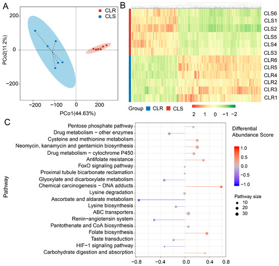
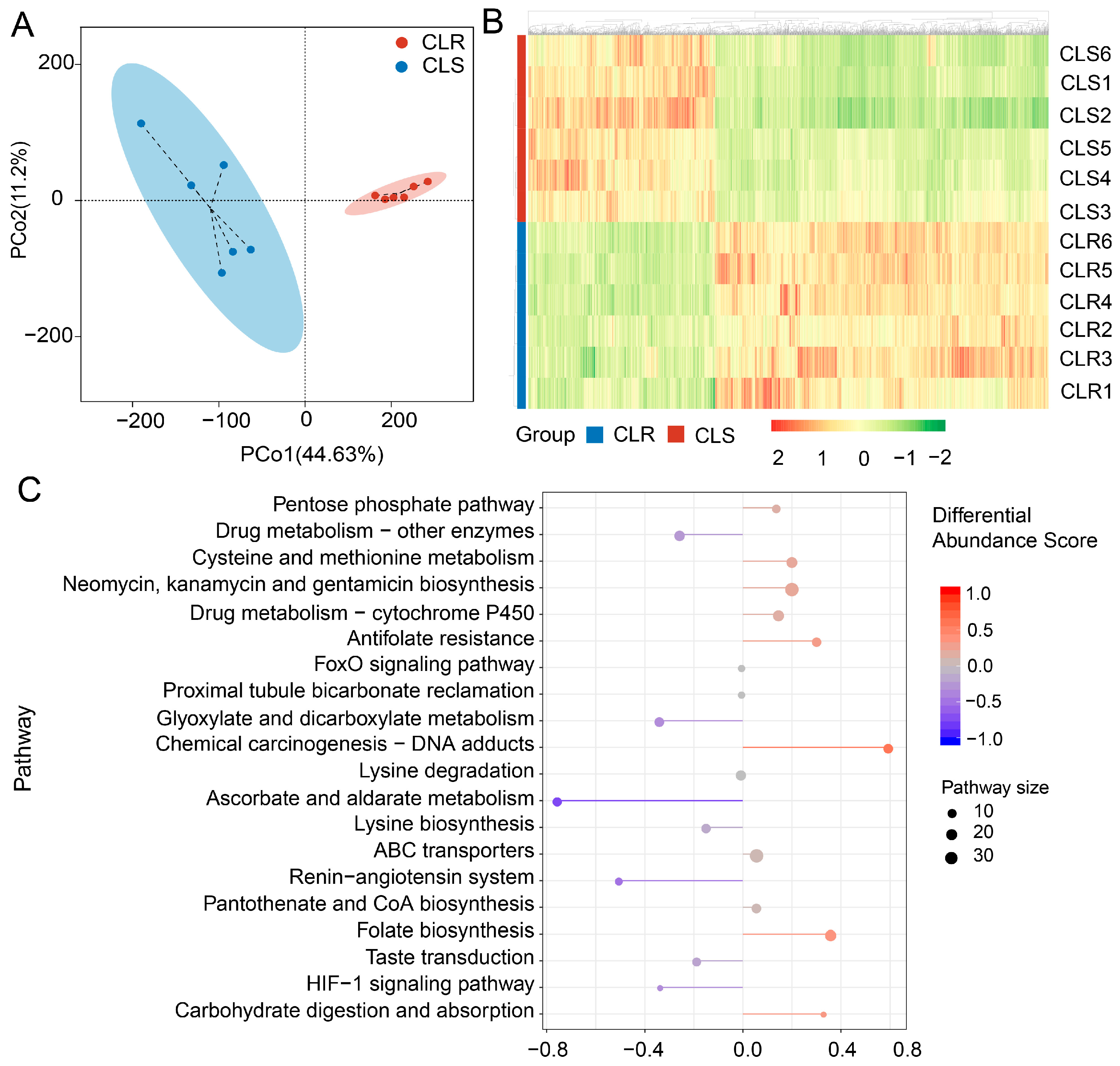
To characterize metabolic alterations associated with resistance, we first compared the metabolite profiles of the resistant strain CLR and the susceptible strain CLS using widely targeted metabolomics. Principal coordinate analysis (PCoA) clearly separated CLR from CLS, with PCo1 and PCo2 explaining 44.63% and 11.20% of the total variation, respectively (55.83% combined), indicating substantial metabolic divergence between the two strains (Figure 1A). The six biological replicates within each strain clustered closely, supporting the robustness of the dataset. Consistently, hierarchical clustering also divided CLR and CLS into two distinct groups and revealed pronounced differences in metabolite accumulation patterns between the resistant and susceptible backgrounds (Figure 1B). Together, these results indicate that resistance in CLR is accompanied by a stable and global shift in metabolic state.
Figure 1.
Global metabolomic differences between clothianidin-resistant (CLR) and susceptible (CLS) strains of the brown planthopper, Nilaparvata lugens. (A) Principal coordinate analysis (PCoA) of metabolite profiles. Each point represents one biological replicate (n = 6 per strain); PCo1 and PCo2 explain 44.63% and 11.20% of the total variation, respectively. (B) Hierarchical clustering heatmap of differential metabolites. Columns represent biological replicates and rows represent metabolites. Colors show row-standardized relative abundance (Z scores), ranging from −2 (green, lower relative abundance) to 2 (red, higher relative abundance). (C) Differential-abundance scores for significantly enriched KEGG pathways. Scores were calculated as (number of upregulated metabolites—number of downregulated metabolites)/(total number of differential metabolites in the pathway) and range from −1 (blue, predominantly downregulated in CLR relative to CLS) to 1 (red, predominantly upregulated). Dot size represents the number of differential metabolites assigned to each pathway (10–30).
Pathway-level differential abundance analysis further showed that the altered metabolites were enriched in several pathways related to detoxification and intermediary metabolism, including the pentose phosphate pathway, drug metabolism by cytochrome P450, drug metabolism by other enzymes, ABC transporters, cysteine and methionine metabolism, glyoxylate and dicarboxylate metabolism, and ascorbate and aldarate metabolism (Figure 1C). These findings suggest that the resistant phenotype is associated not only with detoxification-related changes but also with broader remodeling of core metabolic processes.
3.2. Differential Metabolites Are Mainly Enriched in Central Carbon Metabolism and Detoxification-Related Pathways
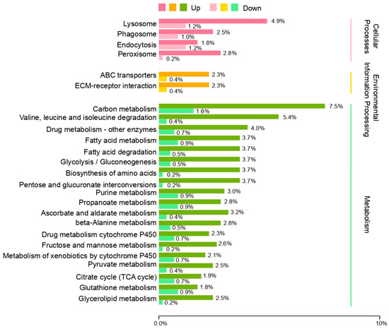
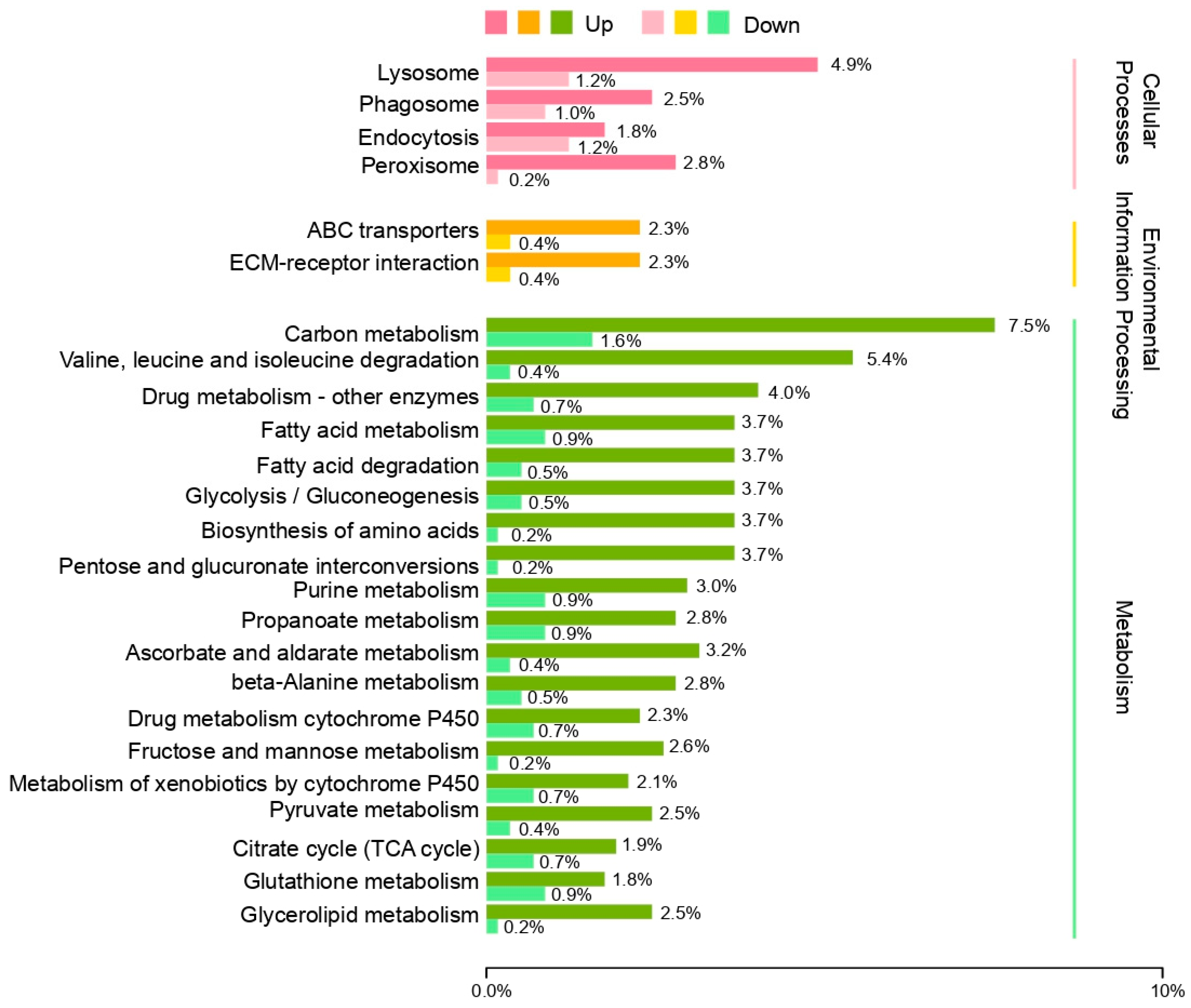
To further define the metabolic pathways altered in CLR, we classified differential metabolites based on KEGG annotations. Most differential metabolites were assigned to metabolism-related pathways, although some also fell into cellular process and environmental information processing categories (Figure 2). Among these, carbon metabolism showed the strongest enrichment, followed by glycolysis/gluconeogenesis, biosynthesis of amino acids, pentose and glucuronate interconversions, fructose and mannose metabolism, pyruvate metabolism, citrate cycle (TCA cycle), glutathione metabolism, metabolism of xenobiotics by cytochrome P450, and ABC transporters (Figure 2). The repeated appearance of these pathways suggests that resistant insects may reprogram energy and carbohydrate metabolism to support detoxification and physiological adaptation.
Figure 2.
KEGG classification of differential metabolites between clothianidin-resistant (CLR) and susceptible (CLS) strains of the brown planthopper, Nilaparvata lugens. Differential metabolites were grouped into cellular process, environmental information processing, and metabolism categories. Bars represent the proportions of upregulated and downregulated metabolites within each pathway.
3.3. Integrated Transcriptome–Metabolome Analysis Identifies Key Resistance-Associated Metabolic Modules
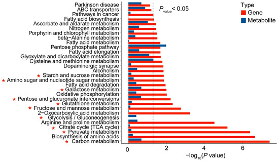
To integrate metabolic and transcriptional changes, differentially expressed genes and differential metabolites were jointly mapped to KEGG pathways. This combined analysis identified carbon metabolism as the most significantly enriched pathway, followed by biosynthesis of amino acids, pyruvate metabolism, citrate cycle (TCA cycle), glycolysis/gluconeogenesis, fructose and mannose metabolism, glutathione metabolism, and pentose and glucuronate interconversions (Figure 3). These recurrently enriched pathways point to coordinated changes in central carbon flux, organic acid metabolism, and reducing-equivalent supply in the resistant strain.
Figure 3.
Integrated KEGG enrichment analysis of differentially expressed genes and differential metabolites in clothianidin-resistant and susceptible strains of the brown planthopper, Nilaparvata lugens. The x-axis shows enrichment significance as −log10 (p value). Red bars indicate gene enrichment and blue bars indicate metabolite enrichment. Only pathways with p < 0.05 are shown. The asterisks indicate pathways related to sugar metabolism and energy metabolism.
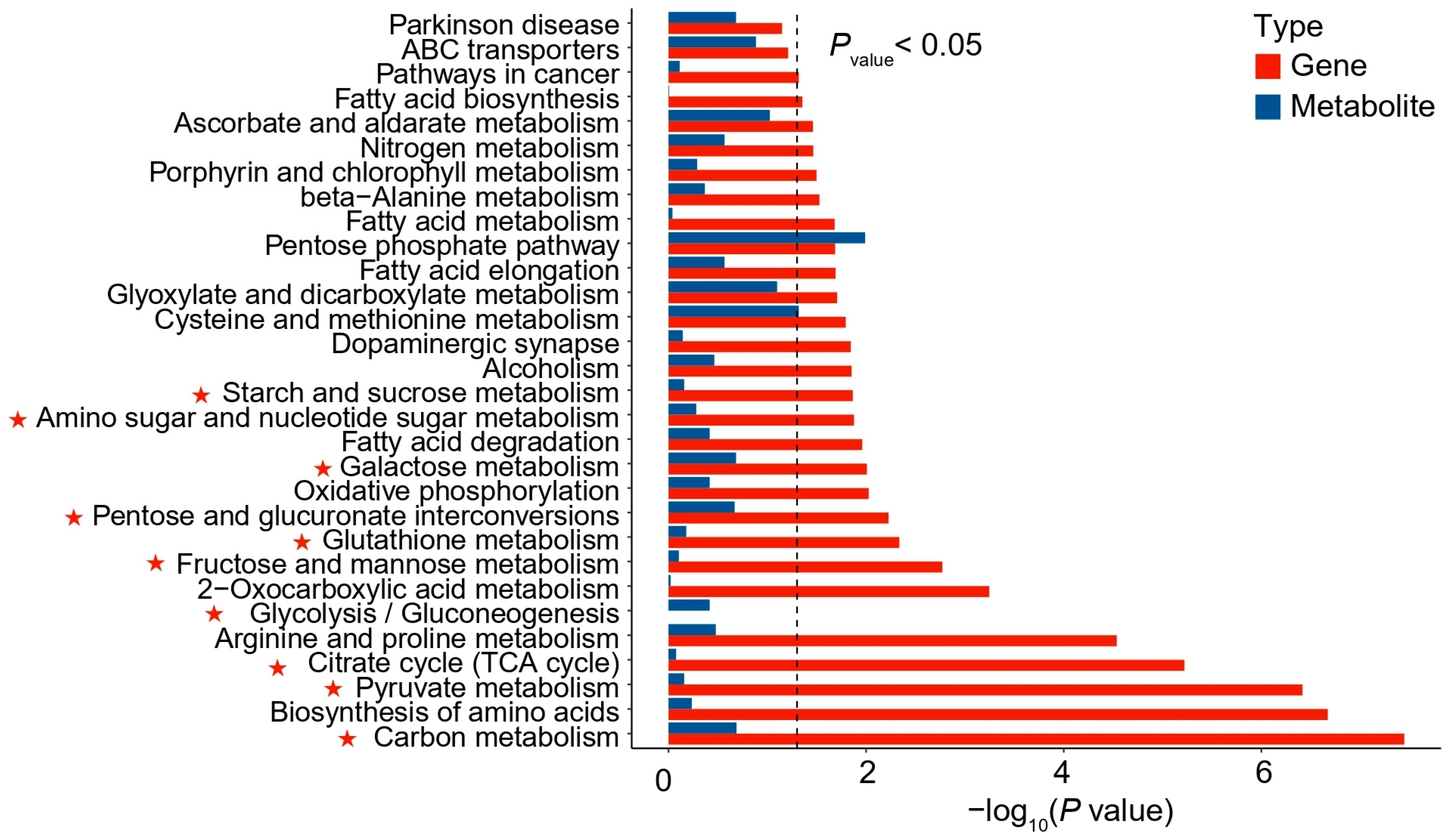
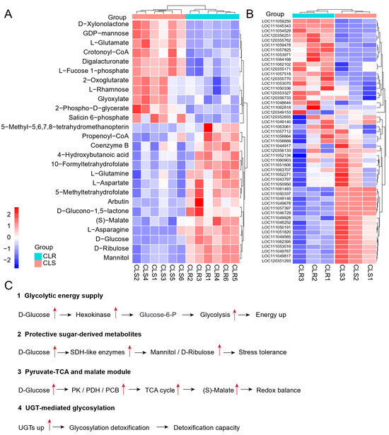
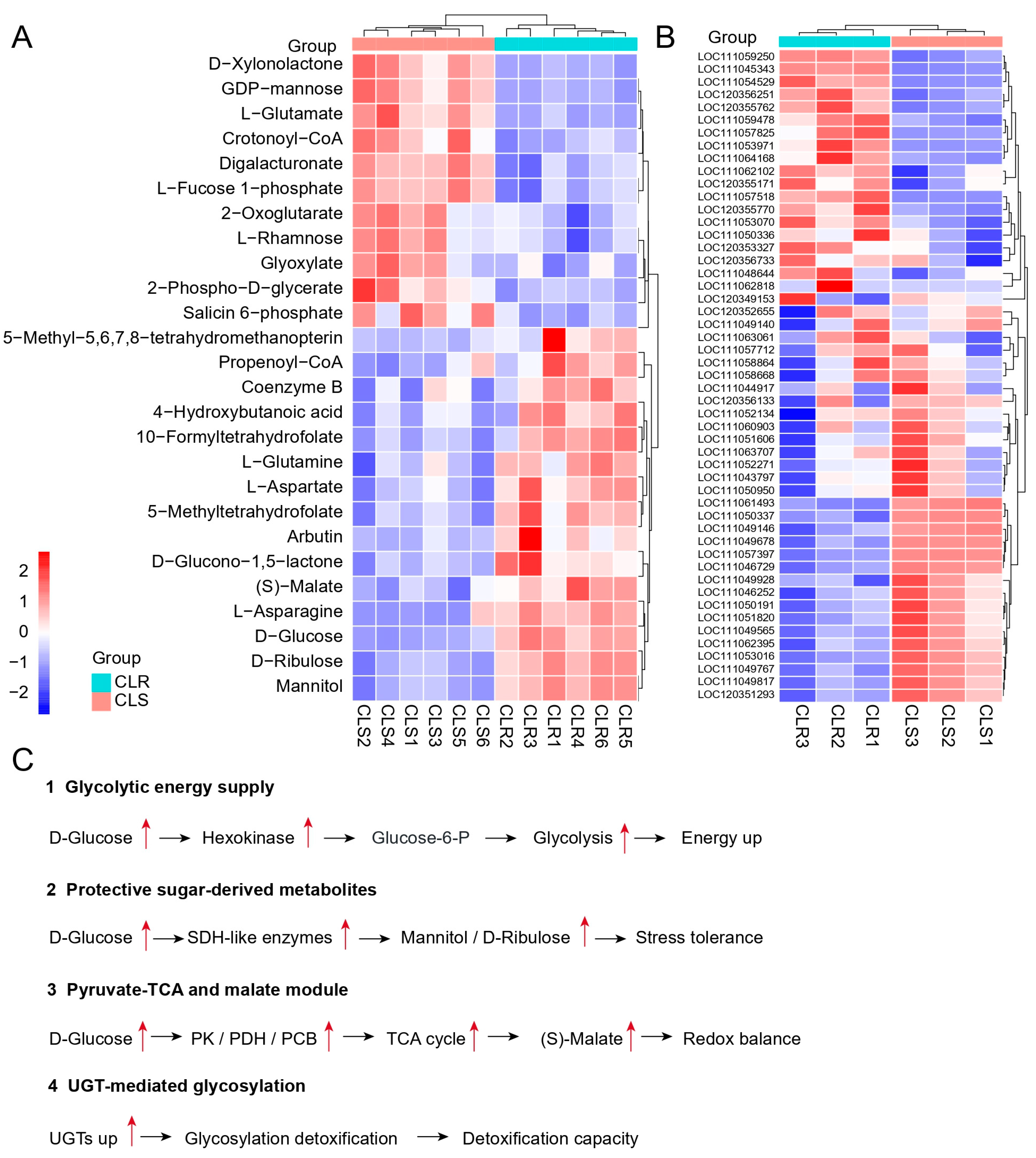
To visualize these relationships in greater detail, we constructed a gene-metabolite-pathway interaction network. The full supplementary network showed that numerous altered genes and metabolites converged on a limited number of core pathways, particularly carbon metabolism, glycolysis/gluconeogenesis, fructose and mannose metabolism, citrate cycle (TCA cycle), and pentose and glucuronate interconversions (Figure S1). Representative metabolites in these modules included D-glucose, 2-phospho-D-glycerate, 2-oxoglutarate, (S)-malate, D-ribulose, mannitol, arbutin, and salicin 6-phosphate (Figure 4A). Representative genes from the same pathways also showed clear strain-specific clustering (Figure 4B). We summarized these interactions into four major functional modules: glycolytic energy supply, protective sugar-derived metabolites, pyruvate-TCA and malate metabolism, and UGT-mediated glycosylation (Figure 4C). These results suggest that resistance in CLR is associated with coordinated remodelling of energy generation, stress-protective carbohydrate metabolism, organic acid turnover, and glycosylation-related detoxification.
Figure 4.
Resistance-associated metabolic modules in clothianidin-resistant (CLR) and susceptible (CLS) strains of the brown planthopper, Nilaparvata lugens. (A) Hierarchical clustering heatmap of representative metabolites in the key metabolic pathways. (B) Hierarchical clustering heatmap of representative differentially expressed genes in the same pathways. In (A) and (B), columns represent biological replicates and rows represent metabolites or genes, respectively. Colors indicate row-standardized relative abundance or expression (Z scores), ranging from −2 (blue, lower relative level) to 2 (red, higher relative level); cyan and salmon bars denote CLR and CLS samples, respectively. (C) Proposed functional model summarizing four resistance-associated modules: glycolytic energy supply, protective sugar-derived metabolites, pyruvate–TCA–malate metabolism, and UGT-mediated glycosylation. Red upward arrows indicate increased metabolite abundance, gene expression, or inferred pathway activity in CLR relative to CLS.
3.4. qRT-PCR Validation Identifies Common Upregulated Candidate Genes in Two Resistant Backgrounds
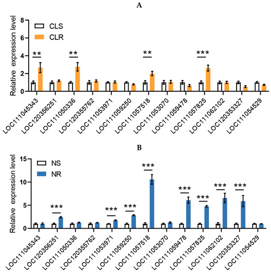
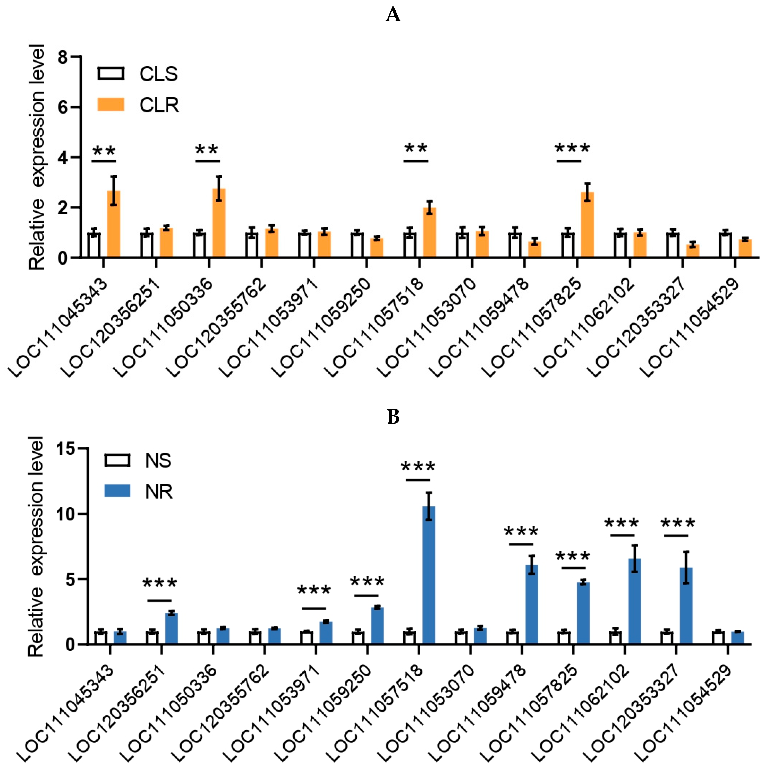
Based on the integrated omics analysis, 13 candidate genes from the major enriched pathways were selected for expression validation. In the clothianidin-resistant strain CLR, qRT-PCR confirmed that several of these genes were differentially expressed relative to CLS, with multiple candidates showing significant upregulation in the resistant background (Figure 5A). To determine whether these candidates also responded in another neonicotinoid-resistant background, we further examined their expression in the nitenpyram-resistant strain NR and its susceptible counterpart NS. Several genes were strongly upregulated in NR relative to NS (Figure 5B).
Figure 5.
qRT-PCR validation of candidate genes in two neonicotinoid-resistant backgrounds of the brown planthopper, Nilaparvata lugens. (A) Relative expression levels of 13 candidate genes in the clothianidin-resistant strain CLR and the susceptible strain CLS. (B) Relative expression levels of the same 13 candidate genes in the nitenpyram-resistant strain NR and the susceptible strain NS. Relative transcript levels were determined by qRT-PCR and calculated using the 2−ΔΔCt method. Data are presented as mean ± SEM. Asterisks indicate significant differences between resistant and susceptible strains (** p < 0.01 and *** p < 0.001).
Among the 13 candidates, two genes, NADP (LOC111057518) and UGT2 (LOC111057825), were consistently upregulated in both resistant backgrounds (Figure 5A,B). Their shared induction in CLR and NR suggested that they may represent common resistance-associated targets across different neonicotinoid-resistant strains. Therefore, these two genes were selected for subsequent functional validation by RNA interference.
3.5. RNAi Validation Supports the Roles of NADP and UGT2 in Neonicotinoid Resistance Maintenance
To confirm the effectiveness of RNA interference prior to bioassays, transcript levels of NADP and UGT2 were quantified after dsRNA treatment. Injection of dsNADP and dsUGT2 markedly reduced the expression of their corresponding target genes relative to the dsEGFP control, indicating that both genes were efficiently silenced under the experimental conditions (Figure S2). These results established the basis for evaluating the effects of gene knockdown on insecticide susceptibility.
To determine whether NADP and UGT2 functionally contribute to resistance, we compared insecticide susceptibility after RNAi treatment. Silencing of NADP significantly reduced the LC50 of nitenpyram from 90.06 mg/L in the dsEGFP group to 49.03 mg/L in the dsNADP group, corresponding to an LCR50 of 1.84 (Table 1). Similarly, the LC50 of clothianidin decreased from 291.77 mg/L to 161.19 mg/L after NADP knockdown, yielding an LCR50 of approximately 1.81 (Table 1). These data indicate that suppression of NADP partially restored susceptibility to both neonicotinoids.
Table 1.
Clothianidin and Nitenpyram LC50 against N. lugens after dsNADP injection.
Consistent with this pattern, silencing of UGT2 also significantly enhanced insecticide susceptibility. After dsUGT2 injection, the LC50 of nitenpyram declined from 98.15 mg/L in the dsEGFP group to 52.01 mg/L, corresponding to an LCR50 of 1.89 (Table 2). For clothianidin, the LC50 decreased from 299.33 mg/L to 163.99 mg/L after UGT2 knockdown, with an LCR50 of 1.83 (Table 2). Taken together, these results show that both NADP and UGT2 contribute to resistance maintenance in brown planthopper and support the conclusion that the resistance phenotype is linked to broader metabolic reprogramming rather than to isolated single-gene changes alone.
Table 2.
Clothianidin and Nitenpyram LC50 against N. lugens after dsUGT2 injection.
4. Discussion
In this study, we found that the clothianidin-resistant strain CLR and the susceptible strain CLS had markedly different metabolic profiles. The altered metabolites were concentrated in central carbon metabolism, glutathione metabolism, xenobiotic metabolism, ABC transport, and glycosylation. Joint transcriptome–metabolome analysis further organized these changes into four interconnected modules: glycolytic energy supply, protective sugar-derived metabolites, pyruvate–TCA–malate metabolism, and UGT-mediated glycosylation. NADP and UGT2 were upregulated in both clothianidin- and nitenpyram-resistant backgrounds, and silencing either gene increased susceptibility to both insecticides. Previous studies have established that CYP6ER1 contributes to resistance to several neonicotinoids in N. lugens, although its relative importance varies among compounds and resistance backgrounds [11,29,30,35,36,37]. Together with these earlier findings, our results indicate that resistance maintenance depends not only on elevated detoxification-enzyme expression but also on the metabolic supply of energy, reducing equivalents, and conjugation substrates.
The concurrent enrichment of carbon metabolism, glycolysis/gluconeogenesis, the TCA cycle, and the pentose phosphate pathway with P450-mediated xenobiotic metabolism, glutathione metabolism, and ABC transport is consistent with the biochemical demands of detoxification. P450 monooxygenase reactions require electrons delivered from NADPH through NADPH-cytochrome P450 reductase; GST reactions depend on reduced glutathione; UGT reactions consume activated UDP-sugars; and some detoxification products are exported by ATP-dependent ABC transporters [9,10,12,13,15,16]. Glycolysis and the TCA cycle sustain ATP production, whereas the pentose phosphate pathway and mitochondrial NADP-dependent dehydrogenases contribute to NADPH generation and glutathione recycling. The resistant strain may therefore redirect carbon flux to meet the energetic and cofactor requirements of P450s, GSTs, UGTs, and transport systems. In other insects, P450s that directly metabolize insecticides likewise depend on sufficient electron supply and a suitable intracellular redox environment [24,25]. Conversely, increased P450 turnover consumes NADPH and can generate reactive oxygen species through catalytic uncoupling. ROS-responsive CncC, JNK-ERK, and AP-1 pathways can in turn regulate P450 transcription [38,39,40], while circadian control of P450 expression and insecticide susceptibility further demonstrates that detoxification capacity is linked to the broader physiological state [41]. High temperature or antibiotic treatment disrupts symbiont-dependent detoxification in N. lugens and consequently increases insecticide susceptibility, providing additional evidence that metabolic and physiological conditions constrain resistance [31,42,43]. Thus, the coordinated alteration of central metabolism and detoxification pathways in our data is consistent with metabolic adaptation to the sustained cost of detoxification. Because metabolite abundance does not directly measure flux, this interpretation should be tested across additional resistant strains by quantifying sugars, organic acids, ATP, NADPH/NADP+, GSH/GSSG, and UDP-sugars, together with stable-isotope tracing of carbon redistribution.
Among the 13 candidate genes selected from the integrated analysis, NADP was upregulated in both clothianidin- and nitenpyram-resistant backgrounds, and its silencing increased susceptibility to both insecticides, supporting a functional association with resistance maintenance. NCBI annotates NADP as mitochondrial NADP-dependent isocitrate dehydrogenase rather than respiratory-chain NADH dehydrogenase; we therefore provisionally refer to it as NADP. This enzyme links the TCA cycle to mitochondrial NADPH metabolism by catalysing the conversion of isocitrate to α-ketoglutarate and contributing to glutathione reduction and mitochondrial redox homeostasis [44]. In light of the concurrent enrichment of the TCA cycle, glutathione metabolism, and xenobiotic metabolism, increased NADP expression could support P450 electron transfer and cellular stress tolerance by sustaining mitochondrial NADPH production, glutathione recycling, and ROS buffering [16,44]. The partial, rather than complete, restoration of susceptibility after knockdown is also consistent with resistance being maintained by multiple metabolic nodes. Measurements of enzyme activity, the isocitrate/α-ketoglutarate ratio, NADPH/NADP+, ROS, and GSH/GSSG would directly test this mechanism.
The expression and functional data for UGT2 closely matched the UGT-mediated glycosylation module identified by the joint analysis. Insect UGTs use UDP-sugars to glycosylate xenobiotics or endogenous metabolites, thereby altering substrate activity, water solubility, and excretion [14,20]. UGT overexpression has also been linked to clothianidin resistance in N. lugens [45]. In our study, pathways related to pentose and glucuronate interconversions were altered, UGT2 was consistently upregulated in two resistant backgrounds, and its silencing increased susceptibility to both clothianidin and nitenpyram. These observations support a model in which carbohydrate metabolism is adjusted to increase UDP-sugar availability and thereby sustain UGT2-mediated conjugation. UGT2 could act directly on the insecticides or their primary metabolites, or it could modify endogenous metabolites that influence redox balance, hormonal signalling, or other detoxification pathways. Its direct substrates remain unresolved and should be identified by heterologous expression, substrate-turnover assays, and LC-MS/MS analysis of glycosylated products.
The altered central metabolic pathways and the RNAi phenotypes of the two candidate genes also point to possible resistance-intervention strategies. If resistant insects depend on elevated ATP and NADPH supply, inhibition of selected energy-producing or reducing-equivalent-generating steps could weaken sustained P450 activity and increase the efficacy of existing insecticides. Because central metabolism is highly conserved, broad-spectrum metabolic inhibitors are better suited initially as mechanistic probes; only insect-selective or tissue-enriched metabolic targets would be appropriate for further development. Nanomaterial-based synergistic approaches have already shown that interference with resistance-associated physiological processes can improve insecticide performance [46]. The sensitization caused by NADP and UGT2 silencing also identifies both genes as potential targets for nucleic-acid-based synergists. Although oral RNAi in N. lugens is limited by nuclease degradation and tissue-delivery efficiency [47], nanoparticle-mediated fusion-dsRNA delivery and co-delivery of dsRNA with insecticides have demonstrated practical routes for improving resistance management [48,49].
In conclusion, our results link neonicotinoid resistance in N. lugens to coordinated remodelling of central metabolism and detoxification. Central carbon metabolism supplies ATP, NADPH, glutathione, and sugar donors to P450, GST, UGT, and ABC transport systems, whereas the energetic and redox burden imposed by detoxification may feed back on detoxification gene expression. NADP and UGT2 connect this metabolic support network to NADPH/redox homeostasis and glycosylation, respectively, as supported by their shared upregulation across resistance backgrounds and the sensitization produced by RNAi. Metabolic resistance in N. lugens is therefore better understood as a coordinated adaptation between resource supply and xenobiotic clearance than as the isolated overexpression of individual detoxification genes. This framework broadens the metabolic basis of neonicotinoid resistance and provides a rationale for identifying synergistic targets within the metabolic support network.
Supplementary Materials
The following supporting information can be downloaded at: https://www.mdpi.com/article/10.3390/insects17090884/s1, Figure S1. Full gene-metabolite-pathway interaction network derived from the integrated transcriptomic and metabolomic analyses. Blue nodes represent genes, orange nodes represent metabolites, and red nodes represent pathways. Gray edges indicate the associations between genes or metabolites and the corresponding pathways. The network summarizes the overall relationships among candidate genes, metabolites, and enriched metabolic pathways. Figure S2. RNAi knockdown efficiency of NADP and UGT2. Relative expression changes in NADP and UGT2 after dsRNA treatment. The dsEGFP treatment served as the control. Each treatment included three independent biological replicates and three technical replicates per biological sample. Data are presented as mean ± SEM and were analyzed using Student’s t-test. Asterisks indicate significant differences from the dsEGFP control (*** p < 0.001). Table S1. Primers used for PCR, RT-qPCR, and dsRNA synthesis in this study.
Author Contributions
G.Z.: conceptualization, methodology, investigation, formal analysis, writing—original draft. M.J.: investigation, data curation, validation. X.C.: investigation, visualization, data curation. L.L.: conceptualization, supervision, funding acquisition, writing—review & editing. All authors have read and agreed to the published version of the manuscript.
Funding
This study was funded by Natural Science Foundation of China (32402415), and Natural Science Foundation of Hubei Province (2025AFB721).
Data Availability Statement
The data supporting the findings of this study are provided in the article and its Supporting Information. Additional raw data are available from the corresponding author upon reasonable request.
Conflicts of Interest
The authors declare no conflicts of interest.
References
- Xue, J.; Zhou, X.; Zhang, C.X.; Yu, L.L.; Fan, H.W.; Wang, Z.; Xu, H.J.; Xi, Y.; Zhu, Z.R.; Zhou, W.W.; et al. Genomes of the Rice Pest Brown Planthopper and Its Endosymbionts Reveal Complex Complementary Contributions for Host Adaptation. Genome Biol. 2014, 15, 521. [Google Scholar] [CrossRef] [Scilit] [PubMed]
- Casida, J.E. Neonicotinoids and Other Insect Nicotinic Receptor Competitive Modulators: Progress and Prospects. Annu. Rev. Entomol. 2018, 63, 125–144. [Google Scholar] [CrossRef] [Scilit] [PubMed]
- Ihara, M.; Matsuda, K. Neonicotinoids: Molecular Mechanisms of Action, Insights into Resistance and Impact on Pollinators. Curr. Opin. Insect Sci. 2018, 30, 86–92. [Google Scholar] [CrossRef] [Scilit] [PubMed]
- Jeschke, P.; Nauen, R.; Schindler, M.; Elbert, A. Overview of the Status and Global Strategy for Neonicotinoids. J. Agric. Food Chem. 2011, 59, 2897–2908. [Google Scholar] [CrossRef] [Scilit] [PubMed]
- Zhang, Y.; Zhu, W.; Wang, Y.; Li, X.; Lv, J.; Luo, J.; Yang, M. Insight of Neonicotinoid Insecticides: Exploring Exposure, Mechanisms in Non-Target Organisms, and Removal Technologies. Pharmacol. Res. 2024, 209, 107415. [Google Scholar] [CrossRef] [Scilit] [PubMed]
- Bass, C.; Denholm, I.; Williamson, M.S.; Nauen, R. The Global Status of Insect Resistance to Neonicotinoid Insecticides. Pestic. Biochem. Phys. 2015, 121, 78–87. [Google Scholar] [CrossRef] [Scilit] [PubMed]
- Hawkins, N.J.; Bass, C.; Dixon, A.; Neve, P. The Evolutionary Origins of Pesticide Resistance. Biol. Rev. 2019, 94, 135–155. [Google Scholar] [CrossRef] [Scilit] [PubMed]
- Sparks, T.C.; Nauen, R. IRAC: Mode of Action Classification and Insecticide Resistance Management. Pestic. Biochem. Phys. 2015, 121, 122–128. [Google Scholar] [CrossRef] [Scilit] [PubMed]
- Li, J.; Zhu, C.; Xu, Y.; He, H.; Zhao, C.; Yan, F. Molecular Mechanism Underlying ROS-Mediated AKH Resistance to Imidacloprid in Whitefly. Insects 2024, 15, 436. [Google Scholar] [CrossRef] [Scilit] [PubMed]
- Nauen, R.; Bass, C.; Feyereisen, R.; Vontas, J. The Role of Cytochrome P450s in Insect Toxicology and Resistance. Annu. Rev. Entomol. 2022, 67, 105–124. [Google Scholar] [CrossRef] [Scilit] [PubMed]
- Bass, C.; Carvalho, R.A.; Oliphant, L.; Puinean, A.M.; Field, L.M.; Nauen, R.; Williamson, M.S.; Moores, G.; Gorman, K. Overexpression of a Cytochrome P450 Monooxygenase, CYP6ER1, Is Associated with Resistance to Imidacloprid in the Brown Planthopper, Nilaparvata lugens. Insect Mol. Biol. 2011, 20, 763–773. [Google Scholar] [CrossRef] [Scilit] [PubMed]
- Dermauw, W.; Van Leeuwen, T. The ABC Gene Family in Arthropods: Comparative Genomics and Role in Insecticide Transport and Resistance. Insect Biochem. Mol. Biol. 2014, 45, 89–110. [Google Scholar] [CrossRef] [Scilit] [PubMed]
- Hayes, J.D.; Flanagan, J.U.; Jowsey, I.R. Glutathione Transferases. Annu. Rev. Pharmacol. Toxicol. 2005, 45, 51–88. [Google Scholar] [CrossRef] [Scilit] [PubMed]
- Meech, R.; Hu, D.G.; McKinnon, R.A.; Mubarokah, S.N.; Haines, A.Z.; Nair, P.C.; Rowland, A.; Mackenzie, P.I. The UDP-Glycosyltransferase (UGT) Superfamily: New Members, New Functions, and Novel Paradigms. Physiol. Rev. 2019, 99, 1153–1222. [Google Scholar] [CrossRef] [Scilit] [PubMed]
- Merzendorfer, H. ABC Transporters and Their Role in Protecting Insects from Pesticides and Their Metabolites. In Advances in Insect Physiology; Elsevier: Amsterdam, The Netherlands, 2014; Volume 46, pp. 1–72. [Google Scholar]
- Zanger, U.M.; Schwab, M. Cytochrome P450 Enzymes in Drug Metabolism: Regulation of Gene Expression, Enzyme Activities, and Impact of Genetic Variation. Pharmacol. Ther. 2013, 138, 103–141. [Google Scholar] [CrossRef] [Scilit] [PubMed]
- Faubert, B.; Solmonson, A.; DeBerardinis, R.J. Metabolic Reprogramming and Cancer Progression. Science 2020, 368, eaaw5473. [Google Scholar] [CrossRef] [Scilit] [PubMed]
- Hanahan, D. Hallmarks of Cancer: New Dimensions. Cancer Discov. 2022, 12, 31–46. [Google Scholar] [CrossRef] [Scilit] [PubMed]
- Yang, H.-C.; Wu, Y.-H.; Yen, W.-C.; Liu, H.-Y.; Hwang, T.-L.; Stern, A.; Chiu, D.T.-Y. The Redox Role of G6PD in Cell Growth, Cell Death, and Cancer. Cells 2019, 8, 1055. [Google Scholar] [CrossRef] [Scilit] [PubMed]
- Lairson, L.L.; Henrissat, B.; Davies, G.J.; Withers, S.G. Glycosyltransferases: Structures, Functions, and Mechanisms. Annu. Rev. Biochem. 2008, 77, 521–555. [Google Scholar] [CrossRef] [Scilit] [PubMed]
- Daborn, P.J.; Yen, J.L.; Bogwitz, M.R.; Le Goff, G.; Feil, E.; Jeffers, S.; Tijet, N.; Perry, T.; Heckel, D.; Batterham, P.; et al. A Single P450 Allele Associated with Insecticide Resistance in Drosophila. Science 2002, 297, 2253–2256. [Google Scholar] [CrossRef] [Scilit] [PubMed]
- Le Goff, G.; Hilliou, F. Resistance Evolution in Drosophila: The Case of CYP6G1. Pest Manag. Sci. 2017, 73, 493–499. [Google Scholar] [CrossRef] [Scilit] [PubMed]
- Djouaka, R.F.; Bakare, A.A.; Coulibaly, O.N.; Akogbeto, M.C.; Ranson, H.; Hemingway, J.; Strode, C. Expression of the Cytochrome P450s, CYP6P3 and CYP6M2 Are Significantly Elevated in Multiple Pyrethroid Resistant Populations of Anopheles Gambiae s.s. from Southern Benin and Nigeria. BMC Genom. 2008, 9, 538. [Google Scholar] [CrossRef] [Scilit] [PubMed]
- Stevenson, B.J.; Bibby, J.; Pignatelli, P.; Muangnoicharoen, S.; O’Neill, P.M.; Lian, L.-Y.; Müller, P.; Nikou, D.; Steven, A.; Hemingway, J.; et al. Cytochrome P450 6M2 from the Malaria Vector Anopheles Gambiae Metabolizes Pyrethroids: Sequential Metabolism of Deltamethrin Revealed. Insect Biochem. Mol. Biol. 2011, 41, 492–502. [Google Scholar] [CrossRef] [Scilit] [PubMed]
- Karunker, I.; Benting, J.; Lueke, B.; Ponge, T.; Nauen, R.; Roditakis, E.; Vontas, J.; Gorman, K.; Denholm, I.; Morin, S. Over-Expression of Cytochrome P450 CYP6CM1 Is Associated with High Resistance to Imidacloprid in the B and Q Biotypes of Bemisia Tabaci (Hemiptera: Aleyrodidae). Insect Biochem. Mol. Biol. 2008, 38, 634–644. [Google Scholar] [CrossRef] [Scilit] [PubMed]
- Patti, G.J.; Yanes, O.; Siuzdak, G. Metabolomics: The Apogee of the Omics Trilogy. Nat. Rev. Mol. Cell Biol. 2012, 13, 263–269. [Google Scholar] [CrossRef] [Scilit] [PubMed]
- Reel, P.S.; Reel, S.; Pearson, E.; Trucco, E.; Jefferson, E. Using Machine Learning Approaches for Multi-Omics Data Analysis: A Review. Biotechnol. Adv. 2021, 49, 107739. [Google Scholar] [CrossRef] [Scilit] [PubMed]
- Subramanian, I.; Verma, S.; Kumar, S.; Jere, A.; Anamika, K. Multi-Omics Data Integration, Interpretation, and Its Application. Bioinform. Biol. Insights 2020, 14, 117793221989905. [Google Scholar] [CrossRef] [Scilit] [PubMed]
- Jin, R.; Mao, K.; Liao, X.; Xu, P.; Li, Z.; Ali, E.; Wan, H.; Li, J. Overexpression of CYP6ER1 associated with clothianidin resistance in Nilaparvata lugens (Stål). Pestic. Biochem. Physiol. 2019, 154, 39–45. [Google Scholar] [CrossRef] [Scilit] [PubMed]
- Mao, K.; Zhang, X.; Ali, E.; Liao, X.; Jin, R.; Ren, Z.; Wan, H.; Li, J. Characterization of nitenpyram resistance in Nilaparvata lugens (Stål). Pestic. Biochem. Physiol. 2019, 157, 26–32. [Google Scholar] [CrossRef] [Scilit] [PubMed]
- Zhang, Y.; Cai, T.; Yuan, M.; Li, Z.; Jin, R.; Ren, Z.; Qin, Y.; Yu, C.; Cai, Y.; Shu, R.; et al. Microbiome variation correlates with the insecticide susceptibility in different geographic strains of a significant agricultural pest, Nilaparvata lugens. npj Biofilms Microbiomes 2023, 9, 2. [Google Scholar] [CrossRef] [Scilit] [PubMed]
- Zhang, B.; Zhang, M.; Jia, X.; Hu, G.; Ren, F.; Fan, X.; Dong, Y. Integrated transcriptome and metabolome dissecting interaction between Vitis vinifera L. and Grapevine fabavirus. Int. J. Mol. Sci. 2023, 24, 3247. [Google Scholar] [CrossRef] [Scilit] [PubMed]
- Livak, K.J.; Schmittgen, T.D. Analysis of Relative Gene Expression Data Using Real-Time Quantitative PCR and the 2−ΔΔCT Method. Methods 2001, 25, 402–408. [Google Scholar] [CrossRef] [Scilit] [PubMed]
- Jing, S.; Wang, F.; Ren, A.; Zheng, F.; Yu, B.; Xu, J.; Liu, Y.; Yang, J.; Chen, R.; Zeng, W.; et al. Identification and functional analysis of three NlCstF genes in Nilaparvata lugens. Insects 2024, 15, 867. [Google Scholar] [CrossRef] [Scilit] [PubMed]
- Pang, R.; Chen, M.; Liang, Z.; Yue, X.; Ge, H.; Zhang, W. Functional Analysis of CYP6ER1, a P450 Gene Associated with Imidacloprid Resistance in Nilaparvata Lugens. Sci. Rep. 2016, 6, 34992. [Google Scholar] [CrossRef] [Scilit] [PubMed]
- Sun, X.; Gong, Y.; Ali, S.; Hou, M. Mechanisms of Resistance to Thiamethoxam and Dinotefuran Compared to Imidacloprid in the Brown Planthopper: Roles of Cytochrome P450 Monooxygenase and a P450 Gene CYP6ER1. Pestic. Biochem. Phys. 2018, 150, 17–26. [Google Scholar] [CrossRef] [Scilit] [PubMed]
- Zhang, H.; Zou, J.; Yang, B.; Zhang, Y.; Liu, Z. Importance of CYP6ER1 Was Different among Neonicotinoids in Their Susceptibility in Nilaparvata Lugens. J. Agric. Food Chem. 2023, 71, 4163–4171. [Google Scholar] [CrossRef] [Scilit] [PubMed]
- Jin, R.; Wang, Y.; He, B.; Zhang, Y.; Cai, T.; Wan, H.; Jin, B.R.; Li, J. Activator Protein-1 Mediated CYP6ER1 Overexpression in the Clothianidin Resistance of Nilaparvata Lugens (Stål). Pest Manag. Sci. 2021, 77, 4476–4482. [Google Scholar] [CrossRef] [Scilit] [PubMed]
- Tang, B.; Cheng, Y.; Li, Y.; Li, W.; Ma, Y.; Zhou, Q.; Lu, K. Adipokinetic Hormone Regulates Cytochrome P450-Mediated Imidacloprid Resistance in the Brown Planthopper, Nilaparvata Lugens. Chemosphere 2020, 259, 127490. [Google Scholar] [CrossRef] [Scilit] [PubMed]
- Zhang, G.; Li, C.; Zeng, Q.; Li, J.; Du, Z.; Geng, T.; He, S.; Li, J.; Guo, L.; Wan, H. JNK-ERK Synergistic Regulation of P450 Gene Expression Confers Nitenpyram Resistance in Nilaparvata Lugens (Stål). J. Agric. Food Chem. 2025, 73, 7695–7703. [Google Scholar] [CrossRef] [Scilit] [PubMed]
- Kang, K.; Yang, P.; Pang, R.; Yue, L.; Zhang, W. Cycle Affects Imidacloprid Efficiency by Mediating Cytochrome P450 Expression in the Brown Planthopper Nilaparvata Lugens. Insect Mol. Biol. 2017, 26, 522–529. [Google Scholar] [CrossRef] [Scilit] [PubMed]
- Tang, T.; Zhang, Y.; Cai, T.; Deng, X.; Liu, C.; Li, J.; He, S.; Li, J.; Wan, H. Antibiotics Increased Host Insecticide Susceptibility via Collapsed Bacterial Symbionts Reducing Detoxification Metabolism in the Brown Planthopper, Nilaparvata lugens. J. Pest Sci. 2021, 94, 757–767. [Google Scholar] [CrossRef] [Scilit]
- Zhang, Y.; Cai, T.; Ren, Z.; Liu, Y.; Yuan, M.; Cai, Y.; Yu, C.; Shu, R.; He, S.; Li, J.; et al. Decline in Symbiont-Dependent Host Detoxification Metabolism Contributes to Increased Insecticide Susceptibility of Insects under High Temperature. ISME J. 2021, 15, 3693–3703. [Google Scholar] [CrossRef] [Scilit] [PubMed]
- Jo, S.H.; Son, M.K.; Koh, H.J.; Lee, S.M.; Song, I.H.; Kim, Y.O.; Lee, Y.S.; Jeong, K.-S.; Kim, W.B.; Park, J.W.; et al. Control of Mitochondrial Redox Balance and Cellular Defense against Oxidative Damage by Mitochondrial NADP+-Dependent Isocitrate Dehydrogenase. J. Biol. Chem. 2001, 276, 16168–16176. [Google Scholar] [CrossRef] [Scilit] [PubMed]
- Zhang, Y.; Liu, C.; Jin, R.; Wang, Y.; Cai, T.; Ren, Z.; Ma, K.; He, S.; Lee, K.S.; Jin, B.R.; et al. Dual Oxidase-dependent Reactive Oxygen Species Are Involved in the Regulation of UGT Overexpression-mediated Clothianidin Resistance in the Brown Planthopper, Nilaparvata lugens. Pest Manag. Sci. 2021, 77, 4159–4167. [Google Scholar] [CrossRef] [Scilit] [PubMed]
- Zeng, Q.; Yu, C.; Chang, X.; Wan, Y.; Ba, Y.; Li, C.; Lv, H.; Guo, Z.; Cai, T.; Ren, Z.; et al. CeO2 Nanohybrid as a Synergist for Insecticide Resistance Management. Chem. Eng. J. 2022, 446, 137074. [Google Scholar] [CrossRef] [Scilit]
- Gao, Y.; Cai, T.; Yu, C.; Zeng, Q.; Wan, Y.; Song, L.; He, S.; Li, J.; Wan, H. A Putative Endonuclease Reduces the Efficiency of Oral RNA Interference in Nilaparvata Lugens. Pest Manag. Sci. 2024, 80, 5771–5779. [Google Scholar] [CrossRef] [Scilit] [PubMed]
- He, D.; Zeng, Q.; Guo, H.; Sun, D.; Zhang, M.; Yu, C.; Xie, Y.; Zhou, H.; Li, J.; He, S.; et al. A Novel Strategy to Combat Insecticide Resistance in Insect Pest with a Nanoparticle-based Fusion DSRNA Delivery System. Pest Manag. Sci. 2025, 81, 6201–6209. [Google Scholar] [CrossRef] [Scilit] [PubMed]
- Hu, C.; Yu, C.; Li, J.; Liu, Y.; Zeng, Q.; Wan, H.; He, S.; Li, Y.; Gao, P.; Lv, Y.; et al. Nanoparticles Co-Delivering Double-Stranded RNA and Insecticide Enhance Control Efficacy and Overcome Pest Resistance. Chem. Eng. J. 2025, 519, 165188. [Google Scholar] [CrossRef] [Scilit]

Disclaimer/Publisher’s Note: The statements, opinions and data contained in all publications are solely those of the individual author(s) and contributor(s) and not of MDPI and/or the editor(s). MDPI and/or the editor(s) disclaim responsibility for any injury to people or property resulting from any ideas, methods, instructions or products referred to in the content. |
© 2026 by the authors. Licensee MDPI, Basel, Switzerland. This article is an open access article distributed under the terms and conditions of the Creative Commons Attribution (CC BY) license.