


Simple Summary
The detoxification system plays an important role in host finding, feeding, and defense against toxic compounds in insects. In this study, four detoxification gene families encoding 146 candidate detoxification genes, including 22 glutathione S-transferases, 20 UDP-glycosyltransferases, 60 cytochrome P450 monooxygenases, and 44 carboxylesterases were identified from the adult antennal transcriptome data in Plagiodera versicolora. Then, the expression of the glutathione S-transferases genes was examined using real-time quantitative PCR. These results will provide valuable information to help explore the detoxification mechanism in P. versicolora.
Abstract
Plagiodera versicolora (Coleoptera: Chrysomelidae), the willow leaf beetle, is a leaf-eating pest that generally occurs on salicaceous trees. However, there is a blank of identification and phylogenetic relationship of the detoxification genes in P. versicolora. Here, we identified four detoxification gene families (glutathione S-transferases: GSTs, UDP-glycosyltransferases: UGTs, cytochrome P450 monooxygenases: CYPs and carboxylesterases: COEs) from the adult antennal transcriptome data. In all, 146 candidate detoxification genes including 22 GSTs, 20 UGTs, 60 CYPs, and 44 COEs were identified. We used quantitative real-time PCR technology to explore the tissue expression patterns of 12 PvGSTs in P. versicolora. The results showed that 7 PvGSTs have significantly high expression in antennae, indicating these PvGSTs may play an important role in degrade and/or inactivate the sex pheromones and host volatiles. The identification and phylogenetics of the detoxification genes in P. versicolora extended the database in Coleoptera and contributed to the subsequent in-depth research for function about detoxification genes.
1. Introduction
In natural environments, insects use complex chemosensory and detoxification systems to find hosts, feed and defend against toxic compounds [1,2,3]. The detoxification system is typically comprised of several genes, mainly including UDP-glycosyltransferases (UGTs), glutathione S-transferases (GSTs), cytochrome P450 monooxygenases (CYPs) and carboxylesterases (COEs) [4,5,6]. These genes also belong to odorant-degrading enzyme family, which is involved in odorant inactivation and xenobiotic processing [7].
GSTs mainly include four families, namely cytosolic GSTs, mitochondrial GSTs and microsomal GSTs [8,9]. Insect cytosolic GSTs are generally classified into six classes, including delta, epsilon, sigma, omega, zeta and theta, with several unclassified classes [10,11]. The amino acid sequences of cytosolic GSTs are usually formed 200 to 250 and are active as either homodimers or heterodimers, whereas microsomal GSTs are typically smaller (about 150 amino acids) and form trimers [12,13,14]. In insects, cytosolic GSTs are generally involved in resistance to insecticides [15,16]. Some GSTs are mainly related to the signal transduction of odor molecules [17].
UGTs in animals are membrane conjugated proteins that occur in the endoplasmic reticulum [18]. UGTs are composed of a variable N-terminus aglycone substrate binding domain, UGT signature motif, and a highly conserved C-terminus UDP-glycoside binding domain. In addition, UGTs generally have a signal peptide (about 20 amino acids) at the N-terminus and a hydrophobic transmembrane domain at the C-terminus, followed by a short cytoplasmic tail [19,20]. Insect UGTs can bind not only a large number of allelochemicals, but also some xenobiotics including insecticides such as chlorpyrifos, imidacloprid [21,22,23].
Cytochrome P450 genes are among the largest families, and are found in almost all organisms, from plants to animals [24,25]. The insect CYPs family can be clustered into four clades: CYP2, CYP3, CYP4, and mitochondrial CYP clades in phylogenetic tree [26]. Insect P450 genes are involved in the acquisition of resistance to a series of toxins, including insecticides and plant-derived toxic compounds [27]. Another important superfamily of COEs also involved in the metabolism of endobiotics and xenobiotics. Insect COEs are divided into three classes of intracellular catalytic, secreted catalytic and neurodevelopmental classes [28,29]. In addition, insect CYPs and COEs play roles in the degradation of pheromone and phytochemical compounds [30,31,32].
The Plagiodera versicolora (Coleoptera: Chrysomelidae) mainly feeds on the leaves of salicaceous trees (willows and poplars) [33,34]. To date, no reports are available on the detoxification gene family in P. versicolora. In this study, we identified GSTs, UGTs, CYPs, and COEs from the adult antennal transcriptome of P. versicolora. We constructed phylogenetic trees and analyzed the phylogenetic relationships of P. versicolora with other Coleoptera species. In addition, we analyzed tissue expression levels of GSTs by performing quantitative real-time PCR technology.
2. Materials and Methods
2.1. Insect Raise and Tissue Collection
In this experiment, the adults of P. versicolora were collected from Sha Lake Park in Wuhan, China. In the insect rearing room, relative humidity was maintained at 70 ± 5%, photoperiod was kept at 12 h light: 12 h dark, and the temperature was maintained at 28 ± 1 °C. Different tissues (antennae and bodies) were collected from male and female adults without treatment of chemical or any substances, respectively. All samples were immediately frozen in liquid nitrogen and stored at −80 °C.
2.2. Total RNA Extraction and cDNA Synthesis
Total RNA samples from antennae and bodies were isolated using TRIzol Reagent (Thermo Scientific, Waltham, MA, USA, Product code: 15596018CN) following the user guide was supplied by company. The concentration of RNA samples was checked using a NanoDrop-2000 (Thermo Scientific, Waltham, MA, USA). Complementary DNA (cDNA) templates were synthesized using a HiScript III 1st Strand cDNA Synthesis Kit (+gDNA wiper) (Vazyme, Nanjing, China) and stored at −20 °C for gene cloning and expression profiling analysis.
2.3. Candidate Gene Identification and Sequence Analysis
Candidate detoxification genes (GST, UGT, P450, and COE) were identified from adult antennal transcriptomes of P. versicolora [35]. Subsequently, the candidate detoxification genes were validated using BLASTX (https://blast.ncbi.nlm.nih.gov/Blast.cgi?PROGRAM=blastx&PAGE_TYPE=BlastSearch&LINK_LOC=blasthome (accessed on 6 November 2025)) in the National Center for Biotechnology Information (NCBI) databases. ORF finder (http://www.ncbi.nlm.nih.gov/gorf/gorf.html (accessed on 6 November 2025)) was used to predict the open reading frame (ORFs) of these candidate genes. The SignalP 2.0 server (http://www.cbs.dtu.dk/services/SignalP-2.0/#submission (accessed on 6 November 2025)) was used to predict the signal peptides. The specific names of P450s use the CYP prefix; the family was indicated by an Arabic numeral, and the subfamily was presented with a capital letter. The specific names of GSTs that have a lower-case letter in each subfamily were named according to the classification of insect GSTs. The structure and binding site of GSTs were predicted by searching the InterPro (http://www.ebi.ac.uk/interpro/search/sequence/ (accessed on 6 November 2025)).
2.4. Phylogenetic Analysis
Different coleopteran insects were used to construct phylogenetic trees for detoxification gene families. In the P450 tree, four Coleoptera species were selected, including Xylotrechus quadripes [36], Tribolium castaneum [37], and P. versicolora. In the UGT tree, four Coleoptera species were chosen, including Dendroctonus ponderosae, Aethina tumida, T. castaneum [20] and P. versicolora. In the GST tree, five Coleoptera were represented, including X. quadripes, Leptinotarsa decemlineata [38], Rhaphuma horsfieldi [36], T. castaneum [39], and P. versicolora. In the COE tree, four Coleoptera were selected, including R. horsfieldi, X. quadripes, Pharsalia antennata [40], and P. versicolora. These protein sequences of detoxification genes were aligned using ClustalX 2.1. Maximum likelihood trees were constructed using FastTree v2.1.11 under the Jones-Taylor-Thornton (JTT) model. The parameter settings are as follows: support values with replicates and CAT approximation with 20 rate categories. In addition, the trees were edited by FigTree v1.4.4 (http://tree.bio.ed.ac.uk/software/figtree/ (accessed on 6 November 2025)). The abbreviation “Pv” is used in the designation of the different genes identified from P. versicolora for construction of the phylogenetic tree.
2.5. Expression Profiling Analysis
At first, we selected all of 14 full-length PvGSTs to investigate their expression using quantitative real-time PCR (qRT-PCR). The PCR efficiencies of primers ranged from 95–105% with high correlation coefficient (R2) values (0.98–0.99) according to the standard. However, two GSTs were substandard primers for qRT-PCR, so we selected 12 PvGSTs for subsequent experiments. We used Beacon Designer 8.14 to design the specific primers (Table S1). The qRT-PCR reaction system was prepared with ChamQ Universal SYBR qPCR Master Mix (Vazyme, Nanjing, China) according to the instructions. The reaction program was set at 95 °C for 30 s, followed by 40 cycles of 95 °C for 5 s and 60 °C for 34 s. Three biological samples for each gene were performed. Gene expression levels were calculated using the 2−ΔΔCT method. The RPS18 reference gene as the normalization genes was selected to evaluate the relative expression level of PvGSTs. Significant differences were calculated using the ANOVA followed by Tukey’s HSD test with SPSS 26.0 software. Significant differences are indicated by different letters above the bars at a significance level of p < 0.05.
3. Results
3.1. KEGG Pathway Analysis
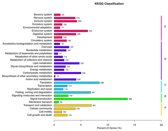
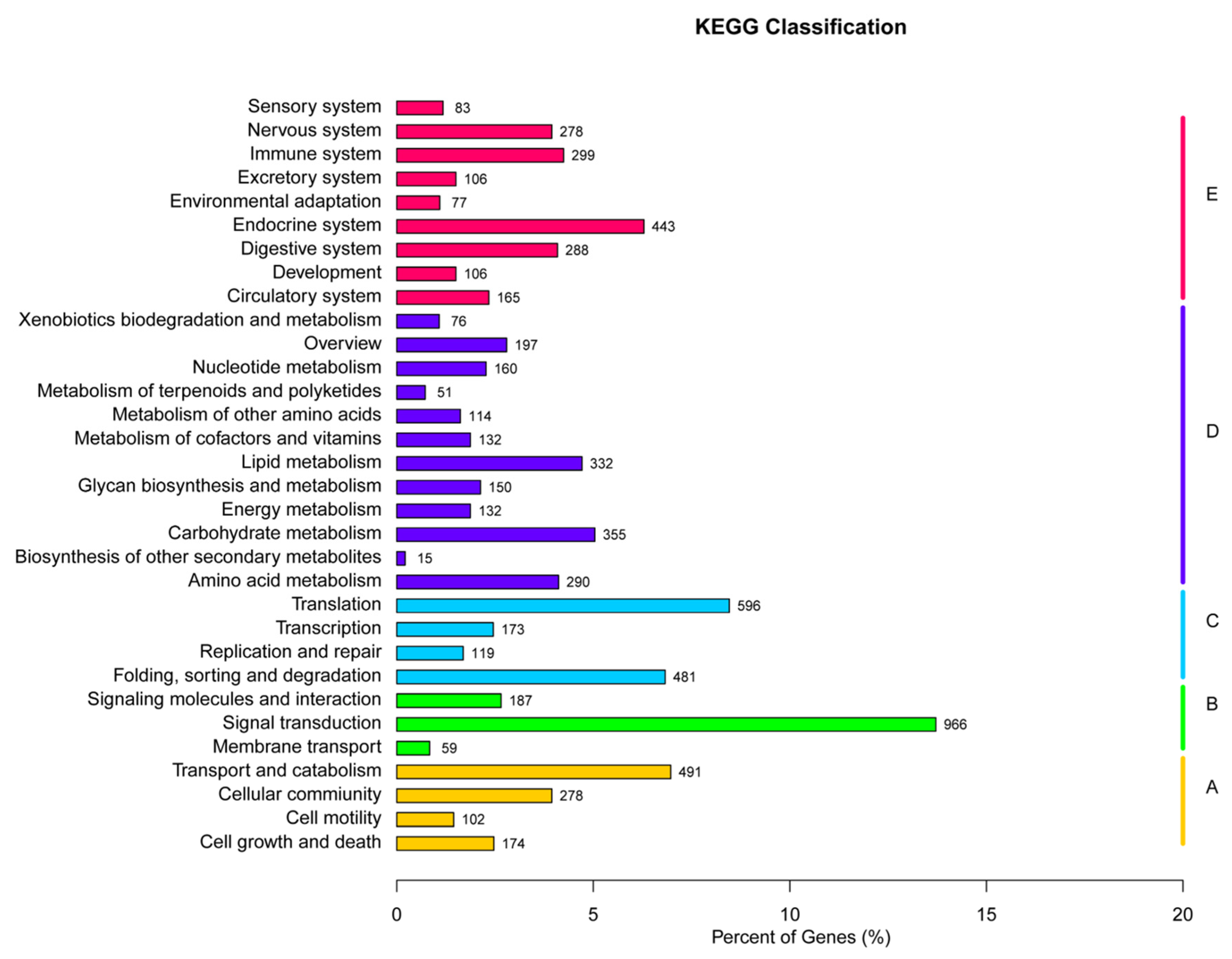
The unigenes were annotated to 6 databases, including Nucleotide Sequence (NT), Non-Redundant Protein Sequence (NR), Swiss-Prot, Gene Ontology (GO), Kyoto Encyclopedia of Genes and Genomes (KEGG), and clusters of eukaryotic Orthologous Groups (KOG). The numbers of annotated unigenes were 6788 in GO, 3847 in KEGG, 7479 in KOG, 11925 in NR, 3642 in NT, and 8108 in Swiss-Prot databases. The unigenes in the KEGG metabolic pathway were divided into 5 branches: 1045 cellular processes, 1212 environmental information processing, 1369 genetic information processing, 2004 metabolism and 1825 organismal systems (Figure 1).
Figure 1.
KEGG pathway classification of unigenes in the transcriptome. A: cellular processes; B: environmental information processing; C: genetic information processing; D: metabolism; E: organismal systems.
3.2. Identification of GSTs
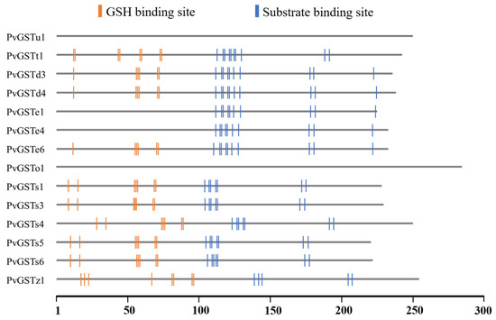
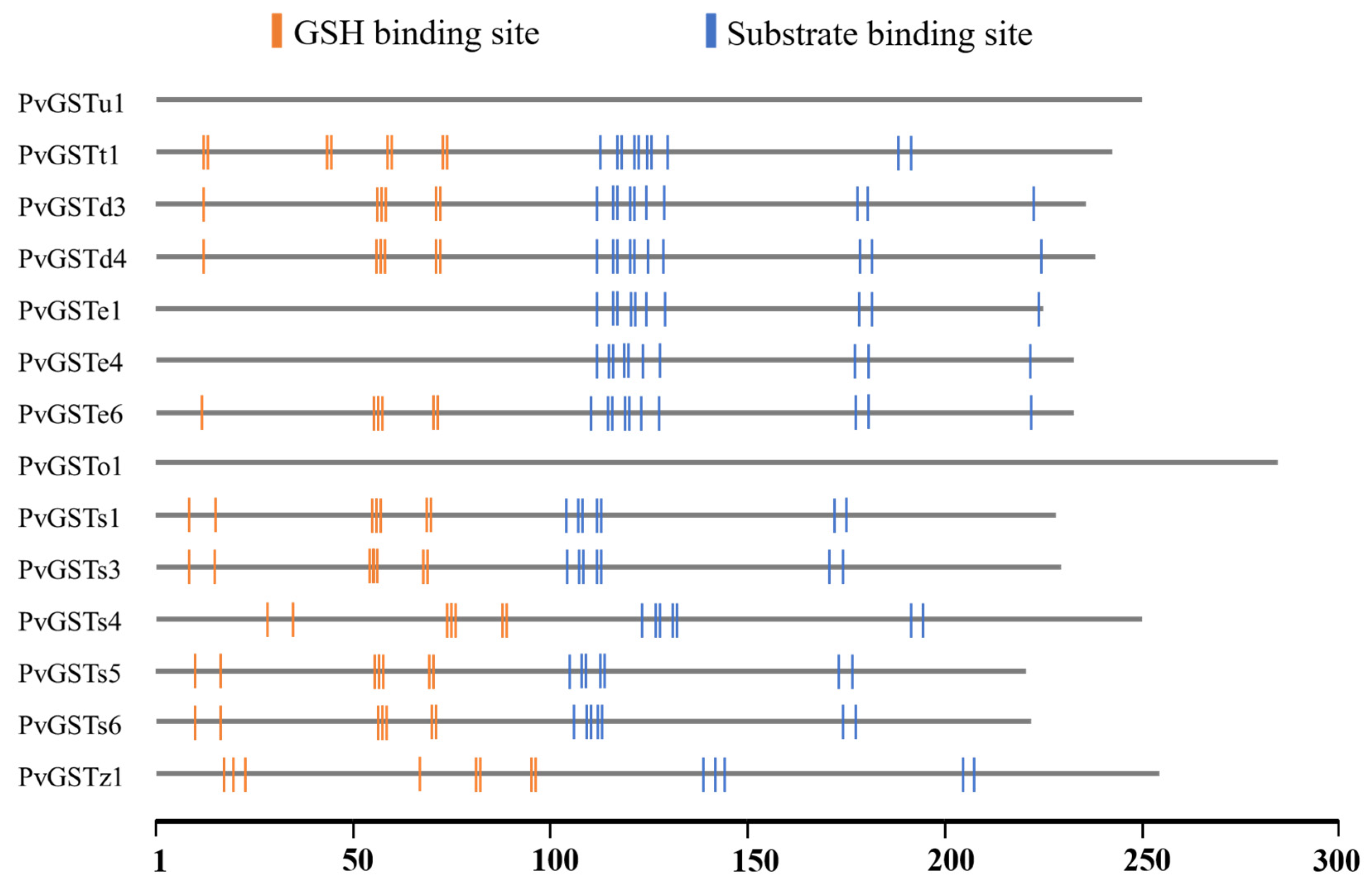
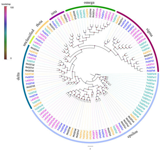
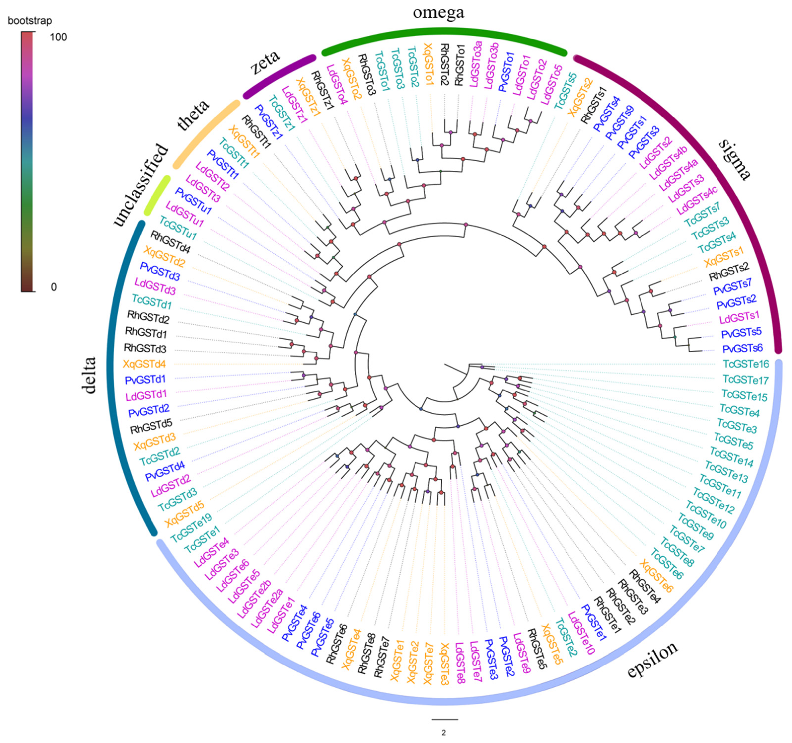
In total, 22 GSTs were identified based on the adult antennal transcriptome of P. versicolora. Fourteen PverGSTs had complete ORFs according to the predicted results, and the proteins ranged from 204 to 263 amino acids (Table S2). Conserved domain prediction revealed that 10 PverGSTs had GSH-binding sites and 12 PvGSTs had substrate-binding sites. In addition, two PvGSTs (GSTe1 and GSTe4) had only substrate-binding sites and two PvGSTs (GSTe1 and GSTe4) lacked both sites (Figure 2). All PverGSTs were named according to the phylogenetic results of P. versicolora with other Coleoptera species. As a result, the 22 PvGSTs were clustered into eight clades, including epsilon (6 PvGSTs), delta (4 PvGSTs), sigma (8 PvGSTs), zeta (1 PvGST), omega (1 PvGST), theta (1 PvGST) and unclassified classes (1 PvGST) (Figure 3).
Figure 2.
Predicted GSH-binding sites (G-sites) indicated by orange vertical lines and substrate-binding sites (H-sites) indicated by blue lines in PverGST proteins.
Figure 3.
Maximum-likelihood tree of GSTs from P. versicolora (Pv), X. quadripes (Xq), L. decemlineata (Ld), R. horsfieldi (Rh), and T. castaneum (Tc). Different species are highlighted with specific color patterns, and P. versicolora genes are shown in blue color.
3.3. Identification of UGTs
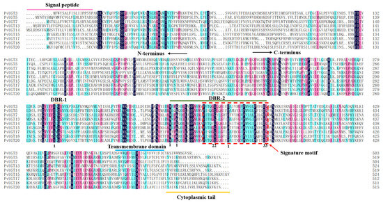
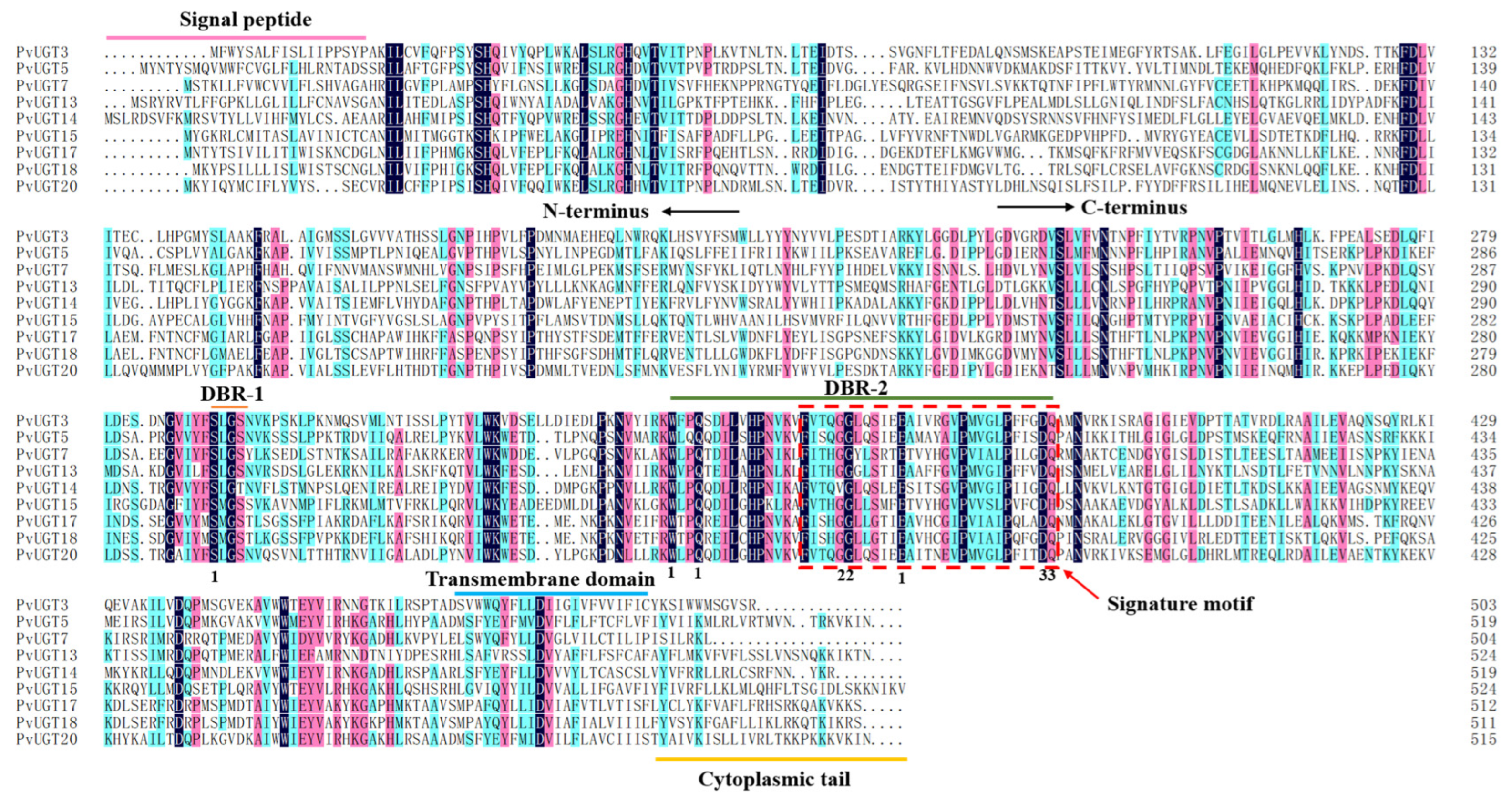
By analyzing the male and female antennal transcriptome dataset, 20 UGTs were identified in P. versicolora. Nine PvUGTs had complete ORFs according to the predicted results, and the encoded proteins ranged from 503 to 535 amino acids; the remaining PvUGTs were partial sequences (Table S3). All 9 full-length PvUGT proteins had a signal peptide at the N-terminus and a transmembrane domain at the C-terminus. Of the 9 full-length PvUGT proteins, in the N-terminus, all of PvUGTs had a signal peptide, and these 9 PvUGTs had a transmembrane domain in the C-terminus. Nine PvUGTs have five nucleotide-binding residues, two phosphate-binding residues and two aglycone-binding residues. In addition, sequence analysis showed that two sugar donor-binding regions (DBR1 and DBR2) and a signature motif were also found in these 9 PvUGTs (Figure 4).
Figure 4.
Alignment of the amino acid sequences of 9 full length UGTs. The signal peptides in the N-terminus and the transmembrane domain in C-terminus are marked in pink and blue color at the top, respectively. The signature motif is marked in red box with dashed line. Two donor binding regions (DBR1 and DBR2) are indicated with red and green colored lines at the top of the alignment, respectively. The different shading colors used to highlight homology level of amino acids in the alignment, as pink ≥ 50%, light blue ≥ 75% and dark blue = 100%. The different number of “1”, “2” and “3” are indicated nucleotide-binding residues, phosphate-binding residues and aglycone-binding residues, respectively.
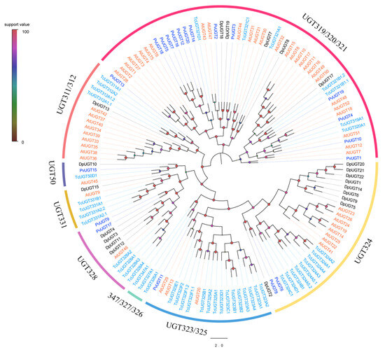
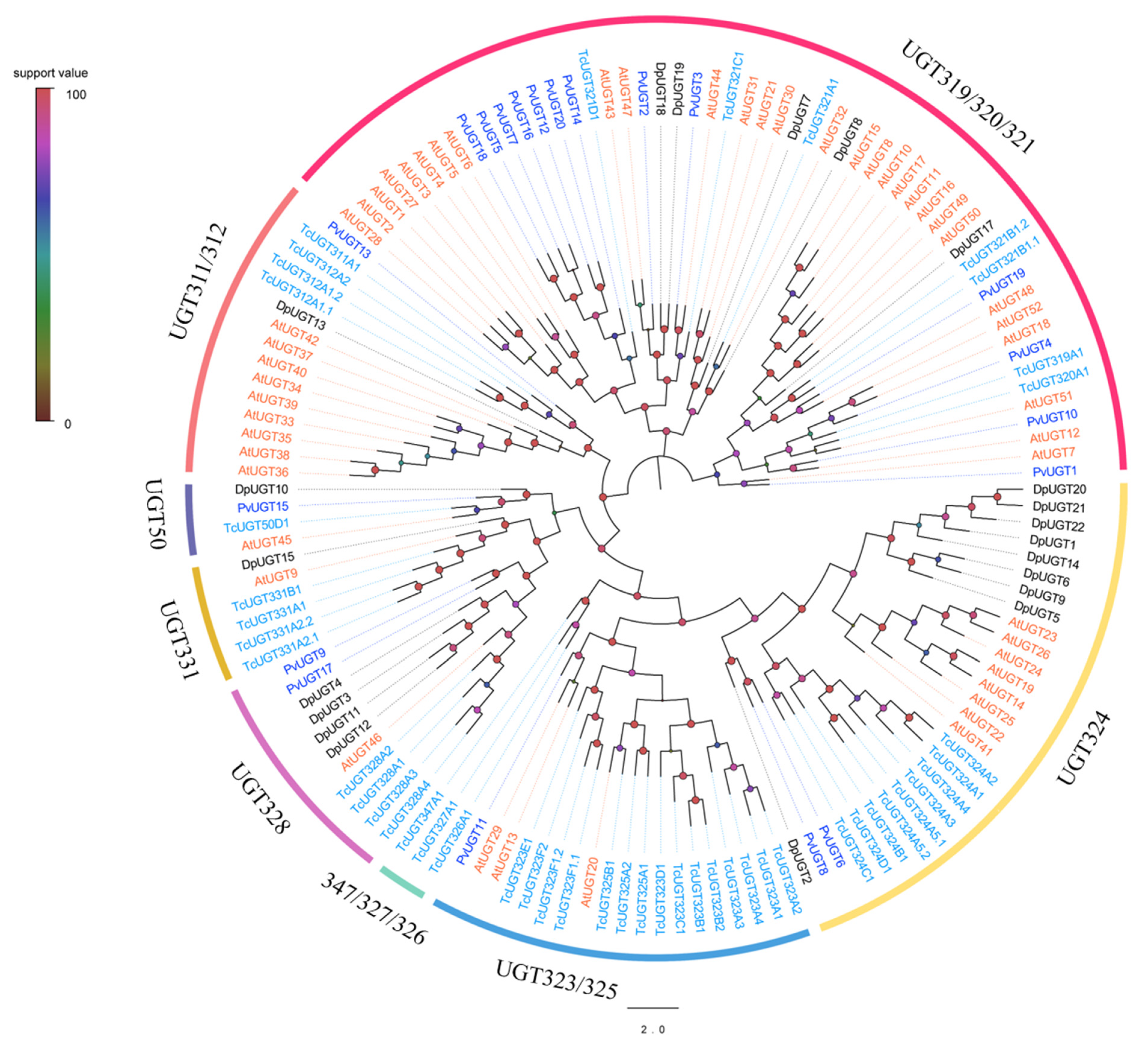
Phylogenetic tree results showed that the 20 UGTs were clustered into eight subfamilies: UGT50, UGT311/312, UGT319/320/321, UGT324, UGT323/325, UGT347/327/326, UGT328 and UGT331. No PverUGTs were found in the UGT331 or UGT347/327/326 subfamilies. Only one member of PvUGT11, PvUGT13 and PvUGT15 were found in the UGT323/325, UGT311/312 and UGT50 subfamilies, respectively. PvUGT9/UGT17 and PvUGT6/UGT8 were clustered into the UGT328 and UGT324 subfamilies, respectively. The UGT319/320/321 subfamilies formed the largest cluster and included PvUGT1, UGT2, UGT3, UGT4, UGT5, UGT7, UGT10, UGT12, UGT14, UGT16, UGT18, UGT19 and UGT20 (Figure 5).
Figure 5.
Maximum-likelihood tree of UGTs from P. versicolora (Pv), D. ponderosae (Dp), A. tumida (At), and T. castaneum (Tc). The different species are highlighted with specific color patterns, and P. versicolora genes are shown in blue color.
3.4. Identification of CYPs
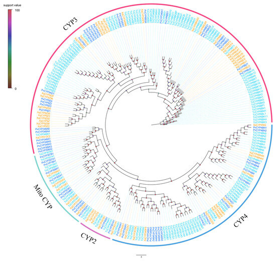
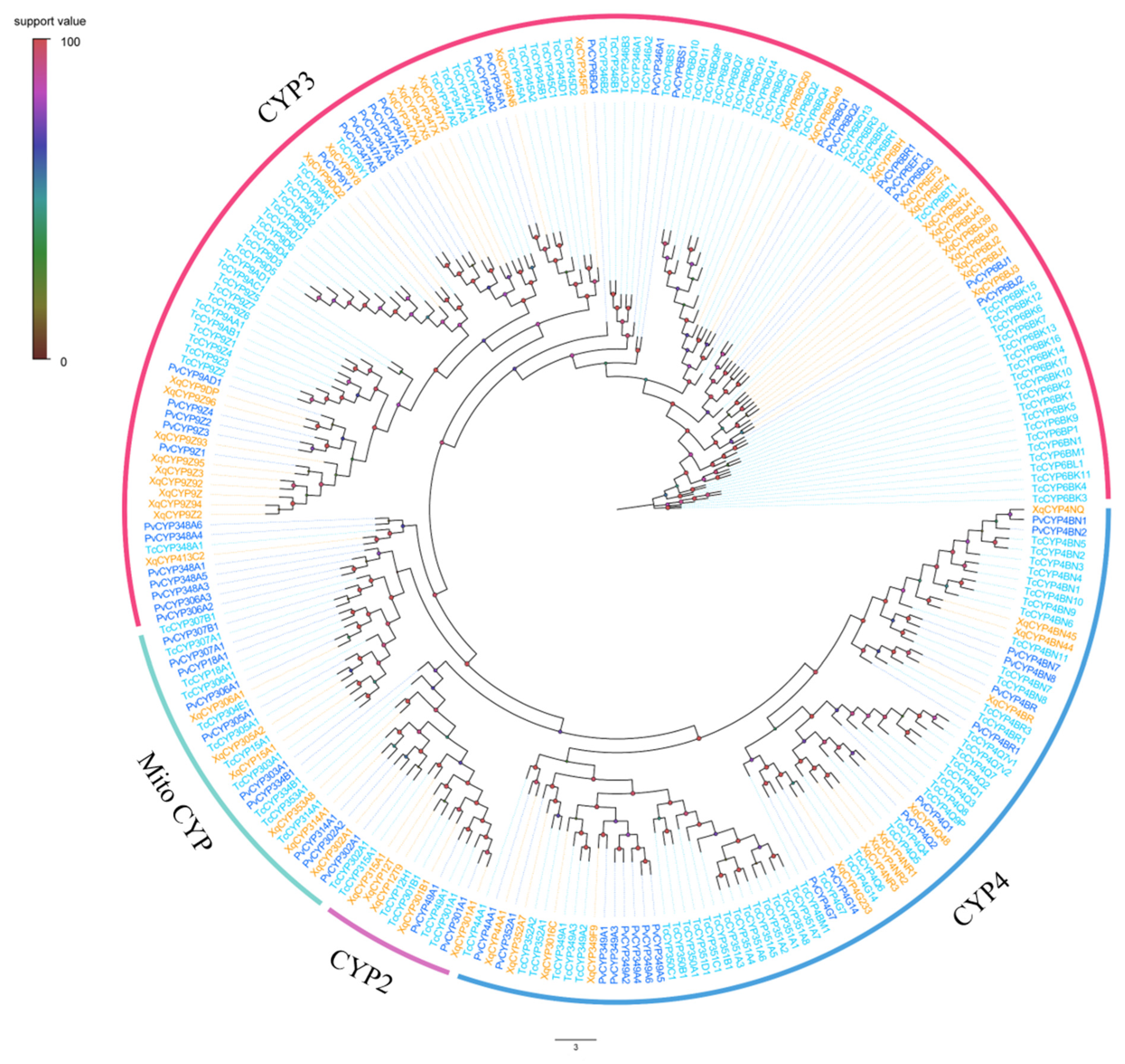
We identified a total of 60 CYPs from the adult antennal transcriptome of P. versicolora. Among the CYP genes, 28 PvCYPs had complete ORFs according to the predicted result, and the protein ranged from 463 to 579 amino acids (Table S4). The remaining 32 PvCYPs were partial sequences due to the absence of 5- or 3-terminals. The results of phylogenetic analysis suggested that CYP genes were clustered into four families: mitochondrial (Mito) members (10 PvCYPs), CYP2 (2 PvCYPs), CYP3 (30 PvCYPs) and CYP4 (18 PvCYPs) (Figure 6).
Figure 6.
Maximum-likelihood tree of CYPs from P. versicolora (Pv), T. castaneum (Tc), and X. quadripes (Xq). Different species are highlighted with specific color patterns, and P. versicolora genes are shown in blue color.
3.5. Identification of COEs
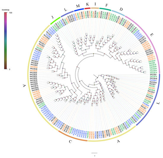
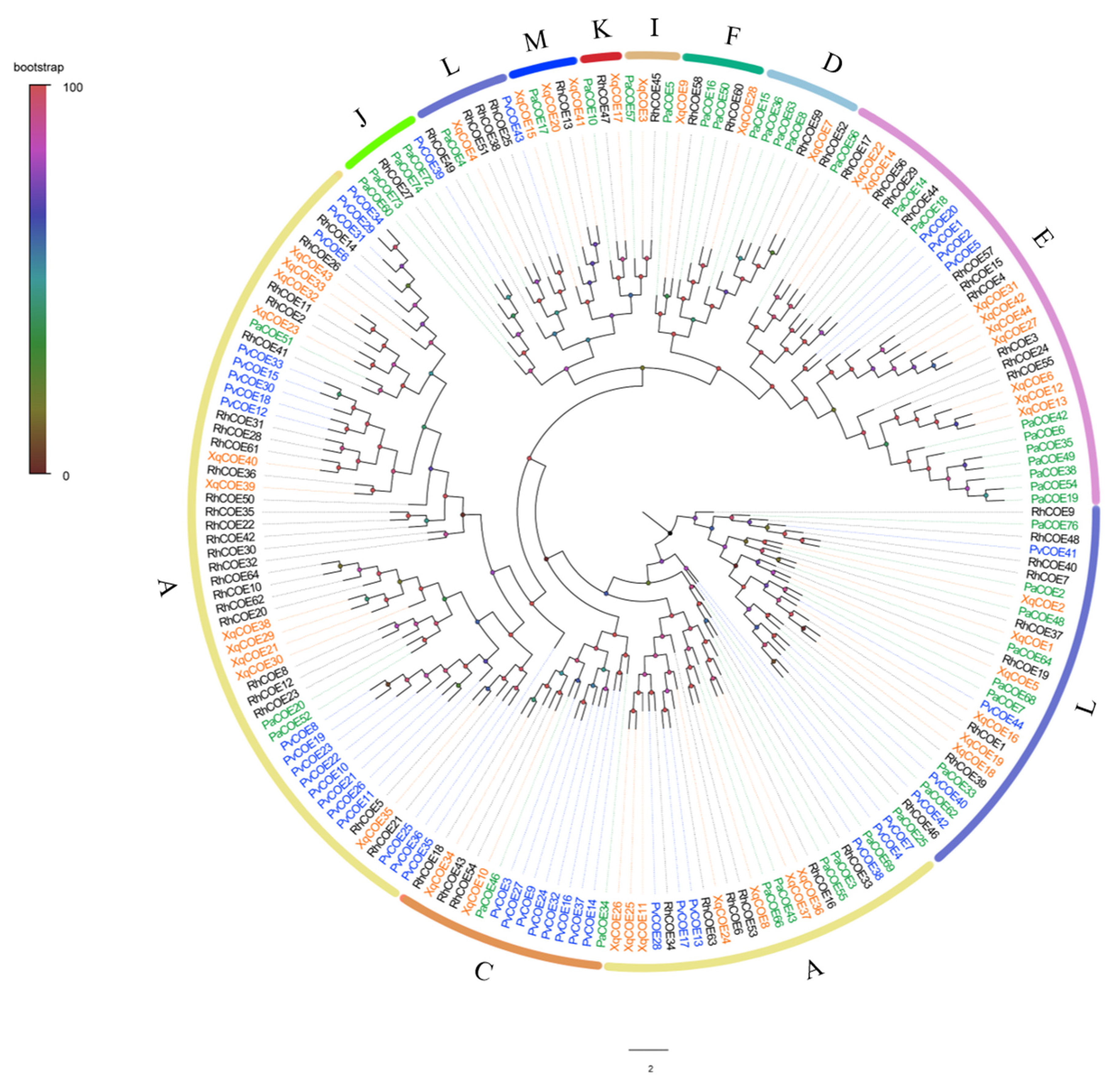
In all 44 COEs were obtained from the transcriptome and 19 PvCOEs have complete ORFs following the predicted result (Table S4). According to the classification system of insect COEs, PvCOEs were grouped into three large classes and 5 clades. Phylogenetic tree results showed that clade A had the largest number of COEs, with 26 PvCOEs, and clade C had 8 PvCOEs in the intracellular catalytic class. The clade E had 4 PvCOEs in the secreted catalytic class, whereas clade L had 5 PvCOEs and clade J had 1 PvCOE in neurodevelopmental class. In addition, no PvCOEs were grouped into the remaining five clades (D, F, I, K, M) (Figure 7).
Figure 7.
Maximum-likelihood tree of COEs from P. versicolora (Pv), R. horsfieldi (Rh), P. antennata (Pa), and X. quadripes (Xq). Different species are highlighted with specific color patterns, and P. versicolora genes are shown in blue. Intracellular catalytic class: A: Coleopteran xenobiotic metabolizing enzymes; C: Microsomal and α-esterases. Secreted catalytic class: D: Integument esterases; E: β- and pheromone esterases; F: Non-lepidopteran juvenile hormone esterases; Neurodevelopmental class: I: Uncharacterized group; J: Acetylcholinesterases; K: Gliotactins; L: Neuroligins; M: Neurotactins.
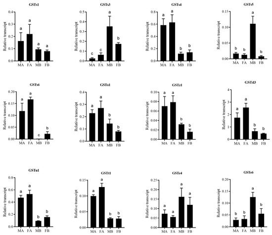
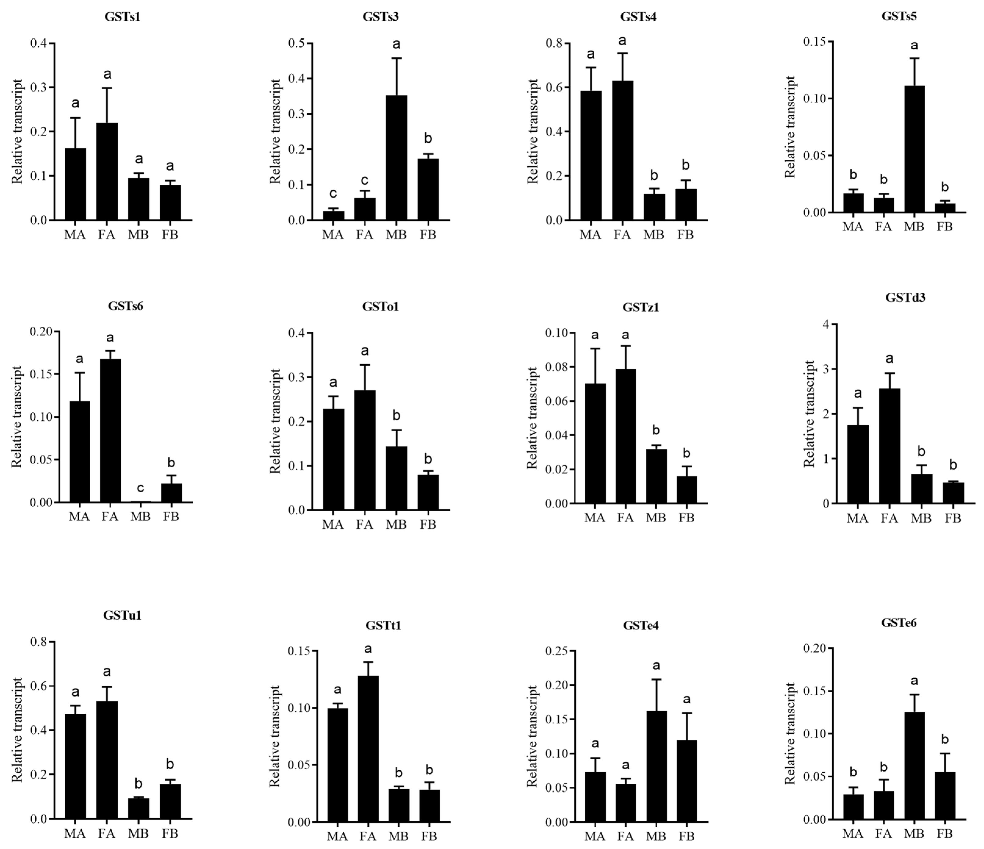
3.6. Expression Profile of GSTs
To explore the expression profile of GSTs, antennae and bodies were selected and tested by qRT–PCR (Figure 8). PvGSTs2 and PvGSTe4 showed no significant difference between antennae and bodies of male and female. PvGSTs3, PvGSTs5 and PvGSTe6 were highly expressed in bodies than in antennae, and further analysis showed that these 3 PvGST were highly expressed in male bodies than in female bodies. PvGSTs4, PvGSTs6, PvGSTo1, PvGSTz1, PvGSTd3, PvGSTu1 and PvGSTt1 had higher expression levels in antennae than in bodies. In addition, expression level of these 7 PvGSTs showed no bias in male and female antennae.
Figure 8.
Expression levels of PvGST genes in different tissues determined by qRT-PCR. MA, male antennae; FA, female antennae; MB, male body without antennae; FB, female body without antennae. Error bars indicate the standard error of three biological replicates. The different letters (a–c) indicate significant differences in different tissues.
4. Discussion
The adult antennal transcriptome of P. versicolora was analyzed, and several detoxification genes were identified in this study. The number of PvGST genes (22 members) was comparable to the number found in the genome and transcriptome of Bombyx mori (23 members) [41], less than 30 GSTs in L. decemlineata [38] and 41 GSTs were identified in T. castaneum [39] based on the whole genome database and NCBI, more than 14 GSTs in Rhodnius prolixus [42] and 10 GSTs in Apis mellifera [5]. The number of PvUGTs (20 members) observed is similar to those found in Holotrichia parallela (20 members) [43], less than 30 UGTs in X. quadripes [44] and more than 13 UGTs in Ostrinia furnacalis [45].
We identified 60 PvCYPs from the antennal transcriptome. This number was lower than that reported in P. antennata (64 CYP genes) [40], A. tumida (116 CYP genes) [46], D. ponderosae (85 CYP genes) [47]. This possibly indicates that more CYP genes may be obtained through further identification in P. versicolora. In addition, 44 PvCOEs were identified from the antennal transcriptome. The number is same as X. quadripes (44 COEs), less than 64 COEs in R. horsfieldi and 72 COEs in L. decemlineata, and more than 35 COEs in Drosophila melanogaster [5]. Meanwhile, these results suggested that the number of detoxification genes varies greatly among different insect species. Pesticides are chemicals that control different pests. These detoxification enzymes (GSTs, UGTs, CYPs and COEs) can reduce the toxicity of pesticides contributed to insect physiology and developmental progress [48].
In the phylogenetic tree of UGTs, some species-specific expansions appeared in P. versicolora, PvUGT319/320/321 subfamily possessed 13 members, including PvUGT1, UGT2, UGT3, UGT4, UGT5, UGT7, UGT10, UGT12, UGT14, UGT16, UGT18, UGT19, and UGT20. Similar patterns were found in other Coleopteran insects, including 8 UGTs of UGT352 in X. quadripes [44], 15 UGTs of UGT352 in P. antennata [49], 8 UGTs of UGT324 in T. castaneum [20], 9 UGTs of UGT312 in A. tumida, and 9 UGTs of UGT324 in D. ponderosae. The results of phylogenetic analyses indicated that this UGT subfamily may have undergone a tandem duplication event.
Unlike odorant-binding proteins and odorant receptor play the fundamental role in the detection and transduction of diverse odor molecules [50]. GSTs that have a highly expressed level in antennae are more likely to degrade and/or inactivate sex pheromones and host volatiles. For example, SzGSTd1 has a specific expression level in the antennae of Sitophilus zeamais and SzGSTd1 functions to selectively degrade capryl alcohol (stored grain volatile) [51]. In the Lepidoptera, many studies have focused on the function of antennae-specific GSTs. PiGSTd1 was antennae-specific and further analysis revealed that PiGSTd1 could degrade and inactivate Z9-12:Ac (sex pheromone component) and aldehyde (host volatiles) of Plodia interpunctella [52]. BmGSTD4 had a specific expression in antennae and a high GSH-binding capacity to 1-chloro-2, 4-dinitrobenzene in B. mori [53]. An antenna specific GST-msolf1 could degrade trans-2-hexenal (green leaf volatile) in Manduca sexta [54]. The qRT-PCR results showed that the expression levels of 7 PvGSTs (GSTs4, GSTs6, GSTo1, GSTz1, GSTd3, GSTu1 and GSTt1) were higher in antennae than in bodies. Thus, it is suggesting that 7 PvGST may be associated as odorant degradation enzyme to ensure the sensitivity of the olfactory system.
5. Conclusions
In conclusion, we obtained 146 detoxification genes from the adult antennae of P. versicolora transcriptome data, including 22 GSTs, 20 UGTs, 60 CYPs, and 44 COEs. Expression profiles showed that 3 PvGST were highly expressed in abdomens, indicating that these PvGSTs may be involved in substance metabolism. In addition, 7 PvGST have high expression levels in the antennae, suggesting that these PvGSTs may take part in odorant perception. This research will be helpful for exploring the detoxification and chemoreception mechanisms of P. versicolora in the future.
Supplementary Materials
The following supporting information can be downloaded at: https://www.mdpi.com/article/10.3390/insects17060643/s1, Table S1: Primers for qRT-PCR of GSTs in P. versicolora. Table S2: The Blastx match of P. versicolora GST genes. Table S3: The Blastx match of P. versicolora UGT genes. Table S4: The Blastx match of P. versicolora CYP genes. Table S5: The Blastx match of P. versicolora COE genes. File S1: The amino acid of GST, UGT, CYP and CXE in the male and female antennae transcriptome of P. versicolora.
Author Contributions
Methodology, X.-L.L., H.-D.S. and Y.-W.P.; Software, H.-D.S. and Y.-W.P.; Formal analysis, X.-L.L., H.-D.S. and Y.-W.P.; Investigation, H.-D.S. and Y.-W.P.; Data curation, X.-L.L., H.-D.S. and H.-N.Z.; Writing—original draft, X.-L.L.; Writing—review & editing, X.-L.L. and H.-N.Z.; Visualization, M.L.; Supervision, X.-L.L., M.L. and H.-N.Z.; Funding acquisition, X.-L.L. and H.-N.Z. All authors have read and agreed to the published version of the manuscript.
Funding
This work was supported by National Natural Science Foundation of China (32301593) and China Postdoctoral Science Foundation (2024M752720).
Data Availability Statement
The data presented in this study are available in this manuscript; further inquiries can be directed to the corresponding author. The raw reads of the transcriptomes in this study have been submitted in the NCBI SRA database, with the BioProject of PRJNA1471247.
Acknowledgments
We thank Ali Hassan (Hubei University) for his assistance with revised the English language.
Conflicts of Interest
The authors declare that they have no conflict of interests.
References
- Field, L.M.; Pickett, J.A.; Wadhams, L.J. Molecular Studies in Insect Olfaction. Insect Mol. Biol. 2000, 9, 545–551. [Google Scholar] [CrossRef] [PubMed]
- Ferry, N.; Edwards, M.G.; Gatehouse, J.A.; Gatehouse, A.M.R. Plant-Insect Interactions: Molecular Approaches to Insect Resistance. Curr. Opin. Biotechnol. 2004, 15, 155–161. [Google Scholar] [CrossRef] [PubMed]
- Després, L.; David, J.-P.; Gallet, C. The Evolutionary Ecology of Insect Resistance to Plant Chemicals. Trends Ecol. Evol. 2007, 22, 298–307. [Google Scholar] [CrossRef] [PubMed]
- Ranson, H.; Claudianos, C.; Ortelli, F.; Abgrall, C.; Hemingway, J.; Sharakhova, M.V.; Unger, M.F.; Collins, F.H.; Feyereisen, R. Evolution of Supergene Families Associated with Insecticide Resistance. Science 2002, 298, 179–181. [Google Scholar] [CrossRef] [PubMed]
- Claudianos, C.; Ranson, H.; Johnson, R.M.; Biswas, S.; Schuler, M.A.; Berenbaum, M.R.; Feyereisen, R.; Oakeshott, J.G. A Deficit of Detoxification Enzymes: Pesticide Sensitivity and Environmental Response in the Honeybee. Insect Mol. Biol. 2006, 15, 615–636. [Google Scholar] [CrossRef] [PubMed]
- Heidel-Fischer, H.M.; Vogel, H. Molecular Mechanisms of Insect Adaptation to Plant Secondary Compounds. Curr. Opin. Insect Sci. 2015, 8, 8–14. [Google Scholar] [CrossRef] [PubMed]
- Younus, F.; Chertemps, T.; Pearce, S.L.; Pandey, G.; Bozzolan, F.; Coppin, C.W.; Russell, R.J.; Maïbèche-Coisne, M.; Oakeshott, J.G. Identification of Candidate Odorant Degrading Gene/Enzyme Systems in the Antennal Transcriptome of Drosophila melanogaster. Insect Biochem. Mol. Biol. 2014, 53, 30–43. [Google Scholar] [CrossRef] [PubMed]
- Hayes, J.D.; Flanagan, J.U.; Jowsey, I.R. Glutathione Transferases. Annu. Rev. Pharmacol. Toxicol. 2005, 45, 51–88. [Google Scholar] [CrossRef] [PubMed]
- Ketterman, A.J.; Saisawang, C.; Wongsantichon, J. Insect Glutathione Transferases. Drug Metab. Rev. 2011, 43, 253–265. [Google Scholar] [CrossRef] [PubMed]
- Sheehan, D.; Meade, G.; Foley, V.M.; Dowd, C.A. Structure, Function and Evolution of Glutathione Transferases: Implications for Classification of Non-Mammalian Members of an Ancient Enzyme Superfamily. Biochem. J. 2001, 360, 1–16. [Google Scholar] [CrossRef]
- Enayati, A.A.; Ranson, H.; Hemingway, J. Insect Glutathione Transferases and Insecticide Resistance. Insect Mol. Biol. 2005, 14, 3–8. [Google Scholar] [CrossRef] [PubMed]
- Board, P.G.; Coggan, M.; Wilce, M.C.; Parker, M.W. Evidence for an Essential Serine Residue in the Active Site of the Theta Class Glutathione Transferases. Biochem. J. 1995, 311, 247–250. [Google Scholar] [CrossRef] [PubMed]
- Toba, G.; Aigaki, T. Disruption of the Microsomal Glutathione S-Transferase-like Gene Reduces Life Span of Drosophila melanogaster. Gene 2000, 253, 179–187. [Google Scholar] [CrossRef] [PubMed]
- Ranson, H.; Rossiter, L.; Ortelli, F.; Jensen, B.; Wang, X.; Roth, C.W.; Collins, F.H.; Hemingway, J. Identification of a Novel Class of Insect Glutathione S-Transferases Involved in Resistance to DDT in the Malaria Vector Anopheles gambiae. Biochem. J. 2001, 359, 295–304. [Google Scholar] [CrossRef]
- Yan, H.; Jia, H.; Gao, H.; Guo, X.; Xu, B. Identification, Genomic Organization, and Oxidative Stress Response of a Sigma Class Glutathione S-Transferase Gene (AccGSTS1) in the Honey Bee, Apis cerana cerana. Cell Stress Chaperones 2013, 18, 415–426. [Google Scholar] [CrossRef] [PubMed]
- Xu, Z.-B.; Zou, X.-P.; Zhang, N.; Feng, Q.-L.; Zheng, S.-C. Detoxification of Insecticides, Allechemicals and Heavy Metals by Glutathione S-Transferase SlGSTE1 in the Gut of Spodoptera litura. Insect Sci. 2015, 22, 503–511. [Google Scholar] [PubMed]
- Ono, H.; Ozaki, K.; Yoshikawa, H. Identification of Cytochrome P450 and Glutathione-S-Transferase Genes Preferentially Expressed in Chemosensory Organs of the Swallowtail Butterfly, Papilio xuthus L. Insect Biochem. Mol. Biol. 2005, 35, 837–846. [Google Scholar] [CrossRef] [PubMed]
- Bock, K.W. Vertebrate UDP-Glucuronosyltransferases: Functional and Evolutionary Aspects. Biochem. Pharmacol. 2003, 66, 691–696. [Google Scholar] [CrossRef] [PubMed]
- Magdalou, J.; Fournel-Gigleux, S.; Ouzzine, M. Insights on Membrane Topology and Structure/Function of UDP-Glucuronosyltransferases. Drug Metab. Rev. 2010, 42, 159–166. [Google Scholar] [PubMed]
- Ahn, S.-J.; Vogel, H.; Heckel, D.G. Comparative Analysis of the UDP-Glycosyltransferase Multigene Family in Insects. Insect Biochem. Mol. Biol. 2012, 42, 133–147. [Google Scholar] [CrossRef] [PubMed]
- Luque, T.; Okano, K.; O’Reilly, D.R. Characterization of a Novel Silkworm (Bombyx mori) Phenol UDP-Glucosyltransferase. Eur. J. Biochem. 2002, 269, 819–825. [Google Scholar] [CrossRef] [PubMed]
- Yang, Z.; Xiao, T.; Lu, K. Contribution of UDP-glycosyltransferases to chlorpyrifos resistance in Nilaparvata lugens. Pestic. Biochem. Physiol. 2023, 190, 105321. [Google Scholar] [CrossRef] [PubMed]
- Tian, F.; Wang, Z.; Li, C.; Liu, J.; Zeng, X. UDP-Glycosyltransferases Are Involved in Imidacloprid Resistance in the Asian Citrus Psyllid, Diaphorina citri (Hemiptera: Lividae). Pestic. Biochem. Physiol. 2019, 154, 23–31. [Google Scholar] [CrossRef] [PubMed]
- Liu, N.; Li, M.; Gong, Y.; Liu, F.; Li, T. Cytochrome P450s—Their Expression, Regulation, and Role in Insecticide Resistance. Pestic. Biochem. Physiol. 2015, 120, 77–81. [Google Scholar] [CrossRef] [PubMed]
- Nauen, R.; Bass, C.; Feyereisen, R.; Vontas, J. The Role of Cytochrome P450s in Insect Toxicology and Resistance. Annu. Rev. Entomol. 2022, 67, 105–124. [Google Scholar] [CrossRef] [PubMed]
- Schuler, M.A.; Berenbaum, M.R. Structure and Function of Cytochrome P450S in Insect Adaptation to Natural and Synthetic Toxins: Insights Gained from Molecular Modeling. J. Chem. Ecol. 2013, 39, 1232–1245. [Google Scholar] [CrossRef] [PubMed]
- Pedra, J.H.F.; McIntyre, L.M.; Scharf, M.E.; Pittendrigh, B.R. Genome-Wide Transcription Profile of Field- and Laboratory-Selected Dichlorodiphenyltrichloroethane (DDT)-Resistant Drosophila. Proc. Natl. Acad. Sci. USA 2004, 101, 7034–7039. [Google Scholar] [CrossRef] [PubMed]
- Montella, I.R.; Schama, R.; Valle, D. The Classification of Esterases: An Important Gene Family Involved in Insecticide Resistance—A Review. Mem. Inst. Oswaldo Cruz 2012, 107, 437–449. [Google Scholar] [CrossRef] [PubMed]
- Tsubota, T.; Shiotsuki, T. Genomic and Phylogenetic Analysis of Insect Carboxyl/Cholinesterase Genes. J. Pestic. Sci. 2010, 35, 310–314. [Google Scholar] [CrossRef]
- Ishida, Y.; Leal, W.S. Rapid Inactivation of a Moth Pheromone. Proc. Natl. Acad. Sci. USA 2005, 102, 14075–14079. [Google Scholar] [CrossRef] [PubMed]
- Keeling, C.I.; Henderson, H.; Li, M.; Dullat, H.K.; Ohnishi, T.; Bohlmann, J. CYP345E2, an Antenna-Specific Cytochrome P450 from the Mountain Pine Beetle, Dendroctonus ponderosae Hopkins, Catalyses the Oxidation of Pine Host Monoterpene Volatiles. Insect Biochem. Mol. Biol. 2013, 43, 1142–1151. [Google Scholar] [CrossRef] [PubMed]
- Chiu, C.C.; Keeling, C.I.; Bohlmann, J. The Cytochrome P450 CYP6DE1 Catalyzes the Conversion of α-Pinene into the Mountain Pine Beetle Aggregation Pheromone Trans-Verbenol. Sci. Rep. 2019, 9, 1477. [Google Scholar] [CrossRef] [PubMed]
- Utsumi, S.; Ando, Y.; Ohgushi, T. Evolution of Feeding Preference in a Leaf Beetle: The Importance of Phenotypic Plasticity of a Host Plant. Ecol. Lett. 2009, 12, 920–929. [Google Scholar] [CrossRef] [PubMed]
- Li, B.; Liu, F.; He, X.; Liu, X.; Lu, M. Temporal Transcriptomic Changes in Willow Leaves Oviposited by Plagiodera versicolora. Integr. Zool. 2024, 19, 784–787. [Google Scholar] [CrossRef]
- Liu, X.; Tong, N.; Wu, Z.; Li, Y.; Ma, M.; Liu, P.; Lu, M. Identification of Chemosensory Genes Based on the Antennal Transcriptomic Analysis of Plagiodera versicolora. Insects 2021, 13, 36. [Google Scholar] [CrossRef] [PubMed]
- Zhao, Y.-J.; Wang, Z.-Q.; Zhu, J.-Y.; Liu, N.-Y. Identification and Characterization of Detoxification Genes in Two Cerambycid Beetles, Rhaphuma horsfieldi and Xylotrechus quadripes (Coleoptera: Cerambycidae: Clytini). Comp. Biochem. Physiol. B Biochem. Mol. Biol. 2020, 243–244, 110431. [Google Scholar] [CrossRef] [PubMed]
- Zhu, F.; Moural, T.W.; Shah, K.; Palli, S.R. Integrated Analysis of Cytochrome P450 Gene Superfamily in the Red Flour Beetle, Triboliumcastaneum. BMC Genom. 2013, 14, 174. [Google Scholar] [CrossRef] [PubMed]
- Han, J.-B.; Li, G.-Q.; Wan, P.-J.; Zhu, T.-T.; Meng, Q.-W. Identification of Glutathione S-Transferase Genes in Leptinotarsa decemlineata and Their Expression Patterns under Stress of Three Insecticides. Pestic. Biochem. Physiol. 2016, 133, 26–34. [Google Scholar] [CrossRef] [PubMed]
- Shi, H.; Pei, L.; Gu, S.; Zhu, S.; Wang, Y.; Zhang, Y.; Li, B. Glutathione S-Transferase (GST) Genes in the Red Flour Beetle, Tribolium castaneum, and Comparative Analysis with Five Additional Insects. Genomics 2012, 100, 327–335. [Google Scholar] [CrossRef] [PubMed]
- Yang, A.-J.; Yin, N.-N.; Chen, D.-L.; Guo, Y.-R.; Zhao, Y.-J.; Liu, N.-Y. Identification and Characterization of Candidate Detoxification Genes in Pharsalia antennata Gahan (Coleoptera: Cerambycidae). Front. Physiol. 2022, 13, 1015793. [Google Scholar] [CrossRef] [PubMed]
- Yu, Q.; Lu, C.; Li, B.; Fang, S.; Zuo, W.; Dai, F.; Zhang, Z.; Xiang, Z. Identification, Genomic Organization and Expression Pattern of Glutathione S-Transferase in the Silkworm, Bombyx mori. Insect Biochem. Mol. Biol. 2008, 38, 1158–1164. [Google Scholar] [CrossRef] [PubMed]
- Schama, R.; Pedrini, N.; Juárez, M.P.; Nelson, D.R.; Torres, A.Q.; Valle, D.; Mesquita, R.D. Rhodnius prolixus Supergene Families of Enzymes Potentially Associated with Insecticide Resistance. Insect Biochem. Mol. Biol. 2016, 69, 91–104. [Google Scholar] [CrossRef] [PubMed]
- Wang, S.; Liu, Y.; Zhou, J.-J.; Yi, J.-K.; Pan, Y.; Wang, J.; Zhang, X.-X.; Wang, J.-X.; Yang, S.; Xi, J.-H. Identification and Tissue Expression Profiling of Candidate UDP-Glycosyltransferase Genes Expressed in Holotrichia parallela Motschulsky Antennae. Bull. Entomol. Res. 2018, 108, 807–816. [Google Scholar] [CrossRef] [PubMed]
- Yin, N.-N.; Zhao, Y.-J.; Zhu, J.-Y.; Liu, N.-Y. Antennal UDP-Glycosyltransferase Genes in the Coffee White Stemborer, Xylotrechus quadripes. J. Asia-Pac. Entomol. 2019, 22, 1145–1153. [Google Scholar] [CrossRef]
- Zhang, L.; Shen, Y.; Jiang, X.; Liu, S. Transcriptomic Identification and Expression Profile Analysis of Odorant-Degrading Enzymes from the Asian Corn Borer Moth, Ostrinia furnacalis. Insects 2022, 13, 1027. [Google Scholar] [CrossRef] [PubMed]
- Evans, J.D.; McKenna, D.; Scully, E.; Cook, S.C.; Dainat, B.; Egekwu, N.; Grubbs, N.; Lopez, D.; Lorenzen, M.D.; Reyna, S.M.; et al. Genome of the Small Hive Beetle (Aethina tumida, Coleoptera: Nitidulidae), a Worldwide Parasite of Social Bee Colonies, Provides Insights into Detoxification and Herbivory. Gigascience 2018, 7, 138. [Google Scholar] [CrossRef] [PubMed]
- Keeling, C.I.; Yuen, M.M.S.; Liao, N.Y.; Docking, T.R.; Chan, S.K.; Taylor, G.A.; Palmquist, D.L.; Jackman, S.D.; Nguyen, A.; Li, M.; et al. Draft Genome of the Mountain Pine Beetle, Dendroctonus ponderosae Hopkins, a Major Forest Pest. Genome Biol. 2013, 14, R27. [Google Scholar] [CrossRef] [PubMed]
- Jaffar, S.; Ahmad, S.; Lu, Y. Contribution of insect gut microbiota and their associated enzymes in insect physiology and biodegradation of pesticides. Front. Microbiol. 2022, 13, 979383. [Google Scholar] [CrossRef] [PubMed]
- Yin, N.; Wang, Z.; Xiao, H. Identification and characterization of UDP-glycosyltransferase genes in a cerambycid beetle, Pharsalia antennata Gahan, 1894 (Coleoptera: Cerambycidae). Diversity 2022, 14, 348. [Google Scholar] [CrossRef]
- Jaffar, S.; Smagghe, G.; Lu, Y. Olfactory receptors in Bactrocera species for sustainable fruit fly management: A review and future perspectives. Physiol. Entomol. 2024, 49, 67–90. [Google Scholar] [CrossRef]
- Xia, D.; Zheng, R.; Huang, J.; Lu, S.; Tang, Q. Identification and Functional Analysis of Glutathione S-Transferases from Sitophilus zeamais in Olfactory Organ. Insects 2022, 13, 259. [Google Scholar] [CrossRef] [PubMed]
- Liu, H.; Tang, Y.; Wang, Q.; Shi, H.; Yin, J.; Li, C. Identification and Characterization of an Antennae-Specific Glutathione s-Transferase from the Indian Meal Moth. Front. Physiol. 2021, 12, 727619. [Google Scholar] [CrossRef] [PubMed]
- Tan, X.; Hu, X.-M.; Zhong, X.-W.; Chen, Q.-M.; Xia, Q.-Y.; Zhao, P. Antenna-Specific Glutathione S-Transferase in Male Silkmoth Bombyx mori. Int. J. Mol. Sci. 2014, 15, 7429–7443. [Google Scholar] [CrossRef] [PubMed]
- Rogers, M.E.; Jani, M.K.; Vogt, R.G. An Olfactory-Specific Glutathione-S-Transferase in the Sphinx Moth Manduca sexta. J. Exp. Biol. 1999, 202, 1625–1637. [Google Scholar] [CrossRef] [PubMed]
Disclaimer/Publisher’s Note: The statements, opinions and data contained in all publications are solely those of the individual author(s) and contributor(s) and not of MDPI and/or the editor(s). MDPI and/or the editor(s) disclaim responsibility for any injury to people or property resulting from any ideas, methods, instructions or products referred to in the content. |
© 2026 by the authors. Licensee MDPI, Basel, Switzerland. This article is an open access article distributed under the terms and conditions of the Creative Commons Attribution (CC BY) license.