


Simple Summary
This study evaluated the therapeutic potential of Scutellaria baicalensis and its active component baicalin against Nosema ceranae infection. The results showed that both the S. baicalensis aqueous extract and baicalin significantly inhibited the proliferation of this pathogen in Apis cerana. Transcriptomic analysis revealed that baicalin promoted the expression of the intestinal chitinase-related genes in infected A. cerana. This enhanced expression is considered a key mechanism that may contribute to improved host capacity for spore elimination. This study provides a theoretical basis for the application of baicalin in the prevention and treatment of N. ceranae, and offers a reference for the development of novel anti-Nosema agents.
Abstract
Nosema ceranae is a common and highly contagious fungal pathogen that primarily infects the gut of adult honeybees, causing nosemosis. As a chronic disease of the digestive system, it poses a global threat to honeybee health and colony sustainability. This study aimed to investigate the inhibitory effects of different concentrations of Scutellaria baicalensis aqueous extract on N. ceranae in the intestines of infected Apis cerana through feeding experiments. In addition, the therapeutic efficacy of its major active component, baicalin, was evaluated, and its potential molecular mechanisms of action were explored. The results showed that, compared with the control group, administration of S. baicalensis aqueous extract at concentrations of 1 mg/mL, 5 mg/mL, and 10 mg/mL significantly reduced midgut spore loads (p < 0.05). Further experiments showed that a 0.5 mg/mL baicalin sucrose solution, prepared with 0.5% (v/v) DMSO as co-solvent, exhibited optimal solubility and significantly inhibited the proliferation of spores in the honeybee midgut. Transcriptomic analysis of A. cerana revealed varying numbers of significantly differentially expressed genes among the baicalin-treated (HG) group, the co-solvent control (DMSO) group, and the blank control (C) group. Four candidate DEGs associated with the effects of baicalin were further identified, namely LOC108003965, LOC108000905, LOC107996681, and CYP4G11. Gene Ontology enrichment analysis showed that, in the comparison between the HG group and the C group, these DEGs were significantly enriched in six functional categories: iron ion binding, phosphoric ester hydrolase activity, heme binding, tetrapyrrole binding, hydrolase activity (acting on ester bonds), and oxidoreductase activity (acting on paired donors, with incorporation or reduction of molecular oxygen). Collectively, these results demonstrate that S. baicalensis aqueous extract effectively inhibits the proliferation of N. ceranae within the host, and its active component, baicalin, exhibits a similar inhibitory effect. The present study proposes a novel strategy in which baicalin may enhance host endogenous chitinase-related activity to target and disrupt the spore wall, offering a new perspective for the prevention and control of honeybee nosemosis.
1. Introduction
Honeybees are of global importance due to their large-scale cultivation, advanced husbandry techniques, and broad applications, providing essential support for ecological stability and sustainable agricultural development [1,2,3]. With the continued expansion of global crop production, honeybees, as highly efficient pollinators, have attracted significant attention for their contributions to agriculture [4,5]. However, alongside the rapid growth of the global beekeeping industry, the prevention and control of honeybee diseases have emerged as critical challenges that cannot be overlooked.
Nosema ceranae is an obligate intracellular fungal pathogen characterized by potent environmental adaptability [6,7,8]. This trait enables year-round infestation of hosts, posing a persistent threat to colony health and sustainability [9]. The pathogenesis of N. ceranae involves the specific infection of midgut epithelial cells in adult honeybees, leading to structural and functional damage and ultimately resulting in nosemosis [10]. This disease is a chronic infectious disorder of the digestive system, characterized by a prolonged incubation period and sustained detrimental effects, and represents a significant threat to both individual honeybees and colony health [11]. N. ceranae is transmitted primarily through the fecal–oral route via contaminated food and water, facilitating its spread within and between colonies [12]. Furthermore, evidence indicates that honeybees can also become infected during activities such as hive cleaning, trophallaxis (food exchange), and mating [13,14,15,16]. Following ingestion, spores germinate in the midgut and inject their sporoplasm into epithelial cells via the polar tube; subsequent intracellular proliferation leads to host cell rupture and the release of new spores, which then infect neighboring cells [17]. During the later stages of infection, spores are excreted in feces and released into the environment, serving as a continuous source of transmission [18].
Facilitated by global apicultural trade and the movement of honeybee colonies, this pathogen has achieved worldwide dissemination and establishment, and has successfully shifted from its original host, Apis cerana, to Apis mellifera [19,20,21,22,23,24,25]. It has now emerged as the primary causative agent of nosemosis in the latter species [26,27].
Studies have demonstrated that, compared to its original host (A. cerana), N. ceranae exhibits greater virulence in its new host A. mellifera [8,26]. Beyond impairing honeybee physiology and reducing lifespan, the pathogen induces energetic stress and suppresses immune and antioxidant defenses in A. mellifera, thereby promoting its own replication [14,28,29,30,31,32]. Therefore, this disease not only threatens the sustainable development of the global beekeeping industry but also poses a serious challenge to agricultural systems that depend on honeybee pollination, with direct implications for ecosystem stability and global food security [33,34]. Fumagillin, historically the only approved agent for the control of nosemosis in honeybees, was widely used in the global beekeeping industry for many years [18]. However, subsequent studies have confirmed its toxicity to honeybees and the potential health risks associated with residues in apiculture-derived products [35]. Consequently, its use has been prohibited in major beekeeping regions, including the European Union [36]. To date, safe, effective, and economically viable alternatives for the treatment of this disease remain lacking.
The control of N. ceranae remains highly challenging, owing not only to its concealed parasitic behavior and complex transmission pathways but also to the unique characteristics of its spores. The spore wall exhibits a unique structure and chemical composition, forming a robust barrier against environmental stressors and pharmacological interventions [37,38]. This allows the spores to maintain long-term infectivity while substantially reducing the efficacy of conventional control strategies.
Throughout the history of human efforts to combat disease, plant-derived medicines have played a vital role. Extensive evidence indicates that plants rich in natural antimicrobial components can be used not only directly in anti-infective therapies but also as valuable resources for developing novel antifungal agents [39]. Gajger et al. [40] found that feeding A. mellifera with “Nozevit” (an oak bark extract) significantly reduced N. apis spore loads in the midgut of infected bees. Maistrello et al. [41] demonstrated that honeybees fed thymol- or resveratrol-supplemented diets exhibited significantly lower Nosema infection rates, with resveratrol also extending bee longevity. Borges and colab [42] further showed that several plant-derived compounds, including sulforaphane (from cruciferous vegetables), cinnamyl alcohol (from oregano oil), and naringenin (from citrus fruits), significantly reduced N. ceranae spore loads in honeybees compared with infected controls. However, these compounds exhibited toxicity to honeybees at higher concentrations. The chemical complexity of plant-derived substances and the variability among extract batches remain key factors limiting their efficacy in controlling N. ceranae infections. Therefore, to achieve consistent therapeutic outcomes, it is essential to identify the active compounds and determine their optimal effective dosages.
This study aimed to evaluate, through feeding experiments, the inhibitory effects of the aqueous extract of Scutellaria baicalensis and its active component baicalin on midgut spore loads in infected honeybees. In addition, transcriptomic analysis was employed to elucidate the pharmacological effects and potential mechanisms of baicalin, thereby providing a theoretical basis for the screening of anti-N. ceranae bioactive compounds.
2. Materials and Methods
2.1. Fungal Spores and Honeybees
In this study, N. ceranae spores were obtained from the Honeybee Research Institute of Jiangxi Agricultural University and purified using the discontinuous Percoll gradient centrifugation [43]. Microscopic examination and PCR verification were performed to confirm the identity of the spores as N. ceranae (see Supplementary Table S1). Fresh spores were used to ensure completion of the infection process within 8 h. Sealed brood frames were collected from three healthy colonies at the same institute and placed in an incubator (35 °C, 75% humidity) to obtain newly emerged A. cerana workers within 24 h. Microscopic examination confirmed that the newly emerged workers were free of infection by spore-forming fungi.
2.2. Medicinal Plant and Bioactive Constituent
Based on previous literature, S. baicalensis and its active constituent baicalin, both known for their antimicrobial properties, were selected as experimental materials in this study. S. baicalensis (dried powder, baicalin content: 10.1%) and baicalin (analytical standard; purity ≥ 98%) were supplied by Pinxuantang Trading Firm (Bozhou, China) and Shanghai Macklin Biochemical Technology Co., Ltd. (Shanghai, China), respectively. Dimethyl sulfoxide (DMSO) (HPLC, Purity: ≥99.9%) was obtained from Shanghai Macklin Biochemical Technology Co., Ltd. (Shanghai, China). Preliminary experiments determined that the maximum safe concentration of S. baicalensis aqueous extract for feeding A. cerana infected with N. ceranae was 10 mg/mL (see Supplementary Figure S1). In addition, based on previous reports [44], the maximum safe concentration of dimethyl sulfoxide (DMSO) in 50% (w/v) sucrose solution for feeding was 0.5% (v/v). Accordingly, the optimal concentration of baicalin solution used in this study was established at 0.5 mg/mL.
Preparation of S. baicalensis extract solution: 1 g of the powder was added to 100 mL of 50% (w/v) aqueous sucrose solution and subjected to ultrasonic extraction at 60 °C for 1 h. The extract was then filtered and diluted with the same sucrose solution to obtain S. baicalensis extract solutions at concentrations of 10 mg/mL (high), 5 mg/mL (medium), and 1 mg/mL (low).
Preparation of baicalin solution: Due to its poor water solubility, 20 mg of baicalin was first dissolved in 200 μL of DMSO, as a co-solvent, followed by dilution with 50% (w/v) sucrose solution to obtain a final concentration of 0.5 mg/mL. A control solution containing DMSO (5 μL/mL) in 50% (w/v) sucrose solution was prepared in parallel for comparison.
2.3. N. ceranae Infection
Based on the spore counts, an appropriate volume of 50% (w/v) sucrose solution was added to a known quantity of spores to prepare a suspension at a concentration of 5 × 10⁴ spores/μ L. Each newly emerged worker honeybee (within 24 h post-eclosion) was orally inoculated with 2 μL of the spore suspension, corresponding to an infection dose of 1 × 10⁵ spores per honeybee. To improve feeding efficiency, all experimental honeybees were starved for 2 h in a constant-temperature incubator prior to inoculation.
2.4. Experimental Groups and Sample Collection
S. baicalensis extract experiment: four experimental groups were established, each comprising 60 honeybees with three biological replicates, for a total of 240 individuals. The feeding regimens were as follows: the control group (C) received 50% (w/v) sucrose solution, while the treatment groups (high, medium, and low concentrations) were provided with 50% (w/v) sucrose solution supplemented with S. baicalensis extract at concentrations of 10 mg/mL, 5 mg/mL, and 1 mg/mL, respectively. The experiment was conducted over 7 days, during which all diets were replaced every 24 h under feeding conditions. At 7 days post-infection (dpi), midgut tissues of individual bees in each group were collected separately for Nosema spore quantification and subsequent analyses.
Baicalin experiment: three experimental groups were established, each comprising 60 honeybees with three biological replicates, for a total of 180 individuals. The feeding regimens were as follows: the control group (C) received 50% sucrose solution; the baicalin-treated group (HG) was administered 50% sucrose solution containing 0.5 mg/mL baicalin (with a DMSO at 5 μL/mL); and the DMSO group (co-solvent control) was provided with 50% sucrose solution containing 5 μL/mL DMSO. The experiment was conducted over 7 days, with diets replaced every 24 h under feeding conditions. At 7 dpi, five worker bees were randomly selected from each replicate for midgut collection and total RNA extraction for transcriptome sequencing. The remaining bees were individually dissected, and their midgut tissues were collected for spore counting and subsequent analyses.
2.5. Spore Quantification and Analysis
Midgut spores from individual honeybees in each group were purified using discontinuous Percoll gradient centrifugation, and the number of mature spores was quantified using a hemocytometer. Statistical analyses were conducted using SPSS 26.0. Data conforming to a normal distribution were evaluated by one-way analysis of variance (ANOVA), while non-normally distributed data were analyzed using the Kruskal–Wallis test.
2.6. RNA-Seq
Total RNA was first extracted from the midgut tissues of infected honeybees in each group using a commercial RNA extraction kit (Beijing TransGen Biotech Co., Ltd., Beijing, China), following the manufacturer’s instructions. Transcriptome sequencing was then performed according to previously described methods [45]. To exclude potential interference from the co-solvent (DMSO) and to accurately identify differentially expressed genes (DEGs) associated with baicalin treatment, pairwise comparisons of gene expression levels were performed for both the A. cerana genome and N. ceranae genes in the honeybee midgut tissues across the different experimental groups, as previously described [45]. Candidate genes were selected based on the following criteria: (1) significant differential expression between the HG and C groups; (2) significant differential expression between the HG and DMSO groups; and (3) no significant differential expression between the DMSO and C groups.
Clean reads were first aligned to the reference genome of A. cerana (assembly version ASM1110058v1) to obtain host-derived data. Unmapped reads were further aligned to the N. ceranae reference genome (assembly version Ncer 3.0) to obtain microsporidian-derived data. Gene expression levels were compared across experimental groups, and DEGs were identified using the edgeR package (v3.24.3). The DEGs were then subjected to Gene Ontology (GO) and Kyoto Encyclopedia of Genes and Genomes (KEGG) enrichment analyses. The screening criteria were set as |log2FoldChange| > 1 and adjusted p < 0.05.
2.7. Validation of DEGs by RT-qPCR
To validate the reliability of the identified DEGs, RT-qPCR was performed to assess the expression of the LOC108000905 gene, using β-actin as the internal reference gene (primer sequences are provided in Table S1). The PCR program consisted of an initial denaturation at 95 °C for 1 min, followed by 40 cycles of 95 °C for 15 s, 55 °C for 30 s, and 72 °C for 45 s. Relative gene expression levels were calculated using the 2−ΔΔCt method. All experiments were conducted with three independent biological replicates, each containing three technical replicates.
3. Results
3.1. Effect of S. baicalensis Extract on Spore Load in Honeybees
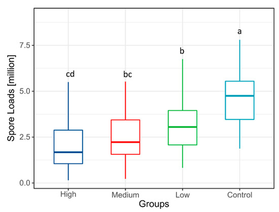
The Kruskal–Wallis test indicated a significant difference among groups (n = 240, H = 96.190, p < 0.001). This indicates that at 7 dpi following the feeding experiment, there were significant differences in intestinal spore levels among different A. cerana groups. Pairwise comparisons further revealed significant differences in spore counts between all S. baicalensis groups (high, medium, and low) and the control group (Figure 1, p < 0.05). Detailed spore count data for each group are provided in Supplementary Table S2.
Figure 1.
The box plot shows the spore load of N. ceranae in Apis cerana from each group under the treatment of S. baicalensis extract. Different letters indicate statistically significant differences between groups (p < 0.05).
3.2. Effect of Baicalin on Spore Load in Honeybees
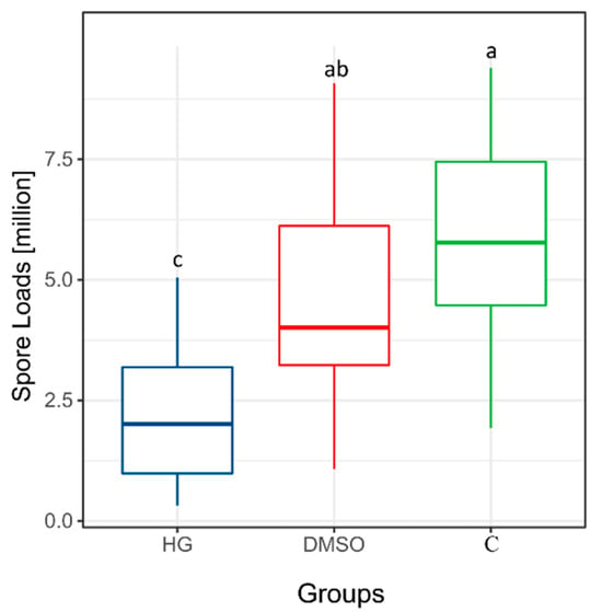
At 7 dpi, the Kruskal–Wallis test indicated significant differences in spore levels among groups (n = 122 after exclusion of invalid data, H = 32.057, p < 0.001). The HG group (baicalin-treated group) exhibited the lowest spore counts, with statistically significant differences compared to both the DMSO group (solvent control) and the C group (blank control) (p < 0.05). In addition, the spore count in the DMSO group was lower than that in the C group; however, this difference was not statistically significant (Figure 2, p > 0.05). Detailed spore count data for each group in the baicalin extract experiment are provided in Supplementary Table S3.
Figure 2.
The box plot shows the spore load of N. ceranae in Apis cerana from each group under the treatment of baicalin. Different letters indicate statistically significant differences between groups (p < 0.05).
3.3. Effects of Baicalin on Host and Parasite mRNA Levels
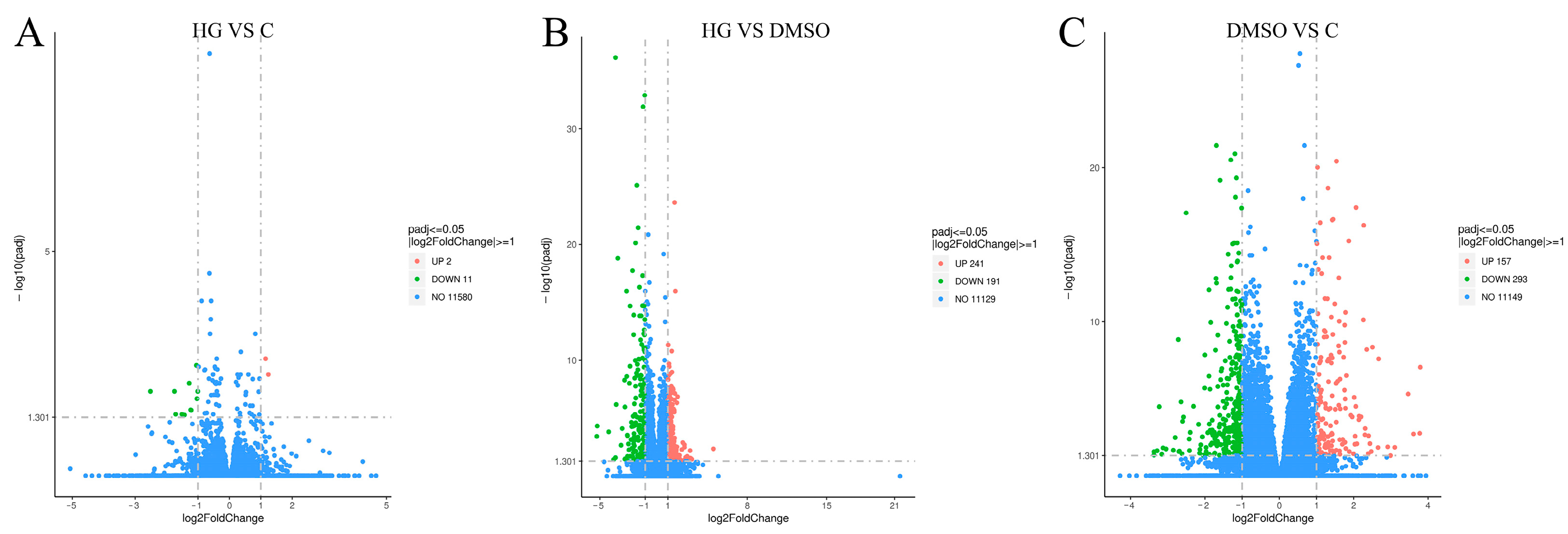
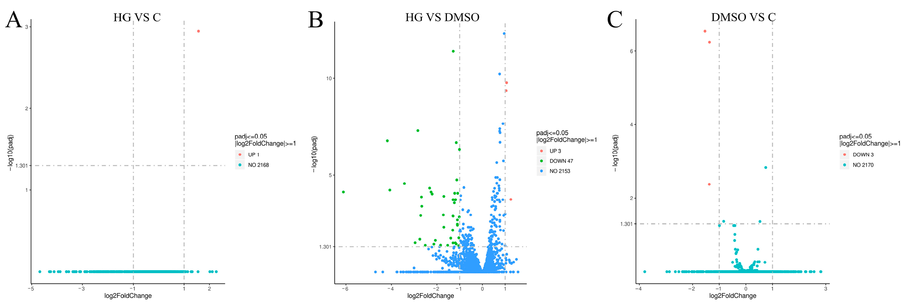
Transcriptome analysis of A. cerana identified 13, 432, and 450 DEGs in the comparisons of the HG group vs. C group (Figure 3A), HG group vs. DMSO group (Figure 3B), and DMSO group vs. C group (Figure 3C), respectively. Similarly, transcriptome analysis of N. ceranae transcriptome identified 1, 50, and 3 significantly differentially expressed genes in the comparisons of HG group vs. C group, HG group vs. DMSO group, and DMSO group vs. C group, respectively (Figure 4).
Figure 3.
The volcano plot shows the comparison results of Apis cerana genes between groups. (A) The volcano plot shows the expression levels of DEGs between the HG group and the C group. (B) The volcano plot shows the expression levels of DEGs between the HG group and the DMSO group. (C) The volcano plot shows the expression levels of DEGs between the DMSO group and the C group.
Figure 4.
The volcano plot shows the comparison results of N. cerana genes between groups. (A) The volcano plot shows the expression levels of DEGs between the HG group and the C group. (B) The volcano plot shows the expression levels of DEGs between the HG group and the DMSO group. (C) The volcano plot shows the expression levels of DEGs between the DMSO group and the C group.
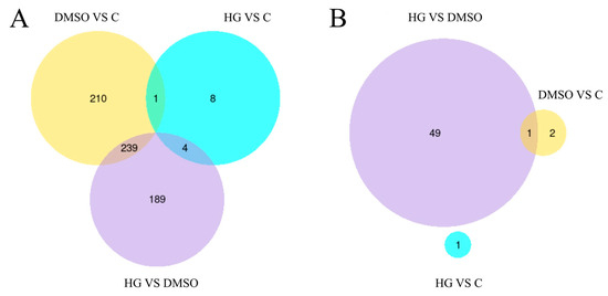
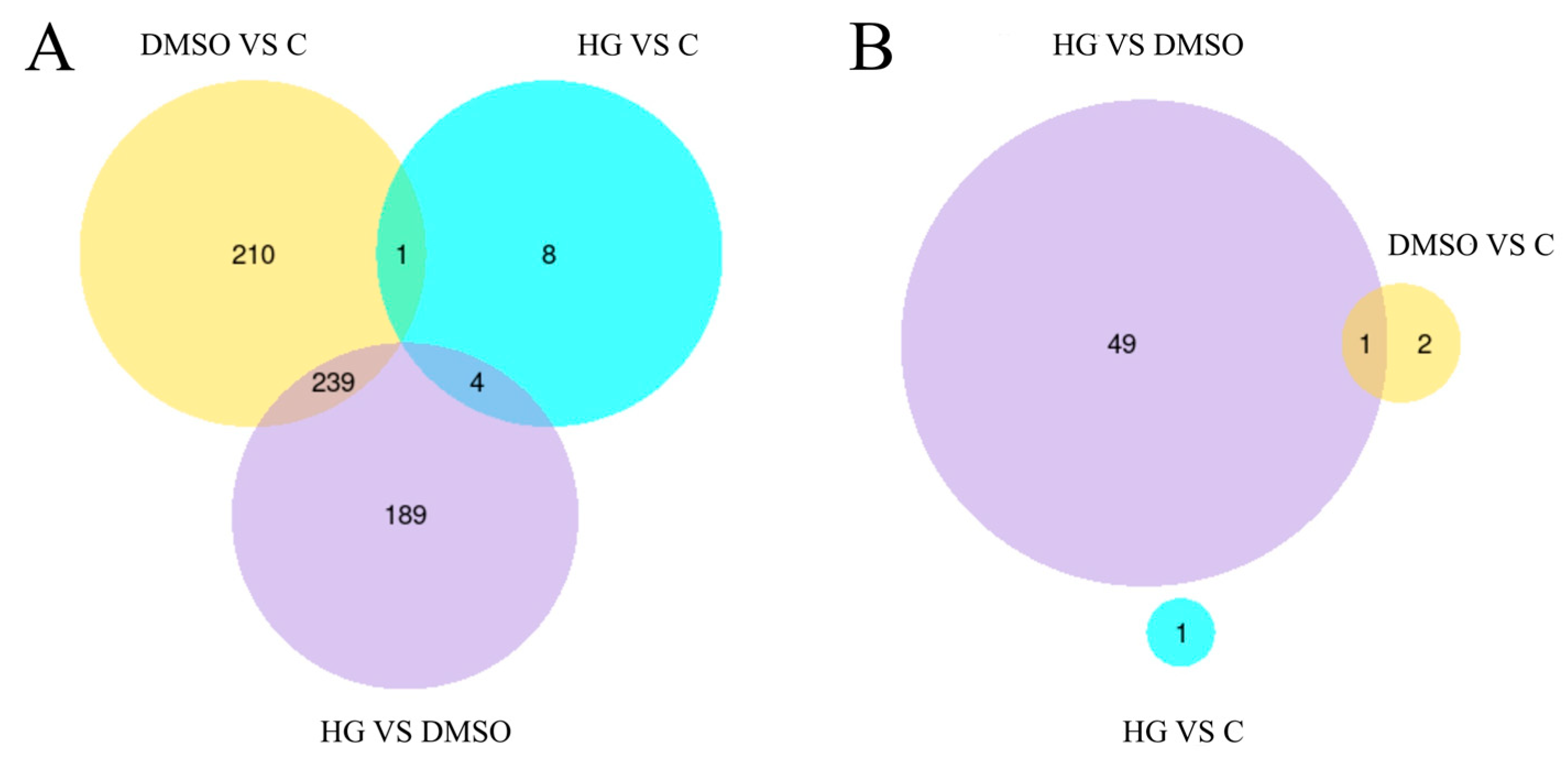
Figure 5A presents the Venn diagram analysis results for the A. cerana genome. Four genes met the selection criteria: LOC108003965, LOC108000905, LOC107996681, and CYP4G11. Figure 5B shows the corresponding Venn diagram analysis results for the N. ceranae genome, in which no differentially expressed genes satisfied the defined criteria.
Figure 5.
The Venn diagrams show the logical relationships among DEGs across groups. (A) The figure shows the Venn analysis of DEGs in Apis cerana among three comparison groups: HG vs. DMSO, HG vs. C, and DMSO vs. C. (B) The figure shows the Venn analysis of DEGs in N. ceranae among three comparison groups: HG vs. DMSO, HG vs. C, and DMSO vs. C. Different colors indicate different comparison combinations.
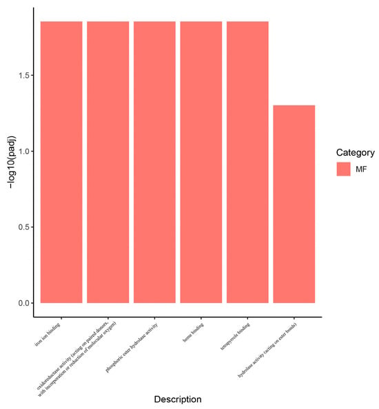
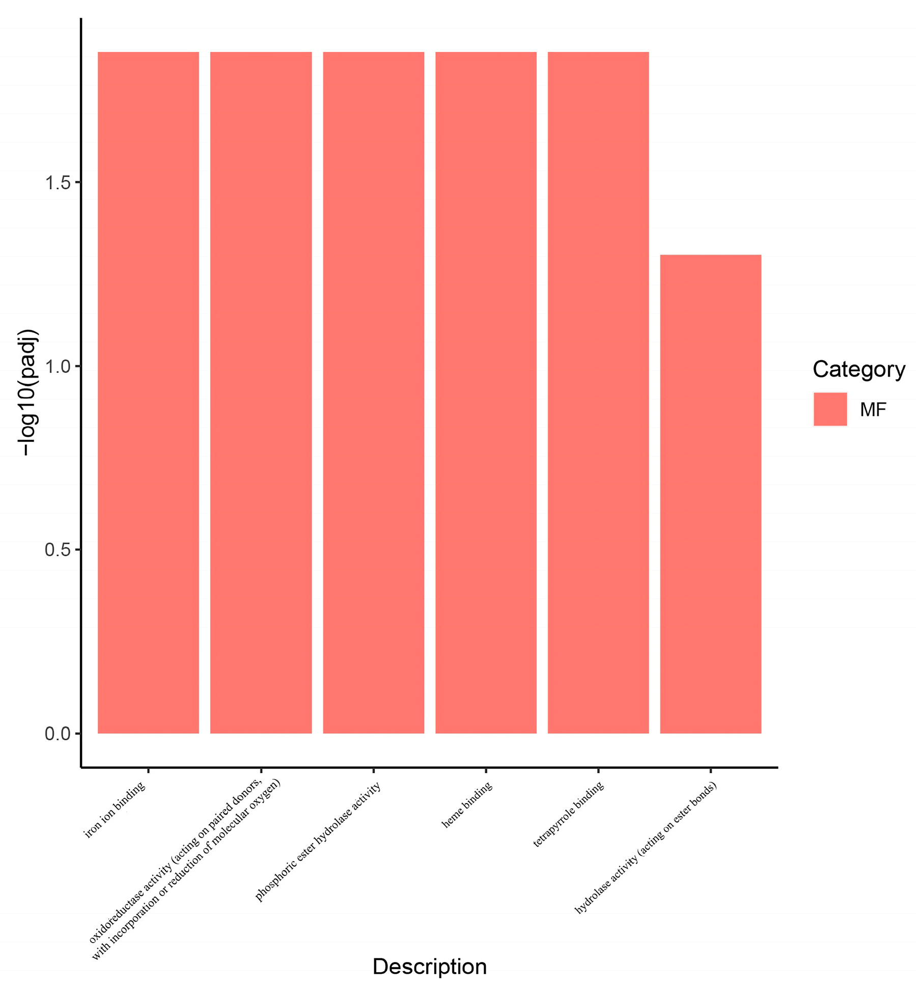
Based on GO and KEGG enrichment analyses of the baicalin-treated group versus the blank control group (HG group vs. C group), we further characterized the functional pathways associated with DEGs in A. cerana. GO enrichment analysis showed that, in the HG vs. C group, DEGs were significantly enriched in six functional categories, including iron ion binding, phosphoric ester hydrolase activity, heme binding, tetrapyrrole binding, hydrolase activity acting on ester bonds, and oxidoreductase activity acting on paired donors with incorporation or reduction of molecular oxygen (Figure 6, padj < 0.05, |log2FoldChange| > 1). In contrast, KEGG enrichment analysis showed no significantly enriched pathways in the HG vs. C group comparison.
Figure 6.
The figure shows the GO analysis plot of DEGs in Apis cerana (HG group vs. C group) related to the effect of baicalin treatment. MF stands for molecular function.
3.4. qRT-PCR Validation of Key DEGs
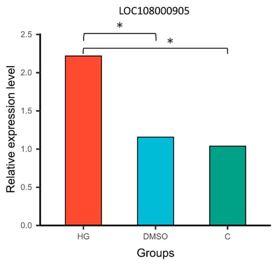
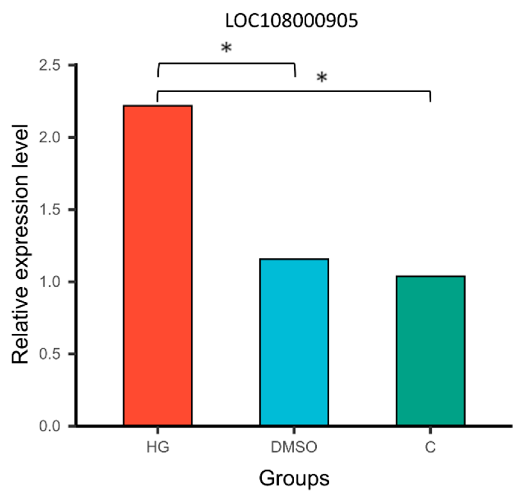
To validate the differential expression of genes among groups, the chitinase-related DEG (LOC108000905) was selected as a representative target. Its expression levels in the midgut of honeybees at 7 days post-infection were quantified using qRT-PCR (see Supplementary Table S5). The qRT-PCR results (Figure 7) demonstrated that LOC108000905 expression was significantly upregulated in the HG group relative to both the C and DMSO groups (p < 0.05), whereas no significant difference was observed between the C and DMSO groups (p > 0.05). These findings were consistent with the transcriptome analysis. Primer sequences used for qRT-PCR analysis are provided in Supplementary Table S4, and the corresponding qPCR data for LOC108000905 are presented in Supplementary Table S5.
Figure 7.
The bar plot shows the relative expression levels of LOC108000905 in the midguts of Apis cerana from each group. Bars with asterisk symbol indicate statistically significant differences (p < 0.05).
4. Discussion
According to Taillon and Andreasen [46], a wide range of compounds, including herbal extracts, can be classified as potential nutraceuticals. These compounds exert their effects through diverse mechanisms to prevent and treat various diseases. Among them, one class comprises compounds with direct antimicrobial activity, which can be used to control N. ceranae infection [47]. These agents may either directly inactivate N. ceranae spores or inhibit their growth and development. Another class includes compounds with anti-inflammatory properties, which are also considered promising for controlling N. ceranae infection [42]. Although these compounds may not directly eliminate spores, their anti-inflammatory effects may alleviate symptoms of N. ceranae infection and enhance the overall health and longevity of infected honeybees. In the present study, administration of S. baicalensis extract at 7 dpi significantly reduced midgut spore loads of honeybees across all treatment groups compared with the blank control group (p < 0.05). Among the tested concentrations, 10 mg/mL was identified as the optimal dose, as it exhibited the strongest inhibitory effect on N. ceranae while remaining safe for A. cerana feeding, based on our preliminary experimental results. The upper limit of the optimal concentration range may be further refined through evaluation of higher concentrations (see Supplementary Figure S1). Additionally, the results of this study showed that baicalin, major bioactive component of S. baicalensis, significantly reduced the midgut spore loads in honeybees at a concentration of 0.5 mg/mL, thereby playing an important role in inhibiting N. ceranae infection.
Transcriptomic analysis revealed significant differential expression of genes for both A. cerana and N. ceranae across the experimental groups. Based on previous research experience, we further identified significant DEGs associated with the effects of baicalin using Venn diagram analysis, and selected these as candidate genes for further investigation. Ho et al. [48] reported that DMSO exerts a protective effect on A. mellifera, improving the survival of infected individuals by enhancing their adaptability or ability to cope with infection, while having no effect on the proliferation of N. ceranae. The authors suggested that DMSO treatment primarily influences gene expression levels in A. mellifera, rather than significantly altering global biochemical pathways or related regulatory modules. This finding is consistent with our results. The screening results identified four significantly differentially expressed honeybee genes associated with the effects of baicalin: LOC108003965, LOC108000905, LOC107996681, and CYP4G11. No corresponding differentially expressed genes were detected in N. ceranae. This may be attributable to the presence of the N. ceranae spore wall, which could act as a physical barrier that limits drug entry, thereby preventing baicalin from exerting a direct effect and resulting in the absence of detectable transcriptomic changes. Based on these findings, we further analyzed the pathways enriched by baicalin-associated A. cerana DEGs in the comparison between the baicalin-treated group and the blank control group (HG vs. C). GO enrichment analysis revealed that these DEGs were significantly enriched in six functional categories: iron ion binding, phosphoric ester hydrolase activity, heme binding, tetrapyrrole binding, hydrolase activity (acting on ester bonds), and oxidoreductase activity (acting on paired donors, with incorporation or reduction of molecular oxygen). In contrast, KEGG enrichment analysis did not identify any significant enriched pathways. This suggests that baicalin may exert its anti-microsporidian effects through mechanisms not captured by KEGG pathway annotations.
Transcriptomic analysis revealed that although LOC108000905 (a DEG) in A. cerana lacks functional annotation, its homologs, such as LOC551600 (Cht5) in A. mellifera (see Supplementary Figure S2) exhibit chitinase activity, suggesting that LOC108000905 may also function as a chitinase. Holt et al. [49,50,51] reported that the seminal fluid of A. mellifera contains antimicrobial components, including chitinase, which degrades chitin, a major structural component of insect exoskeletons and fungal spore cell walls, and is therefore considered a potential factor in the neutralization of N. apis spores. Furthermore, Peng et al. [52] demonstrated that seminal fluid chitinase can weaken and potentially disrupt the chitin-based cell wall of N. apis spores, thereby interfering with the spore life cycle and potentially inducing premature germination. As early as 2016, Grassl et al. [53] identified Cht5 in honeybee seminal fluid. Subsequently, Houdelet et al. [53,54] reported the presence of Cht5 in the honeybee gut, confirming that this enzyme is not restricted to the seminal fluid. Doublet et al. [55] further demonstrated that the expression of the chitinase-related gene Cht5 in A. mellifera is upregulated following Nosema infection, suggesting a potential role in host defense. Consistently, Holt et al. [49] proposed that chitinase is produced in the gut of infected A. mellifera, where it mediates a localized antifungal response against ingested N. apis spores. Altogether, these findings are consistent with our results, showing that baicalin-treated A. cerana exhibited significant upregulation of the Cht5 homolog LOC108000905, accompanied by a marked decrease in midgut Nosema spore load.
Chitin is a primary component of fungal cell walls and plays a critical role in maintaining cell morphology and integrity, as well as mediating key processes during host infection. Chitinases have been widely explored as biological control agents in both medical and agricultural contexts due to their ability to degrade chitin. Previous studies have shown that certain bacterial-derived chitinases can effectively inhibit fungal growth by hydrolyzing chitin in the cell wall, thereby contributing to the control of various fungal diseases [56]. Therefore, chitinase is considered a promising agent for the biological control of fungal pathogens. In this study, we also observed that the expression of CYP4G11 was significantly downregulated in the midguts of honeybees, concomitant with a reduction in spore counts. Previous studies have shown that CYP4G11 is responsive to oxidative stress and may play a role in protecting honeybees against oxidative damage [57]. A plausible explanation for this phenomenon is that the honeybee midgut serves as the primary site of invasion by N. ceranae spores. As the spore load decreases, associated stress factors, such as oxidative stress, are alleviated, leading to reduced expression of CYP4G11. Based on these findings, we propose that baicalin may upregulate the expression of the chitinase homolog LOC108000905 in the intestines of infected honeybees, thereby promoting chitinase production. This, in turn, inhibits the activity and proliferation of N. ceranae spores through degradation of chitin in the spore cell wall. Although several natural products, such as thymol and resveratrol, have been reported to exhibit inhibitory effects against Nosema, baicalin at therapeutic concentrations demonstrates a more favorable safety profile for honeybees, with no observable host mortality [40,41,42].
The above findings suggest that baicalin may promote chitinase-mediated degradation of the spore wall. However, as an obligate intracellular pathogen [17], N. ceranae mainly reproduces within host cells, and the chitin degradation of the spore wall is most likely to occur before host cell invasion or during the formation of new spores. Therefore, we propose that baicalin exerts two main effects in the honeybee gut: a preventive effect by inhibiting spore germination, and a suppressive effect by reducing overall spore burden. In future studies, we will focus on the practical application of baicalin. This includes enhancing its physicochemical properties, such as improving water solubility and bioavailability through salt formation; systematically evaluating its cost-effectiveness, solution stability (e.g., temperature, pH, and storage-related degradation), and field efficacy against N. ceranae; and assessing residue levels and safety in honeybees to ensure the quality and safety of apiculture products.
Overall, these findings provide a theoretical basis for the development and standardized production of anti-Nosema pharmaceuticals, and also highlight the potential of novel enzyme-based strategies targeting spore wall degradation.
5. Conclusions
This study demonstrated for the first time that S. baicalensis and its active component baicalin significantly inhibit the proliferation of N. ceranae in infected honeybees (A. cerana). Furthermore, transcriptomic analysis revealed that baicalin significantly upregulates the expression of the chitinase homologous gene in the host, suggesting its potential as a biological control strategy against N. ceranae infection.
Supplementary Materials
The following supporting information can be downloaded at: https://www.mdpi.com/article/10.3390/insects17050454/s1, Figure S1: Survival curves of baicalin-fed honeybees over 7 days. Figure S2: Diagram of homology analysis of the target gene LOC108000905. Table S1: Microscopic view and PCR verification of N. ceranae spores. Table S2: Scutellaria baicalensis extract experimental spores data. Table S3: Baicalin experimental spores data. Table S4: Primers used for qRT-PCR and their sequences. Table S5: qPCR data of LOC108000905.
Author Contributions
Z.-J.Z. conceived and designed the experiments. X.H. performed the experiments. J.-H.X. and W.-J.J. analysed the data, and X.H. wrote the paper. All authors have read and agreed to the published version of the manuscript.
Funding
This research was supported by the National Key R&D Program Project of China (2022YFD1600202), the Science and Technology Research Project of the Education Department of Jiangxi Province, China (GJJ2207219), Jiangxi Vocational College of Biological Science and Technology Research Fund Project, China (25Y01).
Institutional Review Board Statement
Not applicable.
Data Availability Statement
The data presented in this study are openly available in Transcriptome data at http://www.ncbi.nlm.nih.gov/bioproject/1443012, https://www.ncbi.nlm.nih.gov/bioproject/1445512, reference number PRJNA1443012; PRJNA1445512.
Acknowledgments
The authors are thankful to the Shi-Qing Zhong for their valuable discussions and suggestions on the early version of this work.
Conflicts of Interest
The authors declare no conflicts of interest.
References
- Rader, R.; Howlett, B.G.; Cunningham, S.A.; Westcott, D.A.; Newstrom-Lloyd, L.E.; Walker, M.K.; Teulon, D.A.; Edwards, W. Alternative pollinator taxa are equally efficient but not as effective as the honeybee in a mass flowering crop. J. Appl. Ecol. 2009, 46, 1080–1087. [Google Scholar] [CrossRef] [Scilit]
- Gallai, N.; Salles, J.-M.; Settele, J.; Vaissière, B.E. Economic valuation of the vulnerability of world agriculture confronted with pollinator decline. Ecol. Econ. 2009, 68, 810–821. [Google Scholar] [CrossRef] [Scilit]
- Van der Sluijs, J.P.; Vaage, N.S. Pollinators and global food security: The need for holistic global stewardship. Food Ethics 2016, 1, 75–91. [Google Scholar] [CrossRef] [Scilit]
- Klein, A.-M.; Vaissière, B.E.; Cane, J.H.; Steffan-Dewenter, I.; Cunningham, S.A.; Kremen, C.; Tscharntke, T. Importance of pollinators in changing landscapes for world crops. Proc. R. Soc. B Biol. Sci. 2007, 274, 303–313. [Google Scholar] [CrossRef] [Scilit] [PubMed]
- Aizen, M.A.; Garibaldi, L.A.; Cunningham, S.A.; Klein, A.M. Long-term global trends in crop yield and production reveal no current pollination shortage but increasing pollinator dependency. Curr. Biol. 2008, 18, 1572–1575. [Google Scholar] [CrossRef] [Scilit]
- Fenoy, S.; Rueda, C.; Higes, M.; Martín-Hernández, R.; Del Aguila, C. High-level resistance of Nosema ceranae, a parasite of the honeybee, to temperature and desiccation. Appl. Environ. Microbiol. 2009, 75, 6886–6889. [Google Scholar] [CrossRef] [Scilit]
- Martín-Hernández, R.; Meana, A.; García-Palencia, P.; Marín, P.; Botías, C.; Garrido-Bailón, E.; Barrios, L.; Higes, M. Effect of temperature on the biotic potential of honeybee microsporidia. Appl. Environ. Microbiol. 2009, 75, 2554–2557. [Google Scholar] [CrossRef] [Scilit]
- Higes, M.; García-Palencia, P.; Martín-Hernández, R.; Meana, A. Experimental infection of Apis mellifera honeybees with Nosema ceranae (Microsporidia). J. Invertebr. Pathol. 2007, 94, 211–217. [Google Scholar] [CrossRef] [Scilit]
- Higes, M.; Martín-Hernández, R.; Meana, A. Nosema ceranae in Europe: An emergent type C nosemosis. Apidologie 2010, 41, 375–392. [Google Scholar] [CrossRef] [Scilit]
- Higes, M.; Martín, R.; Meana, A. Nosema ceranae, a new microsporidian parasite in honeybees in Europe. J. Invertebr. Pathol. 2006, 92, 93–95. [Google Scholar] [CrossRef] [Scilit]
- Le Conte, Y.; Navajas, M. Climate change: Impact on honey bee populations and diseases. Rev. Sci. Tech.-Off. Int. Epizoot. 2008, 27, 499–510. [Google Scholar]
- Bailey, L. The epidemiology and control of Nosema disease of the honey-bee. Ann. Appl. Biol. 1955, 43, 379–389. [Google Scholar] [CrossRef] [Scilit]
- Smith, M.L. The honey bee parasite Nosema ceranae: Transmissible via food exchange? PLoS ONE 2012, 7, e43319. [Google Scholar] [CrossRef] [Scilit]
- Martín-Hernández, R.; Botías, C.; Barrios, L.; Martínez-Salvador, A.; Meana, A.; Mayack, C.; Higes, M. Comparison of the energetic stress associated with experimental Nosema ceranae and Nosema apis infection of honeybees (Apis mellifera). Parasitol. Res. 2011, 109, 605–612. [Google Scholar] [CrossRef] [Scilit]
- Huang, W.-F.; Solter, L.F. Comparative development and tissue tropism of Nosema apis and Nosema ceranae. J. Invertebr. Pathol. 2013, 113, 35–41. [Google Scholar] [CrossRef] [Scilit]
- Roberts, K.; Evison, S.; Baer, B.; Hughes, W. The cost of promiscuity: Sexual transmission of Nosema microsporidian parasites in polyandrous honey bees. Sci. Rep. 2015, 5, 10982. [Google Scholar] [CrossRef] [Scilit]
- Gisder, S.; Möckel, N.; Linde, A.; Genersch, E. A cell culture model for Nosema ceranae and Nosema apis allows new insights into the life cycle of these important honey bee-pathogenic microsporidia. Environ. Microbiol. 2011, 13, 404–413. [Google Scholar] [CrossRef] [Scilit]
- Van den Heever, J.P.; Thompson, T.S.; Curtis, J.M.; Ibrahim, A.; Pernal, S.F. Fumagillin: An overview of recent scientific advances and their significance for apiculture. J. Agric. Food Chem. 2014, 62, 2728–2737. [Google Scholar] [CrossRef] [Scilit]
- Wei, X.; Evans, J.D.; Chen, Y.; Huang, Q. Spillover and genome selection of the gut parasite Nosema ceranae between honey bee species. Front. Cell. Infect. Microbiol. 2022, 12, 1026154. [Google Scholar] [CrossRef] [Scilit] [PubMed]
- Klee, J.; Besana, A.M.; Genersch, E.; Gisder, S.; Nanetti, A.; Tam, D.Q.; Chinh, T.X.; Puerta, F.; Ruz, J.M.; Kryger, P. Widespread dispersal of the microsporidian Nosema ceranae, an emergent pathogen of the western honey bee, Apis mellifera. J. Invertebr. Pathol. 2007, 96, 1–10. [Google Scholar] [CrossRef] [Scilit] [PubMed]
- Chen, Y.; Evans, J.D.; Smith, I.B.; Pettis, J.S. Nosema ceranae is a long-present and wide-spread microsporidian infection of the European honey bee (Apis mellifera) in the United States. J. Invertebr. Pathol. 2008, 97, 186–188. [Google Scholar] [CrossRef] [Scilit]
- Plischuk, S.; Martín-Hernández, R.; Prieto, L.; Lucía, M.; Botías, C.; Meana, A.; Abrahamovich, A.H.; Lange, C.; Higes, M. South American native bumblebees (Hymenoptera: Apidae) infected by Nosema ceranae (Microsporidia), an emerging pathogen of honeybees (Apis mellifera). Environ. Microbiol. Rep. 2009, 1, 131–135. [Google Scholar] [CrossRef] [Scilit] [PubMed]
- Williams, G.R.; Shafer, A.B.; Rogers, R.E.; Shutler, D.; Stewart, D.T. First detection of Nosema ceranae, a microsporidian parasite of European honey bees (Apis mellifera), in Canada and central USA. J. Invertebr. Pathol. 2008, 97, 189–192. [Google Scholar] [CrossRef] [Scilit]
- Giersch, T.; Berg, T.; Galea, F.; Hornitzky, M. Nosema ceranae infects honey bees (Apis mellifera) and contaminates honey in Australia. Apidologie 2009, 40, 117–123. [Google Scholar] [CrossRef] [Scilit]
- Higes, M.; Martín-Hernández, R.; Garrido-Bailón, E.; Botías, C.; Meana, A. First detection of Nosema ceranae (Microsporidia) in African Honey bees (Apis mellifera intermissa). J. Apic. Res. 2009, 48, 217–219. [Google Scholar] [CrossRef] [Scilit]
- Paxton, R.J.; Klee, J.; Korpela, S.; Fries, I. Nosema ceranae has infected Apis mellifera in Europe since at least 1998 and may be more virulent than Nosema apis. Apidologie 2007, 38, 558–565. [Google Scholar] [CrossRef] [Scilit]
- Fries, I.; Martin, R.; Meana, A.; García-Palencia, P.; Higes, M. Natural infections of Nosema ceranae in European honey bees. J. Apic. Res. 2006, 45, 230–233. [Google Scholar] [CrossRef] [Scilit]
- Antúnez, K.; Martín-Hernández, R.; Prieto, L.; Meana, A.; Zunino, P.; Higes, M. Immune suppression in the honey bee (Apis mellifera) following infection by Nosema ceranae (Microsporidia). Environ. Microbiol. 2009, 11, 2284–2290. [Google Scholar] [CrossRef] [Scilit]
- Chaimanee, V.; Chantawannakul, P.; Chen, Y.; Evans, J.D.; Pettis, J.S. Differential expression of immune genes of adult honey bee (Apis mellifera) after inoculated by Nosema ceranae. J. Insect Physiol. 2012, 58, 1090–1095. [Google Scholar] [CrossRef] [Scilit]
- Holt, H.L.; Aronstein, K.A.; Grozinger, C.M. Chronic parasitization by Nosema microsporidia causes global expression changes in core nutritional, metabolic and behavioral pathways in honey bee workers (Apis mellifera). BMC Genom. 2013, 14, 799. [Google Scholar] [CrossRef] [Scilit] [PubMed]
- Kurze, C.; Dosselli, R.; Grassl, J.; Le Conte, Y.; Kryger, P.; Baer, B.; Moritz, R.F. Differential proteomics reveals novel insights into Nosema–honey bee interactions. Insect Biochem. Mol. Biol. 2016, 79, 42–49. [Google Scholar] [CrossRef] [Scilit]
- Mayack, C.; Naug, D. Energetic stress in the honeybee Apis mellifera from Nosema ceranae infection. J. Invertebr. Pathol. 2009, 100, 185–188. [Google Scholar] [CrossRef] [Scilit]
- Higes, M.; Martín-Hernández, R.; Botías, C.; Bailón, E.G.; González-Porto, A.V.; Barrios, L.; Del Nozal, M.J.; Bernal, J.L.; Jiménez, J.J.; Palencia, P.G. How natural infection by Nosema ceranae causes honeybee colony collapse. Environ. Microbiol. 2008, 10, 2659–2669. [Google Scholar] [CrossRef] [Scilit]
- Aizen, M.A.; Garibaldi, L.A.; Cunningham, S.A.; Klein, A.M. How much does agriculture depend on pollinators? Lessons from long-term trends in crop production. Ann. Bot. 2009, 103, 1579–1588. [Google Scholar] [CrossRef] [Scilit]
- Huang, W.-F.; Solter, L.F.; Yau, P.M.; Imai, B.S. Nosema ceranae escapes fumagillin control in honey bees. PLoS Pathog. 2013, 9, e1003185. [Google Scholar] [CrossRef] [Scilit] [PubMed]
- Glavinic, U.; Stevanovic, J.; Ristanic, M.; Rajkovic, M.; Davitkov, D.; Lakic, N.; Stanimirovic, Z. Potential of fumagillin and Agaricus blazei mushroom extract to reduce Nosema ceranae in honey bees. Insects 2021, 12, 282. [Google Scholar] [CrossRef] [Scilit] [PubMed]
- Li, X.; Palmer, R.; Trout, J.; Fayer, R. Infectivity of microsporidia spores stored in water at environmental temperatures. J. Parasitol. 2003, 89, 185–188. [Google Scholar] [CrossRef] [Scilit]
- Fayer, R. Infectivity of microsporidia spores stored in seawater at environmental temperatures. J. Parasitol. 2004, 90, 654–657. [Google Scholar] [CrossRef] [Scilit] [PubMed]
- Da, X.; Takahashi, H.; Hein, K.Z.; Morita, E. In Vitro Antifungal Activity of Kampo Medicine Water Extracts against Trichophyton rubrum. Nat. Prod. Commun. 2016, 11, 763. [Google Scholar] [CrossRef] [Scilit]
- Gajger, I.T.; Petrinec, Z.; Pinter, L.; Kozari, Z. Experimental treatment of Nosema disease with “Nozevit” phyto-pharmacological preparation. Am. Bee J. 2009, 149, 485–490. [Google Scholar]
- Maistrello, L.; Lodesani, M.; Costa, C.; Leonardi, F.; Marani, G.; Caldon, M.; Mutinelli, F.; Granato, A. Screening of natural compounds for the control of nosema disease in honeybees (Apis mellifera). Apidologie 2008, 39, 436–445. [Google Scholar] [CrossRef] [Scilit]
- Borges, D.; Guzman-Novoa, E.; Goodwin, P.H. Control of the microsporidian parasite Nosema ceranae in honey bees (Apis mellifera) using nutraceutical and immuno-stimulatory compounds. PLoS ONE 2020, 15, e0227484. [Google Scholar] [CrossRef] [Scilit] [PubMed]
- Chen, Y.P.; Pettis, J.S.; Zhao, Y.; Liu, X.; Tallon, L.J.; Sadzewicz, L.D.; Li, R.; Zheng, H.; Huang, S.; Zhang, X. Genome sequencing and comparative genomics of honey bee microsporidia, Nosema apis reveal novel insights into host-parasite interactions. BMC Genom. 2013, 14, 451. [Google Scholar] [CrossRef] [Scilit]
- Bluhm, W.; Gladbach, D.; Kling, A.; Ruhland, S.; Sack, D.; Schmitzer, S.; Theis, M.; Wollert, S.; Hanewald, N. Limited solubility of test items in regulatory honey bee (Apis mellifera) testing: Potential use of solvents, solubilizers and viscosifiers with aqueous sucrose solution. In Proceedings of the SETAC Europe, Bruxelles, Belgium, 7–11 May 2017. [Google Scholar]
- Han, X.; Xiao, J.H.; Yan, W.Y.; He, X.J.; Zeng, Z.J. A miR-JL-5 inhibitor modulates the proliferation of Nosema ceranae in Apis cerana. Front. Insect Sci. 2025, 5, 1583941. [Google Scholar] [CrossRef] [Scilit]
- Taillon, C.; Andreasen, A. Veterinary nutraceutical medicine. Can. Vet. J. 2000, 41, 231–234. [Google Scholar]
- Borges, D. Control of the Intestinal Parasite Nosema ceranae in Apis mellifera Using Nutraceuticals, Prebiotics and Probiotics. Doctoral Dissertation, University of Guelph, Guelph, ON, Canada, 2015. [Google Scholar]
- Ho, S.T.; Nai, Y.S.; Chang, Z.T.; Chang, J.C.; Hsu, W.C.; Ko, C.Y.; Chen, Y.W.; Yang, Y.L. Dimethyl sulfoxide, an alternative for control of Nosema ceranae infection in honey bees (Apis mellifera). Arch. Insect Biochem. Physiol. 2024, 116, e22099. [Google Scholar] [CrossRef] [Scilit]
- Holt, S.; Cremen, N.; Grassl, J.; Schmid-Hempel, P.; Baer, B. Genetic variation in antimicrobial activity of honey bee (Apis mellifera) seminal fluid. Front. Ecol. Evol. 2021, 9, 755226. [Google Scholar] [CrossRef] [Scilit]
- Vega, K.; Kalkum, M. Chitin, chitinase responses, and invasive fungal infections. Int. J. Microbiol. 2012, 2012, 920459. [Google Scholar] [CrossRef] [Scilit]
- Hamid, R.; Khan, M.A.; Ahmad, M.; Ahmad, M.M.; Abdin, M.Z.; Musarrat, J.; Javed, S. Chitinases: An update. J. Pharm. Bioallied Sci. 2013, 5, 21–29. [Google Scholar] [CrossRef] [Scilit] [PubMed]
- Peng, Y.; Grassl, J.; Millar, A.H.; Baer, B. Seminal fluid of honeybees contains multiple mechanisms to combat infections of the sexually transmitted pathogen Nosema apis. Proc. R. Soc. B Biol. Sci. 2016, 283, 20151785. [Google Scholar] [CrossRef] [Scilit]
- Grassl, J.; Peng, Y.; Baer-Imhoof, B.; Welch, M.; Millar, A.H.; Baer, B. Infections with the sexually transmitted pathogen Nosema apis trigger an immune response in the seminal fluid of honey bees (Apis mellifera). J. Proteome Res. 2017, 16, 319–334. [Google Scholar] [CrossRef] [Scilit]
- Houdelet, C.; Sinpoo, C.; Chantaphanwattana, T.; Voisin, S.N.; Bocquet, M.; Chantawannakul, P.; Bulet, P. Proteomics of anatomical sections of the gut of Nosema-infected Western honeybee (Apis mellifera) reveals different early responses to Nosema spp. isolates. J. Proteome Res. 2020, 20, 804–817. [Google Scholar] [CrossRef] [Scilit] [PubMed]
- Doublet, V.; Poeschl, Y.; Gogol-Döring, A.; Alaux, C.; Annoscia, D.; Aurori, C.; Barribeau, S.M.; Bedoya-Reina, O.C.; Brown, M.J.; Bull, J.C. Unity in defence: Honeybee workers exhibit conserved molecular responses to diverse pathogens. BMC Genom. 2017, 18, 207. [Google Scholar] [CrossRef] [Scilit] [PubMed]
- Downing, K.J.; Thomson, J.A. Introduction of the Serratia marcescens chiA gene into an endophytic Pseudomonas fluorescens for the biocontrol of phytopathogenic fungi. Can. J. Microbiol. 2000, 46, 363–369. [Google Scholar] [CrossRef] [Scilit] [PubMed]
- Shi, W.; Sun, J.; Xu, B.; Li, H. Molecular characterization and oxidative stress response of a cytochrome P450 gene (CYP4G11) from Apis cerana cerana. Z. Naturforschung C 2013, 68, 509–521. [Google Scholar] [CrossRef] [Scilit]
Disclaimer/Publisher’s Note: The statements, opinions and data contained in all publications are solely those of the individual author(s) and contributor(s) and not of MDPI and/or the editor(s). MDPI and/or the editor(s) disclaim responsibility for any injury to people or property resulting from any ideas, methods, instructions or products referred to in the content. |
© 2026 by the authors. Licensee MDPI, Basel, Switzerland. This article is an open access article distributed under the terms and conditions of the Creative Commons Attribution (CC BY) license.