


Simple Summary
Species of Encarsia (Hymenoptera: Aphelinidae) are primary parasitoids of whiteflies and armored scale insects (Hemiptera: Aleyrodidae, Diaspididae, respectively). Several species of Encarsia have been successfully employed in biological control programs against agricultural pests. As the largest genus within the family Aphelinidae, Encarsia currently comprises 473 described species worldwide, including more than 100 species recorded in China. To date, only three mitochondrial genomes have been reported within Aphelinidae, of Encarsia formosa, E. obtusiclava and E. agona. Here we describe two new Encarsia species from Fujian, China, document their host associations, and present the mitochondrial genomes of these two new species as well as E. diaspidicola.
Abstract
Two new species of the genus Encarsia Förster (Hymenoptera: Aphelinidae), E. cinnamomi Wang & Huang, sp.n. and E. ophiopogonis Wang & Huang, sp.n., are described and illustrated from Fujian, China. The mitochondrial genomes of these two new species and E. diaspidicola are sequenced (14,049 bp, 14,746 bp and 14,849 bp, respectively), both showing a strong A + T bias (84.1%, 84.7% and 84.8% respectively). We infer the phylogenies of several Encarsia species of Aphelinidae and other family of Chalcidoidea based on PCG123 datasets using Bayesian inference (BI) and maximum likelihood (ML) methods. ML and BI analysis both support Aphelinidae formed a sister group to Torymidae.
1. Introduction
The genus Encarsia, established by Förster in 1878, is a species-rich genus of the family Aphelinidae (Hymenoptera: Chalcidoidea) comprising 473 described species worldwide, including over 100 recorded species in China [1,2,3,4]. Most of Encarsia species are important natural enemies of armored scale insects (Hemiptera: Diaspididae) and whiteflies (Hemiptera: Aleyrodidae) that infest agricultural crops and ornamental and forestry plants, playing a crucial role in the biological control of these pests [3]. Successful examples include the introduction of E. berlesei (Howard) into Italy to effectively suppress the white peach scale, Pseudaulacaspis pentagona (Targioni-Tozzetti) [5], and the application of E. formosa to control whiteflies worldwide. Encarsia formosa has been successfully used in more than 20 countries such as the United Kingdom, the United States, the Netherlands and India, and its large-scale commercial production has been realized [6,7,8,9,10]. Also, in 1978, E. formosa was introduced from the United Kingdom to China to control whiteflies, and has since become an effective parasitoid against the greenhouse whitefly, Trialeurodes vaporariorum (Westwood), and the sweet potato whitefly, Bemisia tabaci (Gennadius) [9,11,12,13].
With the advancement of molecular biology research, an increasing number of molecular markers have been employed in the systematic classification of Encarsia. Early studies primarily relied on single-gene analyses, such as the mitochondrial genes cytochrome c oxidase subunit I (cox1) and cytochrome c oxidase subunit II (cox2) [14,15,16,17], as well as the nuclear genes 18S ribosomal DNA (18S rDNA) and 28S ribosomal DNA (28S rDNA) [17,18,19]. In recent years, the advent of high-throughput sequencing has facilitated the growing use of mitochondrial genomes in exploring phylogenetic relationships among species.
The insect mitochondrial genome is a closed circular double-stranded DNA molecule, typically ranging from 14 to 20 kb in length. It contains 37 genes: 13 protein-coding genes (PCGs), two rRNA genes (rRNAs), and 22 tRNA genes (tRNAs), along with an A + T-rich region also known as the control region or D-loop region [20,21,22]. The two strands are designated as the J-strand (majority strand), which encodes most genes, and the N-strand (minority strand), which encodes fewer genes [23]. In insects, all 13 PCGs use ATN (ATA, ATT, ATC, ATG) as the start codon and TAA or TAG as the stop codon, although a small number of genes may also use incomplete stop codons such as TA or T [24].
Due to its maternal inheritance, moderate length, and relatively stable structure, the mitochondrial genome has been widely used in species classification and phylogenetic studies [25,26]. Nevertheless, within the family Aphelinidae, mitochondrial genomic data remain scarce. To date, only two species of Encarsia, E. obtusiclava and E. formosa, have had their complete mitochondrial genomes reported, both of which were first described by Zhu et al. [27]. The evolutionary rate of the mitochondrial genome is commonly assessed by the ratio of non-synonymous substitution rate (Ka) to the synonymous substitution rate (Ks). A Ka/Ks value greater than 1 indicates positive selection acting on the protein-coding genes; a value equal to 1 suggests neutral evolution; and a value less than 1 reflects purifying selection [28,29].
In this study, we describe and illustrate two new species, Encarsia cinnamomi Wang & Huang, sp.n. and E. ophiopogonis Wang & Huang, sp.n., collected from Fujian, China, and sequence and assemble the mitochondrial genomes. In addition, we sequence and assemble the mitochondrial genome of E. diaspidicola. Based on PCG123 (the 1st, 2nd, and 3rd codon positions of protein-coding genes) datasets using Bayesian inference (BI) and maximum likelihood (ML) methods, we infer the phylogenetic relationships of several Encarsia species within Aphelinidae and other families of Chalcidoidea. This work adds new mitochondrial genome data for Encarsia, thereby expanding the database and providing a foundation for future species identification. It also contributes to understanding the evolutionary relationships within Aphelinidae and other families of Chalcidoidea.
2. Materials and Methods
2.1. Collection of Parasitoids
Specimens of the cinnamon black fly, Aleurocanthus cinnamomi Takahashi (Aleyrodidae) on camphor tree, Cinnamomum camphora Nees ex Wall and armored scales, Pinnaspis aspidistrae (Signoret) (Diaspididae) on dwarf lilyturf, Ophiopogon japonicus (Linn.f.) Ker-Gawl were collected in Fujian Province, China. The collected specimens were placed in rearing boxes in the laboratory and kept in an artificial climate chamber set at a temperature of 26 ± 1 °C, a humidity of 65 ± 5%, and a photoperiod of 14L:10D, until the parasitic wasps emerged. Encarsia parasitoids reared from these hosts were preserved in 100% ethanol after emergence.
2.2. Photographs, Slides of Parasitoids and Morphological Identification
Prior to slide-mounting, the body color of each specimen was described and photographed. The slide-mounting procedure followed Noyes (1982) [30]. Specimens were photographed using a Nikon DS-Ri2 camera (Tokyo, Japan) and NIS-Elements Dv4.40 software, attached to a Nikon SMZ18 microscope for overall views and a Nikon Ni microscope for detailed structures of slide-mounted specimens. Body length was measured from specimens before slide-mounting, and all other measurements were taken from slide-mounted specimens. Morphological identification was performed based on photographs of adults and slide-mounted specimens, with reference to the works of Huang and Polaszek [4] and Noyes [2]. All type material and the other specimens examined are deposited in the College of Plant Protection, Fujian Agriculture and Forestry University, Fuzhou, China (FAFU).
2.3. DNA Extraction, Library Preparation and Next-Generation Sequencing
Genomic DNA was non-destructively extracted from specimens using the DNeasy Blood and Tissue Kit (QIAGEN GmbH, Hilden, Germany). The wasps remained structurally intact following DNA extraction, and some individuals were used for the preparation of slide-mounted specimens. The extracted DNA was stored at −20 °C, and its concentration was quantified using the Qubit® DNA Assay Kit in a Qubit® 2.0 Fluorometer (Life Technologies, Carlsbad, CA, USA).
A total amount of 1 µg of DNA per sample was used as input to prepare the sequencing libraries. Library construction was performed with the CLEANNGS DNA Kit according to the manufacturer’s protocol, which involved end repairing, A-tailing, and ligation of Illumina adapters. Unique index codes were added to each library for sample multiplexing.
The resulting DNA libraries were sequenced on the Illumina NovaSeq 6000 platform to generate 150 bp paired-end reads.
2.4. Mitochondrial Genome Assembly and Annotation
To ensure the reads were reliable and free from artificial bias in downstream analyses, raw data in FASTQ format were processed through a series of quality control (QC) procedures in-house C scripts. QC standards were the following: (1) removing reads with ≥10% unidentified nucleotides (N); (2) removing reads with >50% bases having a Phred quality score < 5; (3) removing reads with >10 bp not aligned to the adapter, allowing ≤10% mismatches; (4) removing putative PCR duplicates generated during the library construction process. After QC, we used MitoZ v2.4 [31], MEGAHIT v1.2.9 [32] and SPAdes v3.10.1 [33] to assemble the clean data and Pilon v1.3.2 [34] to correct the assembled mitochondrial sequence.
The complete mitochondrial genome was annotated using the MITOS Web Server (https://usegalaxy.eu/?tool_id=mitos2, accessed on 20 October 2025) with the invertebrate mitochondrial genetic code [35]. The putative tRNA genes were confirmed by the tRNAscan-SE (http://lowelab.ucsc.edu/tRNAscan-SE/, accessed on 20 October 2025) [36]. For protein-coding genes that were not annotated, the Open Reading Frame (ORF) search function on the NCBI website was used for re-annotation (https://www.ncbi.nlm.nih.gov/, accessed on 20 October 2025). Finally, the full annotation information of the complete mitochondrial genome was obtained.
2.5. Mitochondrial Genome Sequence Analysis
The base composition and codon usage of the protein-coding genes were calculated using MEGA12 [37] and Geneious v8.0.4 [38]. The AT-skew and GC-skew were computed according to the formulae: AT-skew = (A% − T%)/(A% + T%) and GC-skew = (G% − C%)/(G% + C%) [39]. We used DnaSP v5 [40] to calculate the ratio of the non-synonymous substitution rate (Ka) to the synonymous substitution rate (Ks) of protein-coding genes and thus analyze the evolutionary rate of Encarsia using the mitochondrial genome of Trichopria drosophilae (Platygastroidea) as the outgroup. The relative synonymous codon usage (RSCU) of protein-coding genes was calculated by PhyloSuite v1.2.3 [41], and RSCU plots for each species were generated.
2.6. Phylogenetic Reconstruction
A total of 13 PCGs from 32 mitochondrial genomes were selected for phylogenetic analysis, including 24 species of other families of Chalcidoidea, 6 Encarsia species from Aphelinidae and 2 outgroup species from Cynipoidea: Trichagalma acutissimae, and Diapriidae: Trichopria drosophilae (Table 1). We extracted protein-coding genes using PhyloSuite v1.2.3 [41]. We aligned the protein-coding genes of these 32 species using the G-INS-I algorithm in MAFFT v7.313 [42], then used Gblocks v0.91b [43] to select conserved sites. Substitution saturation tests were performed on the PCG datasets using PhyloSuite v1.2.3 [41]. ModelFinder v2.2.0 [44] was used to select the optimal evolutionary model. When reconstructing the phylogenetic relationships between Aphelinidae and its allied families, maximum likelihood (ML) analysis was employed. Specifically, we used IQ-TREE v1.6.8 [45] with the GTR+F+I+G4 model and 1000 bootstrap replicates. For Bayesian inference (BI) analysis, we used MrBayes v3.2.6 [46] with the GTR+F+I+G4 model. The analysis was run for 10 million generations, with sampling conducted every 1000 generations, and the burn-in rate of the trees was set at 25%. The phylogenetic tree was edited in iTOL v7.2.2 (https://itol.embl.de, accessed on 25 October 2025).
Table 1.
Mitogenomes of Aphelinidae, other families within the Chalcidoidea and outgroups used in this study.
2.7. Terminology, Morphological Measurement and Abbreviations
Terminology and morphological measurement follow Huang (1994) [3] with some modification, and the following abbreviations are used: F1, F2, etc. = funicle antennomeres 1, 2, etc.; C1, C2, etc. = club antennomeres 1, 2, etc.; T1, T2, etc. = gastral tergites 1, 2, etc.
Abbreviations for depositories of specimens are as follows:
| FAFU | College of Plant Protection, Fujian Agriculture and Forestry University, Fuzhou, Fujian, China. |
| IEUN | Istituto de Entomologia Agraria, Universita degli Studi di Napoli, Portici, Italy. |
3. Results
3.1. Species Accounts
Huang & Polaszek (1998) [4] provided a detailed species identification key for females of the 72 known species of genus Encarsia in China (excluding 4 species known only from males). Based on this key, only the modified key couplets for the 2 newly described species are presented herein for identification and comparison.
3.1.1. Modified Key Couplets to the Species of Encarsia (Female) from China by Huang & Polaszek (1998) [4]
| Couplet: | |
| 45 | Face with one or two brown to dark brown cross bands above the toruli (antennal insertions. Side lobes of mesoscutum each with three setae...........................................................................................46 |
| – | Face uniformly pale. Side lobes of mesoscutum each with two setae................................................50 |
| ........................................................................................................................................................................................... | |
| 50 | Mid-lobe of mesoscutum largely pale.................................................................................................50A |
| – | Mid-lobe of mesoscutum dark................................................................................................................51 |
| 50A | Antenna yellow except pedicel. Placoid sensilla on scutellum distinctly closely placed, separated by little more than their own maximum diameter................................................................E. affectata |
| – | Antenna with pedicel slightly brown dorsally, F3, F4 pale brown yellow, other segments brown yellow. Placoid sensilla on scutellum distinctly widely placed, separated by clearly more than their own maximum diameter..................................................................................E. cinnamomi sp.n. |
| Couplet: | |
| 68 | Mid-lobe of mesoscutum with 8 or more setae, and entirely dark. F2 longer than wide and without a longitudinal sensillum......................................................................................................................E. exserta |
| – | Mid-lobe of mesoscutum with 2 or 4 setae. F2 either quadrate or with one longitudinal sensillum...... ......................................................................................................................................................................68A |
| 68A | Mid-lobe of mesoscutum with 4 setae........................................................................................................69 |
| – | Mid-lobe of mesoscutum with 2 setae.........................................................................E. ophiopogonis sp.n. |
3.1.2. Encarsia cinnamomi Wang & Huang, sp.n. (Figure 1 and Figure 2)
Diagnosis. Encarsia cinnamomi sp.n. resembles E. affectata (Silvestri), but can be distinguished from the latter by: (1) antenna with pedicel slightly dark brown dorsally, F3, F4 pale brown yellow, other funiculars brown yellow (E. affectata: antenna yellow except pedicel brown); (2) placoid sensilla on scutellum distinctly widely placed, separated by clearly more than their own maximum diameter (E. affectata: placoid sensilla on scutellum distinctly closely placed, separated by little more than their own maximum diameter).
Figure 1.
Encarsia cinnamomi, sp.n. female. (A) adult in dorsal view; (B) adult in ventral view; (C) mesosoma; (D) fore wing; (E) antennae; (F) head; (G) metasoma.
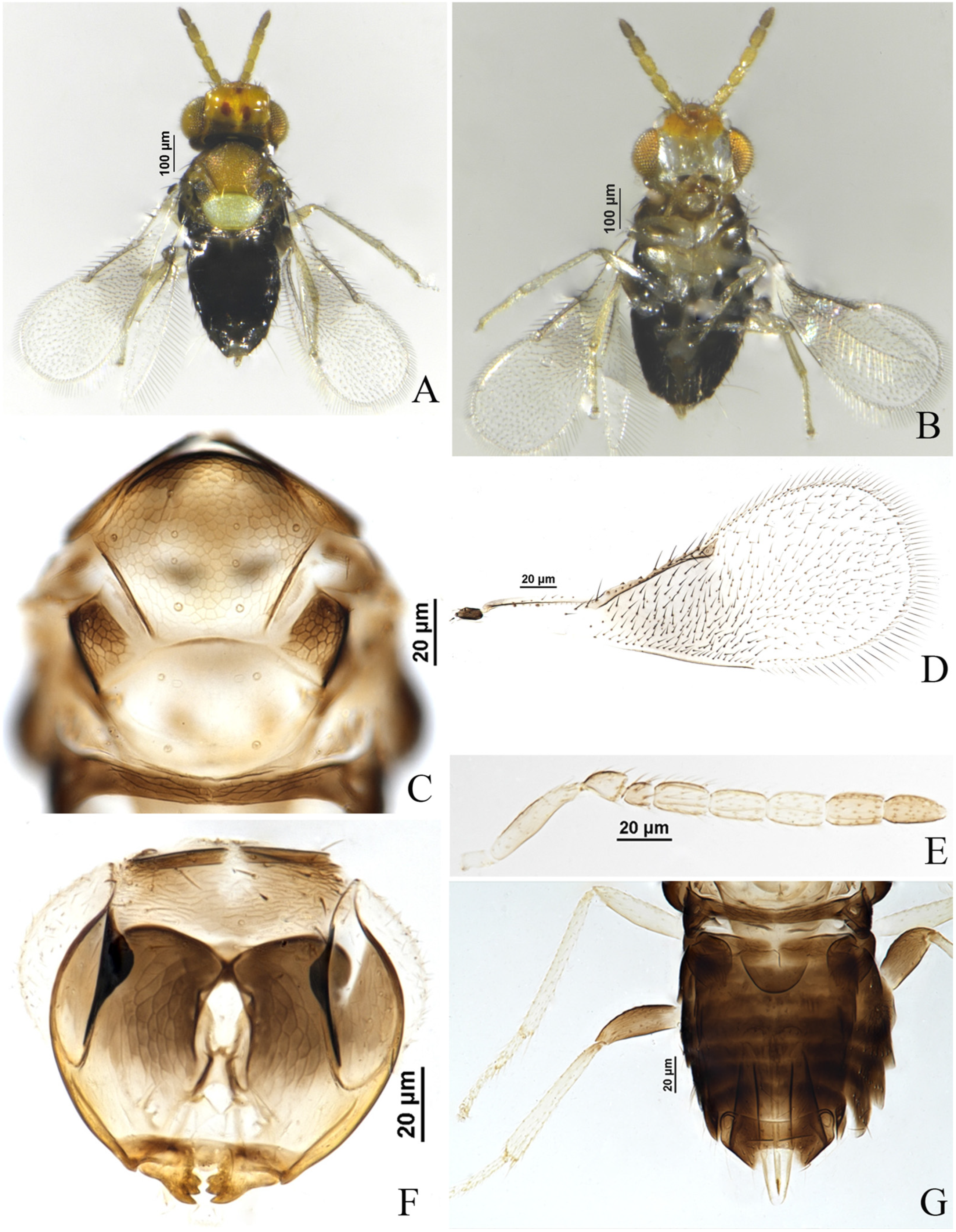
Figure 2.
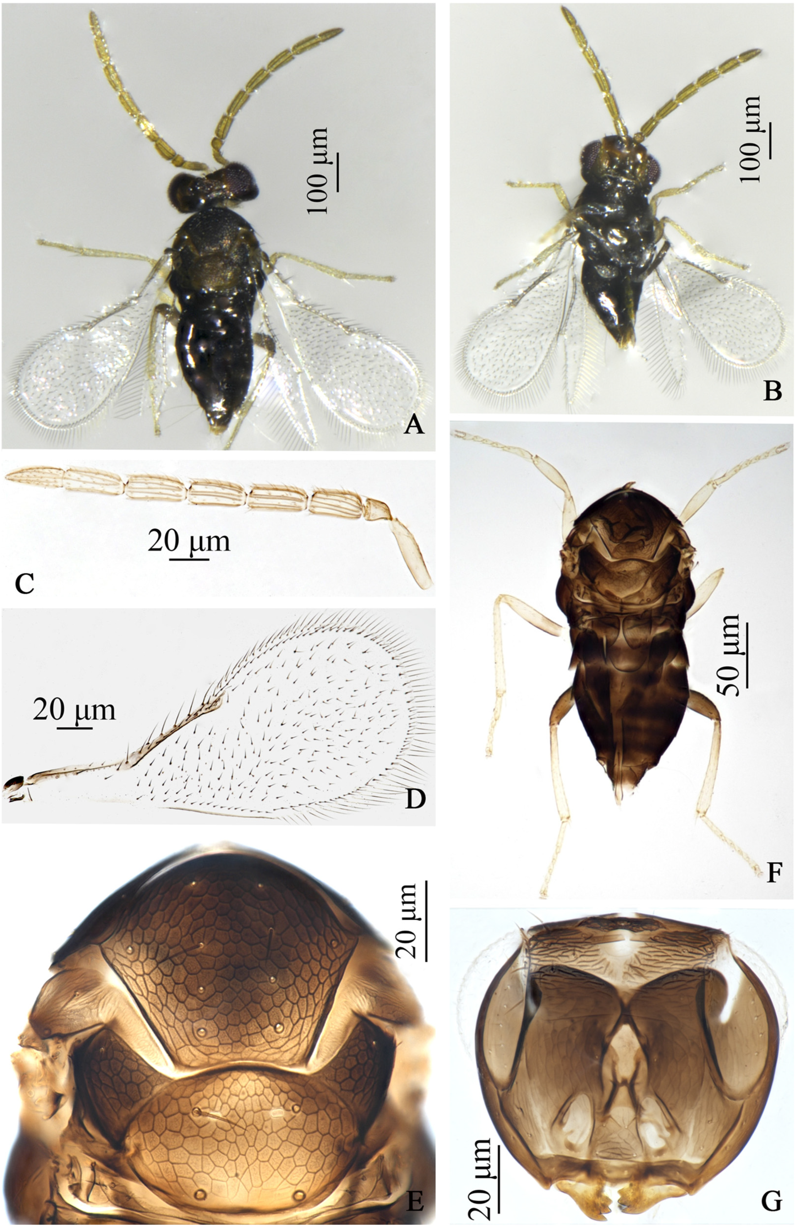
Encarsia cinnamomi, sp.n. male (FZ). (A) adult in dorsal view; (B) adult in ventral view; (C) antennae; (D) forewing; (E) mesosoma; (F) body; (G) head.
Description. Female. Body length 0.70 mm. Color. Head with vertex brown yellow, 2 dark brown spots behind ocellus, face pale, clypeus medially, occiput, mandible apically dark brown; antenna with pedicel slightly dark brown dorsally, F3, F4 pale brown yellow, other funiculars brown yellow; pronotum, axilla mostly, metanotum, propodeum, mesopleuron dark brown, mid-lobe of mesoscutum, parapsides brown yellow and slightly dark anteriorly, scutellum silvery white; abdomen dark brown, T7 pale distally; ovipositor with third valvula dark laterally; fore wing hyaline, area below marginal vein infuscate; legs pale, hind coxae and hind femora dark brown. Head. Eyes setose; mandible with 3 acute teeth; antennal formula 1,1,4,2, F1 shortest, F2–F4 approximately equal in length, C2 slightly longer than C1, except F1, other flagellar segments each with 3–4 longitudinal sensilla. Mesosoma. Mid-lobe of mesoscutum with 10 setae, each parapside with 2 setae, each axilla with 1 seta; scutellum with 2 pairs of setae, distance between the fore pair of setae approximately equal in length to that between the hind pair of setae, placoid sensilla on scutellum distinctly widely placed, separated by clearly more than their own maximum diameter and close to the fore pair of setae. Fore wing. 2.58× as long as broad, marginal fringe 0.26× width of fore wing; submarginal vein with 2 setae, marginal vein with 6–7 setae along anterior margin, basal cell with 2 fine setae. Leg. Tarsal formula 5–5–5. Metasoma. Ovipositor located basally at T3, 1.12× as long as midtibia, third valvula 0.47× length of second valvifer.
Male. Body length 0.68 mm. Similar to female except: body more dark, T7 pale distally; antenna 8-segmented, yellow brown, pedicel shortest, each flagellar segment nearly equal in length, each with 4–5longitudinal sensilla; fore wing hyaline.
Host. Aleurocanthus cinnamomi Takahashi on Cinnamomum camphora Nees ex Wall (Lauraceae).
Distribution. China (Fujian).
Etymology. The new species name is derived from the name of the host whitefly, cinnamomi.
Material. Holotype ♀, China: Fujian, Fuzhou, ex. Aleurocanthus cinnamomi on Cinnamomum camphora, 20.v.2022, coll. Zhigang Dong (FAFU); Paratypes 1♀1♂, same data as holotype.
3.1.3. Encarsia ophiopogonis Wang & Huang, sp.n. (Figure 3)
Diagnosis. Encarsia ophiopogonis sp.n. resembles E. diaspidicola (Silvestri), but can be distinguished from the latter by: (1) mid-lobe of mesoscutum with 2 setae (E. diaspidicola: with 4 setae); (2) antenna dark yellow brown, except the basal 1/2 of scape pale (E. diaspidicola: brown yellow); (3) pedicel 1.20 times as long as F2 (E. diaspidicola: pedicel as long as F2); (4) F2 1.25 times as long as F1 (E. diaspidicola: F2 1.43 times as long as F1).
Figure 3.
Encarsia ophiopogonis, sp.n. female. (A) adult in dorsal view; (B) adult in ventral view; (C) fore wing; (D) antenna; (E) mesosoma; (F) metasoma.
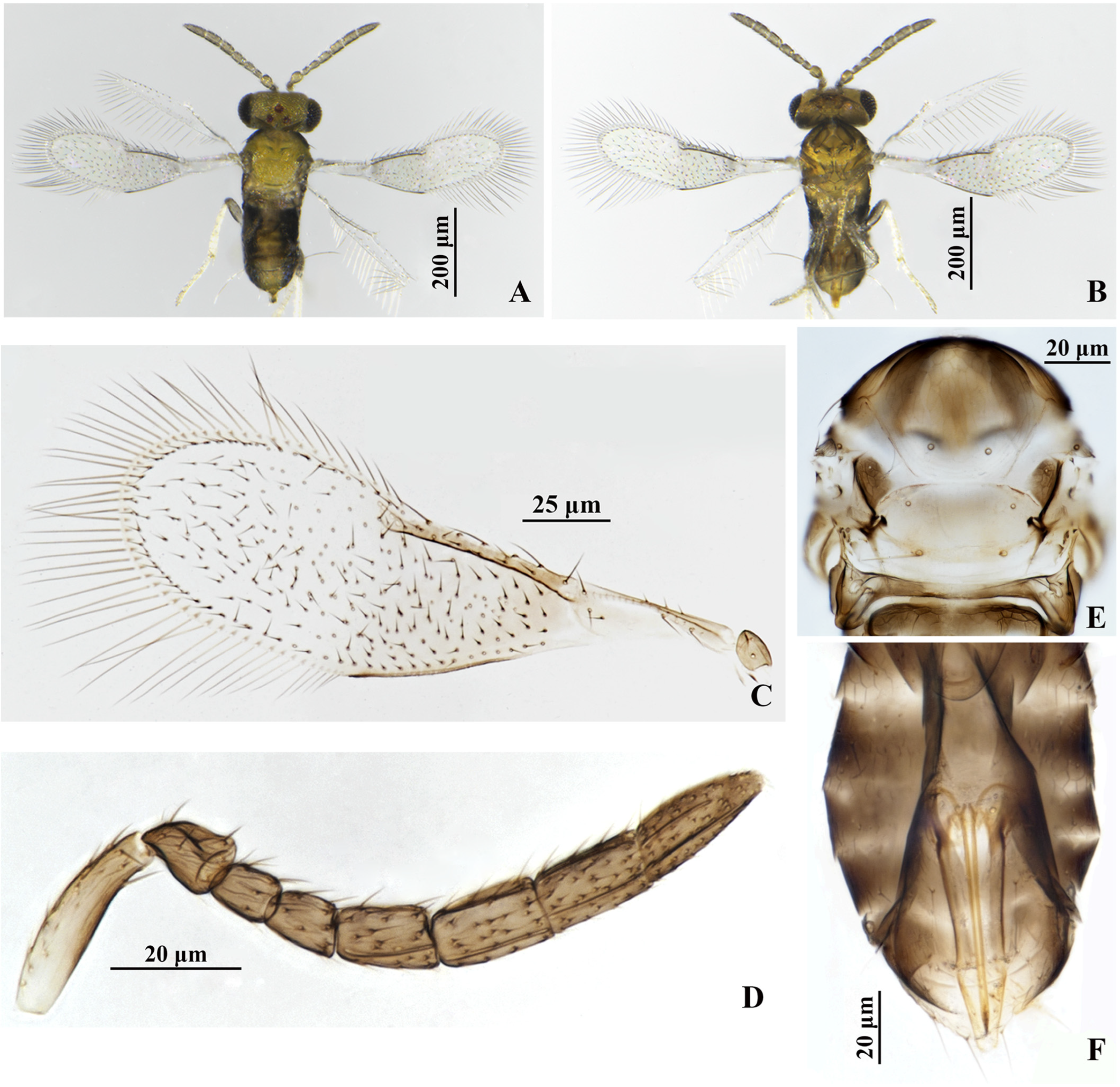
Description. Female. Body length 0.50 mm. Color. Head yellow, except clypeus, hind head brown to dark brown; antenna dark yellow brown, except the basal 1/2 of scape pale; mesosomal terga yellow, except pronotum, mid-lobe of mesoscutum anteriorly, axilla, mesopleuron, propodeum brown; petiole, metasomal tergites brown, T7 yellow distally; ovipositor with third valvula pale; fore wing hyaline, area below marginal vein faintly infuscate; legs pale, hind coxae and hind femora dark brown. Head. Eyes setose; mandible with 3 acute teeth; antennal formula 1,1,3,3, F1 shortest, F2 slightly shorter than F3, C1 nearly equal to C2 in length, shorter than C3; except F1, other flagellar segments each with 1–2 longitudinal sensilla. Mesosoma. Mid-lobe of mesoscutum with 2 setae, each parapside with 1 seta, each axilla with 1 seta; scutellum with 2 pairs of setae, distance between the fore pair of setae longer than that between the hind pair of setae, placoid sensilla on scutellum distinctly widely placed and close to the fore pair of setae. Fore wing. 3.38× as long as broad, marginal fringe 0.71× width of fore wing; submarginal vein with 2 setae, marginal vein with 5 setae along anterior margin. Leg. tarsal formula 5–5–5. Metasoma. Ovipositor located basally at T4, 0.84× as long as midtibia, third valvula 0.50× length of second valvifer.
Male. Unknown.
Host. Pinnaspis aspidistrae (Signoret) on Ophiopogon japonicus (L. f.) Ker Gawl.
Distribution. China (Fujian).
Etymology. The new species name is named after the genus of host plant, Ophiopogon.
Material. Holotype ♀, China: Fujian, Fuzhou, Fujian Agriculture and Forestry University, ex. Pinnaspis aspidistrae (Signoret) on Ophiopogon japonicus (L. f.) Ker Gawl., 30.v.2024, coll. Ye Luo and Zhigang Dong (FAFU). Paratypes 2♀, same data as holotype (FAFU).
Remarks. The new species resembles Encarsia diaspidicola (Silvestri) but distinguished from the latter mainly by setation on the mid-lobe of mesoscutum, as mentioned above. However, it now appears that some errors exist in the description of E. diaspidicola by Huang & Polaszek (1998) [4]. At that time, Huang & Polaszek (1998) [4] examined the types of E. diaspidicola in the IEUN, described the mid-lobe of mesoscutum with 1+1 or 2+2 setae, and pointed out that the setation on the mid-lobe of mesoscutum varies considerably, from 2 setae (1♀), 1+2 setae (5♀), 2+2 setae (5♀) to 1+2+2 setae. Obviously, most of the types are with 4 setae and variation of 4 setae on the mid-lobe of mesoscutum. Moreover, the 4 setae on the mid-lobe of mesoscutum was originally described in E. diaspidicola sp.n. by Silvestri (1909) [48]. So E. diaspidicola should have 4 setae on the mid-lobe of mesoscutum, and the Figure 76 in Huang & Polaszek (1998) [4] should be to illustrate the feature of 4 setae instead of 2 setae on the mid-lobe of mesoscutum.
3.2. Three Mitochondrial Genomes of Encarsia
3.2.1. Mitochondrial Genome Base Composition and Sequence Signature
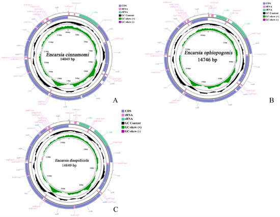
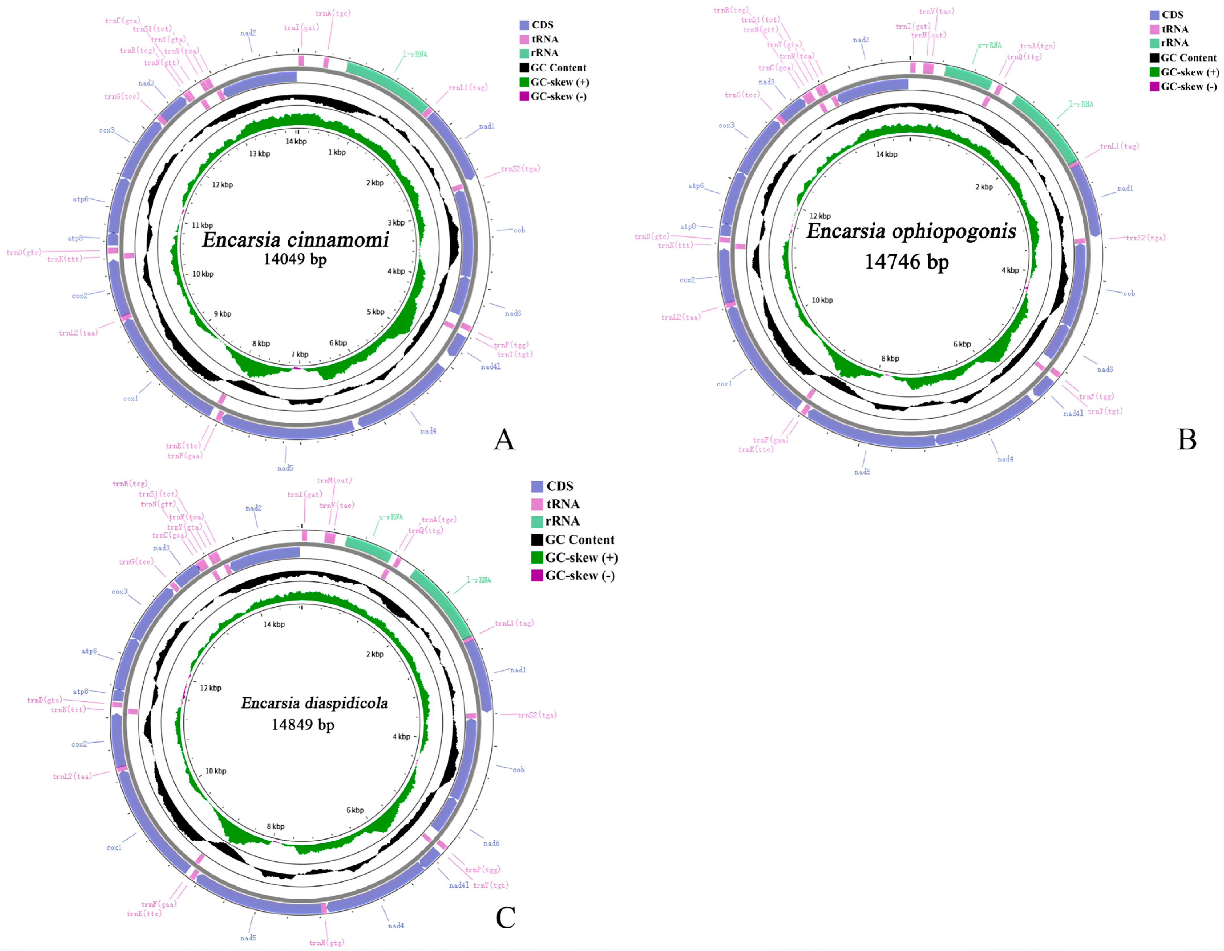
The complete mitochondrial genomes of Encarsia cinnamomi, sp.n., E. ophiopogonis, sp.n. and E. diaspidicola are 14,049 bp, 14,746 bp and 14,849 bp in length, respectively (Table A1, Table A2 and Table A3). The mitogenome of E. cinnamomi, sp.n. comprises 32 genes (13 PCGs, 18 tRNAs, and 1 rRNAs), with 23 genes encoded on the heavy strand (J-strand) and 9 on the light strand (N-strand). The mitogenome of E. ophiopogonis, sp.n. and E. diaspidicola contains the full complement of 37 genes (13 PCGs, 22 tRNAs, and 2 rRNAs), with 27 genes on the J-strand and 10 on the N-strand. Circular maps illustrating the gene arrangements of both mitogenomes are presented in Figure 4A–C. Both genomes exhibit a strong A + T bias, with overall A + T contents of 84.1%, 84.7% and 84.8%, respectively (Table 2). Analysis of the protein-coding genes (PCGs) reveals that the third codon position has the highest A + T content (94.2%, 95.4%, 95.3%), while the second codon position has the lowest (75.2%, 75.4%, 75.6%) (Table 2).
Figure 4.
Maps of the sequenced mitogenome. (A): Encarsia cinnamomi, sp.n.; (B): E. ophiopogonis, sp.n.; (C): E. diaspidicola.
Table 2.
Nucleotide composition of the whole mitogenomes of Encarsia cinnamomi, sp.n., E. ophiopogonis, sp.n. and E. diaspidicola.
3.2.2. Protein-Coding Gene
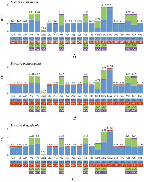
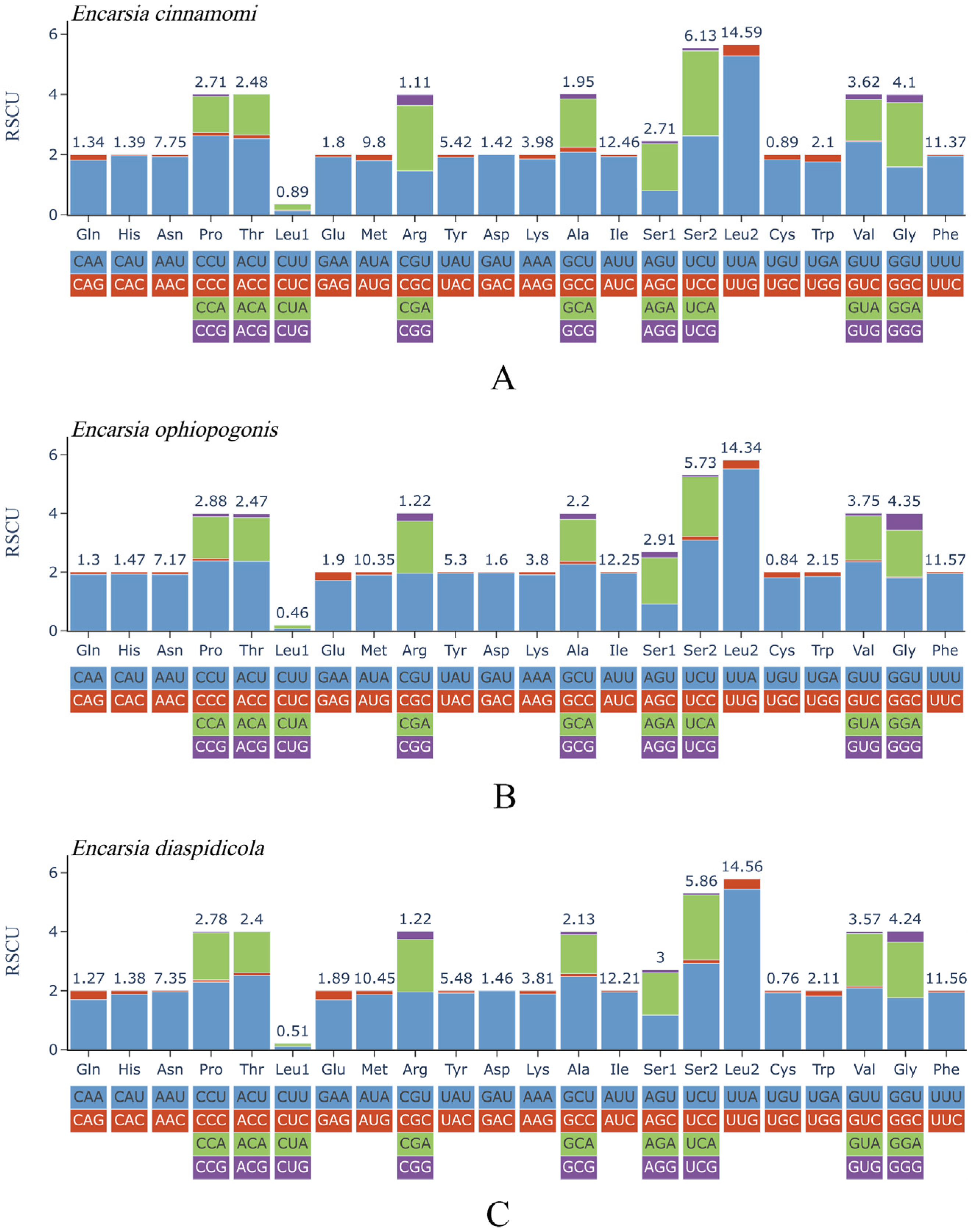
The combined lengths of the 13 protein-coding genes (PCGs) are 10,929 bp in E. cinnamomi, 11,136 bp in E. ophiopogonis and 11,137 bp in E. diaspidicola, with A + T contents of 82.7%, 83.3% and 83.5%, respectively (Table 2). All PCGs in both species use ATN as the start codon. In E. cinnamomi and E. ophiopogonis, all 13 PCGs are terminated with a TAA stop codon. Most PCGs in E. diaspidicola also use TAA, except for nad3 and nad4, which ended with an incomplete termination codon TA and T respectively (Table A1, Table A2 and Table A3). Analysis of relative synonymous codon usage (RSCU) reveals that UUA (Leu2) is the most frequent codon in both species, with RSCU values of 5.28, 5.52 and 5.44, respectively (Figure 5A–C).
Figure 5.
Relative synonymous codon usage of the protein-coding genes. (A): Encarsia cinnamomi, sp.n.; (B): E. ophiopogonis, sp.n.; (C): E. diaspidicola.
The evolutionary rates of the 13 protein-coding genes (PCGs) in three Encarsia species were assessed by calculating the non-synonymous to synonymous substitution rate ratio(Ka/Ks), using Trichopria drosophilae as a reference. In E. cinnamomi sp.n., the Ka/Ks ratios of seven genes were greater than 1; in E. ophiopogonis sp.n., those of nine genes exceeded 1; and in E. diaspidicola, eight genes had Ka/Ks ratios above 1, indicating that these genes were subject to positive selection. Among them, nad2 exhibited the highest evolutionary rate in E. cinnamomi sp.n. and E. diaspidicola (Ka/Ks = 2.1642 and 2.8173, respectively), while atp8 showed the highest evolutionary rate in E. ophiopogonis sp.n. (Ka/Ks = 3.1021). Notably, cox1 exhibited the lowest Ka/Ks values in the three species (0.5146, 0.4185 and 0.4070, respectively), indicating it is under strong purifying selection (Table 3).
Table 3.
Ka/Ks values of the mitochondrial genome protein-coding genes of Encarsia cinnamomi, sp.n., E. ophiopogonis, sp.n. and E. diaspidicola.
3.2.3. Gene Characteristics of tRNAs and rRNAs
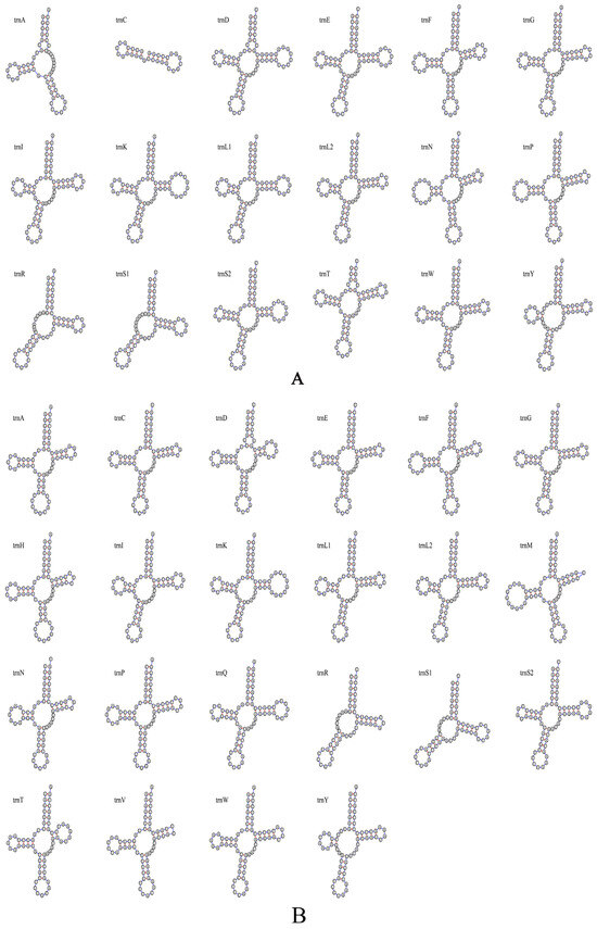
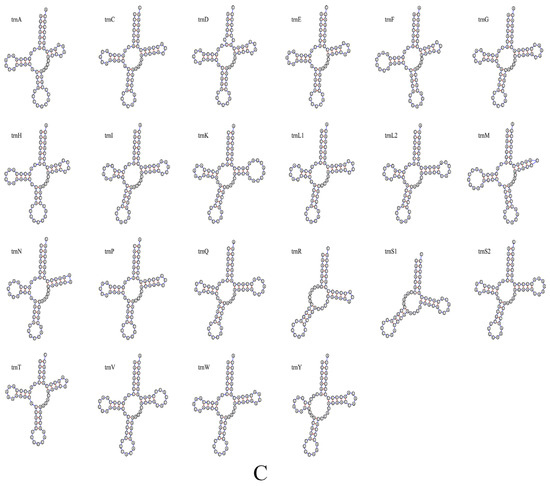
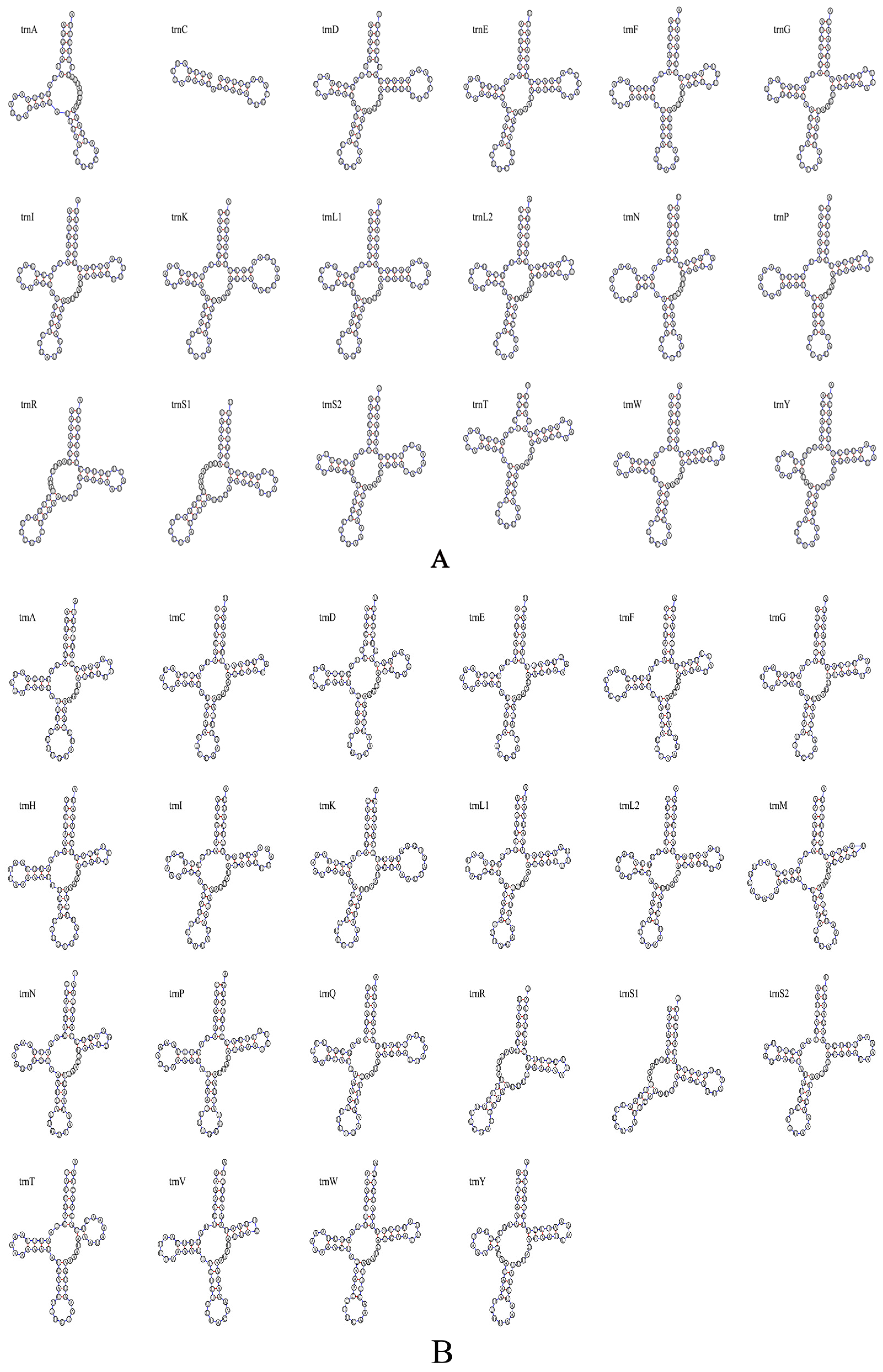
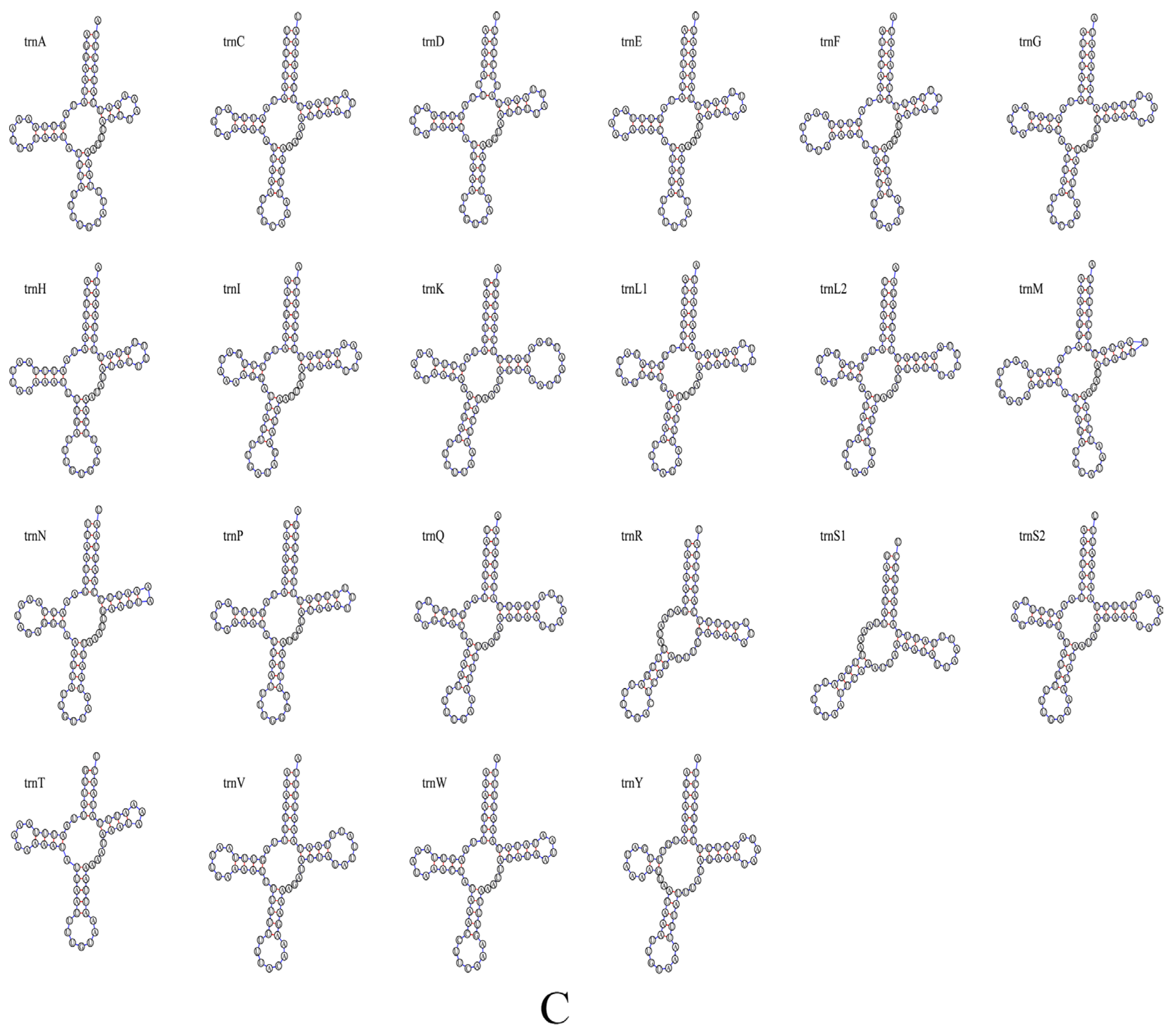
The mitochondrial genome of Encarsia cinnamomi, sp.n. contains 18 tRNA genes, ranging from 28 to 71 bp in length. Most tRNAs exhibit the typical cloverleaf structure except for trnA, trnR and trnS1, which lack the DHU arm, and trnC, which is severely truncated, retaining only the anticodon loop and DHU arm. A total of 9 G-U mismatches are identified in its tRNA structures (Figure 6A). In comparison, the mitogenomes of E. ophiopogonis, sp.n. and E. diaspidicola contains 22 tRNA genes, ranging from 55 to 70 bp in length. All tRNAs form the typical cloverleaf structure, with the exception of trnA and trnS1, both of which lack the DHU arm. Fourteen and ten G-U mismatches are identified in E. ophiopogonis, sp.n. and E. diaspidicola respectively (Figure 6B,C).
Figure 6.
Secondary structure of tRNA gene. (A): Encarsia cinnamomi, sp.n.; (B): E. ophiopogonis, sp.n.; (C): E. diaspidicola.
Regarding rRNA genes, E. cinnamomi, sp.n. possesses a single rRNA gene with a length of 1086 bp, representing 7.7% of the complete mitochondrial genome and an A + T content of 86.2% (Table 2). E. ophiopogonis, sp.n. and E. diaspidicola contain two rRNA genes totaling 1888 bp and 2029 bp, which account for 12.8% and 13.7% of the mitogenome respectively. The small ribosomal RNA gene (rrnS) is located between trnV and trnQ, measuring 606 bp and 750 bp with A + T contents of 87.3% and 88.5%, while the large ribosomal RNA gene (rrnL) is situated between trnA and trnL1, spanning 1282 bp and 1279 bp with A + T contents of 88.2% and 87.9% respectively (Table 2).
3.2.4. Phylogenetic Analysis
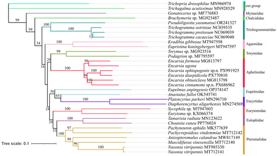
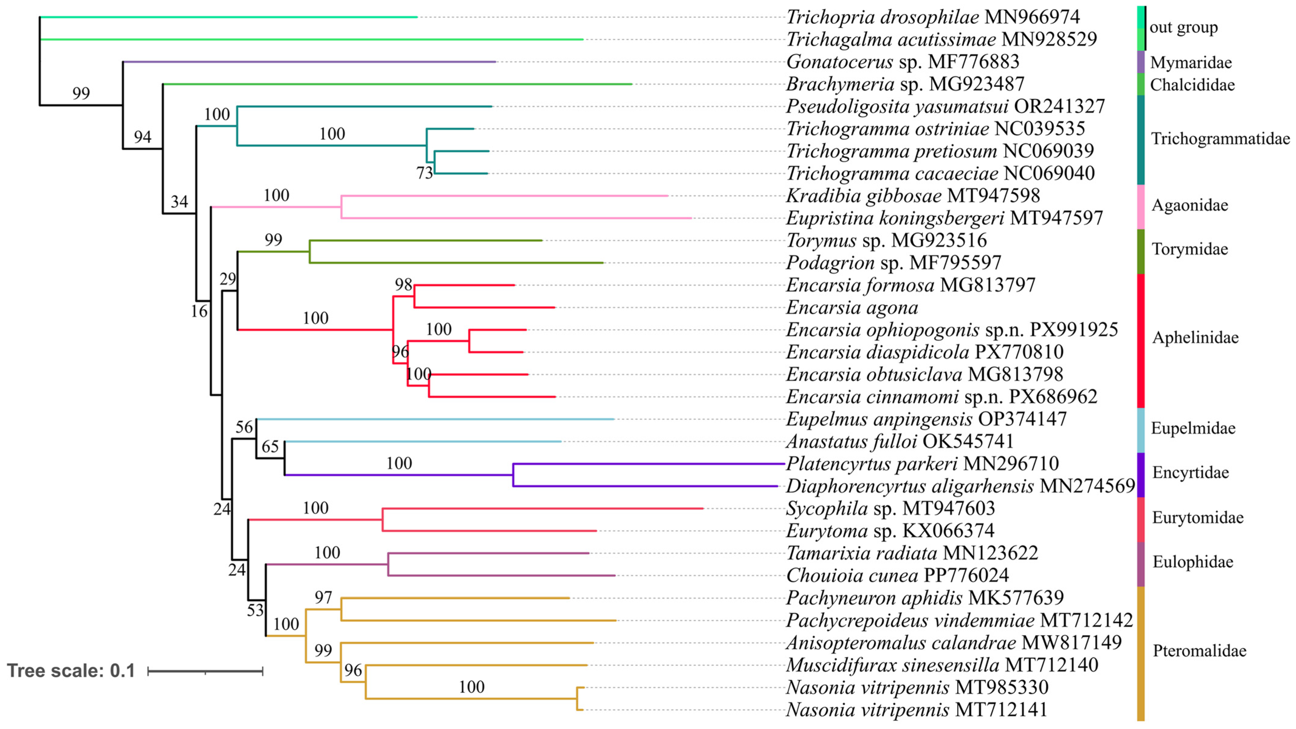
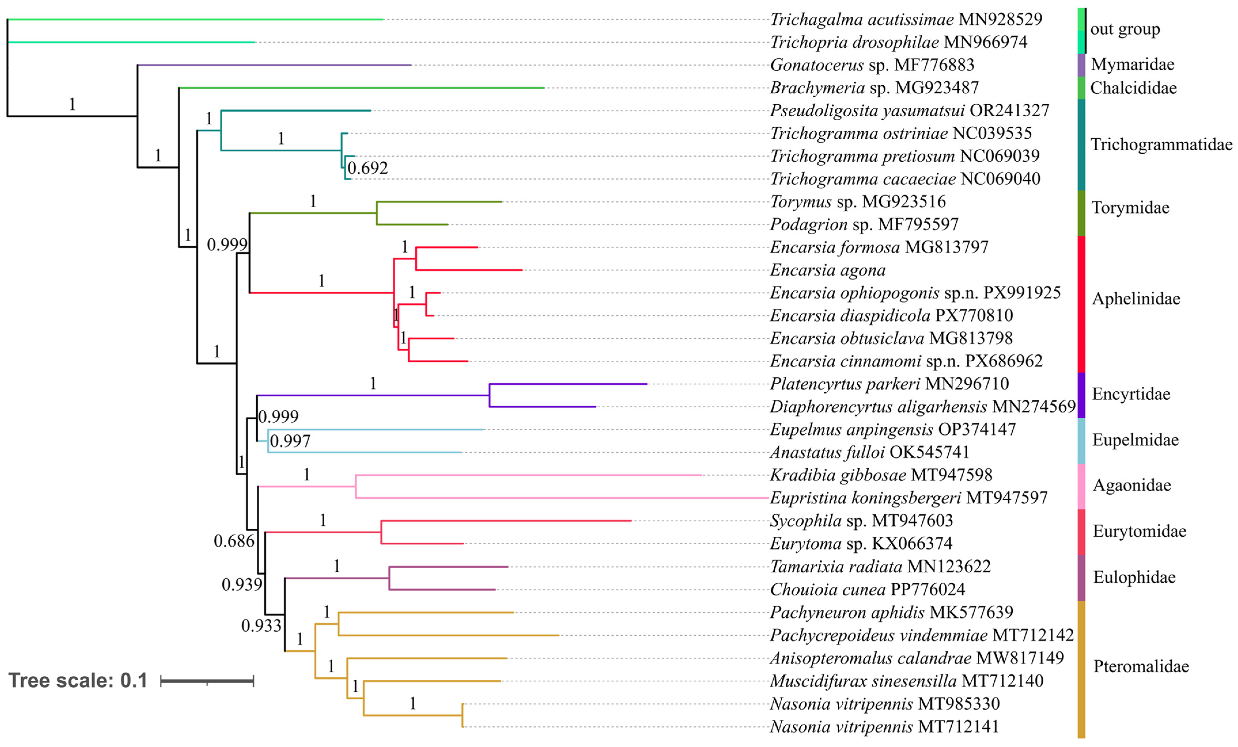
The phylogenetic relationships within Aphelinidae and other families of Chalcidoidea were reconstructed based on three newly sequenced mitochondrial genomes of Encarsia, along with 27 previously recorded mitogenomes in GenBank. Phylogenetic analyses were conducted using both concatenated PCG123 datasets under Bayesian inference (BI) and maximum likelihood (ML) frameworks (Figure 7 and Figure 8). In both ML and BI phylogenetic analyses, Encarsia was supported as a monophyletic group, in which E. ophiopogonis, sp.n. and E. diaspidicola clustered in the same clade with a bootstrap value of 100%. This indicates a close phylogenetic affinity between the two species, which are also extremely similar in morphology. The new species E. cinnamomi sp.n. and E. obtusiclava formed a sister relationship. Meanwhile, the monophyly of each family within Chalcidoidea was strongly supported.
Figure 7.
Phylogenetic tree of Aphelinidae and other families of Chalcidoidea inferred by ML analysis based on 13 PCGs from 30 species. Numbers at the nodes are bootstrap values.
Figure 8.
Phylogenetic tree of Aphelinidae and other families of Chalcidoidea inferred by BI analysis based on 13 PCGs from 30 species. Numbers at the nodes are posterior probabilities.
However, our results regarding the backbone relationships among Chalcidoidea families require cautious interpretation. The ML and BI analyses yielded incongruent topologies for interfamilial relationships (Figure 7 and Figure 8), and crucially, these deeper nodes received consistently low statistical support (e.g., <95% bootstrap) in the ML analysis. This lack of robust resolution, combined with conflicts with more comprehensive phylogenomic studies of Chalcidoidea, precludes drawing definitive conclusions about interfamilial relationships from the current dataset. Despite these limitations, our findings demonstrate the potential of mitogenomic data for resolving relationships within Chalcidoidea. They also underscore that substantially increased taxon sampling, particularly at the family level, is a critical prerequisite for confidently resolving the superfamily’s backbone phylogeny.
4. Discussion
Encarsia ophiopogonis Wang & Huang, sp.n., resembles E. diaspidicola (Silvestri) but is distinguished from the latter mainly by the setation on the mid-lobe of mesoscutum and antennal characteristics, as well as distinct interspecific genetic distances in the mitochondrial genome. The sequence identity of the mitochondrial genomes between these two species is 92.20%, and the genetic distance reaches 7.62%, which has reached the interspecific genetic level. It is generally considered that an insect genetic distance greater than 3% reaches the interspecific level [49]. E. diaspidicola was originally described by Silvestri (1909) [48] with 4 setae on the mid-lobe of mesoscutum. However, it now appears that some errors exist in the description of E. diaspidicola by Huang & Polaszek (1998) [4], in which Figure 76 should illustrate an individual with 4 setae instead of 2 setae on the mid-lobe of mesoscutum, although there is variation of the setae number on the mid-lobe of mesoscutum.
The mitochondrial genome sequence serves as a pivotal molecular marker that is frequently employed in phylogenetic studies to clarify the evolutionary relationships among closely related insect taxa [50,51]. The mitochondrial genomes of the three Encarsia species characterized in this study range from 14,049 to 14,849 bp in length. This length range is comparable to that of the mitochondrial genomes previously reported for Encarsia [27,47], Eupelmidae [52], and Pteromalidae [53], but is consistently shorter than the previously recorded lengths. This discrepancy might be attributable to unsequenced regions in the three mitochondrial genomes analyzed herein. For instance, only 32 genes were identified in E. cinnamomi, sp.n., with 4 tRNA genes and 1 rRNA gene undetected; additionally, the secondary structures of trnC and trnA were found to be incomplete. This may be caused by incomplete sequencing. For E. ophiopogonis, sp.n. and E. diaspidicola, although they contain all 37 genes of the mitochondrial genome, their relatively short length may be attributed to incomplete sequencing of the non-coding regions, whereas E. formosa [27] possesses a considerably long non-coding region. Usually, such species have a high AT content, which makes sequencing more difficult. In Chalcidoidea (Hymenoptera), the AT content typically falls within the range of 75% to 90% [54]. The AT content of these three species described in this study was determined to be 84.1% to 84.8%, which conforms to this typical feature. The mitochondrial genome of E. cinnamomi, sp.n. harbors 23 genes on the J-strand and 9 genes on the N-strand, while those of E. ophiopogonis, sp.n. and E. diaspidicola contain 27 genes on the J-strand and 10 genes on the N-strand. This gene distribution pattern aligns with the general observation across numerous species, in which the majority of coding genes are situated on the J-strand, with only a small proportion on the N-strand [20]. These three species reported in this study all exhibit negative AT-skew and positive GC-skew. This is opposite to the typical pattern of nucleotide composition bias in insect mitochondrial genomes, which is positive AT-skew and negative GC-skew [55], but is consistent with the patterns observed in some previous reports [52,56]. Typically, trnS1 in animals lacks the DHU arm and thus does not possess the canonical cloverleaf structure [20]. The secondary structures of most tRNAs in the three new species are canonical cloverleaf structures, but some gene structural deletions were also observed. In addition to the absence of the DHU arm in trnS1 across all three species, the DHU arm is also missing in trnR; this is consistent with the tRNA secondary structure of the recently reported mitochondrial genome of Encarsia agona [47]; furthermore, the trnC gene of E. cinnamomi, sp.n. has a severe structural deletion.
In the phylogenetic analysis, E. cinnamomi, sp.n., E. ophiopogonis, sp.n. and E. diaspidicola are well separated from the other three known species. Meanwhile, in another study, the single gene cox1 could not separate the species well or reflect the phylogenetic relationships within Aphelinidae [57], which indicates that the mitochondrial genome has great application potential in species identification of Aphelinidae. In both ML and BI phylogenetic analysis, Aphelinidae forms a sister group with Torymidae, which is different from the findings of He et al. (2025) [58], Xing et al. (2021) [59] and Zhao et al. (2021) [60] where it forms a sister group to Trichogrammatidae, and also differs from the findings of Zhu et al. (2018) [27], where it forms a sister group to (Trichogrammatidae + Agaonidae). Morphologically, Aphelinidae is generally close to Encyrtidae and Eulophidae. However, the phylogenetic analysis indicates that it is more closely related to Torymidae. In a recent study, the authors investigated the phylogenetic relationships of Chalcidoidea based on the exons (AHE414) and UCE datasets, and their results recovered Aphelinidae as the sister group to (Encyrtidae + Eunotidae) [61]. Currently, only six mitochondrial genomes of Encarsia are available worldwide (including the three species presented in this study), and mitochondrial genomic data for other genera of Aphelinidae remain entirely absent. To further clarify the phylogenetic relationships within Aphelinidae and among all families of Chalcidoidea, future studies should prioritize expanding taxon sampling for mitochondrial genomic sequencing across these groups.
Author Contributions
Z.W. and J.H. conceived the study research. Y.L., Z.D., X.M., J.G. and Z.W. collected samples, conducted some lab work and analyzed the data. Z.W., Z.D., Y.L. and J.H. wrote the manuscript. S.V.T. modified the manuscript. All authors have read and agreed to the published version of the manuscript.
Funding
This work was funded by National Key R&D Program of China [2023YFE0123000], National Natural Science Foundation of China [32172495].
Data Availability Statement
Data are contained within the article.
Acknowledgments
We are grateful to Xinrui Zhang (FAFU) for collecting some specimens.
Conflicts of Interest
The authors declare no conflicts of interest.
Appendix A
Table A1.
Characteristics of the mitochondrial genome of Encarsia cinnamomi, sp.n.
Table A2.
Characteristics of the mitochondrial genome of Encarsia ophiopogonis, sp.n.
Table A3.
Characteristics of the mitochondrial genome of Encarsia diaspidicola.
References
- Förster, A. Kleine monographien parasitischer Hymenopteren. Verhandlungen Naturhistorischen Ver. Preuss. Rheinl. Westfal. Bonn 1878, 35, 42–82. [Google Scholar]
- Noyes, J.S. Universal Chalcidoidea Database. World Wide Web Electronic Publication. 2019. Available online: https://ucd.chalcid.org (accessed on 20 October 2025).
- Huang, J. Systematic Studies on Aphelinidae of China (Hymenoptera: Chalcidoidea); Chongqing Publishing House: Chongqing, China, 1994; pp. 1–347. [Google Scholar]
- Huang, J.; Polaszek, A. A revision of the Chinese species of Encarsia Förster (Hymenoptera: Aphelinidae): Parasitoids of whiteflies, scale insects and aphids (Hemiptera: Aleyrodidae, Diaspididae, Aphidoidea). J. Nat. Hist. 1998, 32, 1825–1966. [Google Scholar] [CrossRef] [Scilit]
- Cai, Y.C.; Huang, J.; Wang, X.W.; Yang, X.J. Identification and utilization of Encarsia berlesei (Howard) (Hymenoptera: Aphelinidae), an important parasite of Pseudaulacaspis pentagona (Targ.) (Homoptera: Diaspididae). Entomol. J. East. China 1993, 1, 49–53. [Google Scholar]
- Lenteren, J.C. Biological and control in greenhouse. Annu. Rev. Entomol. 1998, 33, 39–69. [Google Scholar]
- Zhao, J.Z.; Zhu, G.R. Research and application of biological control for greenhouse pests abroad. Word Agric. 1990, 9, 43–44. [Google Scholar]
- Singh, V.; Sood, K.A.; Hayat, M. First record of Encarsia formosa Gahan, 1924 (Hymenoptera: Aphelinidae), a parasitoid of the greenhouse whitefly, Trialeurodes vaporariorum (Westwood, 1856) from India. Orient. Insects 2018, 52, 313–317. [Google Scholar] [CrossRef] [Scilit]
- Cheng, H.K.; Tian, Y.Q.; Wei, B.C.; Zhao, J.H.; Ye, C.Q.; Wei, S.X.; Wang, J.Z.; Wang, R.J. A Technique for Commercial-scale Production of Encarsia formosa. Chin. J. Biol. Control 1989, 5, 178–181. [Google Scholar]
- Hoddle, M.S.; Van Driesche, R.G.; Sanderson, J.P. Biology and use of the whitefly parasitoid Encarsia formosa. Annu. Rev. Entomol. 1998, 43, 645–669. [Google Scholar] [CrossRef] [Scilit]
- Tian, Y.Q.; Wei, B.C.; Yan, Y.H. Preliminary Observation on the control of Trialeurodes vaporariorum by introducing Encarsia formosa. Bull. Agric. Sci. Technol. 1979, 9, 34–35. [Google Scholar]
- Wei, S.X.; Wang, J.Z.; Cheng, H.K.; Wang, R.J. Research on the commercial production technology of Encarsia formosa. North. Hortic. 1989, 9, 21–24. [Google Scholar]
- Zhu, G.R.; Qiao, D.L.; Xu, B.Y. Application technology of Encarsia Formosa for Trialeurodes vaporariorum control. Chin. Agric. Sci. Bull. 1993, 9, 52–53. [Google Scholar]
- Monti, M.; Nappo, A.; Giorgini, M. Molecular characterization of closely related species in the parasitic genus Encarsia (Hymenoptera: Aphelinidae) based on the mitochondrial cytochrome oxidase subunit I gene. Bull. Entomol. Res. 2005, 95, 401–408. [Google Scholar] [CrossRef] [Scilit] [PubMed]
- Zhang, X.M.; Xu, H.X.; Wang, S.; Ali, A.; Zhang, F. Molecular and iso-enzymatic identification of the nymphal parasitoid, Encarsia sophia (Hymenoptera: Aphelinidae) of the whitefly, Bemisia tabaci Genn. (Homoptera: Aleyrodidae). J. Environ. Entomol. 2013, 35, 196–203. [Google Scholar]
- Selvaraj, K.; Rameshkumar, A.; Sumalatha, B.V.; Swathi, H.D.; Sardar, S.; Kazmi, S.I. First report of Encarsia cubensis Gahan (Hymenoptera: Aphelinidae) an exotic parasitoid on the neotropical whitefly Aleurotrachelus atratus Hempel (Hemiptera: Aleyrodidae) in India. Phytoparasitica 2023, 51, 255–261. [Google Scholar] [CrossRef] [Scilit]
- Jahan, S.; Lee, K.; Howlader, M.; Bashar, H.; Hasan, G. Molecular identification of parasitoid, Encarsia formosa gahan in Bemisia tabaci (Gennadius) and determination of its secondary endosymbionts. Bangladesh J. Agric. Res. 2014, 39, 563–578. [Google Scholar] [CrossRef] [Scilit]
- Polaszek, A.; Hernández-Suárez, E.; Kresslein, R.L.; Hanson, P.; Linton, Y.M.; MacKenzie-Dodds, J.; Schmidt, S. A revision of the Encarsia mexicana species-group (=Dirphys Howard) (Hymenoptera: Aphelinidae), gregarious endoparasitoids of whiteflies (Hemiptera: Aleyrodidae) in the Neotropical Region. Insects 2023, 14, 570. [Google Scholar] [CrossRef] [Scilit]
- Lahey, Z.; Simmons, A.M.; Andreason, S.A. Encarsia hera Lahey & Andreason (Hymenoptera, Aphelinidae): A charismatic new parasitoid of Aleurocybotus Quaintance & Baker (Hemiptera, Aleyrodidae) from Florida. J. Hymenopt. Res. 2022, 94, 89–104. [Google Scholar] [CrossRef] [Scilit]
- Boore, J.L. Animal mitochondrial genomes. Nucleic Acids Res. 1999, 27, 1767–1780. [Google Scholar] [CrossRef] [Scilit]
- Wolstenholme, D.R.; Clary, D.O. Sequence evolution of Drosophila mitochondria DNA. Genetics 1985, 109, 725–744. [Google Scholar] [CrossRef] [Scilit]
- Sharkey, M.J.; Chapman, E. Ten new genera of Agathidini (Hymenoptera, Braconidae, Agathidinae) from Southeast Asia. ZooKeys 2017, 660, 107–150. [Google Scholar] [CrossRef] [Scilit]
- Simon, C.; Frati, F.; Beckenbach, A.; Crespi, B.; Liu, H.; Flook, P. Evolution, weighting, and phylogenetic utility of mitochondrial gene sequences and a compilation of conserved polymerase chain reaction primers. Ann. Entomol. Soc. Am. 1994, 87, 651–701. [Google Scholar] [CrossRef] [Scilit]
- Clary, D.O.; Wolstenholme, D.R. The mitochondrial DNA molecule of Drosophila yakuba: Nucleotide sequence, gene organization, and genetic code. J. Mol. Evol. 1985, 22, 252–271. [Google Scholar] [CrossRef] [Scilit] [PubMed]
- Ballard, J.W.O.; Rand, D.M. The population biology of mitochondrial DNA and its phylogenetic implications. Annu. Rev. Ecol. Evol. Syst. 2005, 36, 621–642. [Google Scholar] [CrossRef] [Scilit]
- Wei, S.J.; Shi, M.; Sharkey, M.J.; van Achterberg, C.; Chen, X.X. Comparative mitogenomics of Braconidae (Insecta: Hymenoptera) and the phylogenetic utility of mitochondrial genomes with special reference to holometabolous insects. BMC Genom. 2010, 11, 371. [Google Scholar] [CrossRef] [Scilit] [PubMed]
- Zhu, J.C.; Tang, P.; Zheng, B.Y.; Wu, Q.; Wei, S.J.; Chen, X.X. The first two mitochondrial genomes of the family Aphelinidae with novel gene orders and phylogenetic implications. Int. J. Biol. Macromol. 2018, 118, 386–396. [Google Scholar] [CrossRef] [Scilit]
- Hurst, L.D. The Ka/Ks ratio: Diagnosing the form of sequence evolution. Trends Genet. 2002, 18, 486–487. [Google Scholar] [CrossRef] [Scilit]
- Navarro, A.; Barton, N.H. Chromosomal speciation and molecular divergence-accelerated evolution in rearranged chromosomes. Science 2003, 300, 321–324. [Google Scholar] [CrossRef] [Scilit]
- Noyes, J.S. Collecting and preserving chalcid wasps (Hymenoptera: Chalcidoidea). J. Nat. Hist. 1982, 16, 315–334. [Google Scholar] [CrossRef] [Scilit]
- Meng, G.L.; Li, Y.Y.; Yang, C.T.; Liu, S.L. MitoZ: A toolkit for mitochondrial genome assembly, annotation and visualization. Nucleic Acids Res. 2019, 47, e63. [Google Scholar] [CrossRef] [Scilit]
- Li, D.; Liu, C.M.; Luo, R.; Sadakane, K.; Lam, T.W. MEGAHIT: An ultra-fast single-node solution for large and complex metagenomics assembly via succinct de Bruijn graph. Bioinformatics 2015, 31, 1674–1676. [Google Scholar] [CrossRef] [Scilit]
- Bankevich, A.; Nurk, S.; Antipov, D.; Gurevich, A.A.; Dvorkin, M.; Kulikov, A.S.; Lesin, V.M.; Nikolenko, S.I.; Pham, S.; Prjibelski, A.D.; et al. SPAdes: A new genome assembly algorithm and its applications to single-cell sequencing. J. Comput. Biol. 2012, 19, 455–477. [Google Scholar] [CrossRef] [Scilit] [PubMed]
- Walker, B.J.; Abeel, T.; Shea, T.; Priest, M.; Abouelliel, A.; Sakthikumar, S.; Cuomo, C.A.; Zeng, Q.; Wortman, J.; Young, S.K.; et al. Pilon: An integrated tool for comprehensive microbial variant detection and genome assembly improvement. Public Libr. Sci. 2014, 9, e112963. [Google Scholar] [CrossRef] [Scilit] [PubMed]
- Donath, A.; Jühling, F.; Al-Arab, M.; Bernhart, S.H.; Reinhardt, F.; Stadler, P.F.; Middendorf, M.; Bernt, M. Improved annotation of protein-coding genes boundaries in metazoan mitochondrial genomes. Nucleic Acids Res. 2019, 47, 10543–10552. [Google Scholar] [CrossRef] [Scilit]
- Lowe, T.M.; Chan, P.P. tRNAscan-SE On-line: Integrating search and context for analysis of transfer RNA genes. Nucleic Acids Res. 2016, 44, 54–57. [Google Scholar] [CrossRef] [Scilit] [PubMed]
- Sudhir, K.; Glen, S.; Michael, S.; Maxwell, S.; Sudip, S.; Koichiro, T. Mega12: Molecular evolutionary genetic analysis version 12 for adaptive and green computing. Mol. Biol. Evol. 2024, 41, msae263. [Google Scholar] [CrossRef] [Scilit]
- Kearse, M.; Moir, R.; Wilson, A.; Stones-Havas, S.; Cheung, M.; Sturrock, S.; Buxton, S.; Cooper, A.; Markowitz, S.; Duran, C.; et al. Geneious Basic: An integrated and extendable desktop software platform for the organization and analysis of sequence data. Bioinformatics 2012, 28, 1647–1649. [Google Scholar] [CrossRef] [Scilit]
- Perna, N.T.; Kocher, T.D. Patterns of nucleotide composition at fourfold degenerate sites of animal mitochondrial genomes. J. Mol. Evol. 1995, 41, 353–358. [Google Scholar] [CrossRef] [Scilit]
- Librado, P.; Rozas, J. DnaSP v5: A software for comprehensive analysis of DNA polymorphism data. Bioinformatics 2009, 25, 1451–1452. [Google Scholar] [CrossRef] [Scilit]
- Zhang, D.; Gao, F.L.; Jakovli´c, I.; Zou, H.; Zhang, J.; Li, W.X.; Wang, G.T. PhyloSuite: An integrated and scalable desktop platform for streamlined molecular sequence data management and evolutionary phylogenetics studies. Mol. Ecol. Resour. 2020, 20, 348–355. [Google Scholar] [CrossRef] [Scilit]
- Katoh, K.; Standley, D.M. MAFFT multiple sequence alignment software version 7: Improvements in performance and usability. Mol. Biol. Evol. 2013, 30, 772–780. [Google Scholar] [CrossRef] [Scilit]
- Talavera, G.; Castresana, J. Improvement of phylogenies after removing divergent and ambiguously aligned blocks from protein sequence alignments. Syst. Biol. 2007, 56, 564–577. [Google Scholar] [CrossRef] [Scilit]
- Kalyaanamoorthy, S.; Minh, B.Q.; Wong, T.K.F.; von Haeseler, A.; Jermiin, L.S. ModelFinder: Fast model selection for accurate phylogenetic estimates. Nat. Methods 2017, 14, 587–589. [Google Scholar] [CrossRef] [Scilit]
- Nguyen, L.T.; Schmidt, H.A.; von Haeseler, A.; Minh, B.Q. IQ-TREE: A fast and effective stochastic algorithm for estimating maximum-likelihood phylogenies. Mol. Biol. Evol. 2015, 32, 268–274. [Google Scholar] [CrossRef] [Scilit] [PubMed]
- Ronquist, F.; Teslenko, M.; van der Mark, P.; Ayres, D.L.; Darling, A.; Höhna, S.; Larget, B.; Liu, L.; Suchard, M.A.; Huelsenbeck, J.P. MrBayes 3.2: Efficient Bayesian phylogenetic inference and model choice across a large model space. Syst. Biol. 2012, 61, 539–542. [Google Scholar] [CrossRef] [Scilit] [PubMed]
- Polaszek, A.; Otim, M.H.; Briscoe, A.; Court, L.; Court, L.; Macfadyen, S.; Schmidt, S.; Geng, H.; Tay, W.T. Morphological and molecular description, and draft mitogenome, of a new Encarsia species (Hymenoptera: Aphelinidae), a parasitoid of the Bemisia tabaci species complex (Hemiptera: Aleyrodidae) on cassava in Uganda. J. Nat. Hist. 2025, 59, 2441–2461. [Google Scholar] [CrossRef] [Scilit]
- Silvestri, F. Notizie e descrizioni preliminari di insetti parassti della Diaspis pentagona. Atti della Reale Accademia dei Lincei. Rendiconti della Classe di Scienze Fisiche, Matematiche e Naturali 1909, 18, 563–565. [Google Scholar]
- Zhang, H.; Bu, W. Exploring Large-Scale Patterns of Genetic Variation in the COI Gene among Insecta: Implications for DNA Barcoding and Threshold-Based Species Delimitation Studies. Insects 2022, 13, 425. [Google Scholar] [CrossRef] [Scilit]
- Sun, Z.; Zhang, J.; Wang, R.; Xu, Y.J.; Zhang, D.Q. Progress of insect mitochondrial genome. J. Insp. Quar. 2010, 20, 69–73. [Google Scholar]
- Cameron, S.L. Insect Mitochondrial Genomics: A Decade of Progress. Annu. Rev. Entomol. 2025, 70, 83–101. [Google Scholar] [CrossRef] [Scilit]
- Jiang, J.; Wu, T.; Deng, J.; Peng, L. A Compositional Heterogeneity Analysis of Mitochondrial Phylogenomics in Chalcidoidea Involving Two Newly Sequenced Mitogenomes of Eupelminae (Hymenoptera: Chalcidoidea). Genes 2022, 13, 2340. [Google Scholar] [CrossRef] [Scilit]
- Huang, Y.; Yang, Y.; Qi, L.; Hu, H.; Rasplus, J.-Y.; Wang, X. Novel Gene Rearrangement Pattern in Pachycrepoideus vindemmiae Mitochondrial Genome: New Gene Order in Pteromalidae (Hymenoptera: Chalcidoidea). Animals 2023, 13, 1985. [Google Scholar] [CrossRef] [Scilit] [PubMed]
- Liu, H.H.; Li, E.J.; Cao, L.M.; Bao, Q.C.; Wang, X.L.; Xin, X.B.; Yang, Z.Q. Sequencing and analysis of the complete mitochondrial genome of Eurytoma acutibialis (Hymenoptera: Eurytomidae), an important natural enemy insect of Bradybatus sp. (Coleoptera: Curculionidae) in China. Acta Entomol. Sin. 2023, 66, 1233–1245. [Google Scholar]
- Wei, S.J.; Chen, X.X. Progress in research on the comparative mitogenomics of insects. Chin. J. Appl. Entomol. 2011, 48, 1573–1585. [Google Scholar]
- Yi, J.; Wu, H.; Liu, J.; Li, J.; Lu, Y.; Zhang, Y.; Cheng, Y.; Guo, Y.; Li, D.; An, Y. Novel gene rearrangement in the mitochondrial genome of Anastatus fulloi (Hymenoptera Chalcidoidea) and phylogenetic implications for Chalcidoidea. Sci. Rep. 2022, 12, 1351. [Google Scholar] [CrossRef] [Scilit]
- Dong, Z.G.; Ge, J.Q.; Huang, J.; Wang, Z.H. Three New Species of Aphelinus (Hymenoptera: Aphelinidae) from China, with a Note on the japonicus Group. Insects 2025, 16, 1205. [Google Scholar] [CrossRef] [Scilit]
- He, W.; Li, T.T.; Wang, L.Y.; Wu, H.X.; Wang, J.; Zhou, Q. Mitogenomic characterization and comparative analysis of three egg parasitoid wasps parasitizing Nilaparvata lugens (Stål). Insects 2025, 16, 543. [Google Scholar] [CrossRef] [Scilit]
- Xing, Z.P.; Qi, L.Q.; Wang, X.; Chen, L.; Zhu, Y.H.; Huang, Y.X.; Hu, H.Y. Complete mitochondrial genome of a parasitoid, Trichogramma chilonis (Hymenoptera: Chalcidoidea: Trichogrammatidae) and phylogenetic analysis. Mitochondrial DNA B Resour. 2021, 6, 2466–2467. [Google Scholar] [CrossRef] [Scilit]
- Zhao, H.; Chen, Y.; Wang, Z.; Chen, H.; Qin, Y. Two complete mitogenomes of Chalcididae (Hymenoptera: Chalcidoidea): Genome description and phylogenetic implications. Insects 2021, 12, 1049. [Google Scholar] [CrossRef] [Scilit]
- Cruaud, A.; Rasplus, J.Y.; Zhang, J.; Burks, R.; Delvare, G.; Fusu, L.; Gumovsky, A.; Huber, J.T.; Janšta, P.; Mitroiu, M.D.; et al. The Chalcidoidea bush of life: Evolutionary history of a massive radiation of minute wasps. Cladistics 2024, 40, 34–63. [Google Scholar] [CrossRef] [Scilit]

Disclaimer/Publisher’s Note: The statements, opinions and data contained in all publications are solely those of the individual author(s) and contributor(s) and not of MDPI and/or the editor(s). MDPI and/or the editor(s) disclaim responsibility for any injury to people or property resulting from any ideas, methods, instructions or products referred to in the content. |
© 2026 by the authors. Licensee MDPI, Basel, Switzerland. This article is an open access article distributed under the terms and conditions of the Creative Commons Attribution (CC BY) license.