


Simple Summary
In this study, five opsin genes were identified from the genome data of Phortica okadai, and their expression patterns were characterized by quantitative real-time polymerase chain reaction. These results offer a molecular basis for further investigations into the photoreception and visual ecology of P. okadai.
Abstract
In China, Phortica okadai is the only established vector for Thelazia callipaeda and transmits infective larvae to various animals feeding on ocular secretions. Insect opsins are key components of the visual system and play essential roles in host location. However, the opsin repertoire and phylogenetic relationships in P. okadai remain unknown. Here, we identified five opsin genes from adult P. okadai genomic data and profiled their expression across developmental stages and tissues by real-time polymerase chain reaction. The results showed that four opsin genes (PoRh1, PoRh3, PoRh5, and PoRh6) were predominantly expressed in adult compound eyes and peaked during the adult stage, suggesting important roles in vision-dependent behaviors of adult P. okadai. These findings provide a foundation for elucidating the visual mechanisms and host-seeking behavior of P. okadai.
1. Introduction
Phortica okadai (Máca) (Diptera: Drosophilidae) feeds on ocular secretions and conjunctiva of animals [1] and is the only confirmed vector of Thelazia callipaeda (Spirurida: Thelaziidae) in China. It transmits infective larvae to a broad range of hosts, including wild and domestic animals and humans, causing thelaziasis. This infection is characterized by conjunctivitis, purulent discharge, epiphora, lacrimation, photophobia, chemosis, keratitis, and, in severe cases, corneal ulceration [2]. Understanding the host-seeking mechanism of P. okadai is essential for developing effective strategies to control thelaziasis.
Many insects possess elaborate visual systems that enable orientation and navigation, prey capture, habitat selection, and communication in diverse environments [3]. Adult insects perceive light primarily through the compound eyes and ocelli. The compound eyes consist of repeated functional units called ommatidia, each comprising a cornea and crystalline cone overlying a cluster of photoreceptor cells with a light-sensitive rhabdom. Within each ommatidium, variation in the number, morphology, and arrangement of photoreceptors shapes the capacity to detect color and polarized light [4]. Photoreceptors contain abundant rhodopsin, a visual pigment composed of an opsin covalently bound to a chromophore. Whereas most insect visual pigments share a common chromophore, 11-cis-retinal, their absorption maxima are primarily determined by the opsin [5,6,7]. Despite this central role in photoreception, the opsin repertoire of P. okadai remains unexplored.
Insect opsins are a class of transmembrane proteins with a size of typically 30–50 kDa and belong to the G protein-coupled receptor (GPCR) superfamily, consisting of seven transmembrane domains [8]. Opsins are divided into two major categories based on their direct involvement in visual imaging: visual opsins and non-visual opsins. Visual opsins are further categorized by spectral sensitivity: long-wavelength-sensitive opsin (LW opsin), blue light-sensitive opsin (BL opsin), and ultraviolet-sensitive opsin (UV opsin) [9].
Nina E (or Rh1) in Drosophila melanogaster is expressed only in R1-R6 photoreceptors and plays a major role in motion vision [10,11,12,13]. The Rh2 opsin gene, which encodes a violet-light-sensitive pigment, was subsequently identified [14,15,16]. The Rh3 and Rh4 ultraviolet-sensitive opsin genes are expressed in R7 photoreceptor cells [17,18,19], while the Rh5 blue-sensitive opsin gene and the Rh6 long-wavelength-sensitive opsin gene are expressed in R8 photoreceptor cells [20]. Nevertheless, the number of opsins varies among insect species, owing to gene loss or duplication events. These differences highlight the adaptation of insects to their respective living environments [21].
Here, we focus on the opsin genes of P. okadai to lay the foundation for further studies on its host localization and eye-wiring mechanism.
2. Materials and Methods
2.1. Insect Rearing
The P. okadai used in the present study was collected from a field in Zunyi, Guizhou Province, China, and was reared in the laboratory. The insects were maintained in plastic containers measuring 27 cm in length, 20 cm in width, and 10 cm in height, supplied with naturally fermented fruits for three days at a temperature of 28 ± 1 °C, 75 ± 5% relative humidity, and a 16L:8D photoperiod.
2.2. Identification of Opsins
To identify opsin genes in P. okadai, full-length amino acid sequences of opsins from representative Diptera, Hymenoptera, and Lepidoptera were retrieved from GenBank and used as query sequences for local BLASTp searches (E-value cutoff < 1 × 10⁻⁵) against the P. okadai genomic assembly. Furthermore, a Hidden Markov Model (HMM) search was performed against the predicted protein dataset of P. okadai using the rhodopsin-like G-protein-coupled receptor HMM 3.0 profile (Pfam: PF00001). Redundant and incomplete sequences were manually filtered out. The raw genomic data have been deposited in the Genome Sequence Archive (GSA) at the China National Center for Bioinformation/Beijing Institute of Genomics, Chinese Academy of Sciences, under accession number CRA008976 (BioProject: PRJCA013354) [22]. The nucleotide sequences of the five P. okadai opsin genes (PoRh1, PoRh3, PoRh5, PoRh6, and PoRh7) characterized in this study are available in NCBI GenBank under accession numbers QB150445-QB150449.
2.3. Sequencing Analysis and Phylogenetic Tree Construction
The amino acid sequences of the opsin genes of P. okadai were compared using the DNAMAN v6.0 software (Lynnon Biosoft, Quebec, QC, Canada). The theoretical isoelectric points (pI), molecular weight (kDa), and instability indices were predicted with ExPASy ProtParam (https://web.expasy.org/protparam/, accessed on 17 October 2025). The functional sites of opsin were predicted by Prosite Scan (https://npsa-prabi.ibcp.fr/cgi-bin/npsa_automat.pl?page=/NPSA/npsa_proscan.html, accessed on 17 October 2025).
For phylogenetic inference, opsin protein sequences from P. okadai and other representative insects were subjected to neighbor-joining (NJ) analyses. The resulting tree was visualized and annotated using iTOL (https://itol.embl.de/, accessed on 14 October 2026).
2.4. Quantification of Gene Expression Levels by qRT-PCR
Eggs (approximately 2 days old, 60 individuals per biological replicate), larvae (mixed instars, 20 individuals per biological replicate), pupae (approximately 5 days old, 10 individuals per biological replicate), and 3- and 8-day-old adults (10 individuals per biological replicate) of P. okadai were collected for developmental expression analysis. For tissue-specific expression detection, a total of 360 uniformly sized 8-day-old adults were used, with 120 adults per biological replicate (60 females and 60 males) dissected to collect five tissue types, including antennae, abdomen, compound eyes, thorax, and genitalia. All samples at each developmental stage and each tissue type were prepared with three independent biological replicates and three technical quantitative real-time polymerase chain reaction (qRT-PCR) replicates to reduce instrumental and operational variation.
Gene-specific primers were designed based on the open reading frames (ORFs) of the opsin genes using Primer Premier 6.0 software (Table 1). Total RNA was extracted using a total RNA extraction kit (Solarbio, Beijing, China), and cDNA was synthesized with a HiFiScript cDNA Synthesis kit (CoWin Biosciences, Beijing, China). qRT-PCR was performed in a 20 µL reaction volume using SYBR Premix Ex Taq II (Takara Biotechnology, Dalian, China) under the following conditions: 95 °C for 30 s, followed by 40 cycles of 95 °C for 5 s and 55 °C for 30 s. β-actin was used as the internal reference gene to normalize target gene expression [22].
Table 1.
Primers used in this study.
2.5. Statistical Analysis
Relative expression levels were calculated using the 2−ΔΔCT method and expressed as the means ± standard error (SE) [23]. Since these values followed a log-normal distribution and exhibited heterogeneous variance, the fold-change data were log2-transformed prior to statistical analysis to approximate a normal distribution and meet the assumptions for parametric testing. Data analysis and graphing were performed using GraphPad Prism 11. One-way analysis of variance (ANOVA) was used to compare the relative expression of opsin genes in Phortica okadai across different developmental stages. Two-way ANOVA was applied for comparisons across different tissues, followed by Tukey’s honestly significant difference (HSD) post hoc test for pairwise multiple comparisons. The significance threshold was set at α = 0.05. Adjusted p values and 95% confidence intervals of mean differences were reported. Compact letter display was used to indicate significant differences in figures.
3. Results
3.1. Opsin Identification and Sequence Analysis
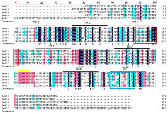
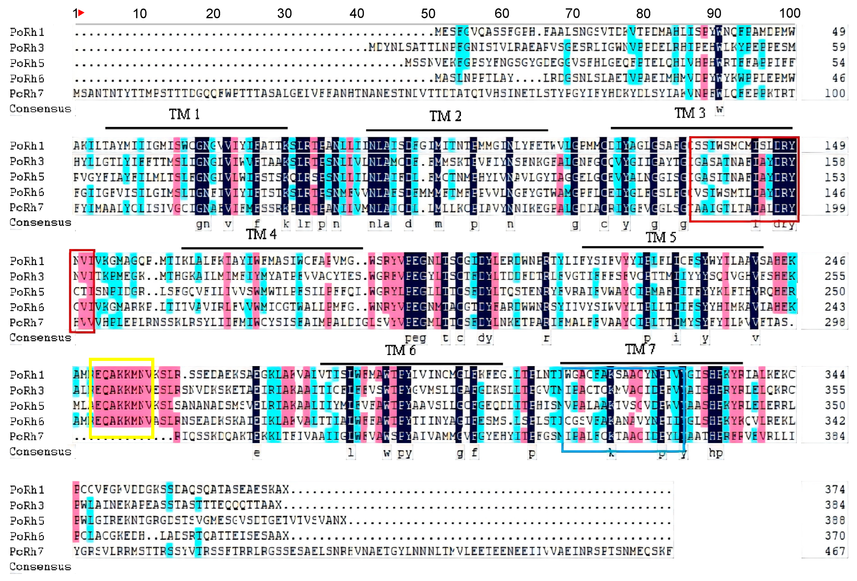
Based on the genomic data, we identified five opsin genes in P. okadai, named PoRh1, PoRh3, PoRh5, PoRh6, and PoRh7. The characteristics of predicted proteins for these five opsin genes are summarized in Table 2. These five opsins encoded 374, 384, 388, 370, and 468 amino acid residues, respectively. Multiple sequence alignments of these five opsins revealed that their functional sites contained the GPCR family signature, a visual pigment (opsin) retinal-binding site, and seven transmembrane (TM) domains (Figure 1).
Table 2.
Sequence features of five opsin genes and their encoded amino acids.
Figure 1.
Sequence alignment of five opsins in P. okadai. TM1-TM7, transmembrane domains from 1 to 7. The G protein-coupled receptor family characteristics indicated are highlighted in red, the visual pigment (opsin) retinal binding site in blue, and the QAKKMNV motif (associated with G-protein binding) in yellow.
3.2. Phylogenetic Analysis of the Opsins
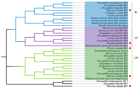
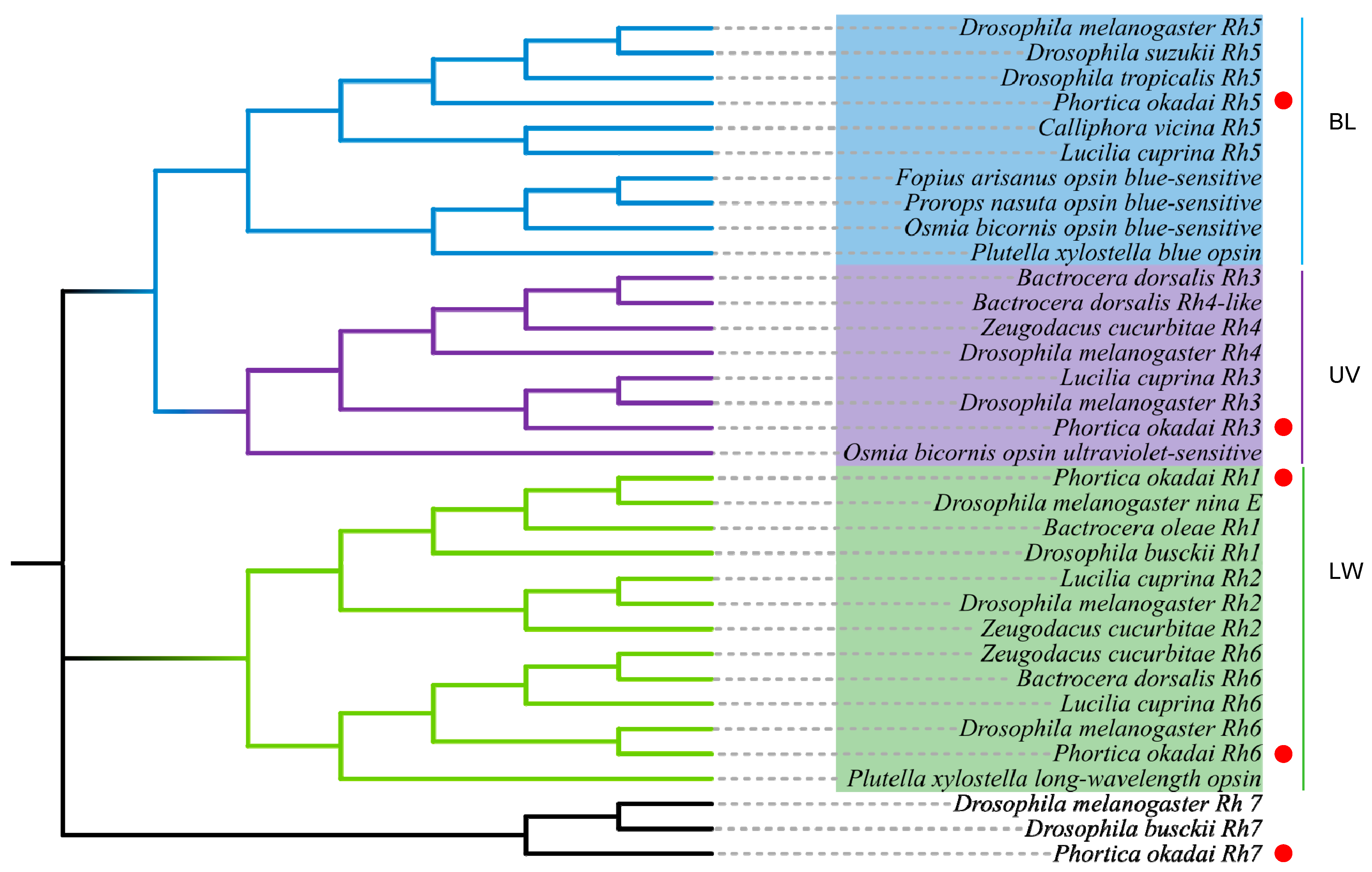
To infer the evolutionary relationships among the five identified PoRh opsins of P. okadai, we performed phylogenetic reconstruction based on opsin amino acid sequences from diverse insect taxa covering Diptera, Lepidoptera, and Hymenoptera. NJ (Figure 2) tree showed that the long-wavelength-sensitive (LW), blue-sensitive (BL), ultraviolet-sensitive (UV), and Rh7 opsins of P. okadai clustered into four clades similar to those of other insects: PoRh1 and PoRh6 both belong to the LW opsin clade, PoRh3 to the UV opsin clade, and PoRh5 to the BL opsin clade. Each P. okadai opsin formed a sister relationship with dipteran orthologs, reflecting strong sequence conservation of these visual photoreceptor genes within Diptera.
Figure 2.
Neighbor-joining tree based on protein sequences of opsins from P. okadai and other insects. The other opsin genes correspond to Diptera, Hymenoptera, and Lepidoptera. Dots denote P. okadai opsin sequences.
3.3. Expression Levels of the Opsins During Different Developmental Stages
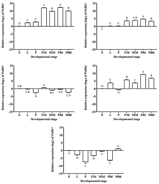
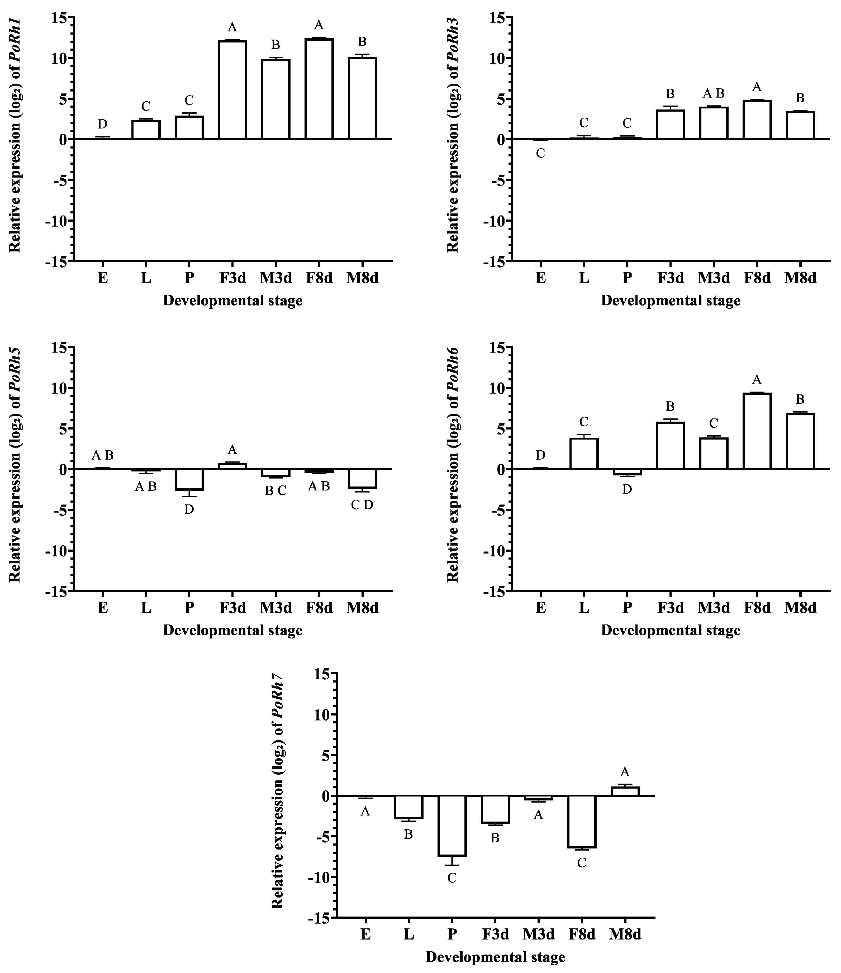
Relative expression levels of five opsin genes (PoRh1, PoRh3, PoRh5, PoRh6, and PoRh7) were quantified across seven developmental stages of P. okadai (eggs, larvae, pupae, 3- and 8-day-old adults of both sexes) using qRT-PCR (Figure 3). Overall, the five genes exhibited distinct stage- and sex-specific expression patterns.
Figure 3.
Relative expression levels of opsin genes in P. okadai at different developmental stages. E, eggs; L, larvae; P, pupae; F3d, 3-day-old female adults; M3d, 3−day−old male adults; F8d, 8−day−old female adults; M8d, 8−day−old male adults. Data are presented as log2−transformed relative expression levels; each value is the mean ± SE of three independent biological replicates. Different letters above the bars indicate significant differences between developmental stages based on a one−way ANOVA test followed by Tukey’s HSD post hoc test (α = 0.05).
PoRh1 and PoRh3 exhibited similar expression patterns, with lower expression in eggs, larvae, and pupae and markedly higher levels in adults, and showed peak expression in 8-day-old females. For PoRh1, expression levels in 3- and 8-day-old females were significantly higher than those in all other developmental stages, while expression in 3- and 8-day-old males was significantly higher than that in eggs, larvae, and pupae. Eggs exhibited the lowest expression, which was significantly lower than that in all other stages. No significant differences were observed between 3- and 8-day-old females, between 3- and 8-day-old males, or between larvae and pupae. For PoRh3, expression levels in 3-day-old males did not differ significantly from those in 8-day-old females, 3-day-old females, or 8-day-old males. Expression in 3-day-old females and 8-day-old males was significantly lower than that in 8-day-old females, yet markedly higher than that in eggs, larvae, and pupae. Eggs, larvae, and pupae formed the lowest-expression group, with no significant differences detected among these three stages.
PoRh5 exhibited relatively low and stable expression across all developmental stages, peaking in 3-day-old females. Eggs, larvae, and 8-day-old females displayed intermediate transcript levels; these groups were not significantly different from 3-day-old females and male adults but were significantly higher than pupae and 8-day-old males. Expression in 8-day-old males was not significantly different from pupae, which had the lowest expression.
PoRh6 showed the highest expression in 8-day-old female adults, significantly higher than in 3-day-old females and 8-day-old males, and both groups had significantly higher transcript abundance than larvae and 3-day-old males. Larvae and 3-day-old males showed significantly higher levels than eggs and pupae. No significant differences were detected between 3-day-old females and 8-day-old males, or between 3-day-old females and larvae, or between eggs and pupae.
PoRh7 exhibited the highest transcript abundance in eggs, 3-day-old males, and 8-day-old males, which was significantly higher than that in larvae and 3-day-old females. These groups had significantly higher transcript levels than pupae and 8-day-old females. No significant differences were detected among eggs, 3-day-old males, and 8-day-old males, between larvae and 3-day-old females, or between pupae and 8-day-old females.
3.4. Expression Pattern of Opsin Genes in Different Tissues
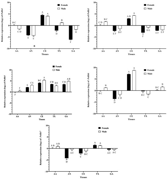
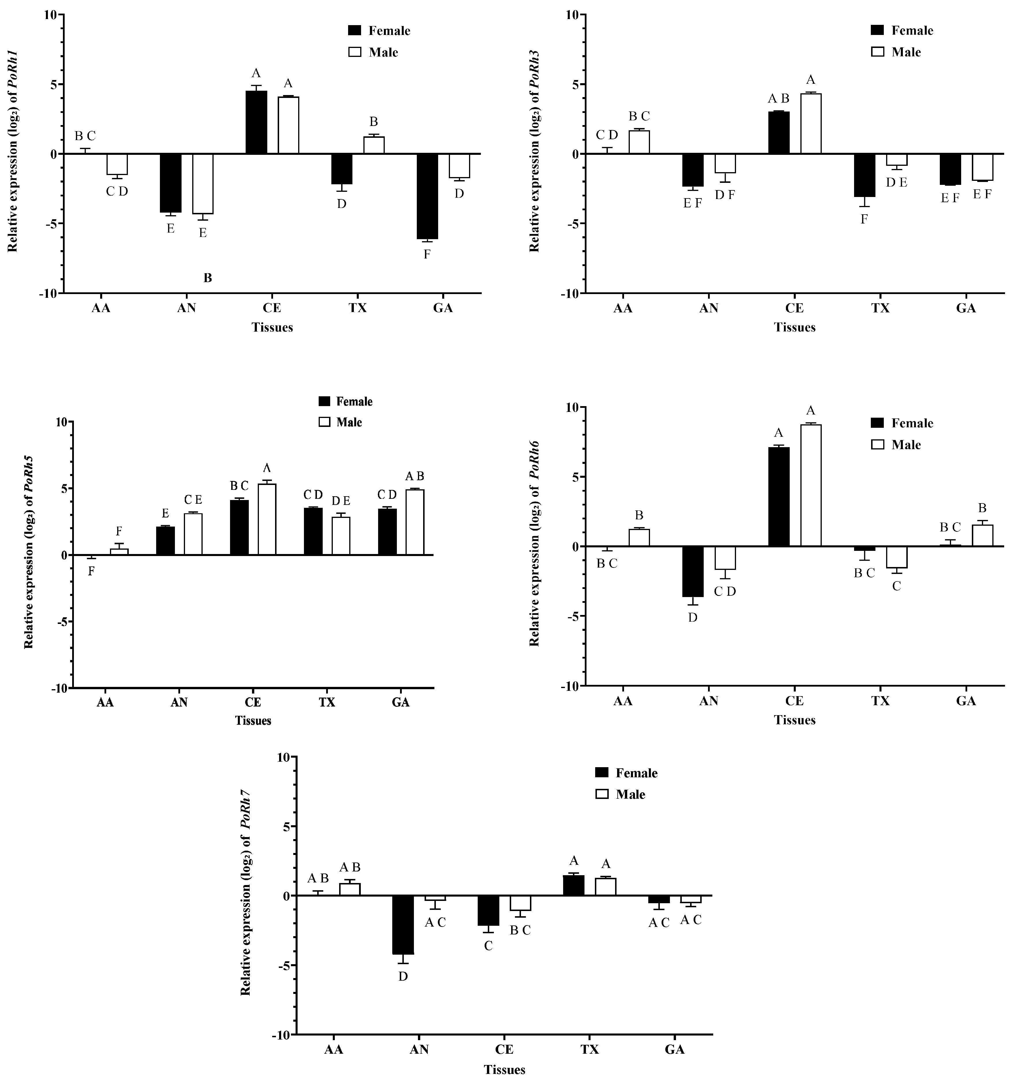
The relative expression levels of the five opsin genes (PoRh1, PoRh3, PoRh5, PoRh6, and PoRh7) were examined in the antennae (AA), abdomen (AN), compound eyes (CE), thorax (TX), and genitalia (GA) of adult female and male P. okadai using qRT-PCR (Figure 4).
Figure 4.
Relative expression levels of opsin genes in different tissues in P. okadai female and male adults. AA, antennae; AN, abdomen; CE, compound eyes; TX, thorax; GA, genitalia. Data are presented as log2−transformed relative expression levels. Each value is the mean ± SE of three independent biological replicates. Different letters above the bars indicate significant differences among tissue–sex groups, as determined by two−way ANOVA followed by Tukey’s HSD post hoc test (α = 0.05).
For PoRh1, PoRh3, PoRh5, and PoRh6, compound eyes exhibited the highest expression in both sexes.
In females, antennae ranked second for PoRh1 and PoRh3 and were significantly higher than the thorax, abdomen, and genitalia. Significant differences were detected across the thorax, abdomen, and genitalia for PoRh1, whereas these three tissues showed comparable transcript abundance for PoRh3. PoRh5 peaked in compound eyes, followed by the thorax, with antennae showing the lowest transcript abundance. For PoRh6, antennae, thorax, and genitalia had similar levels, all significantly higher than the abdomen.
For PoRh1 in males, compound eyes exhibited the highest expression, followed by the thorax, which was significantly higher than the antennae and genitalia. The abdomen showed the lowest expression, significantly lower than all other tissues. For PoRh3, compound eyes were followed by the antennae, which were significantly higher than the abdomen, thorax, and genitalia, with no significant differences detected among these three tissues. For PoRh5, compound eyes had the highest expression, followed by the thorax, which was significantly higher than the genitalia, antennae, and abdomen; the antennae had the lowest expression. In contrast, for PoRh6, expression was highest in compound eyes, followed by the antennae and genitalia, which were significantly higher than the thorax and abdomen; the abdomen showed the lowest expression but did not differ significantly from the thorax.
PoRh7 exhibited the highest transcript abundance in the thorax, followed by the antennae in both sexes. In females, the genitalia showed the next highest expression, followed by the compound eyes and abdomen, with the abdomen showing the lowest expression. In males, the genitalia showed the next highest expression, followed by the abdomen and compound eyes, with the compound eyes showing the lowest expression.
For tissue expression profiles, significantly higher transcript levels in males than in females were observed for PoRh1 in the thorax and genitalia, PoRh3 in the thorax, PoRh5 in compound eyes and genitalia, and PoRh7 in the abdomen. No significant sex-based expression differences were detected in the other examined tissues.
4. Discussion
P. okadai is the primary confirmed vector of T. callipaeda in China, transmitting infective larvae during its feeding activities on host tear fluids [24]. However, the mechanisms underlying host recognition and localization by P. okadai remain unknown. Studies have demonstrated that vision predominates in short-range host location, and opsins are key photoreceptive proteins that mediate visual perception, circadian rhythms, and behavioral responses [25].
To elucidate the molecular basis of visual host recognition in P. okadai, we identified five opsin genes from genomic data and analyzed their expression patterns across different developmental stages and tissues. Multiple-sequence alignment confirmed that all identified PoRh proteins possess the canonical features of insect opsins, including seven transmembrane (7-TM) domains, GPCR family signatures, and opsin retinal-binding sites with a conserved lysine residue in TM7, which are typical characteristics of all opsins [26,27]. PoRh1, PoRh3, PoRh5, and PoRh6 contain the QAKKMNV motif, whereas PoRh7 lacks this motif entirely. The retention of QAKKMNV in PoRh1, PoRh3, PoRh5, and PoRh6 mirrors the pattern observed in conventional visual opsins of numerous insect taxa and underscores the evolutionary conservation of G protein-binding sites in image-forming rhodopsins [21,28]. By contrast, loss of the QAKKMNV motif in PoRh7 is consistent with observations in many insects: in Drosophila melanogaster, Rh7 does not couple to the conventional Gαq-dependent phototransduction cascade despite possessing the typical rhodopsin architecture and instead exhibits a preference for inhibitory-type G-protein subunits, a functional shift partly attributed to the absence of QAKKMNV [29]. We therefore speculate that PoRh7 may have similarly rewired its G-protein coupling specificity.
Molecular phylogenetic analyses identified five opsin protein in P. okadai, encoding long-wavelength (LW), ultraviolet (UV), and blue-sensitive (BL) opsins (Figure 2), which are consistent with those widely accepted in most insects. Notably, PoRh2 and PoRh4, which are present in D. melanogaster, were not detected [17]. Opsin gene loss has been documented in diverse insects; for example, blue-sensitive opsins have been independently lost in multiple Hemiptera (aphids, mirid bugs, planthoppers) and Coleoptera (Tribolium castaneum) [30,31,32], and subterranean beetles show parallel loss or pseudogenization of multiple opsin genes [33]. In T. castaneum, loss of the blue opsin is accompanied by expanded expression of LW opsin across all photoreceptors, suggesting functional compensation [34]. Similarly, the absence of PoRh2 and PoRh4 in P. okadai may be compensated by the retained LW (PoRh1, PoRh6) and UV (PoRh3) opsins, though this hypothesis requires experimental validation.
Overall, PoRh1, PoRh3 and PoRh6 exhibited higher expression in adult compound eyes than in other developmental stages or tissues, suggesting important roles in the visual system of P. okadai that may be associated with visually guided behaviors such as flight, host-seeking, and oviposition-site selection (Figure 3 and Figure 4).
The 3-day-old and 8-day-old adults correspond to the stages prior to and following sexual maturation, respectively. The results suggest that the marked increase in transcript abundance of PoRh3 and PoRh6 in females from 3-day-old to 8-day-old adults may be involved in visual host location for oviposition. A prior study demonstrated that knockdown of Rh6 in Bactrocera minax substantially reduces oviposition on green unripe citrus fruits [35]; nevertheless, whether Rh3 participates in oviposition-site selection awaits further experimental validation. The highest expression of PoRh1 in female compound eyes (Figure S2) is likely because Rh1 is responsible for motion detection and image formation [36].
For PoRh5, no marked differences in expression were observed among eggs, larvae, and adults. In terms of tissue distribution, no significant differences were detected among the compound eye, thorax, and genitalia in females, or between the compound eye and genitalia in males, suggesting that in addition to its canonical visual function, PoRh5 may participate in other physiological processes, such as temperature preference or hearing [37].
PoRh7 was predominantly expressed in eggs, larvae, and pupae (Figure S1), and its expression was mainly localized to the thorax. This thoracic enrichment is consistent with the non-visual role of Rh7. In D. melanogaster, Rh7 is expressed in dorsal clock neurons, including DN1p, DN1a, and DN2 neurons. Rh7 mutants exhibit decreased night activity and fail to adjust their behavior to darkness, while daily activity levels remain similar to controls, suggesting that these effects reflect altered circadian regulation rather than general locomotor impairment [38]. Given that thoracic ganglia are primary motor centers controlling locomotion, we predicted that PoRh7 in P. okadai may be involved in light-dependent modulation of activity patterns, thereby explaining its thoracic enrichment.
Insect opsins function as polymodal sensory receptors rather than exclusively as light sensors, which may explain their expression in non-visual tissues. In Aedes aegypti, op1 and op2 mRNAs are present in neurons in the most distal region of the antenna, where the corresponding opsins function as heat sensors [39]. In Nilaparvata lugens, the long-wavelength opsin gene participates in the regulation of cuticle melanization [40]. These findings collectively support the non-visual roles of opsins in non-visual tissues.
5. Conclusions
In conclusion, we identified five opsin genes from the genomic data of P. okadai. Expression profiling showed that four PoRh genes (excluding PoRh7) were highly expressed in adults and compound eyes. By contrast, PoRh7 exhibited low expression in compound eyes yet was enriched in the thorax. Our findings suggest potential involvement of these opsins in adult-specific visual physiological processes. However, since no gene-functional or behavioral assays were performed in this study, the precise biological functions of these opsins still require validation in future experimental work. This study improves our knowledge of the visual system in P. okadai and may provide insights for future vector control strategies targeting light-dependent behaviors.
Supplementary Materials
The following supporting information can be downloaded at https://www.mdpi.com/article/10.3390/insects17101082/s1.
Author Contributions
Conceptualization, L.W. (Lingjun Wang); Software, Y.L., B.L., and Y.C.; Funding acquisition, J.Z., H.L., Y.W.; Methodology, S.J., J.W., and L.W. (Liang Wang); Writing—original draft, J.Z.; Writing—review and editing, J.Z.; H.L. and L.W. (Liang Wang). All authors have read and agreed to the published version of the manuscript.
Funding
This work was supported by the Fund for Less Developed Regions of the National Natural Science Foundation of China, grant number 82060374; the Natural Science Foundation of Guizhou Province, grant number MS[2025]345; The College Students’ Innovation and Entrepreneurship Training Program of Zunyi Medical University, grant number S202510661479. Guizhou Provincial Postgraduate Education Innovation Program, grant number 2025YJSKYJJ315.
Data Availability Statement
All data is provided in the manuscript.
Acknowledgments
We are very grateful to all members of the Department of Parasitology, Zunyi Medical University, for their assistance during laboratory experiments.
Conflicts of Interest
The authors declare no conflicts of interest.
References
- Wang, S.; Yu, S.J.; Wang, X.L.; Zhao, M.Y.; Deng, S.; Shang, H.; An, C.L. A pediatric case report and literature review of thelaziosis in China. Int. J. Infect. Dis. 2014, 29, 249–250. [Google Scholar] [CrossRef] [Scilit] [PubMed]
- Bezerra-Santos, M.A.; Bernardini, I.; Lia, R.P.; Mendoza-Roldan, J.A.; Beugnet, F.; Pombi, M.; Otranto, D. Phortica oldenbergi (Diptera: Drosophilidae): A new potential vector of the zoonotic Thelazia callipaeda eyeworm. Acta Trop. 2022, 233, 106565. [Google Scholar] [CrossRef] [Scilit] [PubMed]
- Ramos, A.P.; Gustafsson, O.; Labert, N.; Salecker, I.; Nilsson, D.E.; Averof, M. Analysis of the genetically tractable crustacean Parhyale hawaiensis reveals the organisation of a sensory system for low-resolution vision. BMC Biol. 2019, 17, 67. [Google Scholar] [CrossRef] [Scilit] [PubMed]
- Guignard, Q.; Allison, J.D.; Slippers, B. The evolution of insect visual opsin genes with specific consideration of the influence of ocelli and life history traits. BMC Ecol. Evol. 2022, 22, 2. [Google Scholar] [CrossRef] [Scilit] [PubMed]
- Jean-Guillaume, C.B.; Kumar, J.P. Development of the ocellar visual system in Drosophila melanogaster. FEBS J. 2022, 289, 7411–7427. [Google Scholar] [CrossRef] [Scilit] [PubMed]
- Yamaguchi, S.; Heisenberg, M. Photoreceptors and neural circuitry underlying phototaxis in insects. Fly 2011, 5, 333–336. [Google Scholar] [CrossRef] [Scilit] [PubMed]
- Briscoe, A.D.; Chittka, L. The evolution of color vision in insects. Annu. Rev. Entomol. 2001, 46, 471–510. [Google Scholar] [PubMed]
- Thomas, P.S. Structure of rhodopsin and the superfamily of seven-helical receptors: The same and not the same. Curr. Opin. Cell Biol. 2002, 14, 189–195. [Google Scholar] [CrossRef] [Scilit] [PubMed]
- Yuan, F.; Bernard, G.; Le, J.; Briscoe, A.D. Contrasting modes of evolution of the visual pigments in Heliconius butterflies. Mol. Biol. Evol. 2010, 27, 2392–2405. [Google Scholar] [CrossRef] [Scilit] [PubMed]
- Zuker, C.S.; Cowman, A.F.; Rubin, G.M. Isolation and structure of a rhodopsin gene from D. melanogaster. Cell 1985, 40, 851–858. [Google Scholar] [CrossRef] [Scilit] [PubMed]
- O’Tousa, J.E.; Baehr, W.; Martin, R.L.; Hirsh, J.; Pak, W.L.; Meredithe, L.A. The Drosophila ninaE gene encodes an opsin. Cell 1985, 40, 839–850. [Google Scholar] [CrossRef] [Scilit] [PubMed]
- Ostroy, S.E.; Wilson, M.; Pak, W.L. Drosophila rhodopsin: Photochemistry, extraction and differences in the norpAP12 phototransduction mutant. Biochem. Biophys. Res. Commun. 1974, 59, 960–966. [Google Scholar] [CrossRef] [Scilit] [PubMed]
- Harris, W.A.; Stark, W.S.; Walker, J.A. Genetic dissection of the photoreceptor system in the compound eyes of Drosophila melanogaster. J. Physiol. 1976, 256, 415–439. [Google Scholar] [CrossRef] [Scilit] [PubMed]
- Cowman, A.F.; Zuker, C.S.; Rubin, G.M. An opsin gene expressed in only one photoreceptor cell type of the Drosophila eye. Cell 1986, 44, 705–710. [Google Scholar] [CrossRef] [Scilit] [PubMed]
- Feiler, R.; Harris, W.A.; Kirschfeld, K.; Wehrhahn, C.; Zuker, C.S. Targeted misexpression of a Drosophila opsin gene leads to altered visual function. Nature 1988, 333, 737–741. [Google Scholar] [CrossRef] [Scilit] [PubMed]
- Pollock, J.A.; Benzer, S. Transcript localization of four opsin genes in the three visual organs of Drosophila: RH2 is ocellus specific. Nature 1988, 333, 779–782. [Google Scholar] [CrossRef] [Scilit] [PubMed]
- Fryxell, K.J.; Meyerowitz, E.M. An opsin gene that is expressed only in the R7 photoreceptor cell of Drosophila. EMBO J. 1987, 6, 443–451. [Google Scholar] [CrossRef] [Scilit] [PubMed]
- Montell, C.; Jones, K.; Zuker, C.; Rubin, G. A second opsin gene expressed in the ultraviolet-sensitive R7 photoreceptor cells of Drosophila melanogaster. J. Neurosci. 1987, 7, 1558–1566. [Google Scholar] [CrossRef] [Scilit] [PubMed]
- Feiler, R.; Bjornson, R.; Kirschfeld, K.; Mismer, D.; Rubin, G.M.; Smith, D.P.; Socolich, M.; Zuker, C.S. Ectopic expression of ultraviolet-rhodopsins in the blue photoreceptor cells of Drosophila: Visual physiology and photochemistry of transgenic animals. J. Neurosci. 1992, 12, 3862–3868. [Google Scholar] [CrossRef] [Scilit] [PubMed]
- Salcedo, E.; Huber, A.; Henrich, S.; Chadwell, L.W.; Chou, W.H.; Paulsen, R.; Britt, S.G. Blue- and green-absorbing visual pigments of Drosophila: Ectopic expression and physiological characterization of the R8 photoreceptor cell-specific Rh5 and Rh6 rhodopsins. J. Neurosci. 1999, 19, 10716–10726. [Google Scholar] [CrossRef] [Scilit] [PubMed]
- Huang, M.; Meng, J.Y.; Tang, X.; Shan, L.L.; Yang, C.L.; Zhang, C.Y. Identification, expression analysis, and functional verification of three opsin genes related to the phototactic behaviour of Ostrinia furnacalis. Mol. Ecol. 2024, 33, e17323. [Google Scholar] [CrossRef] [Scilit] [PubMed]
- Wang, L.J.; Tang, H.R.; Xie, Z.M.; Li, D.; Yin, C.Z.; Luo, B.; Yan, R.; Sun, W.; Liu, H. Identification and functional characterization of CYP4D2 putatively associated with β-cypermethrin detoxification in Phortica okadai. Genes 2022, 13, 2338. [Google Scholar] [CrossRef] [Scilit] [PubMed]
- Livak, K.J.; Schmittgen, T.D. Analysis of relative gene expression data using real-time quantitative PCR and the 2−ΔΔCT method. Methods 2001, 25, 402–408. [Google Scholar] [CrossRef] [Scilit] [PubMed]
- Luo, Y.; Sun, D.; Xu, Y.J.; Chen, Z.F.; Yan, R.; Zhou, J.; Luo, B.; Long, D.H. Contrasting shifts in potential climatic suitability of two Thelazia callipaeda vectors (Phortica okadai and Phortica variegata) across China and Europe under climate change. Parasites Vectors 2026, 19, 242. [Google Scholar] [CrossRef] [Scilit] [PubMed]
- Wang, Y.H.; Fang, G.Q.; Xu, P.H.; Gao, B.L.; Liu, X.J.; Qi, X.W.; Zhang, G.J.; Cao, S.; Li, Z.H.; Ren, X.M.; et al. Behavioral and genomic divergence between a generalist and a specialist fly. Cell Rep. 2022, 41, 111654. [Google Scholar] [CrossRef] [Scilit] [PubMed]
- Gartner, W.; Towner, P. Invertebrate visual pigments. Photochem. Photobiol. 1995, 62, 1–16. [Google Scholar] [CrossRef] [Scilit] [PubMed]
- Townson, S.M.; Chang, B.S.W.; Salcedo, E.; Chadwell, L.V.; Pierce, N.E.; Britt, S.G. Honeybee blue- and ultraviolet-sensitive opsins: Cloning, heterologous expression in Drosophila, and physiological characterization. J. Neurosci. 1998, 18, 2412–2422. [Google Scholar] [CrossRef] [Scilit] [PubMed]
- Salcedo, E.; Zheng, L.; Phistry, M.; Bagg, E.E.; Britt, S.G. Molecular basis for ultraviolet vision in invertebrates. J. Neurosci. 2003, 23, 10873–10878. [Google Scholar] [CrossRef] [Scilit] [PubMed]
- Kirsch, V.; Reinhard, N.; Hartlieb, H.; Mohr, A.; Rieger, D.; Soba, P.; Helfrich-Förster, C.; Senthilan, P.R. RHODOPSIN 7: An ancient non-retinal photoreceptor for contrast vision, darkness detection, and circadian regulation. eLife 2026, 15, RP109811. [Google Scholar] [CrossRef]
- Feuda, R.; Marlétaz, F.; Bentley, M.A.; Holland, P.W.H. Conservation, duplication, and divergence of five opsin genes in insect evolution. Genome Biol. Evol. 2016, 8, 579–587. [Google Scholar] [CrossRef] [Scilit] [PubMed]
- Xu, P.J.; Lu, B.; Chao, J.T.; Holdbrook, R.; Liang, G.; Lu, Y.H. The evolution of opsin genes in five species of mirid bugs: Duplication of long-wavelength opsins and loss of blue-sensitive opsins. BMC Evol. Biol. 2021, 21, 66. [Google Scholar] [CrossRef] [Scilit] [PubMed]
- Friedrich, M. Parallel losses of blue opsin correlate with compensatory neofunctionalization of UV-opsin gene duplicates in aphids and planthoppers. Insects 2023, 14, 774. [Google Scholar] [CrossRef] [Scilit] [PubMed]
- Tierney, S.M.; Cooper, S.J.B.; Saint, K.M.; Bertozzi, T.; Hyde, J.; Humphreys, W.F.; Austin, A.D. Opsin transcripts of predatory diving beetles: A comparison of surface and subterranean photic niches. R. Soc. Open Sci. 2015, 2, 140386. [Google Scholar] [CrossRef] [Scilit] [PubMed]
- Jackowska, M.; Bao, R.; Liu, Z.Y.; McDonald, E.C.; Cook, T.A.; Friedrich, M. Genomic and gene regulatory signatures of cryptozoic adaptation: Loss of blue-sensitive photoreceptors through expansion of long-wavelength opsin expression in the red flour beetle Tribolium castaneum. Front. Zool. 2007, 4, 24. [Google Scholar] [CrossRef] [Scilit] [PubMed]
- Wang, Y.; Andongma, A.A.; Dong, Y.; Chen, Z.; Xu, P.; Ren, X.; Niu, C.; Krosch, M.N.; Clarke, A.R. Rh6 gene modulates the visual mechanism of host utilization in fruit fly Bactrocera minax. Pest Manag. Sci. 2019, 75, 1621–1629. [Google Scholar] [CrossRef] [Scilit] [PubMed]
- Behnia, R.; Desplan, C. Visual circuits in flies: Beginning to see the whole picture. Curr. Opin. Neurobiol. 2015, 34, 125–132. [Google Scholar] [CrossRef] [Scilit] [PubMed]
- Leungn, N.Y.; Montell, C. Unconventional roles of opsins. Annu. Rev. Cell Dev. Biol. 2017, 33, 241–264. [Google Scholar] [CrossRef] [Scilit] [PubMed]
- Kistenpfennig, C.; Grebler, R.; Ogueta, M.; Hermann-Luibl, C.; Schlichting, M.; Stanewsky, R.; Senthilan, P.R.; Helfrich-Förster, C. A new rhodopsin influences light-dependent daily activity patterns of fruit flies. J. Biol. Rhythms 2017, 32, 406–422. [Google Scholar] [CrossRef] [Scilit] [PubMed]
- Chandel, A.; DeBeaubien, N.A.; Ganguly, A.; Meyerhof, G.T.; Krumholz, A.A.; Liu, J.; Salgado, V.L.; Montell, C. Thermal infrared directs host-seeking behavior in Aedes aegypti mosquitoes. Nature 2024, 633, 615–623. [Google Scholar] [CrossRef] [Scilit] [PubMed]
- Lu, J.B.; Li, Z.D.; Ye, Z.X.; Huang, H.J.; Chen, J.P.; Li, J.M.; Zhang, C.X. Long-wave opsin involved in body color plastic development in Nilaparvata lugens. BMC Genomics 2023, 24, 1–11. [Google Scholar] [CrossRef] [Scilit] [PubMed]

Disclaimer/Publisher’s Note: The statements, opinions and data contained in all publications are solely those of the individual author(s) and contributor(s) and not of MDPI and/or the editor(s). MDPI and/or the editor(s) disclaim responsibility for any injury to people or property resulting from any ideas, methods, instructions or products referred to in the content. |
© 2026 by the authors. Licensee MDPI, Basel, Switzerland. This article is an open access article distributed under the terms and conditions of the Creative Commons Attribution (CC BY) license.