


Diabetes May Modulate the Association Between Age and Optical Coherence Tomography Angiography Parameters: A Serial, Cross-Sectional Study


Abstract
1. Introduction
2. Methods
2.1. Study Sample
2.2. Image Acquisition
2.3. Image Analysis
2.4. Statistical Analysis
3. Results
4. Discussion
Supplementary Materials
Author Contributions
Funding
Institutional Review Board Statement
Informed Consent Statement
Data Availability Statement
Conflicts of Interest
References
- Nguyen, K.H.; Patel, B.C.; Tadi, P. Anatomy, Head and Neck: Eye Retina. In StatPearls; StatPearls Publishing: Treasure Island, FL, USA, 2025. [Google Scholar]
- Jin, Q.; Lei, Y.; Wang, R.; Wu, H.; Ji, K.; Ling, L. A Systematic Review and Meta-Analysis of Retinal Microvascular Features in Alzheimer’s Disease. Front. Aging Neurosci. 2021, 13, 683824. [Google Scholar] [CrossRef]
- Lever, M.; Chen, Y.; Glaser, M.; Unterlauft, J.D.; Lommatzsch, C.; Bechrakis, N.E.; Böhm, M.R.R. Microvascular and Morphologic Changes of the Macula over Lifetime. Life 2022, 12, 568. [Google Scholar] [CrossRef]
- Nag, T.C.; Maurya, M.; Roy, T.S. Age-related changes of the human retinal vessels: Possible involvement of lipid peroxidation. Ann. Anat.-Anat. Anz. 2019, 226, 35–47. [Google Scholar] [CrossRef]
- Emeterio Nateras, O.S.; Harrison, J.M.; Muir, E.R.; Zhang, Y.; Peng, Q.; Chalfin, S.; Gutierrez, J.E.; Johnson, D.A.; Kiel, J.W.; Duong, T.Q. Choroidal blood flow decreases with age: An MRI study. Curr. Eye Res. 2014, 39, 1059–1067. [Google Scholar] [CrossRef][Green Version]
- Polascik, B.W.; Thompson, A.C.; Yoon, S.P.; Powers, J.H.; Burke, J.R.; Grewal, D.S.; Fekrat, S. Association of OCT Angiography Parameters With Age in Cognitively Healthy Older Adults. Ophthalmic Surg. Lasers Imaging Retin. 2020, 51, 706–714. [Google Scholar] [CrossRef]
- Verma, A.; Magesan, K.; Amose, T.; Alagorie, A.R.; Gnanaraj, R.; Sadda, S.R.; Sen, P. Age-related assessment of foveal avascular zone and surrounding capillary networks with swept source optical coherence tomography angiography in healthy eyes. Eye 2022, 36, 1857–1864. [Google Scholar] [CrossRef]
- Iafe, N.A.; Phasukkijwatana, N.; Chen, X.; Sarraf, D. Retinal Capillary Density and Foveal Avascular Zone Area Are Age-Dependent: Quantitative Analysis Using Optical Coherence Tomography Angiography. Investig. Ophthalmol. Vis. Sci. 2016, 57, 5780–5787. [Google Scholar] [CrossRef]
- Chua, J.; Tan, B.; Wong, D.; Garhöfer, G.; Liew, X.W.; Popa-Cherecheanu, A.; Loong Chin, C.W.; Milea, D.; Li-Hsian Chen, C.; Schmetterer, L. Optical coherence tomography angiography of the retina and choroid in systemic diseases. Prog. Retin. Eye Res. 2024, 103, 101292. [Google Scholar] [CrossRef]
- Hoyek, S.; Chaaya, C.; Lemire, C.A.; Halawa, O.; Altamirano, F.; Archer, N.M.; Gonzalez, E.; Patel, N.A. Retinal Imaging Biomarkers and Correlation to Systemic Disease Activity in Pediatric Sickle Cell Disease. Ophthalmol. Sci. 2025, 5, 100774. [Google Scholar] [CrossRef]
- Waheed, N.K.; Rosen, R.B.; Jia, Y.; Munk, M.R.; Huang, D.; Fawzi, A.; Chong, V.; Nguyen, Q.D.; Sepah, Y.; Pearce, E. Optical coherence tomography angiography in diabetic retinopathy. Prog. Retin. Eye Res. 2023, 97, 101206. [Google Scholar] [CrossRef]
- Zhou, J.; Chen, B. Retinal Cell Damage in Diabetic Retinopathy. Cells 2023, 12, 1342. [Google Scholar] [CrossRef]
- Matsunaga, D.R.; Yi, J.J.; De, K.L.O.; Ameri, H.; Puliafito, C.A.; Kashani, A.H. Optical Coherence Tomography Angiography of Diabetic Retinopathy in Human Subjects. Ophthalmic Surg. Lasers Imaging Retin. 2015, 46, 796–805. [Google Scholar] [CrossRef]
- Ashraf, M.; Sampani, K.; Clermont, A.; Abu-Qamar, O.; Rhee, J.; Silva, P.S.; Aiello, L.P.; Sun, J.K. Vascular Density of Deep, Intermediate and Superficial Vascular Plexuses Are Differentially Affected by Diabetic Retinopathy Severity. Investig. Ophthalmol. Vis. Sci. 2020, 61, 53. [Google Scholar] [CrossRef]
- Garg, I.; Uwakwe, C.; Le, R.; Lu, E.S.; Cui, Y.; Wai, K.M.; Katz, R.; Zhu, Y.; Moon, J.Y.; Li, C.Y.; et al. Nonperfusion Area and Other Vascular Metrics by Wider Field Swept-Source OCT Angiography as Biomarkers of Diabetic Retinopathy Severity. Ophthalmol. Sci. 2022, 2, 100144. [Google Scholar] [CrossRef]
- Furino, C.; Montrone, G.; Cicinelli, M.V.; Balestra, S.; Grassi, M.O.; Reibaldi, M.; Boscia, F.; Alessio, G. Optical coherence tomography angiography in diabetic patients without diabetic retinopathy. Eur. J. Ophthalmol. 2020, 30, 1418–1423. [Google Scholar] [CrossRef]
- Ghassemi, F.; Fadakar, K.; Berijani, S.; Babeli, A.; Gholizadeh, A.; Sabour, S. Quantitative assessment of vascular density in diabetic retinopathy subtypes with optical coherence tomography angiography. BMC Ophthalmol. 2021, 21, 82. [Google Scholar] [CrossRef]
- Kaizu, Y.; Nakao, S.; Arima, M.; Wada, I.; Yamaguchi, M.; Sekiryu, H.; Hayami, T.; Ishikawa, K.; Ikeda, Y.; Sonoda, K.-H. Capillary dropout is dominant in deep capillary plexus in early diabetic retinopathy in optical coherence tomography angiography. Acta Ophthalmol. 2019, 97, e811–e812. [Google Scholar] [CrossRef]
- Kasumovic, A.; Matoc, I.; Avdagic, N.; Halimic, T.; Voloder, B.; Muhamedagic, L.; Delic, S.; Sesar, I. Optical Coherence Tomography Angiography Contributions in Classification of Nonproliferative Diabetic Retinopathy. Acta Inform. Med. 2020, 28, 103. [Google Scholar] [CrossRef]
- Ong, J.X.; Kwan, C.C.; Cicinelli, M.V.; Fawzi, A.A. Superficial capillary perfusion on optical coherence tomography angiography differentiates moderate and severe nonproliferative diabetic retinopathy. PLoS ONE 2020, 15, e0240064. [Google Scholar] [CrossRef]
- Sun, Z.; Yang, D.; Tang, Z.; Ng, D.S.; Cheung, C.Y. Optical coherence tomography angiography in diabetic retinopathy: An updated review. Eye 2021, 35, 149–161. [Google Scholar] [CrossRef]
- Zhao, S.; He, J.; Qiu, M.; Liang, X. Changes of blood flow in macular zone of patients with diabetic retinopathy at different stages evaluated by optical coherence tomography angiography. J. Fr. Ophtalmol. 2022, 45, 728–734. [Google Scholar] [CrossRef]
- Nesper, P.L.; Roberts, P.K.; Onishi, A.C.; Chai, H.; Liu, L.; Jampol, L.M.; Fawzi, A.A. Quantifying Microvascular Abnormalities With Increasing Severity of Diabetic Retinopathy Using Optical Coherence Tomography Angiography. Investig. Ophthalmol. Vis. Sci. 2017, 58, BIO307. [Google Scholar] [CrossRef]
- Laotaweerungsawat, S.; Psaras, C.; Haq, Z.; Liu, X.; Stewart, J.M. Racial and ethnic differences in foveal avascular zone in diabetic and nondiabetic eyes revealed by optical coherence tomography angiography. PLoS ONE 2021, 16, e0258848. [Google Scholar] [CrossRef]
- Taha, A.T.; Zhang, Y.S.; Thompson, I.J.B.; Poddar, A.; Keenan, J.D.; Stewart, J.M. Race/Ethnicity Analysis of Vascular Alterations in Optical Coherence Tomography Angiography in Diabetic Patients. Ophthalmic Epidemiol. 2025, 32, 153–162. [Google Scholar] [CrossRef]
- Zhang, Y.S.; Taha, A.T.; Thompson, I.J.B.; Keenan, J.D.; Yang, D.; Wu, J.; Stewart, J.M. Association of Male Sex and Microvascular Alterations on Optical Coherence Tomography Angiography in Diabetes. Transl. Vis. Sci. Technol. 2023, 12, 30. [Google Scholar] [CrossRef]
- Wei, Y.; Jiang, H.; Shi, Y.; Qu, D.; Gregori, G.; Zheng, F.; Rundek, T.; Wang, J. Age-Related Alterations in the Retinal Microvasculature, Microcirculation, and Microstructure. Investig. Ophthalmol. Vis. Sci. 2017, 58, 3804–3817. [Google Scholar] [CrossRef]
- Su, B.; Zhu, X.; Yang, K.; Xiao, Y.; Li, C.; Shi, K.; Qu, J.; Lu, F.; Li, M.; Cui, L. Age- and sex-related differences in the retinal capillary plexus in healthy Chinese adults. Eye Vis. 2022, 9, 38. [Google Scholar] [CrossRef]
- Yoshikawa, Y.; Shoji, T.; Kanno, J.; Ibuki, H.; Ozaki, K.; Ishii, H.; Inami, H.; Shinoda, K. Examination of Age-Related Retinal Vascular Changes in the Macula Using Optical Coherence Tomography Angiography of the Eyes After Cataract Surgery. Clin. Ophthalmol. 2021, 15, 3687–3695. [Google Scholar] [CrossRef]
- Brücher, V.C.; Storp, J.J.; Eter, N.; Alnawaiseh, M. Optical coherence tomography angiography-derived flow density: A review of the influencing factors. Graefes Arch. Clin. Exp. Ophthalmol. 2020, 258, 701–710. [Google Scholar] [CrossRef]
- Munsell, M.K.; Garg, I.; Duich, M.; Zeng, R.; Baldwin, G.; Wescott, H.E.; Koch, T.; Wang, K.L.; Patel, N.A.; Miller, J.B. A normative database of wide-field swept-source optical coherence tomography angiography quantitative metrics in a large cohort of healthy adults. Graefes Arch. Clin. Exp. Ophthalmol. 2023, 261, 1835–1859. [Google Scholar] [CrossRef]
- Wang, X.; Cheng, Z. Cross-Sectional Studies: Strengths, Weaknesses, and Recommendations. Chest 2020, 158, S65–S71. [Google Scholar] [CrossRef]
- Early Treatment Diabetic Retinopathy Study Research Group. Grading diabetic retinopathy from stereoscopic color fundus photographs—An extension of the modified Airlie House classification. ETDRS report number 10. Ophthalmology 1991, 98, 786–806. [Google Scholar] [CrossRef]
- Ho, J.; Dans, K.; You, Q.; Nudleman, E.N.; Freeman, W.R. Comparison of 3 × 3 mm versus 6 × 6 mm Optical Coherence Tomography Angiography Scan Sizes in the Evaluation of Non-proliferative Diabetic Retinopathy. Retina 2019, 39, 259–264. [Google Scholar] [CrossRef]
- Schneider, C.A.; Rasband, W.S.; Eliceiri, K.W. NIH Image to ImageJ: 25 years of image analysis. Nat. Methods 2012, 9, 671–675. [Google Scholar] [CrossRef]
- Durbin, M.K.; An, L.; Shemonski, N.D.; Soares, M.; Santos, T.; Lopes, M.; Neves, C.; Cunha-Vaz, J. Quantification of Retinal Microvascular Density in Optical Coherence Tomographic Angiography Images in Diabetic Retinopathy. JAMA Ophthalmol. 2017, 135, 370–376. [Google Scholar] [CrossRef]
- Li, C.H.; Lee, C.K. Minimum cross entropy thresholding. Pattern Recognit. 1993, 26, 617–625. [Google Scholar] [CrossRef]
- Terheyden, J.H.; Wintergerst, M.W.M.; Falahat, P.; Berger, M.; Holz, F.G.; Finger, R.P. Automated thresholding algorithms outperform manual thresholding in macular optical coherence tomography angiography image analysis. PLoS ONE 2020, 15, e0230260. [Google Scholar] [CrossRef]
- Arganda-Carreras, I.; Fernández-González, R.; Muñoz-Barrutia, A.; Ortiz-De-Solorzano, C. 3D reconstruction of histological sections: Application to mammary gland tissue. Microsc. Res. Tech. 2010, 73, 1019–1029. [Google Scholar] [CrossRef]
- Tokayer, J.; Jia, Y.; Dhalla, A.-H.; Huang, D. Blood flow velocity quantification using split-spectrum amplitude-decorrelation angiography with optical coherence tomography. Biomed. Opt. Express BOE 2013, 4, 1909–1924. [Google Scholar] [CrossRef]
- RStudio Team. RStudio: Integrated Development for R; RStudio Team: Boston, MA, USA, 2020; Available online: https://posit.co/index%2Ephp/download/rstudio-desktop (accessed on 10 May 2026).
- Balaratnasingam, C.; Inoue, M.; Ahn, S.; McCann, J.; Dhrami-Gavazi, E.; Yannuzzi, L.A.; Freund, K.B. Visual Acuity Is Correlated with the Area of the Foveal Avascular Zone in Diabetic Retinopathy and Retinal Vein Occlusion. Ophthalmology 2016, 123, 2352–2367. [Google Scholar] [CrossRef]
- Sim, D.A.; Keane, P.A.; Zarranz-Ventura, J.; Fung, S.; Powner, M.B.; Platteau, E.; Bunce, C.V.; Fruttiger, M.; Patel, P.J.; Tufail, A.; et al. The effects of macular ischemia on visual acuity in diabetic retinopathy. Investig. Ophthalmol. Vis. Sci. 2013, 54, 2353–2360. [Google Scholar] [CrossRef]
- Onishi, A.C.; Nesper, P.L.; Roberts, P.K.; Moharram, G.A.; Chai, H.; Liu, L.; Jampol, L.M.; Fawzi, A.A. Importance of Considering the Middle Capillary Plexus on OCT Angiography in Diabetic Retinopathy. Investig. Ophthalmol. Vis. Sci. 2018, 59, 2167–2176. [Google Scholar] [CrossRef]
- Ong, J.X.; Fawzi, A.A. Perspectives on diabetic retinopathy from advanced retinal vascular imaging. Eye 2022, 36, 319–327. [Google Scholar] [CrossRef]
- Nagaoka, T.; Sato, E.; Takahashi, A.; Sogawa, K.; Yokota, H.; Yoshida, A. Effect of aging on retinal circulation in normotensive healthy subjects. Exp. Eye Res. 2009, 89, 887–891. [Google Scholar] [CrossRef]
- Coscas, F.; Sellam, A.; Glacet-Bernard, A.; Jung, C.; Goudot, M.; Miere, A.; Souied, E.H. Normative Data for Vascular Density in Superficial and Deep Capillary Plexuses of Healthy Adults Assessed by Optical Coherence Tomography Angiography. Investig. Ophthalmol. Vis. Sci. 2016, 57, OCT211–OCT223. [Google Scholar] [CrossRef]
- Park, S.H.; Cho, H.; Hwang, S.J.; Jeon, B.; Seong, M.; Yeom, H.; Kang, M.H.; Lim, H.W.; Shin, Y.U. Changes in the Retinal Microvasculature Measured Using Optical Coherence Tomography Angiography According to Age. J. Clin. Med. 2020, 9, 883. [Google Scholar] [CrossRef]
- You, Q.S.; Chan, J.C.H.; Ng, A.L.K.; Choy, B.K.N.; Shih, K.C.; Cheung, J.J.C.; Wong, J.K.W.; Shum, J.W.H.; Ni, M.Y.; Lai, J.S.M.; et al. Macular Vessel Density Measured With Optical Coherence Tomography Angiography and Its Associations in a Large Population-Based Study. Investig. Ophthalmol. Vis. Sci. 2019, 60, 4830–4837. [Google Scholar] [CrossRef]
- Hashmani, N.; Hashmani, S.; Murad, A.; Baig, N. Macular vascular density at the superficial capillary plexus using the optical coherence tomography angiography. OPTH 2019, 13, 295–302. [Google Scholar] [CrossRef]
- López-Luppo, M.; Catita, J.; Ramos, D.; Navarro, M.; Carretero, A.; Mendes-Jorge, L.; Muñoz-Cánoves, P.; Rodriguez-Baeza, A.; Nacher, V.; Ruberte, J. Cellular Senescence Is Associated With Human Retinal Microaneurysm Formation During Aging. Investig. Ophthalmol. Vis. Sci. 2017, 58, 2832–2842. [Google Scholar] [CrossRef]
- Liao, Y.-L.; Fang, Y.-F.; Sun, J.-X.; Dou, G.-R. Senescent endothelial cells: A potential target for diabetic retinopathy. Angiogenesis 2024, 27, 663–679. [Google Scholar] [CrossRef]
- Habibi-Kavashkohie, M.R.; Scorza, T.; Oubaha, M. Senescent Cells: Dual Implications on the Retinal Vascular System. Cells 2023, 12, 2341. [Google Scholar] [CrossRef]
- Cui, X.; Buonfiglio, F.; Pfeiffer, N.; Gericke, A. Aging in Ocular Blood Vessels: Molecular Insights and the Role of Oxidative Stress. Biomedicines 2024, 12, 817. [Google Scholar] [CrossRef]
- Palochak, C.M.A.; Lee, H.E.; Song, J.; Geng, A.; Linsenmeier, R.A.; Burns, S.A.; Fawzi, A.A. Retinal Blood Velocity and Flow in Early Diabetes and Diabetic Retinopathy Using Adaptive Optics Scanning Laser Ophthalmoscopy. J. Clin. Med. 2019, 8, 1165. [Google Scholar] [CrossRef]
- Fawzi, A.A.; Fayed, A.E.; Linsenmeier, R.A.; Gao, J.; Yu, F. Improved Macular Capillary Flow on Optical Coherence Tomography Angiography After Panretinal Photocoagulation for Proliferative Diabetic Retinopathy. Am. J. Ophthalmol. 2019, 206, 217–227. [Google Scholar] [CrossRef]
- Groh, M.J.; Michelson, G.; Langhans, M.J.; Harazny, J. Influence of age on retinal and optic nerve head blood circulation. Ophthalmology 1996, 103, 529–534. [Google Scholar] [CrossRef]
- Embleton, S.J.; Hosking, S.L.; Roff Hilton, E.J.; Cunliffe, I.A. Effect of senescence on ocular blood flow in the retina, neuroretinal rim and lamina cribrosa, using scanning laser Doppler flowmetry. Eye 2002, 16, 156–162. [Google Scholar] [CrossRef][Green Version]
- Bursell, S.E.; Clermont, A.C.; Kinsley, B.T.; Simonson, D.C.; Aiello, L.M.; Wolpert, H.A. Retinal blood flow changes in patients with insulin-dependent diabetes mellitus and no diabetic retinopathy. Investig. Ophthalmol. Vis. Sci. 1996, 37, 886–897. [Google Scholar]
- Feke, G.T.; Buzney, S.M.; Ogasawara, H.; Fujio, N.; Goger, D.G.; Spack, N.P.; Gabbay, K.H. Retinal circulatory abnormalities in type 1 diabetes. Investig. Ophthalmol. Vis. Sci. 1994, 35, 2968–2975. [Google Scholar]
- Sinclair, S.H.; Miller, E.; Talekar, K.S.; Schwartz, S.S. Diabetes mellitus associated neurovascular lesions in the retina and brain: A review. Front. Ophthalmol. 2022, 2, 1012804. [Google Scholar] [CrossRef]
- Gardner, T.W.; Bek, T. Dissecting the Neurovascular Unit in Human Diabetic Retinal Disease. Diabetes 2024, 73, 1791–1792. [Google Scholar] [CrossRef]
- Mills, S.A.; Jobling, A.I.; Dixon, M.A.; Bui, B.V.; Vessey, K.A.; Phipps, J.A.; Greferath, U.; Venables, G.; Wong, V.H.Y.; Wong, C.H.Y.; et al. Fractalkine-induced microglial vasoregulation occurs within the retina and is altered early in diabetic retinopathy. Proc. Natl. Acad. Sci. USA 2021, 118, e2112561118. [Google Scholar] [CrossRef]
- Hein, M.; Qambari, H.; An, D.; Balaratnasingam, C. Current understanding of subclinical diabetic retinopathy informed by histology and high-resolution in vivo imaging. Clin. Exp. Ophthalmol. 2024, 52, 464–484. [Google Scholar] [CrossRef]
- Cringle, S.J.; Yu, D.Y.; Alder, V.A.; Su, E.N. Retinal blood flow by hydrogen clearance polarography in the streptozotocin-induced diabetic rat. Investig. Ophthalmol. Vis. Sci. 1993, 34, 1716–1721. [Google Scholar]
- Pugliese, G.; Tilton, R.G.; Speedy, A.; Chang, K.; Santarelli, E.; Province, M.A.; Eades, D.; Sherman, W.R.; Williamson, J.R. Effects of very mild versus overt diabetes on vascular haemodynamics and barrier function in rats. Diabetologia 1989, 32, 845–857. [Google Scholar] [CrossRef]
- Bursell, S.E.; Clermont, A.C.; Shiba, T.; King, G.L.; Uusitalo, H. Evaluating Retinal Circulation Using Video Fluorescein Angiography in Control and Diabetic Rats. Curr. Eye Res. 1992, 11, 287–295. [Google Scholar] [CrossRef]
- Albargothy, M.J.; Troendle, E.P.; Laws, R.; Barabas, P.; Steel, D.H.W.; Taggart, M.J.; Curtis, T.M. 3D electron microscopy reveals novel ultrastructural changes in the diabetic retinal neurovascular unit. Diabetologia 2025, 68, 2899–2905. [Google Scholar] [CrossRef]
- Dadzie, A.K.; Le, D.; Abtahi, M.; Ebrahimi, B.; Son, T.; Lim, J.I.; Yao, X. Normalized Blood Flow Index in Optical Coherence Tomography Angiography Provides a Sensitive Biomarker of Early Diabetic Retinopathy. Transl. Vis. Sci. Technol. 2023, 12, 3. [Google Scholar] [CrossRef]
- Simó, R.; Stitt, A.W.; Gardner, T.W. Neurodegeneration in diabetic retinopathy: Does it really matter? Diabetologia 2018, 61, 1902–1912. [Google Scholar] [CrossRef]
- Hwang, T.S.; Jia, Y.; Gao, S.S.; Bailey, S.T.; Lauer, A.K.; Flaxel, C.J.; Wilson, D.J.; Huang, D. Optical Coherence Tomography Angiography Features of Diabetic Retinopathy. Retina 2015, 35, 2371–2376. [Google Scholar] [CrossRef]
- Ueno, Y.; Iwase, T.; Goto, K.; Tomita, R.; Ra, E.; Yamamoto, K.; Terasaki, H. Association of changes of retinal vessels diameter with ocular blood flow in eyes with diabetic retinopathy. Sci. Rep. 2021, 11, 4653. [Google Scholar] [CrossRef]
- Nagaoka, T.; Sato, E.; Takahashi, A.; Yokota, H.; Sogawa, K.; Yoshida, A. Impaired Retinal Circulation in Patients with Type 2 Diabetes Mellitus: Retinal Laser Doppler Velocimetry Study. Investig. Ophthalmol. Vis. Sci. 2010, 51, 6729–6734. [Google Scholar] [CrossRef]
- Yang, X.; Huang, Z.; Xu, M.; Chen, Y.; Cao, M.; Yi, G.; Fu, M. Autophagy in the retinal neurovascular unit: New perspectives into diabetic retinopathy. J. Diabetes 2023, 15, 382–396. [Google Scholar] [CrossRef]
- Kushner-Lenhoff, S.; Li, Y.; Zhang, Q.; Wang, R.K.; Jiang, X.; Kashani, A.H. OCTA Derived Vessel Skeleton Density Versus Flux and Their Associations With Systemic Determinants of Health. Investig. Ophthalmol. Vis. Sci. 2022, 63, 19. [Google Scholar] [CrossRef]
- Hwang, Y.; Takahashi, H.; Won, J.; Yaghy, A.; Marmalidou, A.; Kaiser, S.M.; Jamil, M.U.; Baumal, C.R.; Waheed, N.K.; Fujimoto, J.G. Quantification of Capillary Blood Flow Speeds in Diabetic Retinopathy Using Variable Interscan Time Analysis Optical Coherence Tomography Angiography. Retina 2025, 45, 35–43. [Google Scholar] [CrossRef]
- Zhang, Q.; Gong, D.; Huang, M.; Zhu, Z.; Yang, W.; Ma, G. Recent advances and applications of optical coherence tomography angiography in diabetic retinopathy. Front. Endocrinol. 2025, 16, 1438739. [Google Scholar] [CrossRef]
- Hayati, A.; Abdol Homayuni, M.R.; Sadeghi, R.; Asadigandomani, H.; Dashtkoohi, M.; Eslami, S.; Soleimani, M. Advancing Diabetic Retinopathy Screening: A Systematic Review of Artificial Intelligence and Optical Coherence Tomography Angiography Innovations. Diagnostics 2025, 15, 737. [Google Scholar] [CrossRef]
- Terada, N.; Murakami, T.; Ishihara, K.; Nishikawa, K.; Kawai, K.; Tsujikawa, A. Quantification of dilated deep capillaries in diabetic retinopathy on optical coherence tomography angiography. Sci. Rep. 2023, 13, 17884. [Google Scholar] [CrossRef]
- Sato, R.; Kunikata, H.; Asano, T.; Aizawa, N.; Kiyota, N.; Shiga, Y.; Nishiguchi, K.M.; Kato, K.; Nakazawa, T. Quantitative analysis of the macula with optical coherence tomography angiography in normal Japanese subjects: The Taiwa Study. Sci. Rep. 2019, 9, 8875. [Google Scholar] [CrossRef]
- Rabiolo, A.; Gelormini, F.; Marchese, A.; Cicinelli, M.V.; Triolo, G.; Sacconi, R.; Querques, L.; Bandello, F.; Querques, G. Macular Perfusion Parameters in Different Angiocube Sizes: Does The Size Matter in Quantitative Optical Coherence Tomography Angiography? Investig. Ophthalmol. Vis. Sci. 2018, 59, 231–237. [Google Scholar] [CrossRef]
- Prangel, D.; Prasuhn, M.; Rommel, F.; Grisanti, S.; Ranjbar, M. Comparison of Automated Thresholding Algorithms in Optical Coherence Tomography Angiography Image Analysis. J. Clin. Med. 2023, 12, 1973. [Google Scholar] [CrossRef]
- Quiriconi, P.; Hristov, V.; Aburaya, M.; Greferath, U.; Jobling, A.I.; Fletcher, E.L. The role of microglia in the development of diabetic retinopathy. npj Metab. Health Dis. 2024, 2, 7. [Google Scholar] [CrossRef]
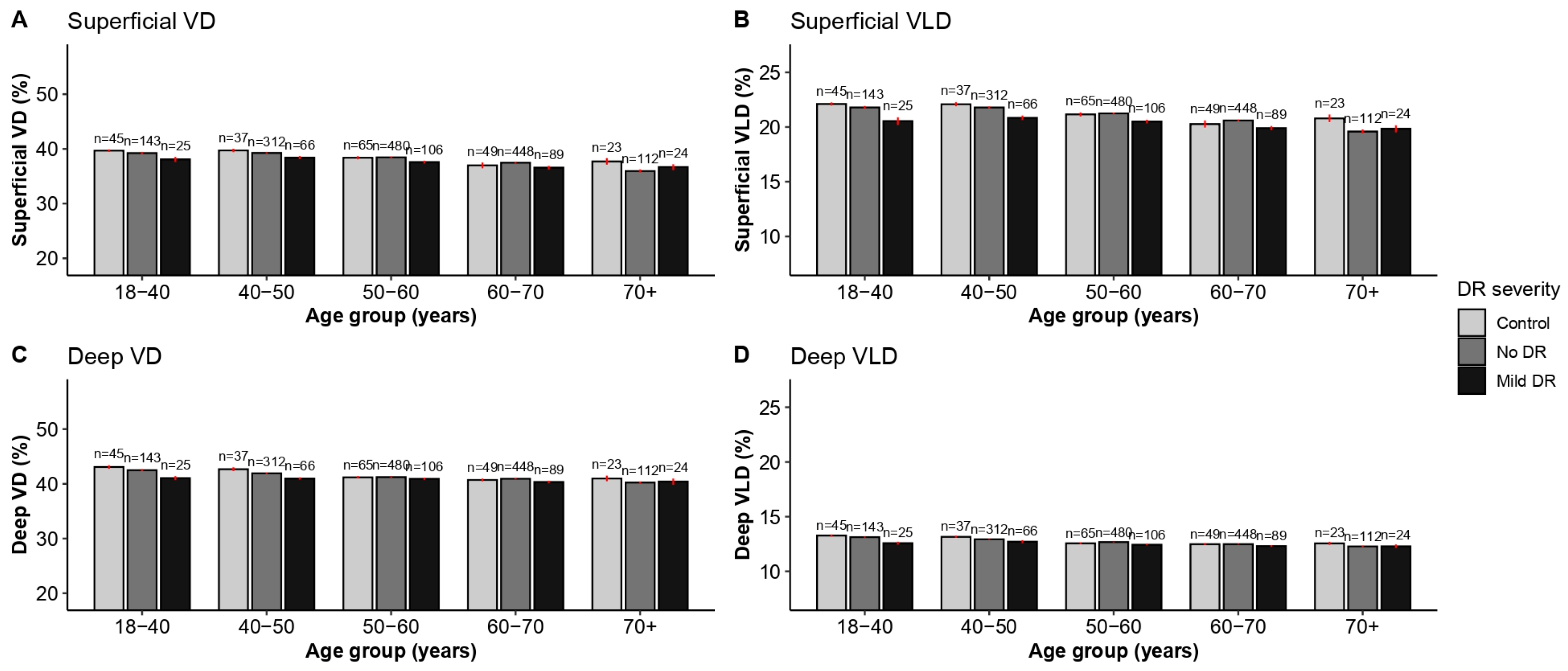
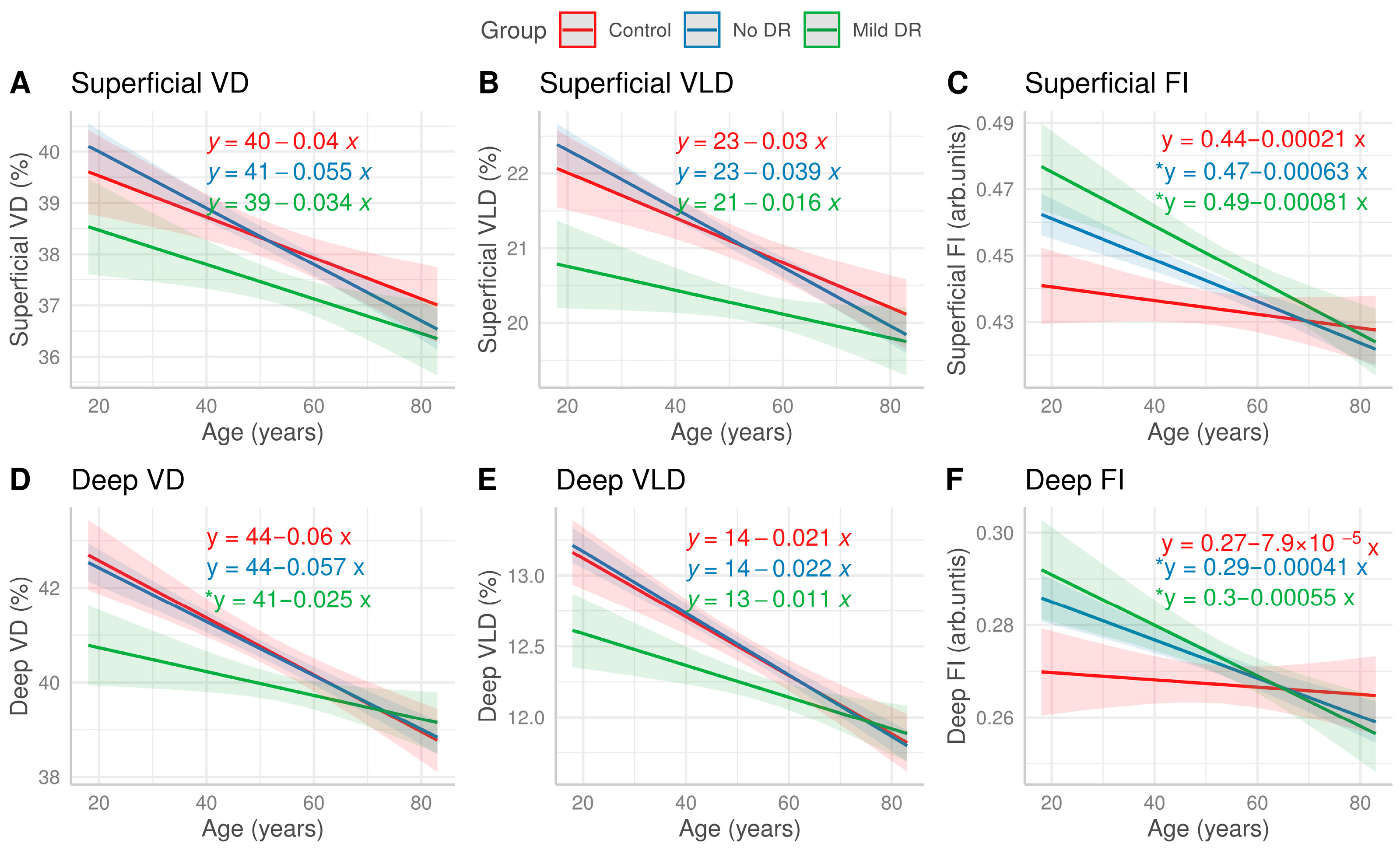

| Diabetic Retinopathy Severity | ||||
|---|---|---|---|---|
| Characteristic | Control, n = 217 | No DR, n = 1352 | Mild DR, n = 286 | Total, n = 1855 |
| Age (years), mean (SD) | 53 (14) | 55 (11) | 56 (11) | 55 (11) |
| Age group (years), n (%) | ||||
| 18–40 | 45 (21) | 130 (9.6) | 22 (7.7) | 197 (11) |
| 40–50 | 37 (17) | 271 (20) | 61 (21) | 369 (20) |
| 50–60 | 64 (29) | 442 (33) | 98 (34) | 604 (33) |
| 60–70 | 49 (23) | 409 (30) | 83 (29) | 541 (29) |
| 70+ | 22 (10) | 100 (7.4) | 22 (7.7) | 144 (7.8) |
| Sex, n (%) | ||||
| Male | 101 (47) | 660 (49) | 148 (52) | 909 (49) |
| Female | 116 (53) | 692 (51) | 138 (48) | 946 (51) |
| Race/ethnicity, n (%) | ||||
| Hispanic | 79 (36) | 555 (41) | 100 (35) | 734 (40) |
| NH Asian | 51 (24) | 352 (26) | 82 (29) | 485 (26) |
| NH Black | 14 (6.5) | 88 (6.5) | 25 (8.7) | 127 (6.8) |
| NH White | 48 (22) | 117 (8.7) | 24 (8.4) | 189 (10) |
| Other | 25 (12) | 240 (18) | 55 (19) | 320 (17) |
| Presence of hypertension, n (%) | 76 (35) | 800 (59) | 179 (63) | 1055 (57) |
| Duration of diabetes (years), mean (SD) | - | 6 (6) | 10 (7) | 7 (6) |
| Hemoglobin A1c (%), mean (SD) | - | 7.87 (3.45) | 8.61 (1.99) | 8.00 (3.25) |
| Axial length (mm), mean (SD) | 23.60 (1.11) | 23.49 (0.98) | 23.46 (1.00) | 23.50 (1.00) |
| LogMAR, mean (SD) | 0.04 (0.07) | 0.09 (0.12) | 0.09 (0.10) | 0.08 (0.11) |
| Signal strength, mean (SD) | 9.76 (0.56) | 9.76 (0.54) | 9.76 (0.51) | 9.76 (0.53) |
| Control (n = 217) | DM No DR (n = 1352) | Mild NPDR (n = 286) | ||||
|---|---|---|---|---|---|---|
| Parameter | Estimate (95% CI) | p-Value | Estimate (95% CI) | p-Value | Estimate (95% CI) | p-Value |
| FAZ area (mm2) | <0.001 (−0.001, 0.001) | 0.777 | <0.001 (−0.000, 0.001) | 0.171 | <0.001 (−0.001, 0.001) | 0.898 |
| FAZ perimeter (mm) | 0.002 (−0.003, 0.007) | 0.567 | 0.002 (−0.000, 0.004) | 0.121 | −0.002 (−0.008, 0.004) | 0.627 |
| Superficial VD (%) | −0.031 (−0.056, −0.007) | 0.026 * | −0.050 (−0.062, −0.038) | <0.001 *** | −0.056 (−0.084, −0.028) | <0.001 *** |
| Superficial VLD (%) | −0.025 (−0.041, −0.010) | 0.003 ** | −0.036 (−0.043, −0.028) | <0.001 *** | −0.029 (−0.047, −0.011) | 0.002 ** |
| Superficial FI | >−0.001 (−0.001, 0.000) | 0.343 | −0.001 (−0.001, −0.001) | <0.001 *** | −0.001 (−0.001, >−0.001) | <0.001 *** |
| Deep FI | >−0.001 (−0.000, 0.000) | 0.774 | >−0.001 (−0.001, −0.000) | <0.001 *** | −0.001 (−0.001, >−0.001) | <0.001 *** |
| Deep VD (%) | −0.051 (−0.070, −0.033) | <0.001 *** | −0.055 (−0.064, −0.045) | <0.001 *** | −0.052 (−0.072, −0.032) | <0.001 *** |
| Deep VLD (%) | −0.018 (−0.024, −0.013) | <0.001 *** | −0.022 (−0.025, −0.019) | <0.001 *** | −0.017 (−0.025, −0.009) | <0.001 *** |
| Age × DM No DR | Age × Mild NPDR | |||||
|---|---|---|---|---|---|---|
| Estimate | 95% CI | p-Value | Estimate | 95% CI | p-Value | |
| FAZ area (mm2) | <0.001 | (−0.001, 0.001) | 0.797 | −0.001 | (−0.002, 0.001) | 0.521 |
| FAZ perimeter (mm) | <0.001 | (−0.005, 0.005) | 0.924 | −0.005 | (−0.011, 0.002) | 0.240 |
| SCP VD (%) | −0.015 | (−0.039, 0.009) | 0.301 | 0.006 | (−0.025, 0.038) | 0.793 |
| SCP VLD (%) | −0.009 | (−0.024, 0.006) | 0.312 | 0.014 | (−0.006, 0.034) | 0.240 |
| SCP FI | >−0.001 | (−0.001, 0) | 0.027 * | >−0.001 | (−0.001, 0) | 0.015 * |
| DCP FI | >−0.001 | (−0.001, 0) | 0.030 * | >−0.001 | (−0.001, 0) | 0.025 * |
| DCP VD (%) | 0.003 | (−0.018, 0.025) | 0.797 | 0.035 | (0.007, 0.063) | 0.029 * |
| DCP VLD (%) | 0.003 | (−0.018, 0.025) | 0.797 | 0.035 | (0.007, 0.063) | 0.059 |
Disclaimer/Publisher’s Note: The statements, opinions and data contained in all publications are solely those of the individual author(s) and contributor(s) and not of MDPI and/or the editor(s). MDPI and/or the editor(s) disclaim responsibility for any injury to people or property resulting from any ideas, methods, instructions or products referred to in the content. |
© 2026 by the authors. Licensee MDPI, Basel, Switzerland. This article is an open access article distributed under the terms and conditions of the Creative Commons Attribution (CC BY) license.
Share and Cite
Taha, A.; Zhang, Y.S.; Ma, C.J.; Stewart, J.M. Diabetes May Modulate the Association Between Age and Optical Coherence Tomography Angiography Parameters: A Serial, Cross-Sectional Study. J. Pers. Med. 2026, 16, 286. https://doi.org/10.3390/jpm16060286
Taha A, Zhang YS, Ma CJ, Stewart JM. Diabetes May Modulate the Association Between Age and Optical Coherence Tomography Angiography Parameters: A Serial, Cross-Sectional Study. Journal of Personalized Medicine. 2026; 16(6):286. https://doi.org/10.3390/jpm16060286
Chicago/Turabian StyleTaha, Abu, Yi Stephanie Zhang, Chu Jian Ma, and Jay M. Stewart. 2026. "Diabetes May Modulate the Association Between Age and Optical Coherence Tomography Angiography Parameters: A Serial, Cross-Sectional Study" Journal of Personalized Medicine 16, no. 6: 286. https://doi.org/10.3390/jpm16060286
APA StyleTaha, A., Zhang, Y. S., Ma, C. J., & Stewart, J. M. (2026). Diabetes May Modulate the Association Between Age and Optical Coherence Tomography Angiography Parameters: A Serial, Cross-Sectional Study. Journal of Personalized Medicine, 16(6), 286. https://doi.org/10.3390/jpm16060286

