Assessment of Optimal Stent Implantation with the Use of Optical Coherence Tomography in Patients with Coronary Artery Disease
 , , ,
, , ,
Abstract
1. Introduction
2. Methods
2.1. Study Population
2.2. Optical Coherence Tomography Image Acquisition and Analysis
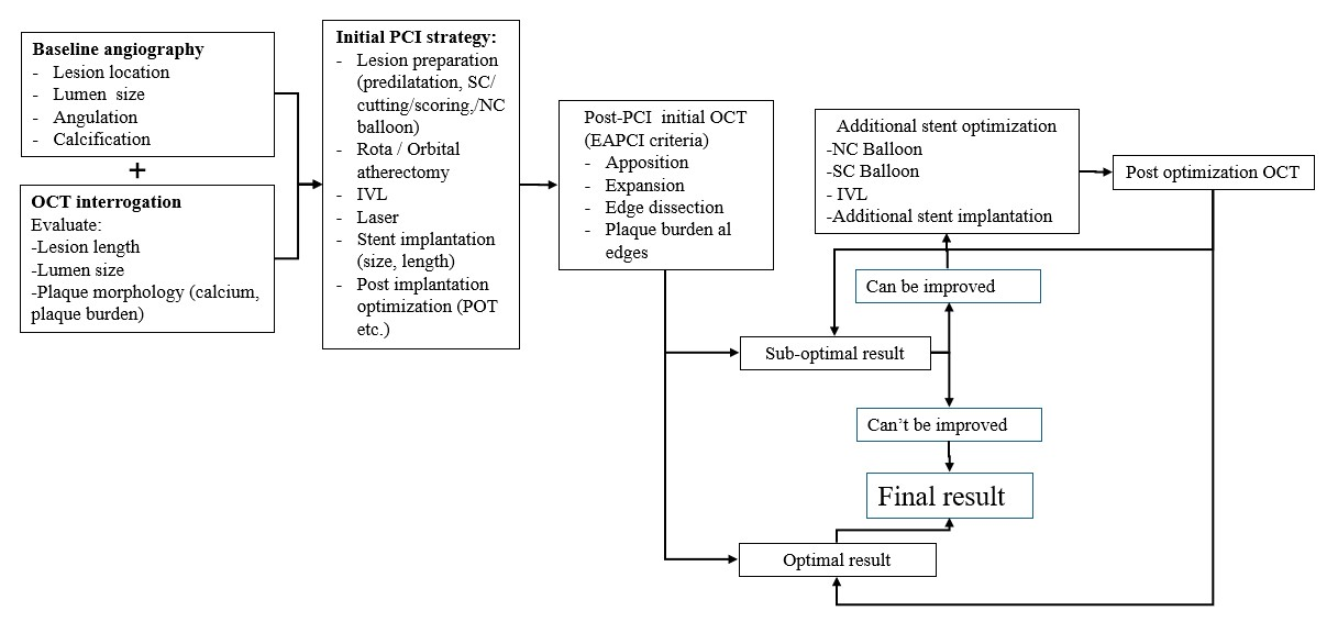
2.3. Study Workflow and OCT-Guided Stent Optimization Protocol
2.4. Endpoints
2.5. Statistical Analysis
3. Results
3.1. Patient Baseline Characteristics
3.2. Lesion and Treatment Characteristics
3.3. OCT Baseline Vessel Characteristics
3.4. Post-PCI OCT Evaluation and Optimization
3.5. OCT Results (Areas, Expansion) After Stent Optimization (Included Also in Table 4)
4. Discussion
5. Limitations
- -
- This is a single-centered study with data collected from a limited number of patients and vessels.
- -
- Left main lesions were not included, as OCT guidance for this subset of lesions is more cumbersome.
- -
- Complex lesions, such as bifurcation lesions or heavily calcified lesions, were not included in this study.
- -
- Our data may not be used to determine which types of lesions OCT may provide valuable information for stent optimization, especially due to the limited number of lesions evaluated.
- -
- Patients with multiple stents (like additional stent implantation) may experience more procedures and a greater risk of adverse outcomes, including the need for surgery, even though in our study we did not have this situation.
6. Conclusions
Supplementary Materials
Author Contributions
Funding
Institutional Review Board Statement
Informed Consent Statement
Data Availability Statement
Conflicts of Interest
References
- Prati, F.; Di Vito, L.; Biondi-Zoccai, G.; Occhipinti, M.; La Manna, A.; Tamburino, C.; Burzotta, F.; Trani, C.; Porto, I.; Ramazzotti, V.; et al. Angiography alone versus angiography plus optical coherence tomography to guide decision-making during percutaneous coronary intervention: The Centro per la Lotta contro l’Infarto-Optimisation of Percutaneous Coronary Intervention (CLI-OPCI) study. EuroIntervention 2012, 8, 823–829. [Google Scholar] [CrossRef] [Scilit]
- Vergallo, R.; Ren, X.; Yonetsu, T.; Kato, K.; Uemura, S.; Yu, B.; Jia, H.; Abtahian, F.; Aguirre, A.D.; Tian, J.; et al. Pancoronary plaque vulnerability in patients with acute coronary syndrome and ruptured culprit plaque: A 3-vessel optical coherence tomography study. Am. Heart J. 2014, 167, 59–67. [Google Scholar] [CrossRef] [Scilit] [PubMed]
- Prati, F.; Romagnoli, E.; La Manna, A.; Burzotta, F.; Gatto, L.; Marco, V.; Fineschi, M.; Fabbiocchi, F.; Versaci, F.; Trani, C.; et al. Long-term consequences of optical coherence tomography findings during percutaneous coronary intervention: The Centro Per La Lotta Contro L’infarto—Optimization Of Percutaneous Coronary Intervention (CLI-OPCI) LATE study. EuroIntervention 2018, 14, e443–e451. [Google Scholar] [CrossRef] [Scilit] [PubMed]
- Sreenivasan, J.; Reddy, R.K.; Jamil, Y.; Malik, A.; Chamie, D.; Howard, J.P.; Nanna, M.G.; Mintz, G.S.; Maehara, A.; Ali, Z.A.; et al. Intravascular Imaging-Guided Versus Angiography-Guided Percutaneous Coronary Intervention: A Systematic Review and Meta-Analysis of Randomized Trials. J. Am. Heart Assoc. 2024, 13, e031111. [Google Scholar] [CrossRef] [Scilit]
- Truesdell, A.G.; Alasnag, M.A.; Kaul, P.; Rab, S.T.; Riley, R.F.; Young, M.N.; Batchelor, W.B.; Maehara, A.; Welt, F.G.; Kirtane, A.J.; et al. Intravascular Imaging During Percutaneous Coronary Intervention: JACC State-of-the-Art Review. J. Am. Coll. Cardiol. 2023, 81, 590–605. [Google Scholar] [CrossRef] [Scilit] [PubMed]
- Ali, Z.A.; Shin, D.; Chaturvedi, A.; Waksman, R. We now have enough evidence to support systematic OCT in daily PCI practice: Pros and cons. EuroIntervention 2024, 20, 533–535. [Google Scholar] [CrossRef] [Scilit]
- Ploscaru, V.; Popa-Fotea, N.M.; Calmac, L.; Itu, L.M.; Mihai, C.; Bataila, V.; Dragoescu, B.; Puiu, A.; Cojocaru, C.; Costin, M.A.; et al. Artificial intelligence and cloud based platform for fully automated PCI guidance from coronary angiography-study protocol. PLoS ONE 2022, 17, e0274296. [Google Scholar] [CrossRef] [Scilit]
- Räber, L.; Mintz, G.S.; Koskinas, K.C.; Johnson, T.W.; Holm, N.R.; Onuma, Y.; Radu, M.D.; Joner, M.; Yu, B.; Jia, H.; et al. Clinical use of intracoronary imaging. Part 1: Guidance and optimization of coronary interventions. An expert consensus document of the European Association of Percutaneous Cardiovascular Interventions. Eur. Heart J. 2018, 39, 3281–3300, Correction in Eur. Heart J. 2019, 40, 308. https://doi.org/10.1093/eurheartj/ehy460. [Google Scholar] [CrossRef] [Scilit]
- Prati, F.; Romagnoli, E.; Burzotta, F.; Limbruno, U.; Gatto, L.; La Manna, A.; Versaci, F.; Marco, V.; Di Vito, L.; Imola, F.; et al. Clinical Impact of OCT Findings During PCI: The CLI-OPCI II Study. JACC Cardiovasc. Imaging 2015, 8, 1297–1305. [Google Scholar] [CrossRef] [Scilit]
- Fujii, K.; Carlier, S.G.; Mintz, G.S.; Yang, Y.M.; Moussa, I.; Weisz, G.; Dangas, G.; Mehran, R.; Lansky, A.J.; Kreps, E.M.; et al. Stent underexpansion and residual reference segment stenosis are related to stent thrombosis after sirolimus-eluting stent implantation: An intravascular ultrasound study. J. Am. Coll. Cardiol. 2005, 45, 995–998. [Google Scholar] [CrossRef] [Scilit]
- Meneveau, N.; Souteyrand, G.; Motreff, P.; Caussin, C.; Amabile, N.; Ohlmann, P.; Morel, O.; Lefrançois, Y.; Descotes-Genon, V.; Silvain, J.; et al. Optical Coherence Tomography to Optimize Results of Percutaneous Coronary Intervention in Patients with Non-ST-Elevation Acute Coronary Syndrome: Results of the Multicenter, Randomized DOCTORS Study (Does Optical Coherence Tomography Optimize Results of Stenting). Circulation 2016, 134, 906–917. [Google Scholar] [CrossRef] [Scilit]
- Wijns, W.; Shite, J.; Jones, M.R.; Lee, S.W.L.; Price, M.J.; Fabbiocchi, F.; Barbato, E.; Akasaka, T.; Bezerra, H.; Holmes, D. Optical coherence tomography imaging during percutaneous coronary intervention impacts physician decision-making: ILUMIEN I study. Eur. Heart J. 2015, 36, 3346–3355. [Google Scholar] [CrossRef] [Scilit]
- Soeda, T.; Uemura, S.; Park, S.J.; Kim, J.S.; Lee, S.J.; Itoh, T.; Yonetsu, T.; Kakuta, T.; Jang, I.K. Incidence and Clinical Significance of Poststent Optical Coherence Tomography Findings: One-Year Follow-Up Study from a Multicenter Registry. Circulation 2015, 132, 1020–1029, Correction in Circulation 2016, 133, e670. https://doi.org/10.1161/CIR.0000000000000429. [Google Scholar] [CrossRef] [Scilit] [PubMed]
- Adriaenssens, T.; Joner, M.; Godschalk, T.C.; Malik, N.; Alfonso, F.; Xhepa, E.; De Cock, D.; Komukai, K.; Tada, T.; Cuesta, J.; et al. Optical Coherence Tomography Findings in Patients with Coronary Stent Thrombosis: A Report of the PRESTIGE Consortium (Prevention of Late Stent Thrombosis by an Interdisciplinary Global European Effort). Circulation 2017, 136, 1007–1021. [Google Scholar] [CrossRef] [Scilit] [PubMed]
- Souteyrand, G.; Amabile, N.; Mangin, L.; Chabin, X.; Meneveau, N.; Cayla, G.; Vanzetto, G.; Barnay, P.; Trouillet, C.; Rioufol, G.; et al. Mechanisms of stent thrombosis analysed by optical coherence tomography: Insights from the national PESTO French registry. Eur. Heart J. 2016, 37, 1208–1216. [Google Scholar] [CrossRef] [Scilit] [PubMed]
- Ali, Z.A.; Maehara, A.; Généreux, P.; Shlofmitz, R.A.; Fabbiocchi, F.; Nazif, T.M.; Guagliumi, G.; Meraj, P.M.; Alfonso, F.; Samady, H.; et al. Optical coherence tomography compared with intravascular ultrasound and with angiography to guide coronary stent implantation (ILUMIEN III: OPTIMIZE PCI): A randomised controlled trial. Lancet 2016, 388, 2618–2628. [Google Scholar] [CrossRef] [Scilit]
- Choi, S.Y.; Witzenbichler, B.; Maehara, A.; Lansky, A.J.; Guagliumi, G.; Brodie, B.; Kellett, M.A., Jr.; Dressler, O.; Parise, H.; Mehran, R.; et al. Intravascular ultrasound findings of early stent thrombosis after primary percutaneous intervention in acute myocardial infarction: A Harmonizing Outcomes with Revascularization and Stents in Acute Myocardial Infarction (HORIZONS-AMI) substudy. Circ. Cardiovasc. Interv. 2011, 4, 239–247. [Google Scholar] [CrossRef] [Scilit]
- Cheneau, E.; Leborgne, L.; Mintz, G.S.; Kotani, J.I.; Pichard, A.D.; Satler, L.F.; Canos, D.; Castagna, M.; Weissman, N.J.; Waksman, R. Predictors of subacute stent thrombosis: Results of a systematic intravascular ultrasound study. Circulation 2003, 108, 43–47. [Google Scholar] [CrossRef] [Scilit]
- Rai, H.; Harzer, F.; Otsuka, T.; Abdelwahed, Y.S.; Antuña, P.; Blachutzik, F.; Koppara, T.; Räber, L.; Leistner, D.M.; Alfonso, F.; et al. Stent Optimization Using Optical Coherence Tomography and Its Prognostic Implications After Percutaneous Coronary Intervention. J. Am. Heart Assoc. 2022, 11, e023493. [Google Scholar] [CrossRef] [Scilit]
- Qiu, F.; Mintz, G.S.; Witzenbichler, B.; Metzger, D.C.; Rinaldi, M.J.; Duffy, P.L.; Weisz, G.; Stuckey, T.D.; Brodie, B.R.; Parvataneni, R.; et al. Prevalence and Clinical Impact of Tissue Protrusion After Stent Implantation: An ADAPT-DES Intravascular Ultrasound Substudy. JACC Cardiovasc. Interv. 2016, 9, 1499–1507. [Google Scholar] [CrossRef] [Scilit]
- Burzotta, F.; Leone, A.M.; Aurigemma, C.; Zambrano, A.; Zimbardo, G.; Arioti, M.; Vergallo, R.; De Maria, G.L.; Cerracchio, E.; Romagnoli, E.; et al. Fractional Flow Reserve or Optical Coherence Tomography to Guide Management of Angiographically Intermediate Coronary Stenosis: A Single-Center Trial. JACC Cardiovasc. Interv. 2020, 13, 49–58. [Google Scholar] [CrossRef] [Scilit] [PubMed]
- Lee, J.M.; Choi, K.H.; Song, Y.B.; Lee, J.Y.; Lee, S.J.; Lee, S.Y.; Kim, S.M.; Yun, K.H.; Cho, J.Y.; Kim, C.J.; et al. Intravascular Imaging-Guided or Angiography-Guided Complex PCI. N. Engl. J. Med. 2023, 388, 1668–1679. [Google Scholar] [CrossRef] [Scilit] [PubMed]
- Vrints, C.; Andreotti, F.; Koskinas, K.C.; Rossello, X.; Adamo, M.; Ainslie, J.; Banning, A.P.; Budaj, A.; Buechel, R.R.; Chiariello, G.A.; et al. 2024 ESC Guidelines for the management of chronic coronary syndromes. Eur. Heart J. 2024, 45, 3415–3537. [Google Scholar] [CrossRef] [Scilit]
- Koskinas, K.C.; Nakamura, M.; Räber, L.; Colleran, R.; Kadota, K.; Capodanno, D.; Wijns, W.; Akasaka, T.; Valgimigli, M.; Guagliumi, G.; et al. Current use of intracoronary imaging in interventional practice—Results of a European Association of Percutaneous Cardiovascular Interventions (EAPCI) and Japanese Association of Cardiovascular Interventions and Therapeutics (CVIT) Clinical Practice Survey. EuroIntervention 2018, 14, e475–e484. [Google Scholar] [CrossRef] [Scilit]
- Low, A.F.; Wongpraparut, N.; Chunhamaneewat, N.; Jeamanukoolkit, A.; Jhung, L.T.; Zhen-Vin, L.; Tan, C.T.; Hwa, H.H.; Rajagopal, R.; Yahya, A.F.; et al. Clinical use of optical coherence tomography during percutaneous coronary intervention and coronary procedures in Southeast Asia: A survey-based expert consensus summary. AsiaIntervention 2023, 9, 25–31. [Google Scholar] [CrossRef] [Scilit] [PubMed]



| All Patients (n = 64) | |
|---|---|
| Age, y | 63.03 ± 0.40 |
| Male sex | 79.69% (51) |
| Diabetes mellitus | 21.87% (14) |
| Hypertension | 84.37% (54) |
| Dyslipidemia | 87.50% (56) |
| Ever smoker | 65.63% (42) |
| History of angina | 53.97% (34) |
| History of ACS | 29.69% (19) |
| LVEF,% | 48.10 ± 6.32 |
| eGFR, mL/min/1.73 m2 | 89.93 ± 17.25 |
| Clinical presentation | |
| STEMI | 53.12% (34) |
| NSTEMI | 6.25% (4) |
| UA | 37.50% (24) |
| Stable angina | 3.13% (2) |
| Number of vessels diseased | |
| Single vessel disease | 26.56% (17) |
| Double vessel disease | 60.94% (39) |
| Triple vessel disease | 12.50% (8) |
| Mean number of vessels | 1.85 ± 0.63 |
| Total Vessels (n = 73) | |
|---|---|
| Lesion location | |
| Left anterior descending artery | 64.38% (47) |
| Right coronary artery | 20.55% (15) |
| Circumflex artery | 8.22% (6) |
| Side branches | 6.85% (5) |
| Number of stents implanted | |
| 1 | 87.67% (64) |
| 2 | 10.96% (8) |
| 3 | 1.37% (1) |
| Mean number of stents | 1.13 ± 0.38 |
| Stent characteristics | |
| Metallic 2nd-generation drug eluting stent | 100% |
| Stent length, mm | 27.94 ± 0.41 |
| Stent diameter, mm2 | 3.08 ± 0.41 |
| Total Vessels (n = 73) | |
|---|---|
| Lumen measurements | |
| Proximal lumen area, mm2 | 7.89 (6.41–10.65) |
| Proximal lumen diameter, mm | 3.17 (2.83–3.61) |
| Distal lumen area, mm2 | 5.53 (4.13–7.38) |
| Distal lumen diameter, mm | 2.70 (2.32–3.06) |
| Average reference lumen area, mm2 | 6.86 (5.76–8.84) |
| Average reference lumen diameter, mm | 2.93 (2.63–3.27) |
| Minimum lumen area, mm2 | 1.49 (1.12–1.93) |
| Minimum lumen diameter, mm | 1.21 (1.11–1.48) |
| Lesion length, mm | 32.60 (24.05–42.30) |
| Calcium characteristics | |
| Presence of calcium | 57.53% (42) |
| Thickness > 0.5 mm | 65.12% (28) |
| Angle > 180° vessel arc | 24.24% (8) |
| Length > 5 mm | 53.49% (23) |
| OCT Post-PCI (n = 73) | OCT Post Optimization (n = 73) | |
|---|---|---|
| Stent expansion index classification, % | ||
| <80% | 42.46% (31) | 36.99% (27) |
| 80–90% | 31.51% (23) | 35.61% (26) |
| >90% | 26.03% (19) | 27.40% (20) |
| Stent measurements | ||
| Minimum stent area > 4.5 mm2 | 82.19% (60) | 87.67% (64) |
| Minimum stent area < 4.5 mm2 | 17.81% (13) | 12.33% (9) |
| Minimum stent area, mm2 | 5.44 (4.61–7.16) | 5.46 (4.72–7.32) |
| Stent expansion index | 81.15 (71.42–90.48) | 81.51 (73.50–91.00) |
| OCT Post-PCI (n = 73) | OCT Post Optimization (n = 73) | |
|---|---|---|
| Suboptimal stent implantation measurements | ||
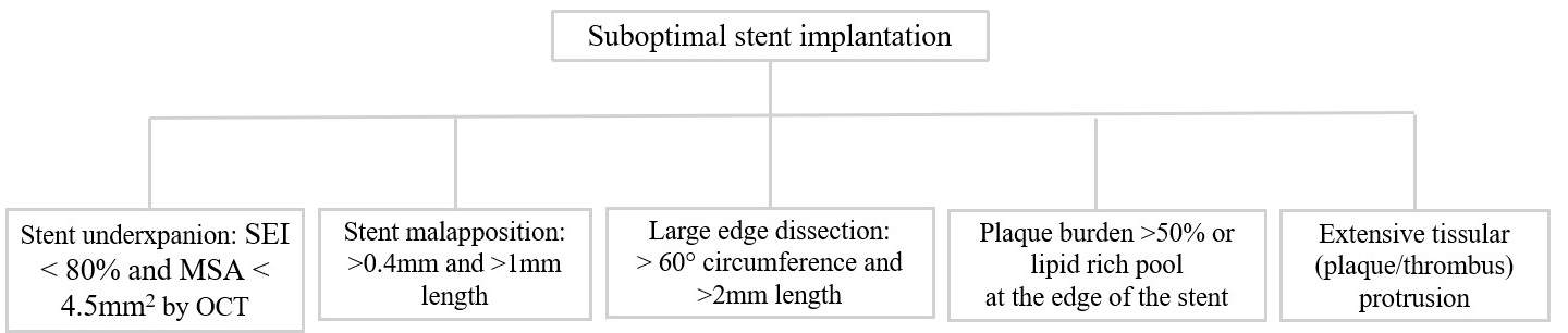
| Total suboptimally stented vessels | 35.61% (26) | 6.84% (5) |
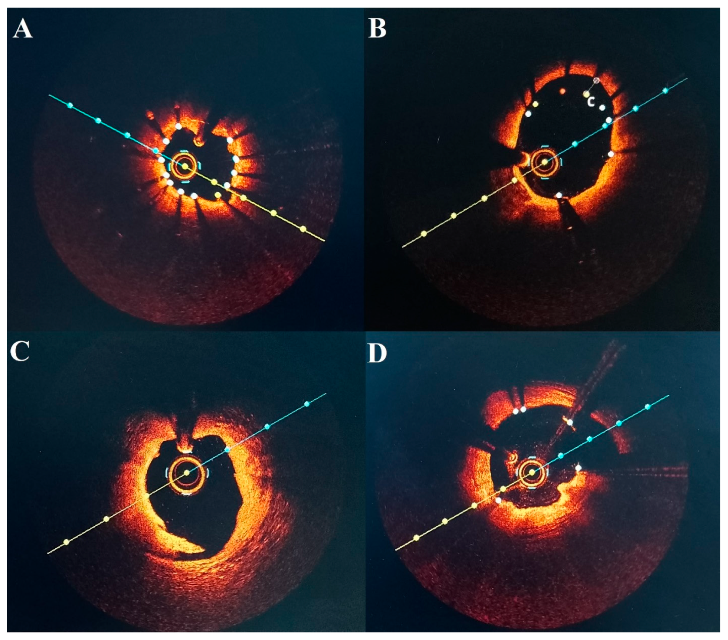
| Stent underexpansion | 9.58% (7) | 5.47% (4) |
| Stent malapposition | 15.06% (11) | 0.0% (0) |
| Edge dissection | 6.84% (5) | 0.0% (0) |
| Plaque burden or lipid-rich pool | 2.73% (2) | 0.0% (0) |
| Tissue protrusion (plaque/thrombus) | 1.36% (1) | 1.36% (1) |
| NC Balloon Diameter, mm | SC Balloon Diameter, mm | Stent Diameter, mm | Pressure, atm | |
|---|---|---|---|---|
| Suboptimal stent implantation | ||||
| Stent underexpansion | 3.07 ± 0.41 | - | - | 18.50 ± 4.01 |
| Stent malapposition | 3.39 ± 0.57 | 3.30 ± 0.97 | - | 17.40 ± 3.13 |
| Edge dissection | - | - | 3.20 ± 0.44 | 15 ± 1.41 |
| Plaque burden or lipid rich pool | - | - | 2.37 ± 0.17 | 15 ± 1.41 |
Disclaimer/Publisher’s Note: The statements, opinions and data contained in all publications are solely those of the individual author(s) and contributor(s) and not of MDPI and/or the editor(s). MDPI and/or the editor(s) disclaim responsibility for any injury to people or property resulting from any ideas, methods, instructions or products referred to in the content. |
© 2026 by the authors. Licensee MDPI, Basel, Switzerland. This article is an open access article distributed under the terms and conditions of the Creative Commons Attribution (CC BY) license.
Share and Cite
Kaperonis, A.; Scafa-Udriște, A.; Mihai, C.; Bataila, V.; Drăgoescu, B.M.; Ploscaru, V.; Zamfir, D.; Popescu, R.; Tonu, D.; Calmac, L. Assessment of Optimal Stent Implantation with the Use of Optical Coherence Tomography in Patients with Coronary Artery Disease. Diagnostics 2026, 16, 813. https://doi.org/10.3390/diagnostics16050813
Kaperonis A, Scafa-Udriște A, Mihai C, Bataila V, Drăgoescu BM, Ploscaru V, Zamfir D, Popescu R, Tonu D, Calmac L. Assessment of Optimal Stent Implantation with the Use of Optical Coherence Tomography in Patients with Coronary Artery Disease. Diagnostics. 2026; 16(5):813. https://doi.org/10.3390/diagnostics16050813
Chicago/Turabian StyleKaperonis, Alexandros, Alexandru Scafa-Udriște, Cosmin Mihai, Vlad Bataila, Bogdan Marian Drăgoescu, Vlad Ploscaru, Diana Zamfir, Radu Popescu, Daniel Tonu, and Lucian Calmac. 2026. "Assessment of Optimal Stent Implantation with the Use of Optical Coherence Tomography in Patients with Coronary Artery Disease" Diagnostics 16, no. 5: 813. https://doi.org/10.3390/diagnostics16050813
APA StyleKaperonis, A., Scafa-Udriște, A., Mihai, C., Bataila, V., Drăgoescu, B. M., Ploscaru, V., Zamfir, D., Popescu, R., Tonu, D., & Calmac, L. (2026). Assessment of Optimal Stent Implantation with the Use of Optical Coherence Tomography in Patients with Coronary Artery Disease. Diagnostics, 16(5), 813. https://doi.org/10.3390/diagnostics16050813