


Abstract
Breast cancer remains the most common female cancer globally, and mammography is pivotal for its early screening and diagnosis, with quality control (QC) as a core guarantee. Mammography QC has advanced from early technical exploration to whole-process management, driven by technological innovation and standardized protocols. This review summarizes the historical evolution, influencing factors, corresponding measures, limitations, and prospects of mammography QC to promote the construction of standardized management systems.
1. Introduction
Since the inaugural global cancer burden assessment was published in the 1980s, breast cancer has consistently ranked as the most frequently diagnosed malignancy among women worldwide, posing a grave threat to female health and imposing a substantial social burden. Over the past three decades, the global incidence rate of breast cancer has increased by 57.8%, with an average annual growth rate of approximately 0.5% [1]. In 2022 alone, there were 2.3 million newly diagnosed cases and 670,000 deaths worldwide [2]. Common imaging examination methods for the breast include mammography, ultrasound, and magnetic resonance imaging (MRI). Mammography uses differences in X-ray absorption among tissues to detect abnormal findings in the breast, such as masses, calcifications, and structural distortions. It is particularly sensitive to microcalcifications and is currently the most evidence-based imaging technology for reducing breast cancer mortality [3]. The American College of Radiology (ACR) recommends annual mammography screening for women starting at age 40 [4], but because the examination involves ionizing radiation, ensuring optimal image quality to reduce radiation exposure is especially crucial. For dense breasts, ultrasound serves as a valuable adjunct to mammography, as it is independent of breast density. However, ultrasound is limited in detecting deep-seated lesions and microcalcifications, and its diagnostic accuracy is highly operator-dependent. In high-risk populations, such as BRCA1/2 mutation carriers, MRI is added to mammography and ultrasound protocols to improve diagnostic sensitivity [5]. Nevertheless, due to high costs, long acquisition times, and a high false-positive rate, MRI is not suitable for routine screening of the general population.
Multimodal imaging optimizes early breast cancer detection by leveraging the strengths of each technique, with mammography remaining a cornerstone of clinical practice. Image quality is the foundation for accurate radiologic diagnosis, defined by technical parameters such as signal-to-noise ratio (SNR), spatial resolution, and contrast [6]. Quality control (QC) in mammography is the core guarantee of high-quality images and is susceptible to multiple factors, including equipment, technologists, physicians, patients, and the environment. Focusing on issues such as inconsistent global protocols, subjective variability, and limited adaptability to emerging technologies in mammography QC, this review systematically traces the historical evolution and key determinants of QC systems, further analyzes current limitations, and envisions the future of precision intelligence, offering insights for the construction of a standardized QC management framework.
2. Historical Development of Mammography QC
From the early use of traditional tungsten-target X-ray tubes for mammography to the emergence of dedicated mammography machines and the subsequent adoption of full-field digital mammography (FFDM) systems, the development of mammography QC has been closely linked to advances in breast imaging technology and the renewal of quality management concepts, evolving from simple initial technical operations to whole-process management today.
2.1. Early Exploration and Technical Foundation of Mammography QC
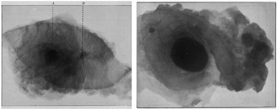
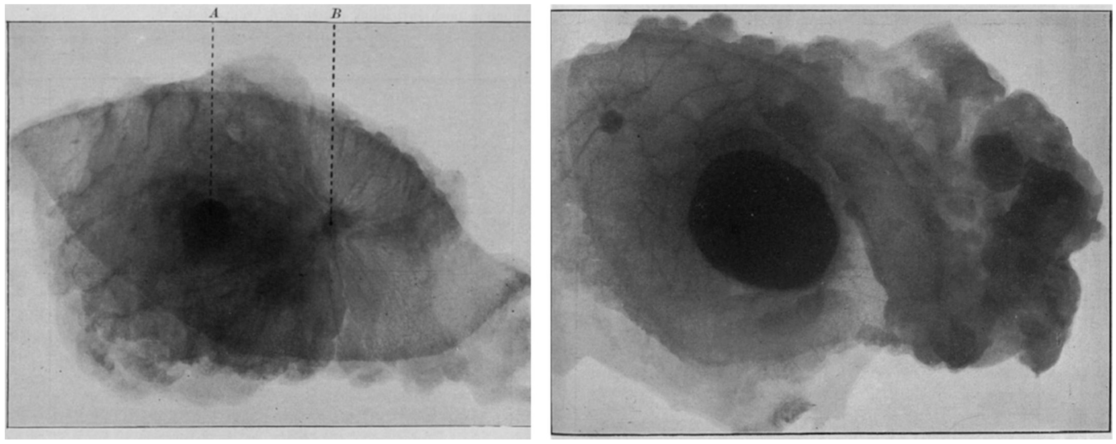
The history of mammography can be traced back to 1913, when Salomon first performed X-ray imaging on breast tissue specimens and confirmed the X-ray characteristics of breast cancer (Figure 1) [7]. In the 1950s, Leborgne identified the diagnostic value of microcalcifications for breast tumors [8] and proposed that image quality could be improved by breast compression and immobilization [9]. However, limited by the performance of equipment at that time, mammography QC was still in the stage of early exploration and technical foundation-laying. The advent of Egan’s mammography technique [10] and the first dedicated mammography machine, Senographe, in 1965 [11] promoted the clinical application of mammography. In 1971, Shapiro et al. reported findings from a randomized controlled trial in which they compared regular mammography screening and clinical breast examination against routine medical care alone. The study revealed a significant reduction in breast cancer mortality in the screened group at 3.5 years, providing the first robust evidence that mammography screening reduces breast cancer mortality [12]. Concurrently, there was a growing recognition that image quality exerts a significant impact on diagnostic accuracy [13].
Figure 1.
Mammographic images of breast tissue specimens by Salomon Albert in 1913. (Left) Chronic mastitis complicated with solid carcinoma. A. Nipple. B. Solid carcinoma with radiating extensions. (Right) A well-circumscribed cancer mass is visualized in the mid-field. Malignant lymphadenopathy is identified within the right axillary adipose tissue. Reprinted with permission from: [7].
2.2. Establishment of a Standardized Mammography QC System
In the 1980s, the ACR launched the first mammography accreditation program, which put forward basic requirements for equipment, radiation dose, and personnel qualification, representing the initial standardized attempt at mammography QC [14]. However, being voluntary in nature, it had relatively few participating institutions in the early stages. In 1992, the U.S. Congress enacted the Mammography Quality Standards Act (MQSA), making the ACR’s accreditation program a mandatory requirement. In addition to the basic requirements, it also required the introduction of final assessment grading and recommendations, patient result notification, follow-up review, and annual inspections of accredited institutions [15]. During this period, significant advancements were made in improving image quality and optimizing radiation dose in mammography [16], which also promoted the reclassification of breast lesions. In 1992, the ACR released the first edition of the Breast Imaging Reporting and Data System (BI-RADS), which standardized the terminology and assessment classification of mammography and built a unified communication framework for radiologists and clinicians [17]. After five updates, BI-RADS has become a global standard (Table 1). The implementation of the MQSA and the development of BI-RADS have greatly promoted the establishment of a standardized mammography QC system, realizing the transition from scattered technical operations to institutionalized quality management.
Table 1.
BI-RADS assessment categories.
2.3. Development of Mammography QC in the Digital Era
The revolutionary digital mammography technology was approved by the U.S. Food and Drug Administration (FDA) in 2000 [19], which enabled post-processing of imaging data and the development of computer-aided detection and diagnosis (CAD) [20] and promoted the shift in QC from the performance assessment of screen-film systems to the refined control of the entire image acquisition–processing–display chain [21]. In 2003, Italian researchers developed the first phantom for periodic QC measurements of digital mammography systems to assess the reproducibility of parameters such as detector linearity, uniformity, image contrast, and spatial resolution [22]. The American College of Radiology Imaging Network (ACRIN) has conducted the Digital Mammography Imaging Screening Trial (DMIST) since 2001, and the FDA has implemented the “Enhancing Quality Using the Inspection Program (EQUIP)” since 2017 to ensure the optimal operation of digital equipment and the effectiveness of image quality [23,24]. Studies have demonstrated that structured audit logs within the EQUIP framework effectively enhance the performance of QC [24], but extra views at screening mammography increased with EQUIP implementation [25]. In March 2023, the FDA released a revised version of the MQSA (“2023 MQSA Final Rule”). A key update mandates that testing facilities provide patients with information about their breast density, thereby helping patients obtain important information that may affect their treatment decisions as completely as possible [26].
3. Factors Influencing Mammography Image Quality and Corresponding QC Measures
All steps of mammography are interconnected, ranging from patient management, breast positioning and compression, and image acquisition to image diagnosis and recommendations, record keeping, and result notification, as well as equipment performance testing, radiation dose optimization, and examination environment improvement [27]. Oversight in any link may affect the final image quality (Table 2).
Table 2.
Factors influencing mammographic image quality, core QC measures, and related studies.
3.1. X-Ray Equipment and Technical Parameters
The performance of mammography equipment and the accurate selection of technical parameters are important foundations for obtaining high-quality images. Appropriate X-ray tube targets and filtration materials have a significant impact on the X-ray spectrum, energy distribution, and image contrast. Combinations of wolfram (W) or molybdenum (Mo) targets with rhodium (Rh) or silver (Ag) filters are common clinical choices. Studies have shown that the W/Rh combination can provide better image quality and visibility of fibers and specks than the W/Ag combination, particularly at tube voltages of 30 and 32 kVp [28]. Meanwhile, breast thickness also affects the selection of target-filter combinations; for thin breasts, the Mo/Mo or Mo/Rh combinations yield better image quality [29]. The kVp and mAs are key parameters that determine the quality and quantity of X-rays. Under different breast thicknesses and tissue types, optimizing exposure parameters can reduce patient radiation dose while improving the image SNR [30]. The AEC system can automatically select the optimal exposure parameters to ensure the consistency of image density, but it is affected by multiple factors such as breast thickness, glandular composition, and body position [31]. In the era of digital mammography, image post-processing and display devices are also crucial links affecting image quality [21]. During a series of complex image processing processes (such as contrast enhancement, edge sharpening, and noise suppression), different algorithms can produce images with varying effects, which may enhance the visibility of lesions or introduce artifacts or distortions [33].
Regular performance evaluation and calibration of X-ray equipment are the focus of mammography QC, including the X-ray generation system (tube voltage, tube current, exposure time), detector performance (SNR, contrast-to-noise ratio, detective quantum efficiency), and image processing and display systems [50,51]. In early practices, image quality quantification was often achieved through manual periodic detection of the visibility of targets in standardized phantoms (such as the ACR-certified phantom, Contrast-Detail Mammography [CDMAM] phantom, and Polymethyl Methacrylate [PMMA] phantom) [34,35]. With technological advancements, the International Atomic Energy Agency (IAEA) proposed the integration of the ATAI to enable remote and automated quality control of mammography equipment in resource-limited regions, representing a new direction in performance monitoring [36]. It is necessary to test the repeatability of the AEC system and the consistency of exposure under different breast thicknesses to obtain appropriate image density under various clinical conditions [32]. In addition, ensuring that the brightness, contrast, and spatial resolution of diagnostic monitors comply with DICOM standards to provide an optimal image interpretation environment is also an important measure to improve the lesion detection rate [21].
3.2. Human Factors and Operational Procedures
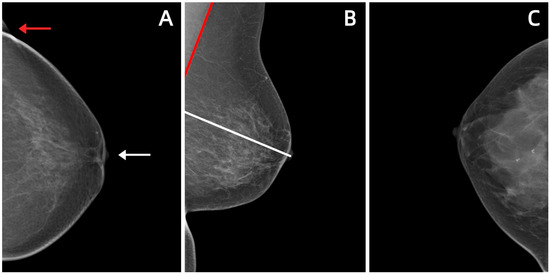
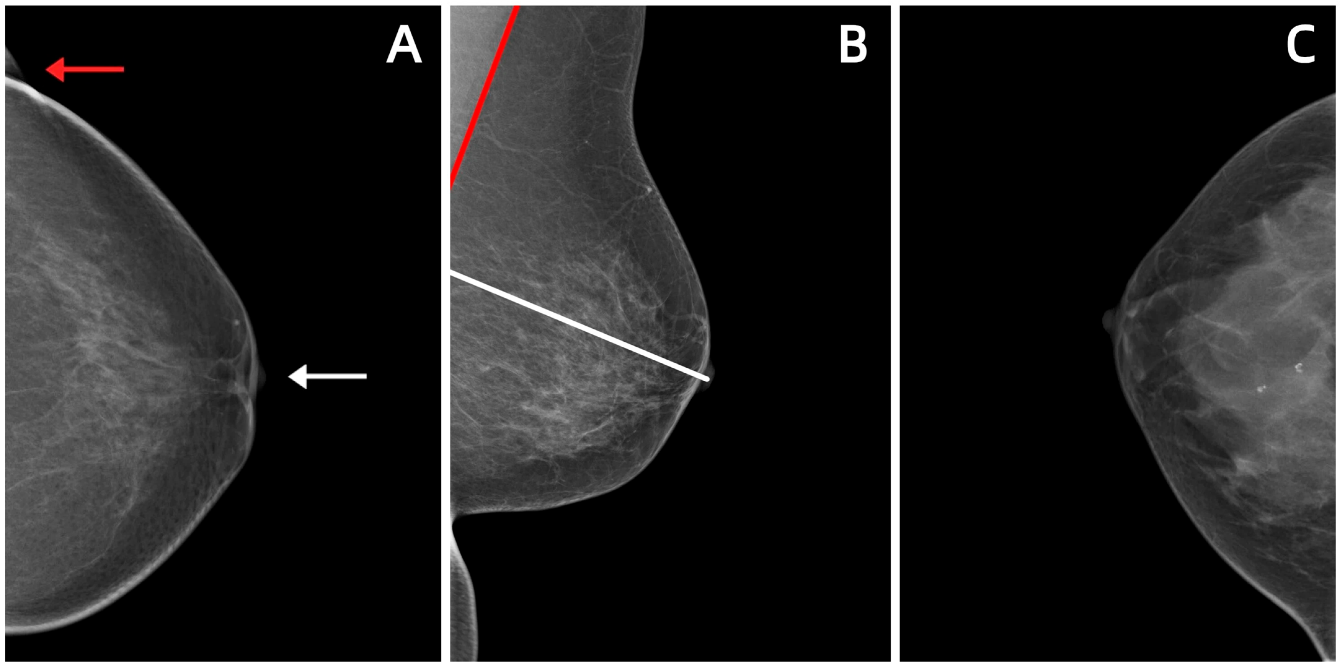
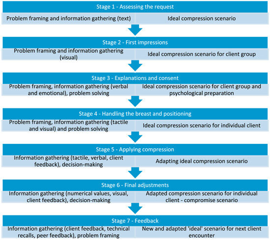
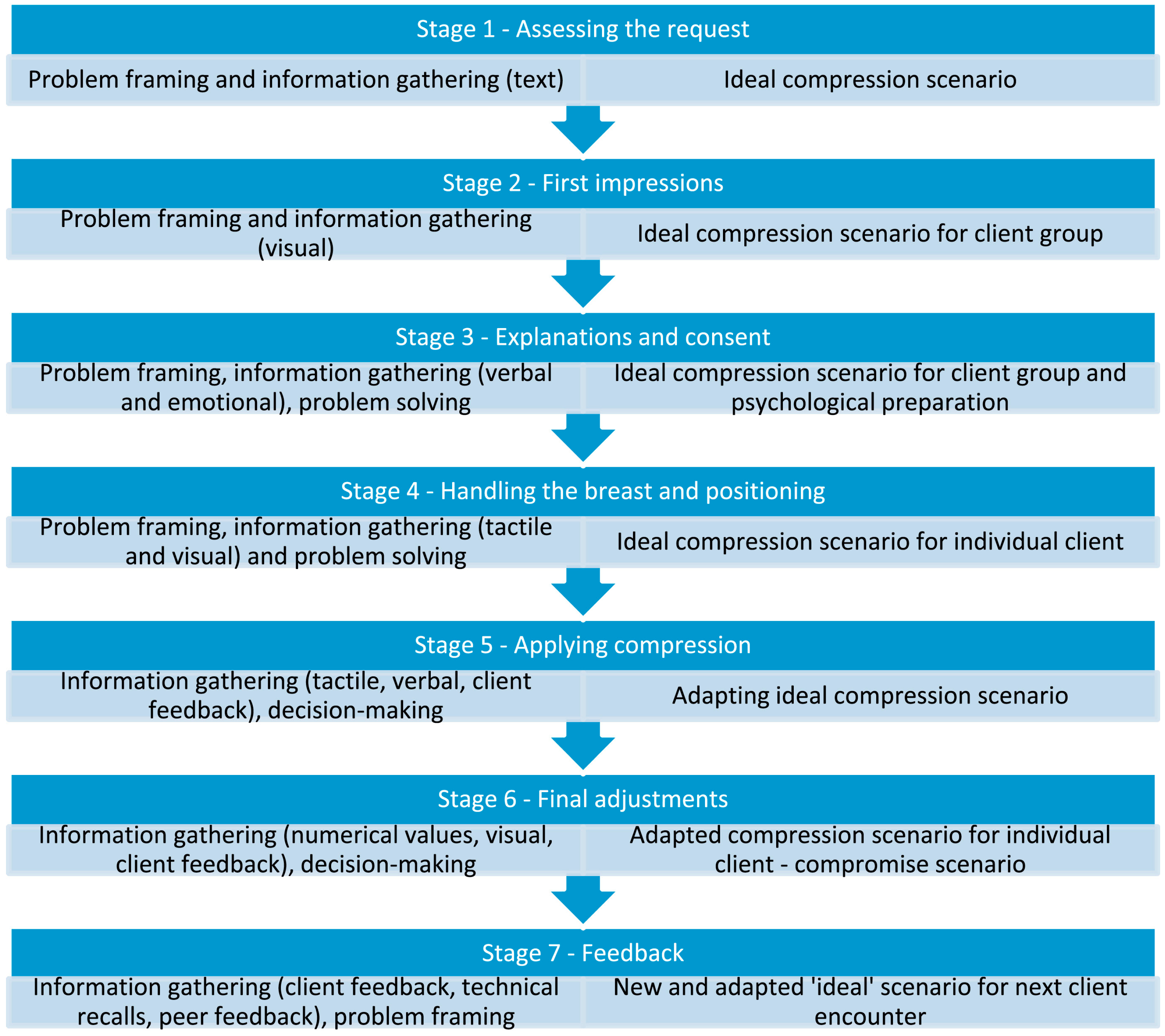
The technician’s professional skills, understanding of equipment performance, and mastery of patient positioning are direct factors in obtaining high-quality images [52]. Two key issues—patient positioning and breast compression—should be noted during mammography procedures, along with selecting appropriate imaging conditions, so as to obtain qualified images that meet diagnostic criteria (Table 3). Correct positioning is crucial for obtaining complete breast tissue images, especially ensuring the inclusion of the posterior part of the breast and the axillary extensions. The FDA has issued a statement pointing out that poor positioning is the main cause of most mammography image defects and clinical misdiagnoses [37]. Common deficiencies include non-tangential display of the nipple, insufficient visualization of the posterior breast tissue, the pectoralis major muscle not extending to the post-nipple line (PNL), and the presence of skin folds (Figure 2) [38,39]. Appropriate compression can not only reduce breast thickness and radiation dose but also homogenize tissue, reduce motion artifacts, and improve image contrast and sharpness [39]. Researchers have proposed a seven-stage breast compression problem-solving model that explains how the ideal compression scenario is identified and adjusted (Figure 3) [53].
Table 3.
Photographic key points and image qualification criteria for CC view and MLO view.
Figure 2.
Common deficiencies in breast positioning during mammography: (A) Nipple not displayed in the tangential position (white arrow); presence of skin folds (red arrow). (B) The PNL (white line) did not reach the lower edge of the pectoralis major muscle (red line). (C) Inadequate visualization of the posterior adipose tissue behind the breast parenchyma.
Figure 3.
The seven-stage continuum mammography compression process model. The left-hand column indicates where different elements of problem analysis occur. The right-hand column identifies how the stages of problem-solving influence the ideal compression scenario. Reprinted with permission from: [53].
To reduce the impact of human factors on image quality, regular training and testing should be conducted to ensure the standardization of operational procedures and the normalization of each examination. A survey involving 251 breast radiologists from 34 European countries showed that more than half of the respondents’ workplaces had established quality assurance measures, but less than one-third were required to participate in regular performance testing. Meanwhile, three-quarters of the respondents believed that mandatory testing would help improve their skills [42]. The fundamental purpose of establishing quality assurance measures lies in the implementation. Professional training on patient positioning techniques, selection of exposure parameters, and mastery of compression force, as well as timely updates to mammography operation guidelines, help improve the operational skills of technologists [43]. In addition, experienced radiologists can review the images acquired by technologists, identify and provide feedback on issues related to positioning, exposure, or post-processing through blind assessment, thereby promoting communication between radiologists and technologists [44].
3.3. Patient-Related Factors
The physiological characteristics and psychological state of patients also indirectly affect image quality. The shape, size, thickness, density, and composition (e.g., the ratio of adipose to glandular tissue) of the breast affect X-ray penetration and image contrast, thereby influencing image quality and diagnostic accuracy. Studies have found that when breast density changes from 6.6% to 33.5%, the probability of breast cancer detection decreases by 61%; when breast compression thickness changes from 46 mm to 66 mm, the probability of breast cancer detection decreases by 42% [45]. Discomfort and anxiety associated with mammography are important reasons why women avoid this examination. In particular, for women with implanted medical devices, not only is the contrast of breast images reduced and the projection of breast tissue and pectoralis major decreased, but patients also experience more pain and anxiety [49]. In addition, slight movement of patients during exposure can lead to image blurring, affect the clarity of microstructures (such as microcalcifications), and increase the risk of false negatives [46]. This is also the most common reason for technical recalls in screening mammography [47].
Improving patients’ compliance during the examination is conducive to improving image quality and promoting more extensive screening. The PAC device allows patients to adjust the compression force by themselves after initial compression by technologists. It can significantly improve the patient experience and achieve image quality similar to that of technologist-controlled compression [40]. It may even increase the additional compression force to reduce breast thickness and radiation dose [54]. Seventy-four percent of patients believe that the PAC device can encourage them to undergo re-examination [41]. Multi-sensory environmental upgrades to mammography rooms, such as playing soothing videos and diffusing light fragrances, are also helpful for improving patients’ psychological status, enabling higher PNL measurement values without increasing compression [48].
4. Limitations and Prospects of Mammography QC
4.1. Lack of Uniform Global QC Protocols
Currently, over 14 QC protocols for mammography are in use globally [55], and QC standards in regions such as the United States, Europe, Canada, Australia, and Japan have different focuses, with variations including the type of tests, phantoms used, image quality criteria, and limiting values. For instance, the digital mammography QC protocol drafted by the European Reference Organisation for Quality Assured Breast Screening and Diagnostic Services (EUREF) physicist team is a recognized European quality standard. Adopted by the European Federation of Organisations in Medical Physics (EFOMP), it is widely implemented in the Netherlands, the UK, Germany, and other countries. This protocol incorporates the CDMAM phantom into the QC testing system and establishes evaluation criteria for key dimensions, including contrast, spatial resolution, radiation dose, image quality, and acquisition repeatability [56]. However, France currently utilizes a protocol established by the French National Agency for Medicines and Health Products Safety (ANSM), which features more lenient QC thresholds and excludes the assessment of tomosynthesis and synthetic 2D (2DS) image quality [57]. This results in a heterogeneous situation and may affect the efficiency and accuracy of large-scale breast cancer screening [58].
In the future, mammography QC needs to pay more attention to international cooperation. By establishing unified examination standards, image metrics, and reporting specifications, the comparison and mutual recognition of examination results across different regions can be promoted. In resource-limited settings, a comprehensive evaluation of social benefits, economic costs, and health impacts should also be considered. This justifies the development of simplified quality control protocols to drive standardization and equity in global breast cancer screening [59].
4.2. Subjective Inconsistency in Image Quality Assessment
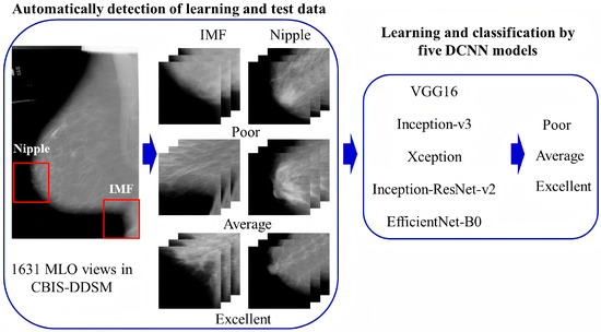
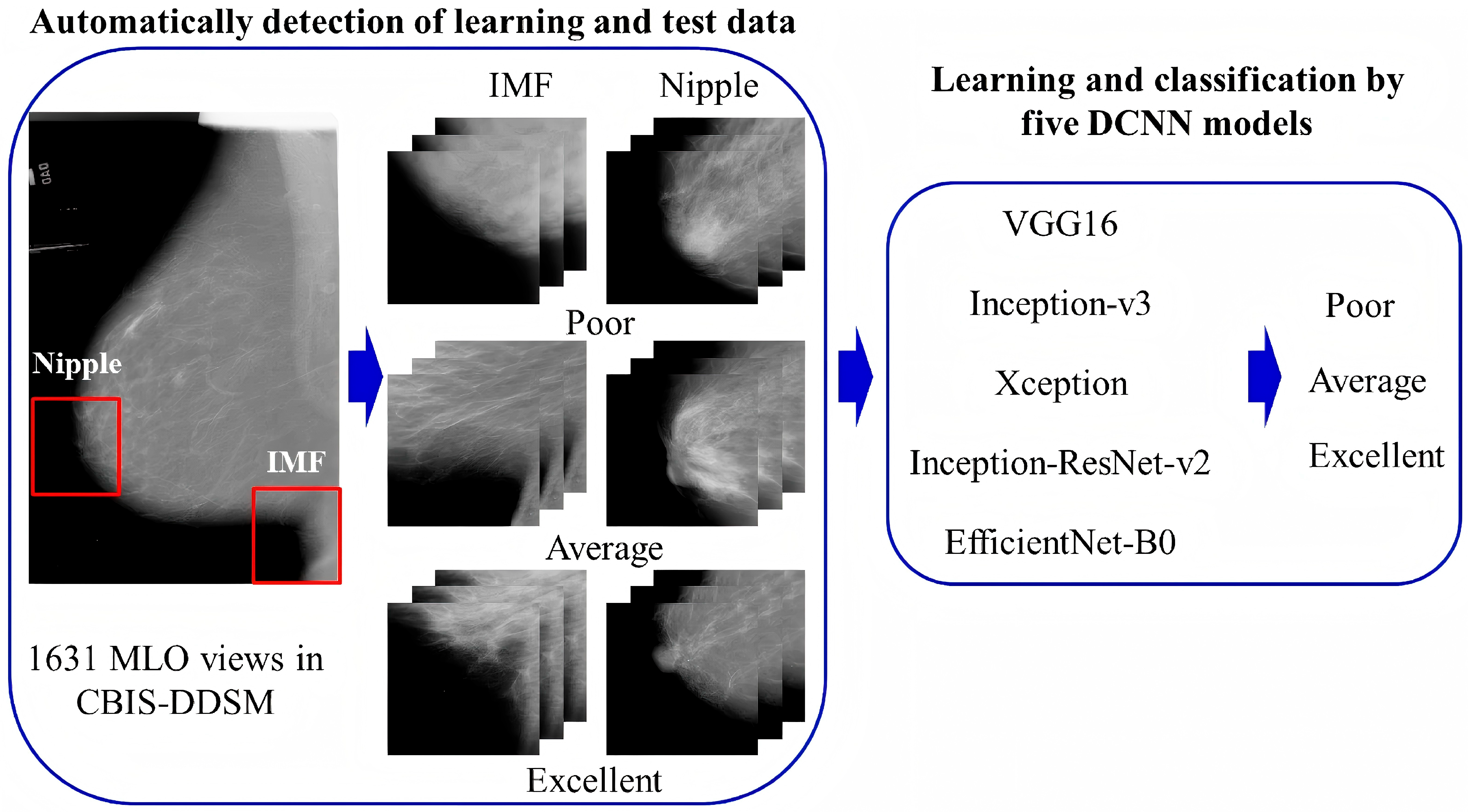
Mammography requires operators to have rich experience, and image quality assessment often relies on the subjective judgment of radiologists or technologists. Although various QC protocols provide basic standards for assessment, studies have shown that there is still subjectivity and inconsistency in quality assessment among different radiologists [60,61]. In recent years, artificial intelligence (AI) has advanced rapidly in breast imaging, with over 20 FDA-approved applications currently available [62]. However, these applications primarily focus on the diagnosis and differentiation of breast diseases [63,64], classification and typing [65,66], and evaluation of treatment efficacy [67,68], with limited use in QC. By analyzing massive image data through deep learning models, AI can automatically assess whether breast positioning is good, with accuracy rates of 96.5% and 93.3%, respectively, in the CC view and MLO view [37]. Other studies have also proposed a deep convolutional neural network (DCNN) classification for the quality control and validation of breast positioning criteria in mammography (Figure 4) [69].
Figure 4.
Working flow of constructing a DCNN model for breast positioning quality control and validation. It mainly includes automated detection of the learning and test data and learning and classification by five DCNN models. Reprinted with permission from: [69].
Nevertheless, these studies are limited to single- or dual-center settings. The training of deep learning models is highly dependent on specific vendors, protocols, or regional datasets, leading to significant data dependency and poor generalization. Moreover, the lack of a unified evaluation framework—where some studies prioritize structural visibility while others focus on geometric accuracy—hinders direct performance comparisons across systems [70]. Therefore, future research should focus on developing a multi-dimensional, comprehensive AI model for evaluating mammography image quality. This model should integrate multiple assessment indicators, such as positioning accuracy, exposure parameters, image contrast, and microcalcification visualization. It is crucial to establish a standardized AI QC dataset for mammography, unify the evaluation criteria for image quality, and conduct multi-center clinical validation of the AI model to enhance its clinical applicability. Moreover, AI should be strictly positioned as an adjunct to radiologists rather than a substitute, preventing over-reliance on automation [57].
4.3. Imbalance Between Image Quality and Radiation Dose
While providing diagnostic information, mammography inevitably involves ionizing radiation exposure. Achieving a balance between the ALARA principle and high-quality images is an ongoing challenge [71]. Excessive pursuit of low doses may sacrifice image quality, leading to missed diagnoses; conversely, excessively high doses will increase the patient’s radiation risk. Existing QC procedures need to ensure that equipment maintains radiation dose at an acceptable level while providing optimal image quality [72].
Balancing diagnostic effects and radiation risks depends on the development of new materials and technologies. For example, compared with traditional broadband mammography, monochromatic X-ray sources are more sensitive for imaging breasts of various sizes and compositions and have lower radiation doses [73]. A composite shielding device composed of a 6 mm lead glass mask and a stainless steel compression plate can reduce radiation exposure to sensitive organs such as the lens and thyroid during mammography [74]. The advancement of new technologies reflects the “patient-centered” QC philosophy. Driven by the advancement of precision medicine, it is crucial to develop personalized dose optimization models based on patient-specific characteristics (such as breast density, thickness, and age), which enable dynamic adjustment of exposure parameters according to individual variations, thereby optimizing the trade-off between image quality and radiation dose [75].
4.4. Inapplicability of Traditional Standards to New Imaging Technologies
With the emergence of new technologies such as digital breast tomosynthesis (DBT) and contrast-enhanced mammography (CEM), traditional QC standards for two-dimensional mammography may no longer be fully applicable. For example, DBT reconstructs three-dimensional breasts through multi-angle X-ray images, which significantly reduces tissue overlap artifacts [76], but it also leads to prolonged imaging time and increased radiation dose. CEM obtains morphological information and blood supply of lesions through dual-energy projection, which can be used for diagnosis and lesion staging, but the use of contrast agents makes the QC process more complex [77].
New imaging technologies require specialized QC protocols. Currently, international QC protocols for DBT have been established by organizations such as EUREF, ACR, and the Australasian College of Physical Scientists and Engineers in Medicine (ACPSEM). The three protocols present a similar set of tests, focusing on collimation tests of light beams, X-ray tube and generator tests, AEC, dose information, image quality, and physical compression. Among them, the ACR standard is more suitable for adoption by other countries due to its high versatility, use of commercial phantoms, and independence from equipment manufacturers [78]. In parallel, countries such as Japan are actively researching and establishing performance assessment and QC procedures for DBT [79]. The French Society of Medical Physics (SFPM) has also established a working group to develop internal QC recommendations for DBT and 2DS images, preparing for the integration of DBT into breast cancer screening programs in the future [80]. Although an independent and comprehensive QC protocol specific to CEM has not yet been established, researchers are validating the utility of commercial and custom-made phantoms for CEM quality control [81,82,83]. Their results revealed that the PMMA phantom exhibited consistent quantification performance, whereas the commercial phantom was superior in visualization [83].
5. Conclusions
Over the past century, mammography and its QC have achieved remarkable development, which is essentially a process driven by the interaction between technological innovation and clinical needs. From the establishment of standards in the early stages to the precise and intelligent QC in the future, its core has always focused on improving image quality, reducing radiation dose, and enhancing patient experience. We have reason to believe that with the continuous development of AI and new imaging technologies, mammography—a traditional yet evolving technology—will benefit more patients.
Author Contributions
H.D.: writing—original draft, conceptualization. Y.Z.: writing—original draft, investigation. M.M.: methodology. Y.J.: supervision. N.Q.: writing—review and editing, funding acquisition. All authors have read and agreed to the published version of the manuscript.
Funding
This work was supported by the Wu Jieping Medical Foundation Special Funding Program for Clinical Research (320.6750.2021-6-33).
Institutional Review Board Statement
Not applicable.
Informed Consent Statement
Not applicable.
Data Availability Statement
No new data were created or analyzed in this study. Data sharing is not applicable to this article.
Conflicts of Interest
The authors declare no conflicts of interest.
References
- Shang, C.; Xu, D. Epidemiology of Breast Cancer. Oncologie 2022, 24, 649–663. [Google Scholar] [CrossRef] [Scilit]
- Kim, J.; Harper, A.; McCormack, V.; Sung, H.; Houssami, N.; Morgan, E.; Mutebi, M.; Garvey, G.; Soerjomataram, I.; Fidler-Benaoudia, M.M. Global patterns and trends in breast cancer incidence and mortality across 185 countries. Nat. Med. 2025, 31, 1154–1162. [Google Scholar] [CrossRef] [Scilit] [PubMed]
- Lauby-Secretan, B.; Scoccianti, C.; Loomis, D.; Benbrahim-Tallaa, L.; Bouvard, V.; Bianchini, F.; Straif, K. Breast-Cancer Screening—Viewpoint of the IARC Working Group. N. Engl. J. Med. 2015, 372, 2353–2358. [Google Scholar] [CrossRef] [Scilit] [PubMed]
- Monticciolo, D.L.; Malak, S.F.; Friedewald, S.M.; Eby, P.R.; Newell, M.S.; Moy, L.; Destounis, S.V.; Destounis, J.W.T.; Hendrick, R.E.; Smetherman, D. Breast Cancer Screening Recommendations Inclusive of All Women at Average Risk: Update from the ACR and Society of Breast Imaging. J. Am. Coll. Radiol. 2021, 18, 1280–1288. [Google Scholar] [CrossRef] [Scilit]
- Comstock, C.E.; Gatsonis, C.; Newstead, G.M.; Snyder, B.S.; Gareen, I.F.; Bergin, J.T.; Rahbar, H.; Sung, J.S.; Jacobs, C.; Harvey, J.A.; et al. Comparison of Abbreviated Breast MRI vs Digital Breast Tomosynthesis for Breast Cancer Detection Among Women with Dense Breasts Undergoing Screening. JAMA 2020, 323, 746–756. [Google Scholar] [CrossRef] [Scilit]
- Boita, J.; van Engen, R.E.; Mackenzie, A.; Tingberg, A.; Bosmans, H.; Bolejko, A.; Zackrisson, S.; Wallis, M.G.; Ikeda, D.M.; Van Ongeval, C.; et al. How does image quality affect radiologists’ perceived ability for image interpretation and lesion detection in digital mammography? Eur. Radiol. 2021, 31, 5335–5343. [Google Scholar] [CrossRef] [Scilit]
- Salomon, A. Beitrge zur Pathologie und Klinik der Mammakarzinome. Arch. Kiln Chir. 1913, 101, 573–668. [Google Scholar]
- Leborgne, R. Diagnosis of tumors of the breast by simple roentgenography; calcifications in carcinomas. Am. J. Roentgenol. Radium Ther. 1951, 65, 1–11. [Google Scholar]
- Leborgne, R. The Breast in Roentgen Diagnosis; Constable: London, UK; Impresora Uruguaya S.A.: Montevideo, Uruguay, 1953. [Google Scholar]
- Egan, R.L. Experience with Mammography in a Tumor Institution: Evaluation of 1000 Studies. Radiology 1960, 75, 894–900. [Google Scholar] [CrossRef] [Scilit]
- Gold, R.H.; Bassett, L.W.; Widoff, B.E. Highlights from the history of mammography. Radiographics 1990, 10, 1111–1131. [Google Scholar] [CrossRef] [Scilit]
- Shapiro, S. Periodic Breast Cancer Screening in Reducing Mortality From Breast Cancer. JAMA 1971, 215, 1777. [Google Scholar] [CrossRef] [Scilit]
- Haus, A.G.; Doi, K.; Metz, C.E.; Bernstein, J. Image quality in mammography. Radiology 1977, 125, 77–85. [Google Scholar] [CrossRef] [Scilit]
- Hendrick, R.E. Standardization of image quality and radiation dose in mammography. Radiology 1990, 174, 648–654. [Google Scholar] [CrossRef] [Scilit]
- Houn, F.; Elliott, M.L.; McCrohan, J.L. The Mammography Quality Standards Act of 1992. History and philosophy. Radiol. Clin. N. Am. 1995, 33, 1059–1065. [Google Scholar] [CrossRef] [Scilit] [PubMed]
- Suleiman, O.H.; Spelic, D.C.; McCrohan, J.L.; Symonds, G.R.; Houn, F. Mammography in the 1990s: The United States and Canada. Radiology 1999, 210, 345–351. [Google Scholar] [CrossRef] [Scilit] [PubMed]
- Burnside, E.S.; Sickles, E.A.; Bassett, L.W.; Rubin, D.L.; Lee, C.H.; Ikeda, D.M.; Mendelson, E.B.; Wilcox, P.A.; Butler, P.F.; D’Orsi, C.J. The ACR BI-RADS® Experience: Learning From History. J. Am. Coll. Radiol. 2009, 6, 851–860. [Google Scholar] [CrossRef] [Scilit] [PubMed]
- Newell, M.S.; Destounis, S.V.; Leung, J.W.T.; DeMartini, W.B.; Lee, C.H.; Eby, P.R. ACR BI-RADS® v2025 Manual; American College of Radiology: Reston, VA, USA, 2025. [Google Scholar]
- White, J. FDA Approves System for Digital Mammography. JNCI J. Natl. Cancer Inst. 2000, 92, 442. [Google Scholar] [CrossRef] [Scilit]
- Feig, S.A.; Yaffe, M.J. Digital Mammography, Computer-Aided Diagnosis, and Telemammography. Radiol. Clin. N. Am. 1995, 33, 1205–1230. [Google Scholar] [CrossRef] [Scilit]
- Young, K.C.; Engen, R.V.; Bosmans, H.; Jacobs, J.; Zanca, F. Quality Control in Digital Mammography. In Digital Mammography; Bick, U., Diekmann, F., Eds.; Medical Radiology; Springer: Berlin/Heidelberg, Germany, 2010; pp. 33–54. [Google Scholar] [CrossRef] [Scilit]
- Gennaro, G.; Baldelli, P.; Di Maggio, C.; Taibi, A.; Gambaccini, M. A first experience of quality control in full field digital mammography. In Digital Mammography; Peitgen, H.-O., Ed.; Springer: Berlin/Heidelberg, Germany, 2003; pp. 81–84. ISBN 978-3-642-63936-4. [Google Scholar]
- Pisano, E.D.; Gatsonis, C.A.; Yaffe, M.J.; Hendrick, R.E.; Tosteson, A.N.A.; Fryback, D.G.; Bassett, L.W.; Baum, J.K.; Conant, E.F.; Jong, R.A.; et al. American College of Radiology Imaging Network digital mammographic imaging screening trial: Objectives and methodology. Radiology 2005, 236, 404–412. [Google Scholar] [CrossRef] [Scilit]
- Hawley, J.R.; Ha, C.; Cai, L.; Taylor, C.R. The FDA Enhancing Quality Using the Inspection Program (EQUIP) Breast Imaging Quality Initiative. 5-Year Clinical Experience. J. Breast Imaging 2024, 6, 388–396. [Google Scholar] [CrossRef] [Scilit]
- Falcon, S.; Funaro, K.; Williams, A.; Niell, B.L. Effect of the EQUIP initiative on additional images performed at screening mammography: A pilot study. Clin. Imaging 2021, 73, 13–17. [Google Scholar] [CrossRef] [Scilit] [PubMed]
- Lerner, D.L. Highlights of the 2023 Amendments to the MQSA Implementing Regulations. Radiology 2024, 312, e242203. [Google Scholar] [CrossRef] [Scilit] [PubMed]
- IAEA. Worldwide Implementation of Digital Mammography Imaging; International Atomic Energy Agency: Havertown, PA, USA, 2023; ISBN 978-92-0-126923-2. [Google Scholar]
- Alkhalifah, K.; Brindabhan, A.; Alsaeed, R. Effect of exposure factors on image quality in screening mammography. Radiography 2017, 23, e99–e102. [Google Scholar] [CrossRef] [Scilit] [PubMed]
- Kim, C.; Kang, S.-S.; Kim, J.-H.; Lee, J.-S. Evaluation of Image Quality using Monte Carlo Simulation in Digital Mammography System. J. Korea Contents Assoc. 2014, 14, 247–254. [Google Scholar] [CrossRef] [Scilit]
- Williams, M.; Raghunathan, P.; Seibert, J.; Kwan, A.; Lo, J.; Samei, E.; Ranger, N.; Fajardo, L.; McGruder, A.; Maxwell, S.; et al. TU-B-M100J-01: Optimizing Mammography Image Quality and Dose: X-Ray Spectrum and Exposure Parameter Selection. Med. Phys. 2007, 34, 2540–2541. [Google Scholar] [CrossRef] [Scilit]
- Kattar, Z.A.; Balaa, H.E.; Harb, H.; Raad, N.; Hodroj, N.; Sleiman, F.; Omayrat, S. Influence of the acquisition parameters on the image quality in digital mammography. In Proceedings of the 2015 International Conference on Advances in Biomedical Engineering (ICABME); Beirut, Lebanon, 16–18 September 2015, IEEE: Piscataway, NJ, USA, 2015; pp. 33–36. [Google Scholar] [CrossRef] [Scilit]
- Szczepura, K.; Dos Reis, C.S. Image Quality, System Optimisation and Quality Control. In Digital Mammography; Mercer, C., Hogg, P., Kelly, J., Eds.; Springer International Publishing: Cham, Switzerland, 2022; pp. 233–245. [Google Scholar] [CrossRef] [Scilit]
- Fausto, A.M.F.; Lopes, M.C.; De Sousa, M.C.; Furquim, T.A.C.; Mol, A.W.; Velasco, F.G. Optimization of Image Quality and Dose in Digital Mammography. J. Digit. Imaging 2017, 30, 185–196. [Google Scholar] [CrossRef] [Scilit]
- Figl, M.; Homolka, P.; Osanna-Elliott, A.; Semturs, F.; Kaar, M.; Hummel, J. Conversion factors between human and automatic readouts of CDMAM phantom images of CR mammography systems. Phys. Med. Biol. 2016, 61, N514–N521. [Google Scholar] [CrossRef] [Scilit]
- Oberhofer, N. SU-E-I-147: New Digital Mammography Equipment: Physical Characterization and Image Quality Evaluation. Med. Phys. 2011, 38, 3430. [Google Scholar] [CrossRef] [Scilit]
- Mora, P.; Pfeiffer, D.; Zhang, G.; Bosmans, H.; Delis, H.; Razi, Z.; Arreola, M.; Tsapaki, V. The IAEA remote and automated quality control methodology for radiography and mammography. J. Appl. Clin. Med. Phys. 2021, 22, 126–142. [Google Scholar] [CrossRef] [Scilit]
- Brahim, M.; Westerkamp, K.; Hempel, L.; Lehmann, R.; Hempel, D.; Philipp, P. Automated Assessment of Breast Positioning Quality in Screening Mammography. Cancers 2022, 14, 4704. [Google Scholar] [CrossRef] [Scilit]
- Feigin, K. Quality assurance in Mammography: An overview. Eur. J. Radiol. 2023, 165, 110935. [Google Scholar] [CrossRef] [Scilit]
- Bassett, L.W.; Farria, D.M.; Bansal, S.; Farquhar, M.A.; Wilcox, P.A.; Feig, S.A. Reasons for Failure of a Mammography Unit at Clinical Image Review in the American College of Radiology Mammography Accreditation Program. Radiology 2000, 215, 698–702. [Google Scholar] [CrossRef] [Scilit] [PubMed]
- Dontchos, B.N.; Edmonds, C.E.; Mercaldo, S.F.; Miles, R.C.; Chu, K.F.; Lehman, C.D. Patient-Assisted Compression in Screening Mammography: Patient Experience and Image Quality. J. Breast Imaging 2019, 1, 192–198. [Google Scholar] [CrossRef] [Scilit] [PubMed]
- Balleyguier, C.; Cousin, M.; Dunant, A.; Attard, M.; Delaloge, S.; Arfi-Rouche, J. Patient-assisted compression helps for image quality reduction dose and improves patient experience in mammography. Eur. J. Cancer 2018, 103, 137–142. [Google Scholar] [CrossRef] [Scilit] [PubMed]
- Michalopoulou, E.; Clauser, P.; Gilbert, F.J.; Pijnappel, R.M.; Mann, R.M.; Baltzer, P.A.T.; Chen, Y.; Fallenberg, E.M. A survey by the European Society of Breast Imaging on radiologists’ preferences regarding quality assurance measures of image interpretation in screening and diagnostic mammography. Eur. Radiol. 2023, 33, 8103–8111. [Google Scholar] [CrossRef] [Scilit]
- Tirada, N.; Li, G.; Dreizin, D.; Robinson, L.; Khorjekar, G.; Dromi, S.; Ernst, T. Digital Breast Tomosynthesis: Physics, Artifacts, and Quality Control Considerations. RadioGraphics 2019, 39, 413–426. [Google Scholar] [CrossRef] [Scilit]
- Sá Dos Reis, C.; Gremion, I.; Richli Meystre, N. Consensus about image quality assessment criteria of breast implants mammography using Delphi method with radiographers and radiologists. Insights Imaging 2020, 11, 56. [Google Scholar] [CrossRef] [Scilit]
- Strandberg, R.; Illipse, M.; Czene, K.; Hall, P.; Humphreys, K. Influence of mammographic density and compressed breast thickness on true mammographic sensitivity: A cohort study. Sci. Rep. 2023, 13, 14194. [Google Scholar] [CrossRef] [Scilit]
- Abdullah, A.K.; Kelly, J.; Thompson, J.D.; Mercer, C.E.; Aspin, R.; Hogg, P. The impact of simulated motion blur on lesion detection performance in full-field digital mammography. Br. J. Radiol. 2017, 90, 20160871. [Google Scholar] [CrossRef] [Scilit]
- Martaindale, S.; Moseley, T.; Santiago, L.; Huang, M.; Sullivan, C.; Bassett, R.L.; Whitman, G. Analysis of Technical Repeat Studies in Screening Mammography. J. Breast Imaging 2023, 5, 416–424. [Google Scholar] [CrossRef] [Scilit]
- Sarquis-Kolber, S.; Schilling, K.; Hanney, W.J.; Kolber, M.J. The Association of Mammography Environment and Image Quality. J. Breast Imaging 2020, 2, 436–442. [Google Scholar] [CrossRef] [Scilit] [PubMed]
- Paap, E.; Witjes, M.; Van Landsveld-Verhoeven, C.; Pijnappel, R.M.; Maas, A.H.E.M.; Broeders, M.J.M. Mammography in females with an implanted medical device: Impact on image quality, pain and anxiety. BJR 2016, 89, 20160142. [Google Scholar] [CrossRef] [Scilit] [PubMed]
- Williams, M.B.; Yaffe, M.J.; Maidment, A.D.A.; Martin, M.C.; Seibert, J.A.; Pisano, E.D. Image Quality in Digital Mammography: Image Acquisition. J. Am. Coll. Radiol. 2006, 3, 589–608. [Google Scholar] [CrossRef] [Scilit] [PubMed]
- Siegel, E.; Krupinski, E.; Samei, E.; Flynn, M.; Andriole, K.; Erickson, B.; Thomas, J.; Badano, A.; Seibert, J.A.; Pisano, E.D. Digital Mammography Image Quality: Image Display. J. Am. Coll. Radiol. 2006, 3, 615–627. [Google Scholar] [CrossRef] [Scilit]
- Bassett, L.W. Determinants of Quality in Mammography. Surg. Oncol. Clin. N. Am. 1997, 6, 213–232. [Google Scholar] [CrossRef] [Scilit]
- Nightingale, J.M.; Murphy, F.J.; Robinson, L.; Newton-Hughes, A.; Hogg, P. Breast compression—An exploration of problem solving and decision-making in mammography. Radiography 2015, 21, 364–369. [Google Scholar] [CrossRef] [Scilit]
- Arenas, N.; Alcantara, R.; Posso, M.; Louro, J.; Perez-Leon, D.; Ejarque, B.; Arranz, M.; Maiques, J.; Castells, X.; Macià, F.; et al. Comparison of technical parameters and women’s experience between self-compression and standard compression modes in mammography screening: A single-blind randomized clinical trial. Eur. Radiol. 2022, 32, 7480–7487. [Google Scholar] [CrossRef] [Scilit]
- Reis, C.; Pascoal, A.; Sakellaris, T.; Koutalonis, M. Quality assurance and quality control in mammography: A review of available guidance worldwide. Insights Imaging 2013, 4, 539–553. [Google Scholar] [CrossRef] [Scilit]
- Gennaro, G.; Avramova-Cholakova, S.; Azzalini, A.; Luisa Chapel, M.; Chevalier, M.; Ciraj, O.; de Las Heras, H.; Gershan, V.; Hemdal, B.; Keavey, E.; et al. Quality Controls in Digital Mammography protocol of the EFOMP Mammo Working group. Phys. Med. 2018, 48, 55–64. [Google Scholar] [CrossRef] [Scilit]
- Heid, P. Quality control in mammography within organized breast cancer screening programs: Benefits and risks. Imag. Femme 2025, 35, 125–129. [Google Scholar] [CrossRef] [Scilit]
- Ng, K.-H.; Jamal, N.; DeWerd, L. Global quality control perspective for the physical and technical aspects of screen-film mammography—Image quality and radiation dose. Radiat. Prot. Dosim. 2006, 121, 445–451. [Google Scholar] [CrossRef] [Scilit] [PubMed]
- Chotai, N.; Renganathan, R.; Uematsu, T.; Wang, J.; Zhu, Q.; Rahmat, K.; Pradaranon, V.; Fong, J.C.; Choridah, L.; Chang, J.M. Breast Cancer Screening in Asian Countries: Epidemiology, Screening Practices, Outcomes, Challenges, and Future Directions. Korean J. Radiol. 2025, 26, 743–758. [Google Scholar] [CrossRef] [Scilit] [PubMed]
- Funaro, K.; Niell, B. Variability in Mammography Quality Assessment After Implementation of Enhancing Quality Using the Inspection Program (EQUIP). J. Breast Imaging 2021, 3, 168–175. [Google Scholar] [CrossRef] [Scilit] [PubMed]
- Albeshan, S.M.; Alashban, Y.; Al Tahan, F.M.; Al-enezi, S.; Alnaimy, N.; Shubayr, N.; Eliraqi, F. Mammography image quality evaluation in breast cancer screening: The Saudi experience. J. Radiat. Res. Appl. Sci. 2022, 15, 100467. [Google Scholar] [CrossRef] [Scilit]
- Taylor, C.R.; Monga, N.; Johnson, C.; Hawley, J.R.; Patel, M. Artificial Intelligence Applications in Breast Imaging: Current Status and Future Directions. Diagnostics 2023, 13, 2041. [Google Scholar] [CrossRef] [Scilit]
- Rodriguez-Ruiz, A.; Lång, K.; Gubern-Merida, A.; Broeders, M.; Gennaro, G.; Clauser, P.; Helbich, T.H.; Chevalier, M.; Tan, T.; Mertelmeier, T.; et al. Stand-Alone Artificial Intelligence for Breast Cancer Detection in Mammography: Comparison with 101 Radiologists. J. Natl. Cancer Inst. 2019, 111, 916–922. [Google Scholar] [CrossRef] [Scilit]
- McKinney, S.M.; Sieniek, M.; Godbole, V.; Godwin, J.; Antropova, N.; Ashrafian, H.; Back, T.; Chesus, M.; Corrado, G.S.; Darzi, A.; et al. International evaluation of an AI system for breast cancer screening. Nature 2020, 577, 89–94. [Google Scholar] [CrossRef] [Scilit]
- Wu, M.; Zhong, X.; Peng, Q.; Xu, M.; Huang, S.; Yuan, J.; Ma, J.; Tan, T. Prediction of molecular subtypes of breast cancer using BI-RADS features based on a “white box” machine learning approach in a multi-modal imaging setting. Eur. J. Radiol. 2019, 114, 175–184. [Google Scholar] [CrossRef] [Scilit]
- Polónia, A.; Campelos, S.; Ribeiro, A.; Aymore, I.; Pinto, D.; Biskup-Fruzynska, M.; Veiga, R.S.; Canas-Marques, R.; Aresta, G.; Araújo, T.; et al. Artificial Intelligence Improves the Accuracy in Histologic Classification of Breast Lesions. Am. J. Clin. Pathol. 2021, 155, 527–536. [Google Scholar] [CrossRef] [Scilit]
- Wang, X.; Xie, T.; Luo, J.; Zhou, Z.; Yu, X.; Guo, X. Radiomics predicts the prognosis of patients with locally advanced breast cancer by reflecting the heterogeneity of tumor cells and the tumor microenvironment. Breast Cancer Res. 2022, 24, 20. [Google Scholar] [CrossRef] [Scilit]
- Gu, J.; Tong, T.; He, C.; Xu, M.; Yang, X.; Tian, J.; Jiang, T.; Wang, K. Deep learning radiomics of ultrasonography can predict response to neoadjuvant chemotherapy in breast cancer at an early stage of treatment: A prospective study. Eur. Radiol. 2022, 32, 2099–2109. [Google Scholar] [CrossRef] [Scilit] [PubMed]
- Watanabe, H.; Hayashi, S.; Kondo, Y.; Matsuyama, E.; Hayashi, N.; Ogura, T.; Shimosegawa, M. Quality control system for mammographic breast positioning using deep learning. Sci. Rep. 2023, 13, 7066. [Google Scholar] [CrossRef] [Scilit] [PubMed]
- Spuur, K.M.; Singh, C.L.; Mousa, D.A.; Chau, M.T. Automated Software Evaluation in Screening Mammography: A Scoping Review of Image Quality and Technique Assessment. Curr. Oncol. 2025, 32, 571. [Google Scholar] [CrossRef] [Scilit] [PubMed]
- Noor, D.K.A.M.; Norsuddin, D.N.M.; Izuan, D.M.; Mazrouei, D.N.A.; Kaabi, M.F.A. Exploring the Association between Mammography Dose Optimization and Image Quality. J. Med. Imaging Radiat. Sci. 2024, 55, 101572. [Google Scholar] [CrossRef] [Scilit]
- Iqeilan, N.; Al-Naemi, H.M. Quality control program of X-ray mammography. Phys. Medica 2016, 32, 194. [Google Scholar] [CrossRef] [Scilit]
- Silver, E.H.; Shulman, S.D.; Rehani, M.M. Innovative monochromatic x-ray source for high-quality and low-dose medical imaging. Med. Phys. 2021, 48, 1064–1078. [Google Scholar] [CrossRef] [Scilit]
- Jeon, K.-S.; Kim, J.-H.; Koo, B.-Y. Radiation dose reduction with an eye and thyroid shielding device during mammography. Radiat. Phys. Chem. 2026, 238, 113214. [Google Scholar] [CrossRef] [Scilit]
- Van Strien-Knippenberg, I.S.; Arjangi-Babetti, H.; Timmermans, D.R.M.; Schrauwen, L.; Fransen, M.P.; Melles, M.; Damman, O.C. Communicating the results of risk-based breast cancer screening through visualizations of risk: A participatory design approach. BMC Med. Inform. Decis. Mak. 2024, 24, 78. [Google Scholar] [CrossRef] [Scilit]
- Friedewald, S.M.; Rafferty, E.A.; Rose, S.L.; Durand, M.A.; Plecha, D.M.; Greenberg, J.S.; Hayes, M.K.; Copit, D.S.; Carlson, K.L.; Cink, T.M.; et al. Breast cancer screening using tomosynthesis in combination with digital mammography. JAMA 2014, 311, 2499–2507. [Google Scholar] [CrossRef] [Scilit]
- Coffey, K.; Jochelson, M.S. Contrast-Enhanced Mammography in Breast Cancer Screening. Eur. J. Radiol. 2022, 156, 110513. [Google Scholar] [CrossRef] [Scilit]
- Pramayuni, M.; Palupi, D. Quality control of digital breast tomosynthesis: A review on current protocols. In Proceedings of the The International Conference On Advanced Material and Technology (Icamt) 2021, South Tangerang, Indonesia, 14–16 December 2021; p. 040002. [Google Scholar]
- Shinohara, N.; Akiyama, S.; Ito, T.; Okada, S.; Chiba, Y.; Negishi, T.; Hirofuji, Y.; Kunitomo, H. Examination of the Quality Control Items for Digital Breast Tomosynthesis System in Japan. Nihon Hoshasen Gijutsu Gakkai Zasshi 2021, 77, 478–486. [Google Scholar] [CrossRef] [Scilit]
- Voyeau-Gautier, M.; Villani, N.; Guillerminet, C.; Michel, C.; Sage, J. 52 SFPM working group on DBT internal quality control. Phys. Medica 2018, 56, 29. [Google Scholar] [CrossRef] [Scilit]
- Zhang, C.; Fu, J. A new breast phantom suitable for digital mammography, contrast-enhanced digital mammography and digital breast tomosynthesis. Phys. Med. Biol. 2023, 68, 045015. [Google Scholar] [CrossRef] [Scilit]
- Kim, J.-H.; Kessell, M.; Taylor, D.; Hill, M.; Burrage, J.W. The verification of the utility of a commercially available phantom combination for quality control in contrast-enhanced mammography. Phys. Eng. Sci. Med. 2024, 47, 1491–1499. [Google Scholar] [CrossRef] [Scilit]
- Ramírez, D.I.; Castillo, J.P.; Galván, H.A.; Brandan, M.E. Evaluation of commercial and custom-made phantoms for quality control in contrast-enhanced mammography. In Proceedings of the Eighteenth Mexican Symposium On Medical Physics, Guanajuato, México, 20–22 March 2024; p. 030004. [Google Scholar]

Disclaimer/Publisher’s Note: The statements, opinions and data contained in all publications are solely those of the individual author(s) and contributor(s) and not of MDPI and/or the editor(s). MDPI and/or the editor(s) disclaim responsibility for any injury to people or property resulting from any ideas, methods, instructions or products referred to in the content. |
© 2026 by the authors. Licensee MDPI, Basel, Switzerland. This article is an open access article distributed under the terms and conditions of the Creative Commons Attribution (CC BY) license.