


Abstract
Background/Objectives: This exploratory in vitro study evaluated the effects of topical facial products on quantitative pixel-level parameters of intraoral radiographic images under different exposure conditions using a photostimulable phosphor (PSP) system. Methods: Six groups were tested: inorganic sunscreen, organic sunscreen, BB cream, CC cream, silver (Ag) sulfadiazine 1%, and an untreated control. To isolate the individual radiographic signature of each formulation from anatomical confounders, the products were applied at a dose of 2 mg/cm2 to a homogeneous silicone soft-tissue phantom and imaged under three protocols (60 kVp/7 mA/0.080 s, pediatric anterior; 60 kVp/7 mA/0.160 s, adult anterior; 63 kVp/6 mA/0.200 s, adult posterior) with three independent acquisitions per group and protocol (54 images). Six parameters were measured in ImageJ: brightness, uniformity, signal-to-noise ratio, histogram clipping, noise, and uniformity index. Data were analyzed by two-way ANOVA with Dunnett post hoc testing and Benjamini–Hochberg (BH) correction. Results: Compared to the control, the BB cream showed significant differences in uniformity (higher SD), noise (higher), and uniformity index (higher; i.e., greater uniformity among the ROI means); the CC cream showed significant differences in histogram clipping (higher), noise (higher), and uniformity index (higher) (all BH-adjusted p ≤ 0.011). Silver sulfadiazine increased histogram clipping (BH-adjusted p = 0.002), while the inorganic sunscreen altered the uniformity index (BH-adjusted p = 0.042). The organic sunscreen did not differ from the control in the primary analysis. Neither exposure protocol nor the group-by-protocol interaction reached significance, although the study was powered only for large effects and exposure dependence cannot be excluded. Conclusions: Products containing metal oxides or silver sulfadiazine produced formulation-specific changes in quantitative imaging parameters, whereas the organic sunscreen tested in this study showed no significant difference from the control. This study did not test whether the observed differences are clinically visible or whether software processing can render them visible. The findings serve as a reference for subsequent research involving anatomical phantoms and observer-based studies.
1. Introduction
A radiograph that cannot be interpreted has to be repeated, and repeat exposures remain one of the most avoidable sources of dose in dental practice. Image quality is therefore both a diagnostic and a radiation-protection concern [1]. This is the basis of the ALADA (As Low As Diagnostically Acceptable) principle [2,3]. Image quality in intraoral radiography depends mainly on the detector, the geometry of positioning, and the exposure parameters [4,5].
In radiology, image quality is evaluated through interrelated quantitative parameters (brightness, noise, signal-to-noise ratio (SNR), uniformity, and histogram clipping) [1]; changes in these parameters have the potential to affect the imaging chain. Evaluating these parameters under varying exposure conditions allows the dependence of the findings on the exposure settings to be explored [1,6].
In addition to these technical factors, patient-related external factors can also alter quantitative imaging parameters. Cosmetic products used for aesthetic or protective purposes contain compounds that alter X-ray attenuation [7]. Inorganic sunscreens, which contain zinc oxide or titanium dioxide [8], can create artificial opacities via increased photoelectric absorption [7]. In medical radiology literature, zinc-based cream residues have been reported to mimic mammographic microcalcifications [9], and ultraviolet-protective lotions produce screen–film cassette artifacts [10]. Deodorants and talcum powder have also been listed among topical products known to mimic breast calcifications [9]. These findings suggest that the formulation, rather than the product category, may be the more relevant determinant of the observed effects; for UV-filter mixtures, formulation is known to change the behavior of individual ingredients [11].
This distinction is particularly important because these products are frequently used together in clinical practice. 1% silver sulfadiazine cream is applied to wounds in facial burns and blast injuries [12]. Burn rehabilitation approaches include topical moisturizers and silicone gels for scar management [13], inorganic sunscreens (TiO2/ZnO) for photoprotection [14], and cosmetic camouflage creams to support psychosocial recovery [15], a product category that includes the BB/CC creams studied here (Section 2.2). Consequently, in patients undergoing intraoral imaging following burn or trauma care, silver (Z = 47), zinc (Z = 30), iron (Z = 26), and titanium (Z = 22) may be present in or near the path of the radiographic beam. However, the effects of these products on intraoral radiographic imaging parameters remain unclear. Since the attenuation contribution of each compound is unknown, testing the products individually is a necessary first step. Therefore, this study evaluated the individual radiographic signature of each product rather than assessing combination or layered applications.
Since investigating these effects directly in patients raises ethical and standardization concerns, in vitro phantom models are widely used in dental radiology research to ensure controlled reproducibility while avoiding unnecessary patient exposure [16]. In this study, a homogeneous simulation phantom devoid of anatomical structures (bone, dentin, and enamel) was deliberately chosen; this design aimed to characterize each product’s radiographic signature by isolating it from anatomical confounders. Assessing clinical diagnostic impact requires subsequent studies using anatomical phantoms, to be conducted after the effects of individual products have been determined. There is only one previous in vitro study examining the effects of sunscreens on dental radiographic image parameters. That study also used an empty phantom devoid of anatomical structures [7]; however, the effects of silver sulfadiazine, BB/CC creams, and the exposure protocol have not yet been investigated.
This exploratory in vitro study evaluated the effects of cosmetic/dermatological topical products commonly used in burn/trauma rehabilitation (inorganic/organic sunscreens, BB creams, CC creams, and silver sulfadiazine) on the quantitative pixel-level parameters of intraoral radiographic images, assessing six objective parameters across three exposure conditions. The null hypothesis stated that these products do not significantly affect image parameters, regardless of the exposure scenario.
2. Materials and Methods
2.1. Study Design and Sample Size
This in vitro study evaluated the effects of five cosmetic/dermatological topical products (inorganic and organic sunscreen, BB cream, CC cream, and silver sulfadiazine) versus a control on quantitative parameters of intraoral radiographic images. The products were evaluated individually to isolate the radiographic effects of each formulation and minimize potential confounding from their simultaneous presence. No human or animal subjects were involved, and ethical committee approval was therefore not required.
A pilot study of 18 images (six groups × three protocols) gave a large effect size for noise (Cohen’s f = 1.28), which in G*Power (Version 3.1.9.7) (one-way ANOVA, α = 0.05, power = 0.80, k = 6) indicated n = 2 per group. Given the potential inflation of effect-size estimates derived from small pilot samples, this estimate was not considered sufficient as the sole basis for the final sample-size determination. Therefore, we adopted n = 3 per cell as the final sample size. Each of the three replicates per cell represented an independent product application and image acquisition, not a repeated read of a single image. Therefore, the study in question should be regarded as exploratory in vitro research with a sensitivity (80% power, α = 0.05, N = 54) limited to effects of approximately f ≥ 0.53 for product group, f ≥ 0.45 for exposure scenario, and f ≥ 0.62 for their interaction. The pilot images were not included in the 54 analyzed images. The findings should be considered preliminary; “null” results (i.e., the absence of a significant difference) regarding the exposure protocol or the group-protocol interaction should be interpreted as inconclusive rather than as evidence of the absence of an effect. To draw clinical inferences, these findings must be validated through more comprehensive studies utilizing anatomically realistic phantoms and observer-based assessments.
2.2. Preparation of Study Materials
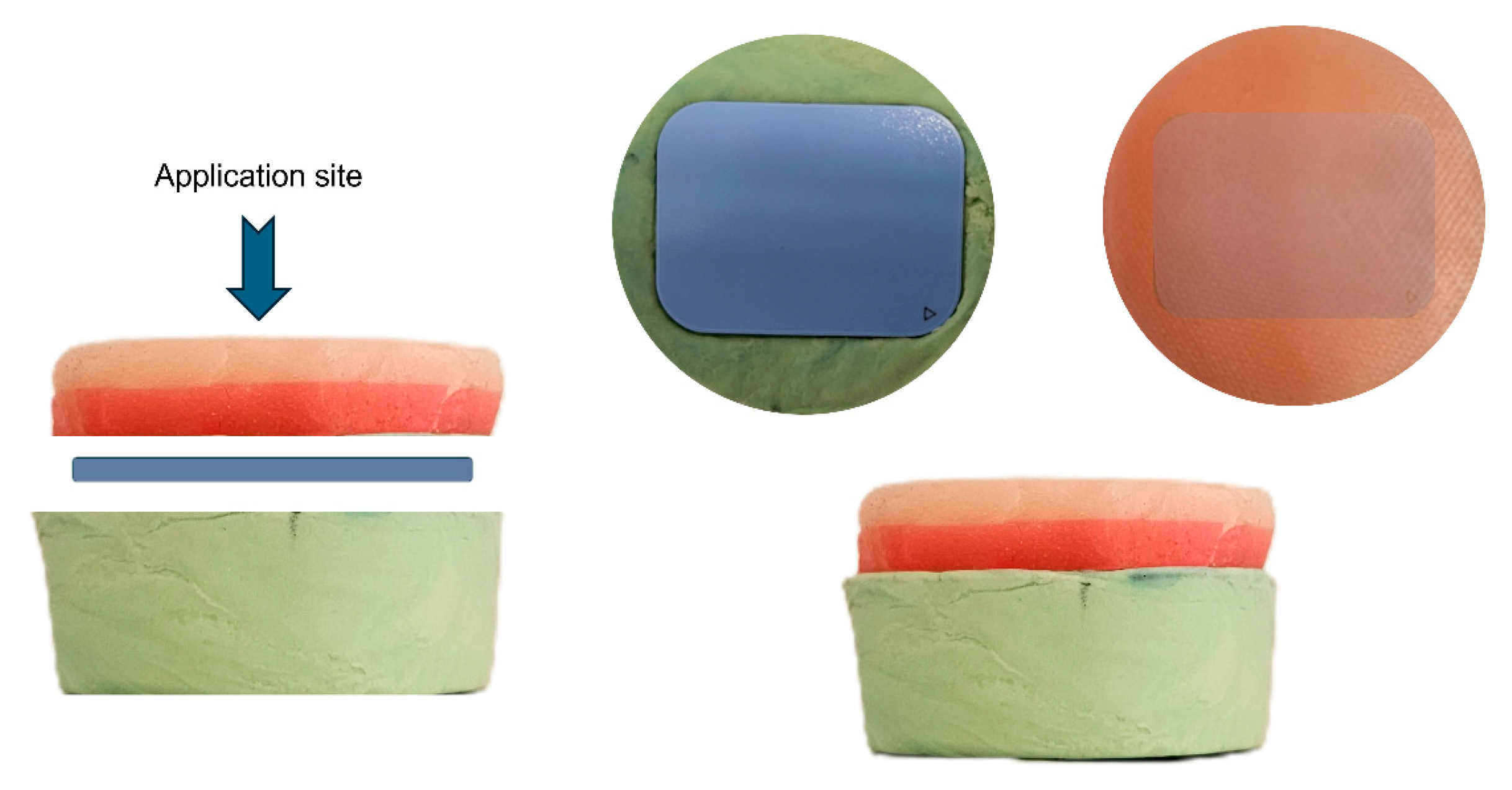
A simulation setup was constructed to provide a standardized and repeatable acquisition geometry. A circular base 65 mm in diameter and 35 mm thick was formed from putty-consistency polyvinyl siloxane impression material (Zetaplus®, Zhermack SpA, Badia Polesine, Italy) as a support layer. A photostimulable phosphor (PSP) plate (Planmeca size-2, 41 mm × 31 mm) was placed on this base, and a commercial suture-training pad of platinum-catalyzed (addition-cured) polydimethylsiloxane (PraticMed®, Ankara, Türkiye) (diameter 50 mm, thickness 10 mm, Shore A hardness: 10) was placed on top of it as a soft tissue surrogate.
Silicone elastomers are widely used to reproduce the mechanical properties of skin [17,18] and were selected here for dimensional stability and repeatable positioning rather than for verified radiographic tissue equivalence. This setup allowed the PSP plate to be held in a stable and repeatable position between the silicone support and suture training material (Figure 1). The test products were applied to the upper surface of the material at a standard dose (2 mg/cm2).
Figure 1.
Experimental simulation setup (not to scale). (Left): exploded side view; (Right): assembled side view and top views of the PSP plate and the product-covered surface. Green, polyvinyl siloxane support base (diameter 65 mm, thickness 35 mm); blue, PSP plate (41 mm × 31 mm); pink and red layers, silicone suture-training pad used as the soft-tissue surrogate (diameter 50 mm, thickness 10 mm). Products were applied to the upper surface (application site); the PSP plate was oriented perpendicular to the central X-ray beam.
Six groups were tested: control; organic-filter sunscreen (L’Occitane en Provence®, Manosque, France); inorganic-filter sunscreen (MISSHA®, Seoul, South Korea); BB and CC cream (Pure Beauty®, Istanbul, Türkiye); and silver sulfadiazine 1% (Silverdin®, Deva Holding, Istanbul, Türkiye). The key radiographically relevant components and their atomic numbers are listed in Table 1. All products were applied at 2 mg/cm2 (approximately 25 mg, precision-weighed, over the area of the phantom directly overlying the PSP plate, 41 mm × 31 mm, approximately 12.7 cm2) for consistency with Nejaim et al. [7] and then pressed with an epoxy plate to form a uniform film.
Table 1.
Radiographically relevant key component profile of the topical products based on INCI label information.
2.3. Image Acquisition
Image acquisitions were performed each morning before clinical use to avoid PSP degradation from ambient light/heat and device-heating current fluctuations, using a Planmeca ProX™ intraoral X-ray unit (Planmeca, Helsinki, Finland) and PSP system under three exposure protocols corresponding to the device’s predefined clinical protocols by anatomical region and patient age (Table 2). The anterior region protocols (A and B) also reflect the frequency of trauma assessment in these regions in maxillofacial trauma/burn cases.
Table 2.
Exposure scenarios used for radiographic image acquisition.
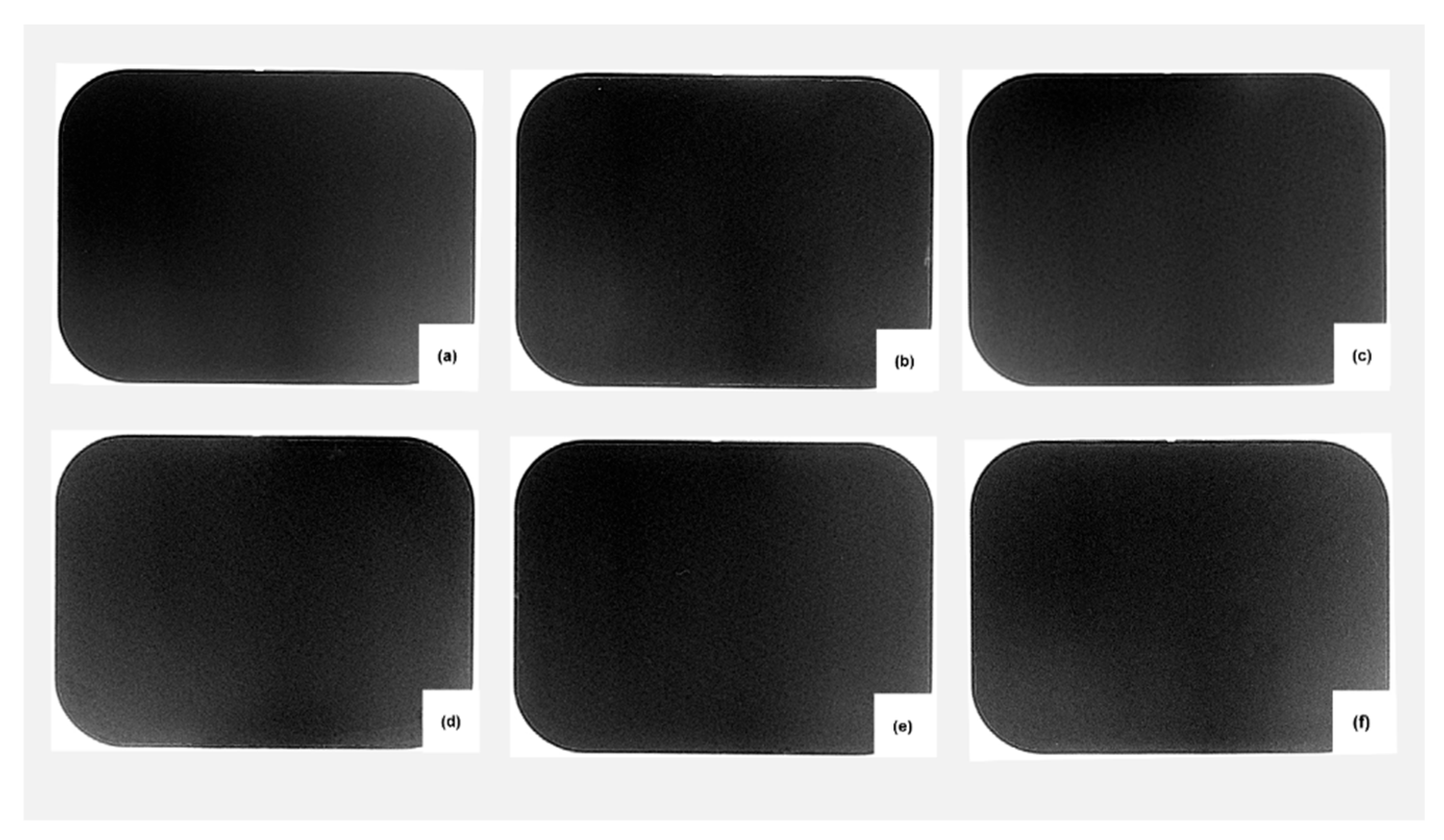
The unit operated at a total filtration of ≥2.5 mm Al equivalent (at 70 kV, per manufacturer specification) with a nominal focal spot of 0.4 mm. Beam collimation was provided by the standard circular cone (60 mm diameter) with a source-to-skin distance (SSD) of 200 mm (short 20 cm cone); with the 10 mm silicone layer on the plate, the focus-to-receptor distance was approximately 210 mm. The PSP plate was positioned on the silicone phantom using a parallel technique, oriented perpendicular to the central X-ray beam; it was ensured that any rotational deviation did not exceed ±3°. Exposure factors were set manually, and no automatic exposure control was used. A single new size-2 PSP plate (Planmeca, 41 mm × 31 mm, 35 µm pixel size) was used for all image acquisitions in this study; the use of a single plate throughout the experiment was chosen to eliminate inter-plate variability as a potential source of confounding. Prior to use, the plate was inspected under standard lighting for scratches, wear, coating damage, and residual latent signals, and its integrity was maintained throughout the experiment. The order of product groups was randomized within each exposure scenario using a random list, and each product application–exposure–scanning sequence was performed as an independent measurement. The time interval between exposure and scanning was standardized to approximately 1 min across all groups and scenarios. After each scan, the plate was erased using the Planmeca ProScanner’s built-in automatic erasure function; no external erasure unit or manual light exposure was used. The scanner was operated in its high-resolution mode (35 µm pixel pitch) with the manufacturer’s scanner-specific calibration files installed; the authors applied no further gain, flat-field or other correction, although firmware-level processing inside the scanner that cannot be disabled by the user cannot be excluded. Images were exported as 16-bit TIFF files (0–65,535 grayscale range), preserving the detector’s full native dynamic range. No image enhancement filters were applied during acquisition, scanning, or export: sharpening, edge enhancement, noise reduction, contrast/brightness adjustment, gamma correction, and histogram equalization were either disabled or not invoked in the ProScanner software (version 4.6.2.R; Planmeca Oy, Helsinki, Finland). Consequently, the exported TIFF represents the earliest detector output accessible to the user and is referred to as the “raw image” throughout the article. Representative raw images from Scenario B are shown in Figure 2.
Figure 2.
Representative raw PSP images acquired under exposure scenario B after application of the tested topical products: (a) control, (b) inorganic sunscreen, (c) organic sunscreen, (d) BB cream, (e) CC cream, and (f) silver sulfadiazine. These images illustrate the unprocessed radiographs used for quantitative image analysis and are shown at the default display window; the differences reported in this study are quantitative and are not visually apparent in these images.
2.4. Image Quality Assessment and Measurements
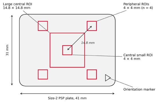
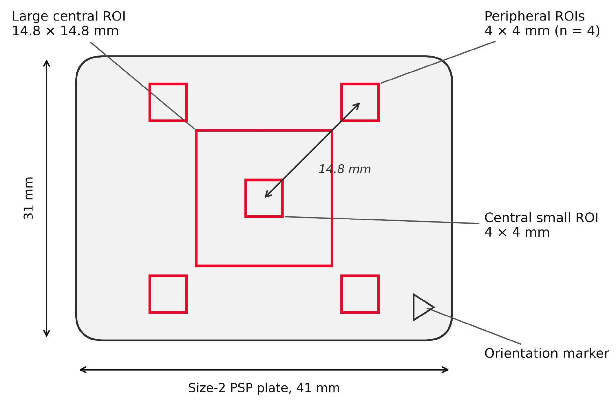
Region-of-interest (ROI) pixel density measurement is an established and reproducible method in dental radiology research, with the mean and SD of pixel density serving as objective image quality indicators. The images were analyzed in ImageJ [19] (v1.54g, NIH, Bethesda, MD, USA) using the exported TIFFs at their native 16-bit depth (0–65,535 grayscale range) and calibrated to the spatial resolution embedded in the file metadata (725 dpi, 35 µm/pixel). Following Miranda-Viana et al. [20], a center-ROI distance of 14.8 mm was used, with five small 4 mm × 4 mm ROIs (center plus four diagonal directions) and one large 14.8 mm × 14.8 mm central ROI (Figure 3). The mean and SD of each of the five small ROIs, from which noise and the uniformity index were calculated, are provided for every image in Supplementary Table S3.
Figure 3.
Schematic illustration of the central and peripheral ROI placement used for grayscale analysis in ImageJ. Brightness, uniformity, SNR, and histogram clipping were measured in the large central ROI; noise and the uniformity index were calculated from the five small 4 mm × 4 mm ROIs (the central small ROI and the four peripheral ROIs).
All ROIs were positioned using manually entered pixel coordinates (Edit > Selection > Specify). Because the ROI coordinates were fixed across all images, repeated measurements of the same image were deterministic; therefore, the intra-rater ICC was not calculated. All ROIs were measured simultaneously (ImageJ ROI Manager, Multi Measure) by a single observer blinded to the group. The primary and derived parameters, formulas, and interpretations are presented in Table 3 [21,22]. Noise and uniformity follow the SD-based definitions of Miranda-Viana et al. [20] and Nejaim et al. [7]. Because these SD values include structured components (beam non-uniformity, receptor non-uniformity, and the distribution of the applied product) in addition to random noise, they are interpreted as SD-based estimates of grayscale variation and not as pure random noise. SNR (mean/SD of the large central ROI) inherits the same limitation, and uniformity (SD) and SNR are derived from the same ROI and are therefore not independent. More generally, SNR is a function of brightness and uniformity (SNR = brightness ÷ uniformity), noise and the uniformity index are both derived from the same five small ROIs, and brightness, uniformity, SNR, and clipping are all derived from the same large central ROI. The six parameters were therefore not treated as independent lines of evidence, and agreement between related parameters is not interpreted as independent confirmation. Histogram clipping counts pixels at either end of the 16-bit scale (0 and 65,535) together; its interpretation as a detector saturation or floor effect is an assumption.
Table 3.
Primary and derived image quality parameters used for radiographic image analysis.
2.5. Statistical Analysis
Analyses were performed using IBM SPSS Statistics v28.0 with α = 0.05. The raw data for all 54 images are provided in Supplementary Table S1. Normality was assessed via the Shapiro–Wilk test on model residuals. Brightness and Uniformity met the normality assumption and were analyzed using two-way ANOVA on raw values, with Group and Scenario treated as fixed factors. SNR, Clipping, Noise, and Uniformity Index did not meet the assumptions and underwent rank transformation prior to two-way ANOVA; the same rank-transformed values were consistently used for all three tests in each model (Group main effect, Scenario main effect, and Group × Scenario interaction). Rank transformation was used as a distribution-free alternative for the four outcomes that violated the normality assumption. No separate re-analysis using the Aligned Rank Transform was conducted; a single, consistent rank-based approach was applied. Because rank-based interaction tests can be unreliable with small cell sizes (n = 3 per cell), the Group × Scenario interaction is regarded as exploratory, and the primary inference rests on the Group main effect and the Dunnett contrasts against the control. Homogeneity of variance across the 18 cells was examined with Levene’s test (mean-centered; n = 3 per cell): it was rejected for brightness (p = 0.017) and, for the rank-transformed values, for clipping (p = 0.002), noise (p = 0.001) and UI (p = 0.014), whereas the median-centered (Brown–Forsythe) version did not reject for any outcome (all p > 0.7); with only three images per cell, neither version is a powerful check. Because the design is balanced, the ANOVA F tests are comparatively robust to unequal variances, and Welch t-tests against the control (on raw values for brightness and uniformity and on rank-transformed values for SNR, clipping, noise and UI; Benjamini–Hochberg [BH]-adjusted across the same 30 comparisons) were run as a sensitivity analysis (Supplementary Table S2). Each product group was compared against the Control group using Dunnett’s post hoc test. The Benjamini–Hochberg false discovery rate (FDR) correction [23] was applied globally across all 30 pairwise comparisons (5 product-vs-Control comparisons × 6 outcome parameters) rather than separately for each parameter or product. Thus, the FDR was controlled across the entire family of comparisons. The resulting BH-adjusted p-values are reported as q-values. Effect sizes were reported as partial η2 alongside F-statistics and degrees of freedom.
3. Results
3.1. Descriptive Statistics
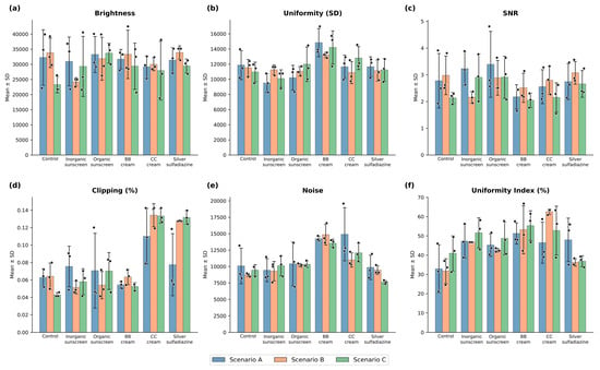
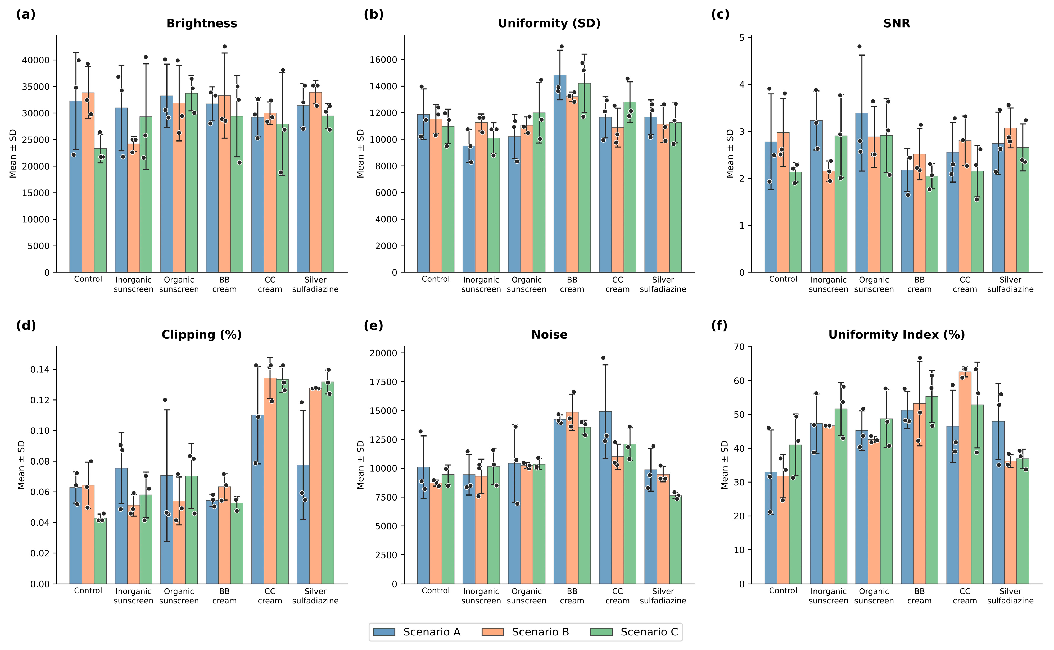
The BB cream exhibited higher noise values (greater pixel-level variation) and higher uniformity index values (greater uniformity among the five small ROI means) compared to the control across all scenarios (Table 4). Histogram clipping was highest in the CC cream and silver sulfadiazine groups, which may reflect a more pronounced detector saturation/floor effect (an interpretation that was not tested; see Section 2.4). The organic sunscreen showed values close to the control for all parameters (Figure 4). In the representative images (Figure 2), no differences between the control and the product groups are visually apparent; the differences reported below are quantitative.
Table 4.
Descriptive statistics of radiographic image quality parameters across the experimental groups and exposure scenarios (A, B, C) (mean ± SD).
Figure 4.
Distribution of image quality parameters across the study groups. (a) brightness, (b) uniformity (SD), (c) signal-to-noise ratio (SNR), (d) histogram clipping (%), (e) noise, and (f) uniformity index (%). Bars show mean ± SD; dots show individual images (n = 3 per group and scenario).
3.2. Two-Way ANOVA: Group and Exposure Effects
Two-way ANOVA (Table 5) showed a significant product group effect on uniformity, clipping, noise, and uniformity index (all p < 0.001), but not on brightness (p = 0.550) or SNR (p = 0.116). In this study, no statistically significant effect of the exposure protocol was found for any parameter (F = 0.239–1.835, all p > 0.05); however, since the study was powered only to detect large effects, small or moderate protocol effects cannot be ruled out. No group-by-protocol interaction reached significance, although the interaction for clipping approached the threshold (F = 2.092, p = 0.052).
Table 5.
Effects of product group, exposure scenario, and their interaction on radiographic image quality parameters: two-way ANOVA analysis.
3.3. Post Hoc Comparisons with Control
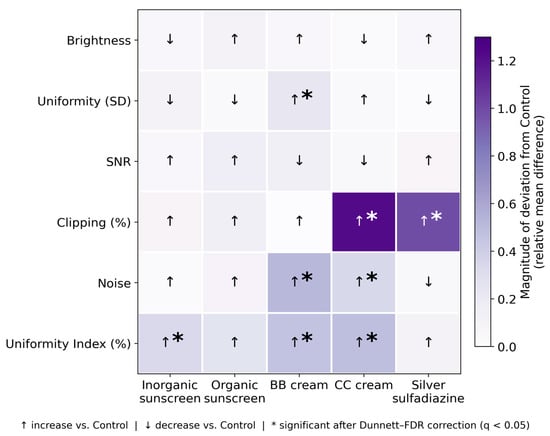
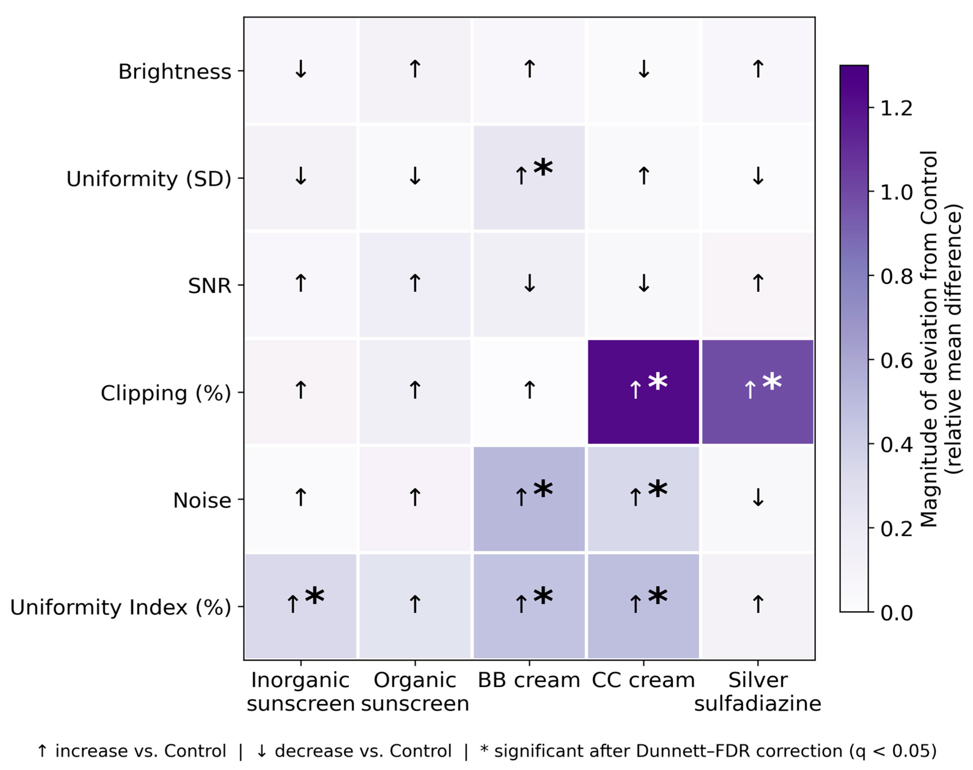
The Dunnett post hoc test with FDR correction (Table 6) showed that significant differences from the control were product-specific. Dunnett-adjusted p-values are given first, followed by the BH-adjusted p-values on which the FDR decisions are based. The BB cream differed from the control in Uniformity (higher SD than control; p = 0.003; BH-adjusted p = 0.011), Noise (higher; p < 0.001; BH-adjusted p < 0.001), and UI (higher, indicating greater uniformity among the five small ROI means; p < 0.001; BH-adjusted p = 0.002). The CC cream differed in Clipping (higher; p < 0.001; BH-adjusted p < 0.001), Noise (higher; p < 0.001; BH-adjusted p = 0.002), and UI (higher; p < 0.001; BH-adjusted p = 0.002). Silver sulfadiazine differed from the control only in Clipping (higher; p < 0.001; BH-adjusted p = 0.002). The inorganic sunscreen differed only in UI (higher; p = 0.011; BH-adjusted p = 0.042). The organic sunscreen did not show a significant difference from the control in any parameter, including the uniformity index (Dunnett-adjusted p = 0.096; BH-adjusted p = 0.321); the Welch sensitivity analysis (Supplementary Table S2) identified the same eight significant comparisons. The relative mean differences (pooled across the three exposure scenarios) and their directionality across all parameters and groups are summarized in Figure 5, with CC cream and silver sulfadiazine showing the largest deviations (histogram clipping), and BB cream affecting the widest range of parameters. Although uniformity (the SD of the central ROI) differed significantly between groups while brightness did not, the derived SNR did not reach statistical significance (p = 0.116); given the modest sample size per cell (n = 3), this should be read as inconclusive and not as evidence that signal-to-noise is unaffected.
Table 6.
Dunnett Post hoc Results after FDR * Correction.
Figure 5.
Heat map summarizing the direction and relative magnitude of changes in radiographic image parameters across experimental groups. Color intensity represents the relative mean difference, (product group mean − control mean)/control mean. Arrows indicate increase (↑) or decrease (↓) relative to control. For the uniformity index (UI), an increase indicates greater uniformity among the five small ROI means. Asterisks indicate statistically significant differences after Dunnett–FDR correction (BH-adjusted (q < 0.05, where q denotes the Benjamini–Hochberg-adjusted p-value)). Values are pooled across the three exposure scenarios (n = 9 per group).
4. Discussion
Across six parameters and three exposure protocols, products containing metal oxides or silver sulfadiazine produced formulation-specific changes in quantitative imaging parameters; the clinical significance of these changes was not evaluated in this study. No statistically significant effect of the exposure protocol was detected; however, as the study was powered only to detect large effects, this finding should not be interpreted as evidence of protocol independence.
A distinction must be made between statistical and clinical significance: although the product group had large effects on uniformity, clipping, noise, and the uniformity index (partial η2 = 0.486–0.675), brightness, which directly corresponds to the displayed gray value, did not differ significantly between the groups (p = 0.550; partial η2 = 0.101). The largest difference in brightness between a pooled group mean and the control mean was approximately 12 gray levels in 8-bit equivalent terms (about 3150 of 65,535 units; organic sunscreen), whereas differences between individual product–scenario cell means reached approximately 40 levels, and the control group itself differed by approximately 41 levels between scenarios B and C. Whether these differences are clinically perceptible was not evaluated in this study and requires observer-based studies.
Nejaim et al. [7] tested sunscreens under a single exposure protocol. We added three products used in burn and trauma care, measured six parameters rather than three, and repeated the acquisitions under three exposure protocols, for a total of 54 images. Notably, the “inorganic sunscreen” tested by Nejaim et al. lists iron oxide pigments (CI 77491, CI 77492, CI 77499) in its INCI composition, placing it closer in radiographic terms to our BB and CC cream groups than to classic mineral filters containing only TiO2 and ZnO. The differences in parameter-level effect profiles between the two studies may therefore reflect formulation heterogeneity as much as methodological differences and reinforce that “inorganic sunscreen” is not a radiographically homogeneous category.
The literature on the radiographic artifacts of cosmetic and dermatological products is limited; existing studies focus primarily on how gold facial threads degrade panoramic image quality and how filler materials mimic odontogenic pathology on CBCT scans [24,25].
Within-cell variability proved higher than expected for a phantom setup with fixed geometry. This trend was particularly evident in the control group. Given that positioning was controlled using fixed pixel coordinates and a ±3° rotation criterion, the primary sources of this variation likely include the time-dependent fading of the PSP latent image (affecting the exposure-to-scanning interval) and residual signals persisting between erasure cycles. Furthermore, while the use of a single PSP plate throughout the study was a deliberate choice to eliminate inter-plate variability, it could not entirely rule out cumulative plate degradation over the course of 54 exposures. This variability reduced the study’s precision and represents an area for methodological improvement.
Mechanistically, BB and CC creams share titanium dioxide, aluminum hydroxide, and iron oxide pigments. The higher local noise observed in the BB cream can be interpreted as a radiographic reflection of the heterogeneous particle distribution within the formulation. Its higher uniformity index, in contrast, indicates smaller differences among the five small ROI means (greater regional uniformity). The two findings are not contradictory because they describe different spatial scales: noise reflects pixel-level variation within each small ROI, whereas the uniformity index reflects differences in mean signal between ROIs. Why the BB cream reduced the between-ROI differences was not investigated; as particle size, distribution, film thickness, and actual metal oxide content were not measured in this study, these explanations are hypotheses requiring testing rather than proven mechanisms. Radiopacity is known to depend on both material type and thickness [26]. Similarly, the more pronounced histogram clipping observed in the CC cream may be consistent with the presence of a denser and more homogeneous iron oxide layer. Metal oxides (such as cerium, tantalum, and niobium oxides) have been shown to increase the radiopacity of dental adhesives [27]. Whether these observed parameter changes impact clinical diagnostic accuracy was not assessed in this study; anatomical phantom and observer-based ROC studies are necessary to elucidate this issue.
The inorganic sunscreen (ZnO + TiO2) differed significantly from the control only in the uniformity index, where a higher value indicates greater uniformity among the five small ROI means; the clinical relevance of this difference was not assessed. The findings are consistent with the literature on particle coating/dispersion effects [8] and the interaction of TiO2/ZnO nanoparticles with the stratum corneum [28,29]. Our surface-only in vitro setup likely underestimates actual skin penetration; furthermore, reference skin thickness data come from a Korean ultrasonographic cohort [30], which limits the generalizability of these values. The increase in clipping observed in silver sulfadiazine may be consistent with the high atomic number of silver (Z = 47) and its strong photoelectric absorption; this was not tested. This finding is relevant for future research because the product is frequently used in facial burn/trauma care, including in pediatric patients, for whom the ALADA principle is particularly emphasized [2,3].
The organic sunscreen tested in this study did not show a significant difference from the control in any of the measured parameters in the primary analysis; this finding is consistent with the low atomic number of the elements in organic UV filters (mainly carbon, hydrogen and oxygen), which suggests negligible photoelectric absorption at dental X-ray energies (not tested here), and aligns with the results reported by Nejaim et al. [7]. Patients with darker skin tones tend to prefer chemical filters over mineral formulations because the latter leave a visible white residue [31]; whether this preference has any bearing on image parameters cannot be inferred from a single organic sunscreen. The absence of a detectable effect related to the exposure protocol cannot be taken as evidence that these findings generalize across technical configurations, because the study lacks the statistical power to demonstrate this independence.
The clinical scenario motivating this study (the combined use of silver sulfadiazine, an inorganic sunscreen, and BB/CC cream in burn or trauma rehabilitation) raises the possibility of a cumulative effect arising from the coexistence of components with high atomic numbers (Ag = 47, Zn = 30, Fe = 26, Ti = 22). However, in this study, the products were tested individually, and scenarios involving combinations or layered applications were not evaluated. The observed individual effects position this possibility as a hypothesis requiring verification and establish it as a priority research question for future studies.
A critical point regarding the clinical significance of this study is the distinction between the raw and the processed images presented to the clinician. While current PSP and CMOS detectors have a depth of 12–16 bits (4096–65,536 gray levels), the human eye can discern only approximately 60 shades within this range [32]. Software therefore compresses the dynamic range for display, and this compression may bring weak signals (including topical product residues) up to the contrast level of anatomic structures. Our findings were obtained from 16-bit TIFF images, which were necessary for methodological consistency; however, current digital radiography software may apply automatic histogram equalization, unsharp masking, and noise reduction algorithms before image display. It is well established in digital image processing that unsharp masking can amplify local pixel gradients and generate overshoot artifacts, such as dark halos around edges [22]. An edge-enhancement filter cannot theoretically distinguish a local density difference caused by titanium dioxide particles from a true anatomical margin; it may accentuate both in the same manner. This aligns with warnings in standard oral radiology literature stating that subjective image enhancement does not improve the accuracy of image interpretation and that what benefits one diagnostic task may reduce image quality for another [33], so the benefit of such filters depends on the diagnostic task (enhancement filters improved caries detection in one study [34]).
The concern lies not in the formation of the artifact itself, but in the possibility that such signals in processed images could be mistaken for pathological findings. However, this study did not evaluate processed images, nor did it involve observer assessments or measurements of diagnostic accuracy. Consequently, the potential for clinical error, including the risk of misinterpretation or unnecessary repeat imaging in pediatric or trauma patients, remains a hypothesis that cannot be directly inferred from the study’s data. Conversely, such processing could also reduce or mask the differences measured here, which was likewise not tested.
Similarly, the possibility that silver sulfadiazine-induced histogram compression might mask diagnostic information during automated dynamic range normalization, and the question of whether deep learning-based systems for detecting caries and periapical lesions, which rely on local pixel gradients, are affected by residues of topical products, remain open research questions awaiting investigation through anatomical phantom studies and observer-based evaluations.
The methodological strengths of this study include a six-parameter quantitative assessment framework, a standardized ROI template with fixed pixel coordinates that minimizes observer variation, calibrated gray-value measurements at native TIFF bit depth, and evaluation across three clinically relevant exposure protocols.
This study has several limitations. First, a homogeneous simulation phantom devoid of anatomical structures (bone, dentin, enamel) was used; this choice was made to isolate the product’s radiographic signature from anatomical confounders, but it limits the direct extrapolation of findings to clinical diagnostic tasks. Future studies using phantoms containing skulls, bone blocks, or teeth are necessary to test clinical relevance. In addition, the silicone soft-tissue surrogate was chosen for dimensional stability and repeatable positioning and was not validated as radiographically equivalent to human soft tissue (its attenuation was not compared with tissue-equivalent reference materials), so the magnitude of the observed effects may not transfer directly to clinical intraoral radiography, where the beam traverses facial skin, soft tissue, and bone. Second, as the study was powered only to detect large effects (n = 3 per group), small or moderate effects may have gone undetected, and the negative results obtained regarding exposure protocols should be interpreted as inconclusive. Third, only a single PSP system was tested, and other digital detector technologies (e.g., CMOS) might exhibit different behavior [35]; PSP systems also differ in microstructure and image uniformity [36]. Fourth, a single application dose (2 mg/cm2) was used, failing to reflect real-world variations in application quantity [28,29]. Fifth, the effects of drying, radiation dose, diagnostic accuracy, and combined product use were not evaluated [2,3,7]. Sixth, since only one commercial product was tested in each category, the results cannot be generalized to all organic sunscreens, inorganic sunscreens, BB creams, or CC creams. Seventh, a single PSP plate was used throughout the study; this design was chosen to eliminate inter-plate variability but cannot rule out cumulative plate degradation over the course of 54 exposures, and no post hoc correction for signal drift (e.g., latent-image fading or plate degradation) was applied. Eighth, the three replicates per cell are technical replicates obtained from a single phantom, a single PSP plate, a single X-ray unit, and a single lot of each product, so the results apply to this experimental setup. Ninth, the six image parameters are mathematically related and partly derived from overlapping regions of interest (Section 2.4), so they do not represent independent outcomes. Tenth, no observer-based diagnostic assessment was performed, so differences in pixel-level measurements cannot be equated with clinically meaningful image degradation. Additionally, a standard rank-based two-way ANOVA was used instead of the aligned rank transform for interaction tests on rank-transformed parameters; because rank-based interaction tests can be unreliable with small cell sizes, the Group × Scenario interaction results should be regarded as exploratory. Priority extensions for future studies include observer-based ROC tests using anatomical phantoms, CMOS comparisons, combined product scenarios, and cumulative dosimetry.
Although it is premature to derive direct clinical practice recommendations from this study, the findings may help guide future research questions. Questions suggested for evaluation in subsequent studies include: does observer-based diagnostic accuracy differ between patients using BB/CC creams or silver sulfadiazine and controls; does skin cleansing prior to radiography eliminate this difference; are artifacts observed in processed images also present in raw images and clinically distinguishable; and are AI-based diagnostic systems affected by residues of topical products? The answers to these questions will determine whether the current in vitro findings can be translated into clinical practice.
5. Conclusions
Within the scope of this exploratory in vitro study, all tested products (one per category) except the organic sunscreen produced measurable changes in the quantitative pixel-level parameters of intraoral radiographic images, with the pattern of change varying across formulations. The organic sunscreen showed no significant difference from the control. No statistically significant effect related to the exposure protocol was detected; however, as the study was powered only to detect large effects, an exposure-dependent effect cannot be ruled out. Whether the observed differences are clinically perceptible, and whether software processing could render these differences visible, was not tested in this study. These findings serve as a reference for future studies involving anatomically realistic phantoms, observer-based ROC analyses, comparisons between processed and raw images, and the use of different detector technologies (CMOS).
Supplementary Materials
The following supporting information can be downloaded at: https://www.mdpi.com/article/10.3390/diagnostics16223885/s1, Table S1: Raw numerical data for the 54 images (6 product groups × 3 exposure scenarios × 3 replicates); Table S2: Sensitivity analysis: Welch (unequal-variance) t-tests of each product group versus the control, for each of the six parameters (30 comparisons); Table S3: Mean and SD of the grayscale values of each of the five small ROIs (ROI C = central small ROI; ROI 1–4 = the four peripheral small ROIs, 4 mm × 4 mm each) for each image, with the noise and uniformity index values calculated from them.
Author Contributions
Conceptualization, O.B.; Methodology, O.B.; Formal Analysis, O.B.; Investigation, E.K.; Resources, E.K.; Writing—Original Draft Preparation, O.B.; Writing—Review and Editing, O.B. and E.K. All authors have read and agreed to the published version of the manuscript.
Funding
This research received no external funding.
Institutional Review Board Statement
Not applicable.
Informed Consent Statement
Not applicable.
Data Availability Statement
The original contributions presented in this study are included in the article/Supplementary Materials. Further inquiries can be directed to the corresponding author.
Acknowledgments
The authors thank Elif Bilgir for her methodological contributions. During the preparation of this manuscript, the authors used Claude Opus 4.8 (Anthropic, San Francisco, CA, USA) and Gemini 3.6 Flash (Google LLC, Mountain View, CA, USA) for English language editing. The authors have reviewed and edited the output and take full responsibility for the content of this publication.
Conflicts of Interest
The authors declare no conflicts of interest.
Abbreviations
The following abbreviations are used in this manuscript:
| ACR | American College of Radiology |
| Ag | Silver |
| ALADA | As Low As Diagnostically Acceptable |
| ANOVA | Analysis of Variance |
| BB | Blemish Balm |
| BH | Benjamini–Hochberg |
| CBCT | Cone-Beam Computed Tomography |
| CC | Color Correcting |
| CMOS | Complementary Metal–Oxide–Semiconductor |
| FDR | False Discovery Rate |
| GV | Gray Value |
| ICC | Intraclass Correlation Coefficient |
| INCI | International Nomenclature of Cosmetic Ingredients |
| PA | Protection Grade of UVA |
| PIU | Percent Integral Uniformity |
| PSP | Photostimulable Phosphor |
| ROC | Receiver Operating Characteristic |
| ROI | Region of Interest |
| SD | Standard Deviation |
| SNR | Signal-to-Noise Ratio |
| SPF | Sun Protection Factor |
| UI | Uniformity Index |
References
- Williams, M.B.; Krupinski, E.A.; Strauss, K.J.; Breeden, W.K.; Rzeszotarski, M.S.; Applegate, K.; Wyatt, M.; Bjork, S.; Seibert, J.A. Digital Radiography Image Quality: Image Acquisition. J. Am. Coll. Radiol. 2007, 4, 371–388. [Google Scholar] [CrossRef] [Scilit] [PubMed]
- FDI World Dental Federation. FDI Policy Statement on Radiation Safety in Dentistry. Int. Dent. J. 2014, 64, 289–290. [Google Scholar] [CrossRef] [Scilit] [PubMed]
- Mendonça, R.P.d.; Estrela, C.; Bueno, M.R.; Carvalho, T.C.A.S.G.; Estrela, L.R.d.A.; Chilvarquer, I. Principles of Radiological Protection and Application of ALARA, ALADA, and ALADAIP: A Critical Review. Braz. Oral Res. 2025, 39, e14. [Google Scholar] [CrossRef] [Scilit] [PubMed]
- Ardila, C.M.; Vivares-Builes, A.M.; Pineda-Vélez, E. Image Quality, Radiation Dose, and Patient Comfort Associated with Wireless Sensors in Digital Radiography: A Systematic Review. Dent. J. 2024, 12, 267. [Google Scholar] [CrossRef] [Scilit] [PubMed]
- Tompe, A.; Sargar, K. X-Ray Image Quality Assurance. In StatPearls; StatPearls Publishing: Treasure Island, FL, USA, 2022. [Google Scholar]
- Wenzel, A.; Møystad, A. Work Flow with Digital Intraoral Radiography: A Systematic Review. Acta Odontol. Scand. 2010, 68, 106–114. [Google Scholar] [CrossRef] [Scilit] [PubMed]
- Nejaim, Y.; Bregolin, G.S.N.; Suekane, N.H.; Gonzalez, E.F.; Valente, F.R.F.; Fontenele, R.C. Does Facial Sunscreen Usage Impact Radiographic Image Quality and Radiation Dose? An In Vitro Study. Oral Radiol. 2025, 41, 475–484. [Google Scholar] [CrossRef] [Scilit] [PubMed]
- Manaia, E.B.; Kaminski, R.C.K.; Corrêa, M.A.; Chiavacci, L.A. Inorganic UV Filters. Braz. J. Pharm. Sci. 2013, 49, 201–209. [Google Scholar] [CrossRef] [Scilit]
- Suen, K.; Ho, L.; Chow, K.; Wong, L.; Leong, L. Calamine Lotion: Mimicking Intramammary Calcifications. Australas. Radiol. 2007, 51, B43–B44. [Google Scholar] [CrossRef] [Scilit] [PubMed]
- Cupp, S.L.; Beideck, D.J.; Maidment, A.D.A. Ultraviolet Protectants: Causative Agents for Screen and Image Artifacts in Radiography. Radiology 2003, 227, 119–123. [Google Scholar] [CrossRef] [Scilit] [PubMed]
- Ginzburg, A.L.; Blackburn, R.S.; Santillan, C.; Truong, L.; Tanguay, R.L.; Hutchison, J.E. Zinc Oxide-Induced Changes to Sunscreen Ingredient Efficacy and Toxicity under UV Irradiation. Photochem. Photobiol. Sci. 2021, 20, 1273–1285. [Google Scholar] [CrossRef] [Scilit] [PubMed]
- Ebrahimi, A.; Kalantar Motamedi, M.H. Application of Silver Sulfadiazine Cream with Early Surgical Intervention in Patients Suffering from Combined Burn-Blast Injury Facial Tattoos. Trauma Mon. 2012, 17, 255–258. [Google Scholar] [CrossRef] [Scilit] [PubMed]
- Anthonissen, M.; Daly, D.; Janssens, T.; Van den Kerckhove, E. The Effects of Conservative Treatments on Burn Scars: A Systematic Review. Burns 2016, 42, 508–518. [Google Scholar] [CrossRef] [Scilit] [PubMed]
- Mancuso, J.B.; Maruthi, R.; Wang, S.Q.; Lim, H.W. Sunscreens: An Update. Am. J. Clin. Dermatol. 2017, 18, 643–650. [Google Scholar] [CrossRef] [Scilit] [PubMed]
- Magi, S.; Arena, D.; Lanzoni, A.; Stella, M.; Depetris, N.; Sarzi, L.; Massazza, G. Camouflage in the Rehabilitation Process of Burn Scars. Burns Open 2021, 5, 258–261. [Google Scholar] [CrossRef] [Scilit]
- Yeom, H.-G.; Kim, J.-E.; Huh, K.-H.; Yi, W.-J.; Heo, M.-S.; Lee, S.-S. Development and Validation of a Clinical Phantom Reproducing Various Lesions for Oral and Maxillofacial Radiology Research. Imaging Sci. Dent. 2023, 53, 345–353. [Google Scholar] [CrossRef] [Scilit] [PubMed]
- Prasomsin, W.; Prastowo, R.B.; Okhawilai, M.; Jubsilp, C.; Rimdusit, S. Development of a Suture Pad for Medical Training from Silk Fiber Reinforced Polydimethylsiloxane Composite. Eng. J. 2021, 25, 71–79. [Google Scholar] [CrossRef] [Scilit]
- Dąbrowska, A.K.; Rotaru, G.-M.; Derler, S.; Spano, F.; Camenzind, M.; Annaheim, S.; Stämpfli, R.; Schmid, M.; Rossi, R.M. Materials Used to Simulate Physical Properties of Human Skin. Skin Res. Technol. 2016, 22, 3–14. [Google Scholar] [CrossRef] [Scilit] [PubMed]
- Schneider, C.A.; Rasband, W.S.; Eliceiri, K.W. NIH Image to ImageJ: 25 Years of Image Analysis. Nat. Methods 2012, 9, 671–675. [Google Scholar] [CrossRef] [Scilit] [PubMed]
- Miranda-Viana, M.; Fontenele, R.C.; Nogueira-Reis, F.; Farias-Gomes, A.; Oliveira, M.L.; Freitas, D.Q.; Haiter-Neto, F. DICOM File Format Has Better Radiographic Image Quality than Other File Formats: An Objective Study. Braz. Dent. J. 2023, 34, 150–157. [Google Scholar] [CrossRef] [Scilit] [PubMed]
- American College of Radiology. Magnetic Resonance Imaging Quality Control Manual; American College of Radiology: Reston, VA, USA, 2015. [Google Scholar]
- Gonzalez, R.; Woods, R. Digital Image Processing, 4th ed.; Pearson Education Limited: Harlow, UK, 2018. [Google Scholar]
- Benjamini, Y.; Hochberg, Y. Controlling the False Discovery Rate: A Practical and Powerful Approach to Multiple Testing. J. R. Stat. Soc. Ser. B Stat. Methodol. 1995, 57, 289–300. [Google Scholar] [CrossRef] [Scilit]
- Ching, M.E.; Joan, L.Z.Y.; Nambiar, P. Cosmetic “Gold Thread Therapy”: A Nuisance That Decreases the Diagnostic Quality of a Dental Panoramic Image. Case Rep. Dent. 2021, 2021, 4077930. [Google Scholar] [CrossRef] [Scilit] [PubMed]
- Alsufyani, N.A.; Alsufyani, M.A. Radiographic Features of Facial Cosmetic Material: Report of Two Cases. Case Rep. Dent. 2021, 2021, 7308636. [Google Scholar] [CrossRef] [Scilit] [PubMed]
- Yaylacı, A.; Karaarslan, E.S.; Hatırlı, H. Evaluation of the Radiopacity of Restorative Materials with Different Structures and Thicknesses Using a Digital Radiography System. Imaging Sci. Dent. 2021, 51, 261. [Google Scholar] [CrossRef] [Scilit] [PubMed]
- Balhaddad, A.A.; Garcia, I.M.; Mokeem, L.; Alsahafi, R.; Collares, F.M.; Sampaio de Melo, M.A. Metal Oxide Nanoparticles and Nanotubes: Ultrasmall Nanostructures to Engineer Antibacterial and Improved Dental Adhesives and Composites. Bioengineering 2021, 8, 146. [Google Scholar] [CrossRef] [Scilit] [PubMed]
- Smijs, T.; Pavel, S. Titanium Dioxide and Zinc Oxide Nanoparticles in Sunscreens: Focus on Their Safety and Effectiveness. Nanotechnol. Sci. Appl. 2011, 4, 95–112. [Google Scholar] [CrossRef] [Scilit] [PubMed]
- Addor, F.A.S.; Barcaui, C.B.; Gomes, E.E.; Lupi, O.; Marçon, C.R.; Miot, H.A. Sunscreen Lotions in the Dermatological Prescription: Review of Concepts and Controversies. An. Bras. Dermatol. 2022, 97, 204–222. [Google Scholar] [CrossRef] [Scilit] [PubMed]
- Jeong, K.M.; Seo, J.Y.; Kim, A.; Kim, Y.C.; Baek, Y.S.; Oh, C.H.; Kye, Y.C. Ultrasonographic Analysis of Facial Skin Thickness in Relation to Age, Site, Sex, and Body Mass Index. Skin Res. Technol. 2023, 29, e13426. [Google Scholar] [CrossRef] [Scilit] [PubMed]
- Song, H.; Beckles, A.; Salian, P.; Porter, M.L. Sunscreen Recommendations for Patients with Skin of Color in the Popular Press and in the Dermatology Clinic. Int. J. Womens Dermatol. 2021, 7, 165–170. [Google Scholar] [CrossRef] [Scilit] [PubMed]
- Udupa, H.; Mah, P.; Dove, S.B.; McDavid, W.D. Evaluation of Image Quality Parameters of Representative Intraoral Digital Radiographic Systems. Oral Surg. Oral Med. Oral Pathol. Oral Radiol. 2013, 116, 774–783. [Google Scholar] [CrossRef] [Scilit] [PubMed]
- Lam, E.W.N.; Mallya, S.M. (Eds.) White and Pharoah’s Oral Radiology: Principles and Interpretation, 9th ed.; Elsevier: St. Louis, MO, USA, 2024; p. 55. [Google Scholar]
- Shokri, A.; Kasraei, S.; Lari, S.; Mahmoodzadeh, M.; Khaleghi, A.; Musavi, S.; Akheshteh, V. Efficacy of Denoising and Enhancement Filters for Detection of Approximal and Occlusal Caries on Digital Intraoral Radiographs. J. Conserv. Dent. 2018, 21, 162–168. [Google Scholar] [CrossRef] [Scilit] [PubMed]
- Kamburoğlu, K.; Samunahmetoğlu, E.; Eratam, N.; Sönmez, G.; Karahan, S. Clinical Comparison of Intraoral CMOS and PSP Detectors in Terms of Time Efficiency, Patient Comfort, and Subjective Image Quality. Imaging Sci. Dent. 2022, 52, 93. [Google Scholar] [CrossRef] [Scilit] [PubMed]
- Souza-Pinto, G.N.d.; Nejaim, Y.; Gomes, A.F.; Canteras, F.B.; Freitas, D.Q.; Haiter-Neto, F. Evaluation of the Microstructure, Chemical Composition, and Image Quality of Different PSP Receptors. Braz. Oral Res. 2022, 36, e130. [Google Scholar] [CrossRef] [Scilit] [PubMed]

Disclaimer/Publisher’s Note: The statements, opinions and data contained in all publications are solely those of the individual author(s) and contributor(s) and not of MDPI and/or the editor(s). MDPI and/or the editor(s) disclaim responsibility for any injury to people or property resulting from any ideas, methods, instructions or products referred to in the content. |
© 2026 by the authors. Licensee MDPI, Basel, Switzerland. This article is an open access article distributed under the terms and conditions of the Creative Commons Attribution (CC BY) license.