


Abstract
Background and Clinical Significance: Atypical hemolytic uremic syndrome (aHUS) is a rare, life-threatening, complement-mediated thrombotic microangiopathy (TMA) that may occur de novo after kidney transplantation. Because its manifestations overlap with more common causes of allograft dysfunction, including rejection, calcineurin inhibitor toxicity, and infection, diagnosis can be challenging. The disease may progress rapidly, making early clinical suspicion and timely etiologic evaluation essential. Case Presentation: We report a 52-year-old man who developed abrupt, severe graft dysfunction approximately 10 years after ABO-incompatible living-donor kidney transplantation. His serum creatinine had remained approximately 2.0 mg/dL after treatment for suspected chronic active antibody-mediated rejection but increased to 6.57 mg/dL at outpatient follow-up and to 8.47 mg/dL on admission after a recent norovirus infection. Initial evaluation considered common post-transplant causes of graft dysfunction, including rejection, calcineurin inhibitor toxicity, and infection-related injury. However, as renal dysfunction progressed and concurrent anemia and thrombocytopenia became evident, TMA was suspected, and a targeted workup was undertaken. Elevated lactate dehydrogenase, decreased haptoglobin, fragmented red blood cells, and allograft biopsy findings supported TMA. No HLA class I or II antibodies were detected, C4d staining was negative, ADAMTS13 activity was preserved, and testing for Shiga toxin-producing Escherichia coli was negative. Genetic testing identified a heterozygous CFH variant (c.3329G>C, p.Gly1110Ala), classified as a variant of uncertain significance. Based on the clinical, hematologic, and histopathologic findings, suspected aHUS was considered the most likely diagnosis. However, ABMR-associated TMA could not be completely excluded. Plasma exchange was complicated by anaphylaxis, and eculizumab was initiated. Hematologic abnormalities subsequently resolved, although renal function did not recover and the patient remained dialysis-dependent. Conclusions: This case highlights the importance of considering TMA when graft dysfunction progresses rapidly or remains unexplained, even many years after kidney transplantation. Early recognition of microangiopathic features and prompt evaluation for the underlying etiology are particularly important when complement-mediated aHUS is suspected, because established acute-on-chronic allograft injury may limit renal recovery despite hematologic response to C5 inhibition.
1. Introduction
Atypical hemolytic uremic syndrome (aHUS) is a rare and life-threatening subtype of thrombotic microangiopathy (TMA), primarily associated with genetic or acquired abnormalities in regulation of the alternative complement pathway [1,2]. This dysregulation leads to uncontrolled complement activation, endothelial injury, and subsequent TMA. Recognizing aHUS is diagnostically challenging but therapeutically important. After kidney transplantation, TMA can be triggered by multiple factors, including infections and medications, particularly in genetically susceptible individuals [3,4]. Plasma exchange, a conventional therapy for TMA, is less effective in complement-mediated aHUS after kidney transplantation. Eculizumab, an anti-C5 monoclonal antibody, inhibits terminal complement activation, thereby preventing further endothelial injury and promoting hematologic and, in some patients, renal recovery [5,6].
2. Case Report
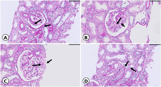
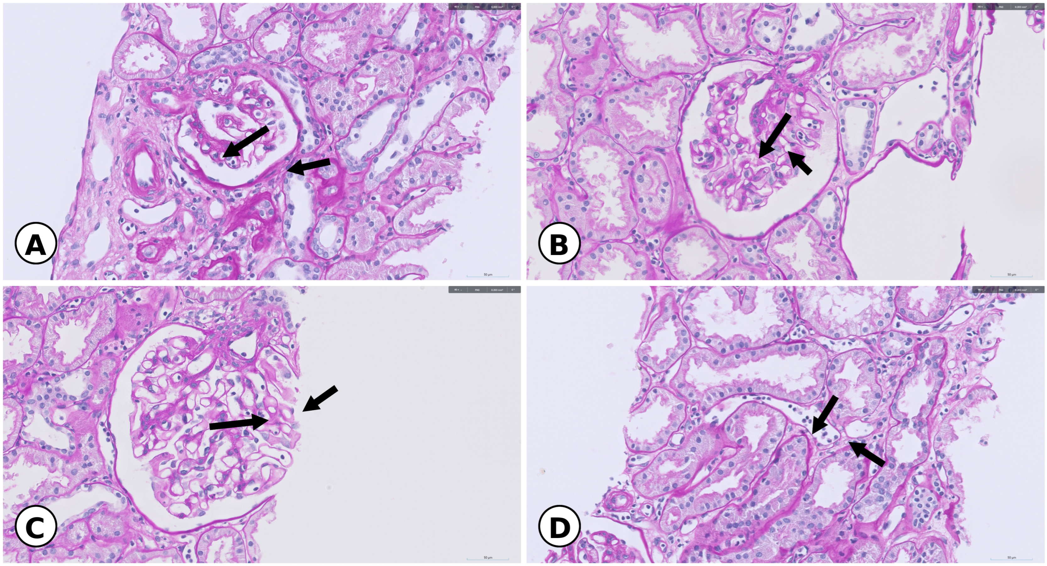
A 52-year-old man presented with a sudden and severe elevation in serum creatinine (sCr). Approximately 10 years before the current episode, he had undergone ABO-incompatible living-donor kidney transplantation from his sister, with immunoglobulin A (IgA) nephropathy recorded as the primary renal disease. For the first 8 years after transplantation, the patient maintained stable graft function with a baseline sCr of approximately 1.03 mg/dL. In the eighth year, he experienced a decline in allograft function with biopsy findings most compatible with the Banff 2022 chronic active antibody-mediated rejection (caAMR) category. Under the Banff 2022 framework, the eighth-year biopsy showed lesion scores of i1, t0, ti1, vx, g2, ci1, ct1, cg2, ptc3, c4d1 (3% of peritubular capillaries by immunohistochemistry), mm2, cvx, ah2, aah2, i-IFTA3, and pvl0 (MVI score: g + ptc = 5) [7]. The biopsy also showed immune complex-mediated glomerulonephritis suggestive of recurrent IgA nephropathy (IgA 1+; Oxford M0 E1 S1 T0 C0) (Figure 1). Anti-HLA antibody testing with a Luminex single-antigen bead assay (LABScreen, Class II, One Lambda, West Hills, CA, USA)), performed on serum collected on the biopsy day (five days before methylprednisolone pulse therapy and plasma exchange and ten days before rituximab), detected anti-DQB1*02:02 antibody (MFI 2308), but donor specificity could not be determined because donor HLA-DQ typing was unavailable. Geographical distance and health-related limitations prevented the living donor from attending for additional HLA-DQ typing. Although caAMR was the favored Banff 2022 category, concurrent glomerulonephritis suggestive of recurrent IgA nephropathy limited the attribution of glomerular lesions to antibody-mediated rejection and therefore precluded definitive classification. He was treated with methylprednisolone pulse therapy and plasma exchange, followed by rituximab, after which serum creatinine generally remained approximately 2.0–2.8 mg/dL; the last documented value before the norovirus infection was approximately 2.0 mg/dL.
Figure 1.
Histopathological findings of the allograft biopsy performed in the eighth post-transplant year (PAS stain, ×400). (A,B) Glomeruli demonstrating glomerulitis (g). (C) Relatively preserved glomerular architecture for comparison. (D) Peritubular capillaritis (ptc). Black arrows mark representative histologic features or comparison structures.
Before the acute episode, an earlier outpatient assessment had shown a hemoglobin level of 10.8 g/dL and a platelet count of 150 × 103/µL. Mycophenolate was intermittently withheld because of diarrhea, including before admission. During the early hospitalization, the recorded immunosuppression regimen included tacrolimus 4 mg twice daily (admission trough level, 2.5 ng/mL) and prednisolone 5 mg daily; mycophenolate was subsequently resumed. Outpatient tacrolimus trough levels before admission had varied between approximately 4 and 11 ng/mL rather than remaining persistently low; subsequent inpatient levels were approximately 5–6 ng/mL after dose adjustments. Two to three weeks before the admission in the tenth post-transplant year, the patient developed a norovirus infection and subsequently recovered clinically. At routine outpatient follow-up, however, sCr had abruptly increased to 6.57 mg/dL, prompting admission for evaluation of acute allograft dysfunction. On admission, sCr was 8.47 mg/dL. Blood pressure at the initial evaluation was 135/76 mmHg. Urinalysis showed 3+ protein and 0–2 red blood cells per high-power field. As severe renal dysfunction persisted, concurrent anemia (Hb, 7.6 g/dL) and thrombocytopenia (platelet count, 110 × 103/µL), together with elevated lactate dehydrogenase (LDH, 629 U/L; reference, 120–250 U/L) and decreased haptoglobin (14.2 mg/dL; reference, 40–280 mg/dL), raised suspicion for TMA and prompted a focused diagnostic evaluation. During the initial TMA evaluation, C3 was 72.5 mg/dL (reference, 90–180 mg/dL) and C4 was 28.8 mg/dL (reference, 10–40 mg/dL). Approximately 3 weeks later, immediately before the first eculizumab infusion, C3 and C4 had decreased to 36.7 and 12.9 mg/dL, respectively, and CH50 was 25.2 U/mL (reference, 46.7–63.3 U/mL). Soluble C5b-9 was not measured.
Serial schistocyte proportions ranged from 0.16% to 0.52%, below the >1% threshold considered suspicious for TMA in the ICSH recommendations. However, the combination of anemia, thrombocytopenia, elevated LDH, decreased haptoglobin, severe allograft dysfunction, and characteristic biopsy findings supported TMA despite the low schistocyte percentages [8].
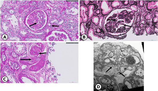
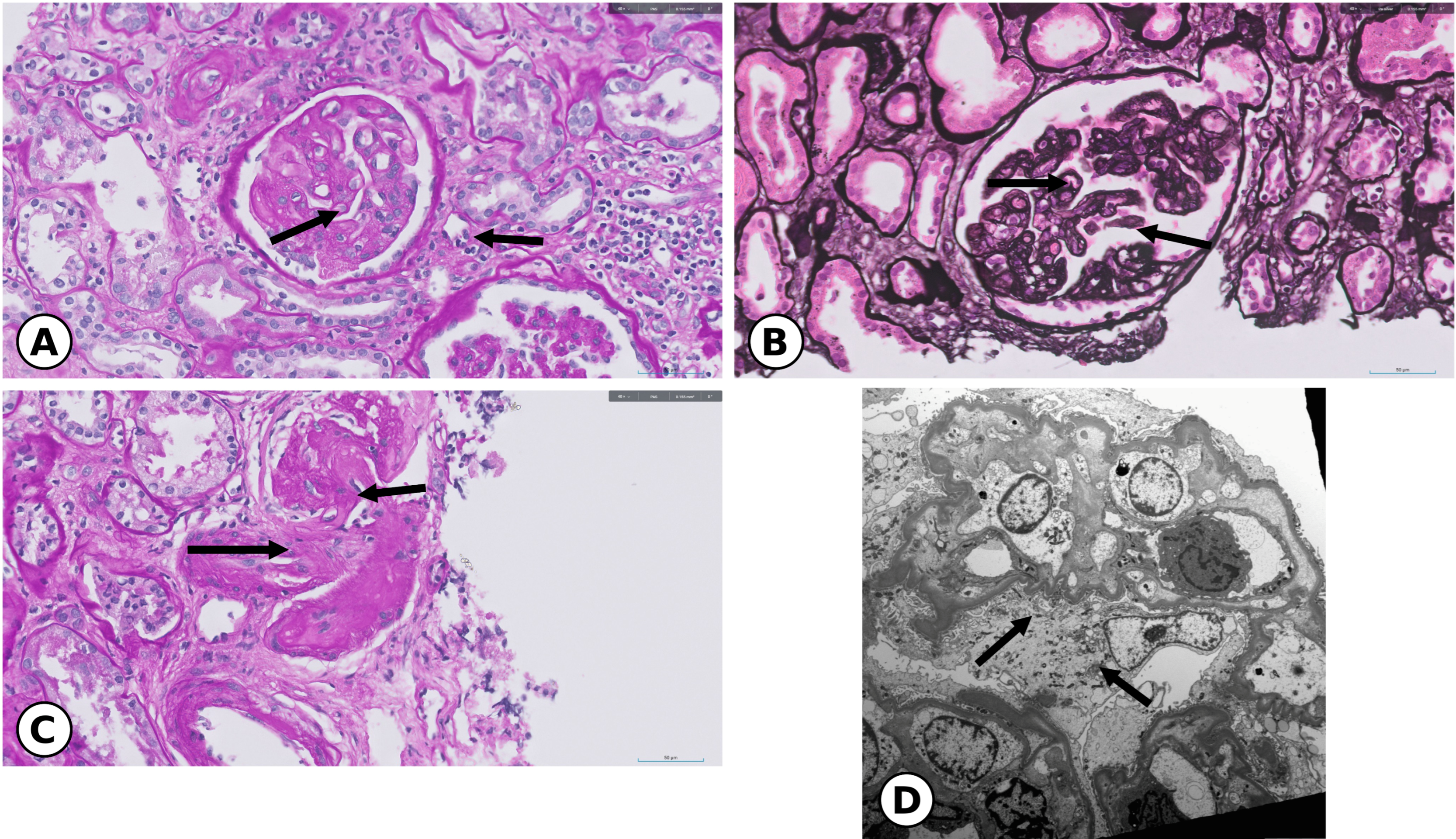
The allograft biopsy obtained during the acute episode in the tenth post-transplant year was interpreted as showing TMA, including acute and chronic microcirculatory injury, along with severe interstitial fibrosis, tubular atrophy, and focal segmental glomerulosclerosis (Figure 2). Under the Banff 2022 framework, the acute biopsy showed lesion scores of i0, t0, ti3, v0, g3, ci3, ct3, cg3, ptc3, c4d0, mm0, cv0, ah2, i-IFTA3, t-IFTA0, aah2, ptcml0, and pvl0 (MVI score: g + ptc = 6). C4d staining was negative (0%). Anti-HLA antibody testing with a Luminex single-antigen bead assay (LABScreen, One Lambda), performed three days before biopsy and five days before plasma exchange, detected no HLA class I or II antibodies. Non-HLA antibody testing was not performed. The findings were most consistent with the Banff 2022 descriptive phenotype of “MVI, DSA-negative and C4d-negative” [7]. Immunofluorescence showed weak granular staining for IgA, IgM, and C3 (±), whereas IgG, C4, and C1q were negative. C9 staining was not documented. Only three glomeruli were available for evaluation, below the Banff biopsy adequacy criteria; this limits the representativeness of the reported glomerular scores and the confidence of the Banff 2022 descriptive phenotype assignment. Active recurrent IgA nephropathy was not established.
Figure 2.
Histopathological findings during the acute TMA episode. (A) Glomerular mesangiolysis, capillary-loop aneurysm, and endothelial swelling (PAS, ×400). (B) Glomerular basement membrane (GBM) double contours (silver stain, ×400). (C) Arteriolar endothelial injury and wall thickening (PAS, ×400). (D) Subendothelial neo-GBM formation and GBM double contours (electron microscopy). Black arrows indicate the representative lesions.
ADAMTS13 activity was preserved at 71.2%, making severe ADAMTS13-deficient thrombotic thrombocytopenic purpura unlikely. Testing for Shiga toxin-producing Escherichia coli was negative, as was the direct antiglobulin test. Anti-factor H antibody testing was reported negative. Finally, a genetic panel for aHUS identified a heterozygous missense variant in the complement factor H (CFH) gene (c.3329G>C, p.Gly1110Ala). Although this variant is classified as a variant of uncertain significance (VUS), an in silico tool (PolyPhen-2) predicted it to be deleterious. The sequencing panel did not include C5 and was limited in its ability to detect CFHR copy-number or structural variants.
Given the clinical suspicion of aHUS, treatment was initiated with plasma exchange and high-dose methylprednisolone therapy. Plasma exchange was attempted four times during the initial week of treatment, using fresh frozen plasma; the fourth procedure was stopped after anaphylaxis with urticaria, gastrointestinal symptoms, hypotension, and oxygen desaturation requiring intramuscular epinephrine. The second C3/C4 measurements were obtained 14 days after the fourth plasma exchange attempt (which was aborted), and more than 14 days after the last completed session. Meningococcal vaccination (Menveo) and oral ciprofloxacin prophylaxis were initiated 3 days before the first eculizumab infusion. Antibiotic prophylaxis was continued for approximately three months, initially with ciprofloxacin and subsequently with azithromycin. Eculizumab was administered as induction therapy and then maintenance therapy: 900 mg intravenously weekly for the first four doses, followed by 1200 mg every 2 weeks.
Before eculizumab initiation, the patient had undergone two temporary hemodialysis sessions but was not yet receiving maintenance dialysis. Intermittent hemodialysis was subsequently required, and regular thrice-weekly maintenance hemodialysis was established approximately seven months after the first dose, without sustained recovery of allograft function.
Following eculizumab administration, gradual improvement in hematologic parameters was observed. Eculizumab was continued despite persistent renal impairment and subsequent dialysis dependence, as gradual improvement in LDH and haptoglobin suggested possible control of ongoing TMA activity. A total of 34 doses were administered over approximately 14 months. Treatment was discontinued after substantial hematologic stabilization, with insurance reimbursement limitations and the financial burden of continued therapy also contributing to the decision. During approximately 3 years and 10 months of follow-up after discontinuation, no clinical or laboratory evidence of recurrent TMA was documented.
3. Discussion
Atypical hemolytic uremic syndrome (aHUS) is a complement-mediated thrombotic microangiopathy (TMA) that can arise in association with genetic or acquired abnormalities of complement regulation and may be triggered by infection, pregnancy, or organ transplantation [2,4,5,9,10]. In kidney transplant recipients, de novo TMA may involve multiple factors, including antibody-mediated rejection (ABMR), calcineurin inhibitor exposure, infection, and complement dysregulation, making the underlying etiology difficult to establish [2,3,11,12].
The diagnosis of aHUS is primarily clinical and requires recognition of TMA while evaluating competing causes. Kidney biopsy can be particularly useful when systemic hematologic manifestations are incomplete or when several causes of graft dysfunction coexist. Complement studies and genetic testing may support etiologic classification, but their results must be interpreted in the overall clinical context [1,2,5,13]. Histologic findings such as endothelial swelling, mesangiolysis, capillary or arteriolar thrombi, subendothelial widening, and glomerular basement membrane double contours indicate microvascular endothelial injury but are not specific to its cause. Thus, biopsy findings support the diagnosis of TMA without establishing its etiology.
In the present case, negative HLA class I/II antibody testing and C4d staining argued against ABMR-associated TMA; however, this mechanism could not be completely excluded given the previous suspicion of chronic active antibody-mediated rejection (caAMR) and marked microvascular inflammation. The decreases in both C3 and C4, although C4 remained within the reference range, raised the possibility of classical pathway involvement, but the predominant complement activation pathway could not be determined from these measurements. Acute TMA may have been superimposed on chronic endothelial injury in an allograft previously suspected of caAMR, although the relative contributions of these processes remain uncertain [7,14]. The acute biopsy contained only three glomeruli, which limited the representativeness of the g3 and cg3 scores, the interpretation of the marked microvascular inflammation, and the assignment of the Banff 2022 descriptive phenotype of “MVI, DSA-negative and C4d-negative.” Non-HLA antibody testing was not performed, which further limited exclusion of ABMR-associated TMA.
An association between immunoglobulin A nephropathy (IgAN) and TMA has been reported. El Karoui et al. described predominantly arterial and arteriolar TMA lesions in IgAN, frequently associated with hypertension; prominent glomerular lesions of the type observed in the present case were not a major feature of their cohort [15]. Our patient did not have severe hypertension at the initial evaluation, whereas mesangiolysis and glomerular basement membrane double contours were prominent alongside arteriolar endothelial injury. Although the eighth-year allograft biopsy suggested recurrent IgAN and was followed by treatment, active recurrent IgAN was not established at the time of the subsequent TMA episode. Overall, these observations did not provide convincing evidence that active recurrent IgAN was the principal cause of this episode. Associations between IgAN and the CFH/CFHR genomic region, including the protective association of CFHR3–CFHR1 deletion with IgAN, have also been reported [16]. This association is distinct from the CFH p.Gly1110Ala variant of uncertain significance identified in our patient and does not establish a shared causal mechanism.
Hematologic parameters recovered at different rates. LDH and haptoglobin improved gradually, with fluctuations, before eventual normalization. Persistent anemia in the setting of advanced allograft dysfunction and subsequent dialysis is not, by itself, specific evidence of ongoing microangiopathic hemolysis. Variable timing of hematologic recovery during complement inhibition has been reported [17]. Although serial post-treatment complement functional measurements were unavailable and complete C5 blockade could not be confirmed or drug resistance entirely excluded [18], the slow but sustained improvement in biochemical indices made complete clinical nonresponse less likely. Severe pre-existing interstitial fibrosis and tubular atrophy also limited the potential for renal recovery. Because maintenance dialysis had not been established when eculizumab was initiated, the possibility of recovery of residual allograft function could not be excluded. Nevertheless, whether eculizumab delayed dialysis dependence cannot be determined from this course. The progressive hematologic improvement during treatment suggested potential benefit despite the poor renal prognosis. On this basis, eculizumab was continued because the improving indices suggested possible control of TMA activity, even though renal recovery remained uncertain [19,20].
Plasma exchange is appropriate when thrombotic thrombocytopenic purpura (TTP) remains a clinically significant possibility while ADAMTS13 results are pending, and it may also be considered if eculizumab is not immediately available [1,2]. Eculizumab inhibits C5 at a common downstream step of complement activation; thus, terminal complement activation can be inhibited even when the predominant upstream activation pathway has not been established [5]. In this patient, changes in C3 and C4 did not identify the predominant pathway, but hematologic indices improved during eculizumab treatment. Because C5 blockade increases the risk of invasive meningococcal infection, vaccination and appropriate antimicrobial prophylaxis are important components of care [5,10]. The duration of eculizumab treatment should be individualized, and decisions about discontinuation should take into account genetic background, previous relapses, the extent of renal recovery, and persistent triggers [1,9,10].
Importantly, complement dysregulation is not limited to patients undergoing transplantation for known aHUS. Le Quintrec et al. identified CFH or CFI abnormalities in 29% of kidney transplant recipients with de novo TMA, suggesting that transplantation may reveal previously unrecognized complement susceptibility. Most reported episodes occur early after transplantation: the Korean consensus describes a peak within the first several months, and 67% of cases in the Le Quintrec series occurred within 3 months. However, onset ranged from the immediate post-transplant period to 6 years, indicating that a prolonged interval does not completely exclude transplant-associated complement-mediated TMA [2,12].
In this context, the onset of suspected aHUS approximately 10 years after transplantation is a notable feature of our case. This interval extends well beyond the usual early post-transplant period and exceeds the latest onset reported in the Le Quintrec series. The preceding norovirus infection may have served as an additional inflammatory trigger, although its contribution remains uncertain. This distinction is important because complement activation may be the primary driver of TMA in some transplant recipients, whereas in others it may amplify pre-existing endothelial injury [12,14].
Overall, this case offers two practical implications. First, consideration of complement-mediated TMA should not be determined solely by the time elapsed since transplantation. Second, hematologic improvement following C5 blockade and renal recovery should be evaluated as related but distinct treatment outcomes. Earlier recognition may allow preservation of potentially reversible renal tissue, whereas very late presentation with advanced chronic injury may permit control of active microangiopathy without recovery of allograft function [5,6].
Author Contributions
Investigation, J.Y.M., Y.J.J. and H.S.K.; resources, H.S.K.; data curation, J.Y.M. and H.S.K.; writing—original draft preparation, J.Y.M.; writing—review and editing, J.Y.M. and Y.J.J. All authors have read and agreed to the published version of the manuscript.
Funding
This research received no external funding.
Institutional Review Board Statement
Institutional Review Board review and approval were not required at Asan Medical Center because this manuscript describes the clinical course of a single patient treated as part of routine clinical care.
Informed Consent Statement
Informed consent for publication was obtained from the patient.
Data Availability Statement
All relevant clinical findings are presented in this article. Additional patient-level medical records are not publicly available because of patient privacy and confidentiality restrictions.
Conflicts of Interest
The authors declare no conflicts of interest.
References
- Fakhouri, F.; Schwotzer, N.; Frémeaux-Bacchi, V. How I diagnose and treat atypical hemolytic uremic syndrome. Blood 2023, 141, 984–995. [Google Scholar] [CrossRef] [Scilit] [PubMed]
- Lee, H.; Kang, E.; Kang, H.G.; Kim, Y.H.; Kim, J.S.; Kim, H.-J.; Moon, K.C.; Ban, T.H.; Oh, S.W.; Jo, S.K.; et al. Consensus regarding diagnosis and management of atypical hemolytic uremic syndrome. Korean J. Intern. Med. 2020, 35, 25–40. [Google Scholar] [CrossRef] [Scilit] [PubMed]
- Cho, W.; Jo, S.K.; Jung, C.W.; Kim, M.G. Characteristics and management of thrombotic microangiopathy in kidney transplantation. Korean J. Transplant. 2023, 37, 11–18. [Google Scholar] [CrossRef] [Scilit] [PubMed]
- Ávila, A.; Gavela, E.; Sancho, A. Thrombotic microangiopathy after kidney transplantation: An underdiagnosed and potentially reversible entity. Front. Med. 2021, 8, 642864. [Google Scholar] [CrossRef] [Scilit] [PubMed]
- Goodship, T.H.J.; Cook, H.T.; Fakhouri, F.; Fervenza, F.C.; Frémeaux-Bacchi, V.; Kavanagh, D.; Nester, C.M.; Noris, M.; Pickering, M.C.; de Córdoba, S.R.; et al. Atypical hemolytic uremic syndrome and C3 glomerulopathy: Conclusions from a Kidney Disease: Improving Global Outcomes (KDIGO) Controversies Conference. Kidney Int. 2017, 91, 539–551. [Google Scholar] [CrossRef] [Scilit] [PubMed]
- Brocklebank, V.; Walsh, P.R.; Smith-Jackson, K.; Hallam, T.M.; Marchbank, K.J.; Wilson, V.; Bigirumurame, T.; Dutt, T.; Montgomery, E.K.; Malina, M.; et al. Atypical hemolytic uremic syndrome in the era of terminal complement inhibition: An observational cohort study. Blood 2023, 142, 1371–1386. [Google Scholar] [CrossRef] [Scilit] [PubMed]
- Naesens, M.; Roufosse, C.; Haas, M.; Lefaucheur, C.; Mannon, R.B.; Adam, B.A.; Aubert, O.; Böhmig, G.A.; Callemeyn, J.; Groningen, M.C.-V.; et al. The Banff 2022 Kidney Meeting Report: Reappraisal of microvascular inflammation and the role of biopsy-based transcript diagnostics. Am. J. Transplant. 2024, 24, 338–349. [Google Scholar] [CrossRef] [Scilit] [PubMed]
- Zini, G.; d’Onofrio, G.; Erber, W.N.; Lee, S.-H.; Nagai, Y.; Basak, G.W.; Lesesve, J.; International Council for Standardization in Hematology (ICSH). 2021 update of the 2012 ICSH Recommendations for identification, diagnostic value, and quantitation of schistocytes: Impact and revisions. Int. J. Lab Hematol. 2021, 43, 1264–1271. [Google Scholar] [CrossRef] [Scilit] [PubMed]
- Java, A. Atypical hemolytic uremic syndrome: Diagnosis, management, and discontinuation of therapy. Hematol. Am. Soc. Hematol. Educ. Program 2024, 2024, 200–205. [Google Scholar] [CrossRef] [Scilit] [PubMed]
- Kang, H.G.; Hsu, D.; Kato, N.; Kim, J.S.; Okumi, M.; Tseng, M.; Tu, K.; Yap, D.Y.H.; Lim, W.H. Management of atypical haemolytic uraemic syndrome with triggers: Diagnostic and treatment algorithms from an Asia-Pacific perspective. Nephrology 2025, 30, e70116. [Google Scholar] [CrossRef] [Scilit] [PubMed]
- Mirioglu, S.; Morelle, J.; Efe, O.; Hurdogan, O.; Dirim, A.B.; Kumru, G.; Lentine, K.L.; Caliskan, Y. Thrombotic microangiopathy after kidney transplantation: Diagnosis and management strategies. Clin. Kidney J. 2026, 19, sfag018. [Google Scholar] [CrossRef] [Scilit] [PubMed]
- Le Quintrec, M.; Lionet, A.; Kamar, N.; Karras, A.; Barbier, S.; Büchler, M.; Fakhouri, F.; Provost, F.; Fridman, W.H.; Thervet, E.; et al. Complement mutation-associated de novo thrombotic microangiopathy following kidney transplantation. Am. J. Transplant. 2008, 8, 1694–1701. [Google Scholar] [CrossRef] [Scilit] [PubMed]
- Leon, J.; LeStang, M.B.; Sberro-Soussan, R.; Servais, A.; Anglicheau, D.; Frémeaux-Bacchi, V.; Zuber, J. Complement-driven hemolytic uremic syndrome. Am. J. Hematol. 2023, 98, S44–S56. [Google Scholar] [CrossRef] [Scilit] [PubMed]
- Dessaix, K.; Bontoux, C.; Aubert, O.; Grünenwald, A.; Soussan, R.S.; Zuber, J.; Van Huyen, J.-P.D.; Anglicheau, D.; Legendre, C.; Bacchi, V.F.; et al. De novo thrombotic microangiopathy after kidney transplantation in adults: Interplay between complement genetics and multiple endothelial injury. Am. J. Transplant. 2024, 24, 1205–1217. [Google Scholar] [CrossRef] [Scilit] [PubMed]
- El Karoui, K.; Hill, G.S.; Karras, A.; Jacquot, C.; Moulonguet, L.; Kourilsky, O.; Frémeaux-Bacchi, V.; Delahousse, M.; Van Huyen, J.-P.D.; Loupy, A.; et al. A clinicopathologic study of thrombotic microangiopathy in IgA nephropathy. J. Am. Soc. Nephrol. 2012, 23, 137–148. [Google Scholar] [CrossRef] [Scilit] [PubMed]
- Xie, J.; Kiryluk, K.; Li, Y.; Mladkova, N.; Zhu, L.; Hou, P.; Ren, H.; Wang, W.; Zhang, H.; Chen, N.; et al. Fine mapping implicates a deletion of CFHR1 and CFHR3 in protection from IgA nephropathy in Han Chinese. J. Am. Soc. Nephrol. 2016, 27, 3187–3194. [Google Scholar] [CrossRef] [Scilit] [PubMed]
- Fakhouri, F.; Hourmant, M.; Campistol, J.M.; Cataland, S.R.; Espinosa, M.; Gaber, A.O.; Menne, J.; Minetti, E.E.; Provôt, F.; Rondeau, E.; et al. Terminal complement inhibitor eculizumab in adult patients with atypical hemolytic uremic syndrome: A single-arm, open-label trial. Am. J. Kidney Dis. 2016, 68, 84–93. [Google Scholar] [CrossRef] [Scilit] [PubMed]
- Nishimura, J.I.; Yamamoto, M.; Hayashi, S.; Ohyashiki, K.; Ando, K.; Brodsky, A.L.; Noji, H.; Kitamura, K.; Eto, T.; Takahashi, T.; et al. Genetic variants in C5 and poor response to eculizumab. N. Engl. J. Med. 2014, 370, 632–639. [Google Scholar] [CrossRef] [Scilit] [PubMed]
- Duineveld, C.; Bouwmeester, R.N.; Wijnsma, K.L.; Bemelman, F.; van der Heijden, J.; Berger, S.; Heuvel, L.v.D.; van de Kar, N.C.; Wetzels, J.F.; van Zuijlen, A.; et al. Eculizumab rescue therapy in patients with recurrent atypical hemolytic uremic syndrome after kidney transplantation. Kidney Int. Rep. 2023, 8, 715–726. [Google Scholar] [CrossRef] [Scilit] [PubMed]
- Min, E.K.; Kim, H.J.; Kim, S.; Jung, M.; Kim, J.S.; Han, S.H.; Huh, K.H. The first successful eculizumab rescue therapy of a kidney transplant recipient with atypical hemolytic uremic syndrome in South Korea: A case report. Korean J. Transplant. 2023, 37, 57–62. [Google Scholar] [CrossRef] [Scilit] [PubMed]
Disclaimer/Publisher’s Note: The statements, opinions and data contained in all publications are solely those of the individual author(s) and contributor(s) and not of MDPI and/or the editor(s). MDPI and/or the editor(s) disclaim responsibility for any injury to people or property resulting from any ideas, methods, instructions or products referred to in the content. |
© 2026 by the authors. Licensee MDPI, Basel, Switzerland. This article is an open access article distributed under the terms and conditions of the Creative Commons Attribution (CC BY) license.