


Abstract
Background/Objectives: Patient-specific three-dimensional (3D) models may clarify complex anatomy in gynaecological cancer surgery. This scoping review mapped applications of virtual and printed models, and examined their reported contributions to surgical planning and outcomes. Methods: The review followed Joanna Briggs Institute guidance along with the Preferred Reporting Items for Systematic Reviews and Meta-Analyses extension for Scoping Reviews. PubMed, Scopus and Embase were searched on 4 March 2026 for English- or Spanish-language reports. Following a broad gynaecological search, an oncology-focused synthesis examined patient-specific models linked to surgical anatomy or planning. Eligibility assessment and data charting were each performed independently by two reviewers. Findings were synthesised descriptively without formal risk-of-bias appraisal or quantitative pooling. Results: Fourteen studies published between 2016 and 2026 were included: nine evaluated virtual models and five incorporated physical printing. Evidence comprised comparative observational studies, small feasibility or prospective studies, and case reports. Computed tomography and magnetic resonance imaging were the main imaging sources. Applications included tumour mapping, depiction of neural and vascular anatomy, and planning complex or recurrent tumour resections. Some comparative studies reported shorter operative times and lower blood losses, whereas others found improved anatomical assessments without significant perioperative differences. Any additional advantage of printing over virtual visualisation remained uncertain. Interpretation was limited by small samples, heterogeneous methods, subjective evaluations, and one mixed malignant and non-malignant cohort. Conclusions: Patient-specific 3D modelling may assist planning in selected complex gynaecological oncology procedures. Evidence of improved clinical outcomes remains insufficient. Prospective comparative evaluations should establish appropriate indications, and determine whether additional anatomical information produces meaningful patient benefit.
1. Introduction
Three-dimensional (3D) modelling technologies, originally developed from two-dimensional computed tomography (CT) imaging in the late 1970s [1], have evolved into increasingly relevant tools in contemporary surgical practice. By transforming conventional imaging modalities such as CT and magnetic resonance imaging (MRI) into patient-specific anatomical models (either virtual or physical), these technologies provide a more intuitive representation of anatomical structures and pathological processes, compared with traditional two-dimensional visualisation [2]. Applications in neurosurgical, hepatobiliary, oesophagogastric, vascular and complex pelvic procedures have been described [3,4,5,6,7,8]. The literature also addresses patient education, surgical training, and anatomical learning [9,10,11], alongside planning in orthopaedics and surgical oncology [12,13]. These applications share a need to communicate spatial information, although their clinical objectives and requirements for validation differ.
Evidence supporting routine implementation remains limited by heterogeneous indications, reconstruction methods and outcome measures. Production costs, segmentation accuracy, and integration into clinical workflows also influence adoption. Reviews across surgical specialties describe potential advantages for anatomical understanding and planning, while identifying limitations in comparative evidence [2,5,14,15]. Successful model production therefore requires evaluation against the clinical task for which the model is intended.
In gynaecological oncology, surgical planning depends on relationships between disease and anatomical structures that influence the feasibility and extent of resection. Ultrasound, CT, and MRI remain central to assessment, but complex spatial relationships can be difficult to appreciate in recurrent disease, in distorted anatomy, or in tumours involving adjacent organs. Patient-specific 3D models have been explored to depict tumour extent and vascular and urinary relationships relevant to operative decisions [16,17,18]. Reviews of women’s health and gynaecological applications cover a broad range of conditions and uses [15,19,20,21]; the evidence specifically addressing oncological surgery warrants focused examination. This scoping review therefore mapped patient-specific virtual and printed models in gynaecological oncology, examining their clinical indications, contributions to surgical planning, reported outcomes, and evidence gaps. The review also considered how anatomical feasibility, perceived usefulness, and measured clinical outcomes differ when judging their potential role in surgical care.
2. Materials and Methods
2.1. Protocol and Review Framework
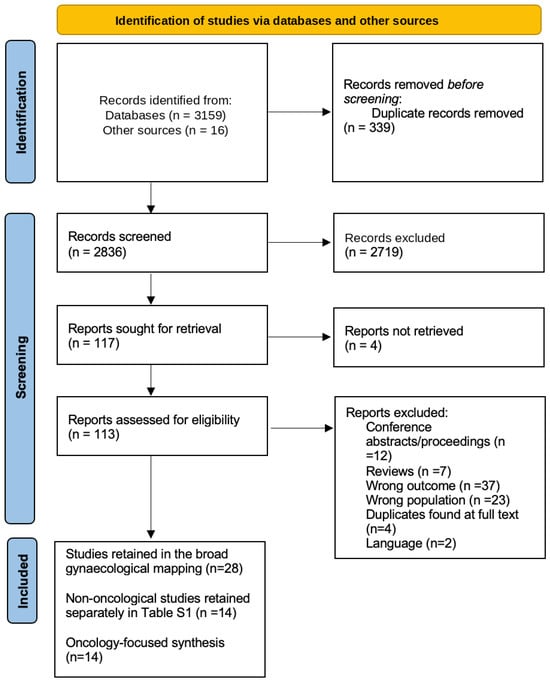
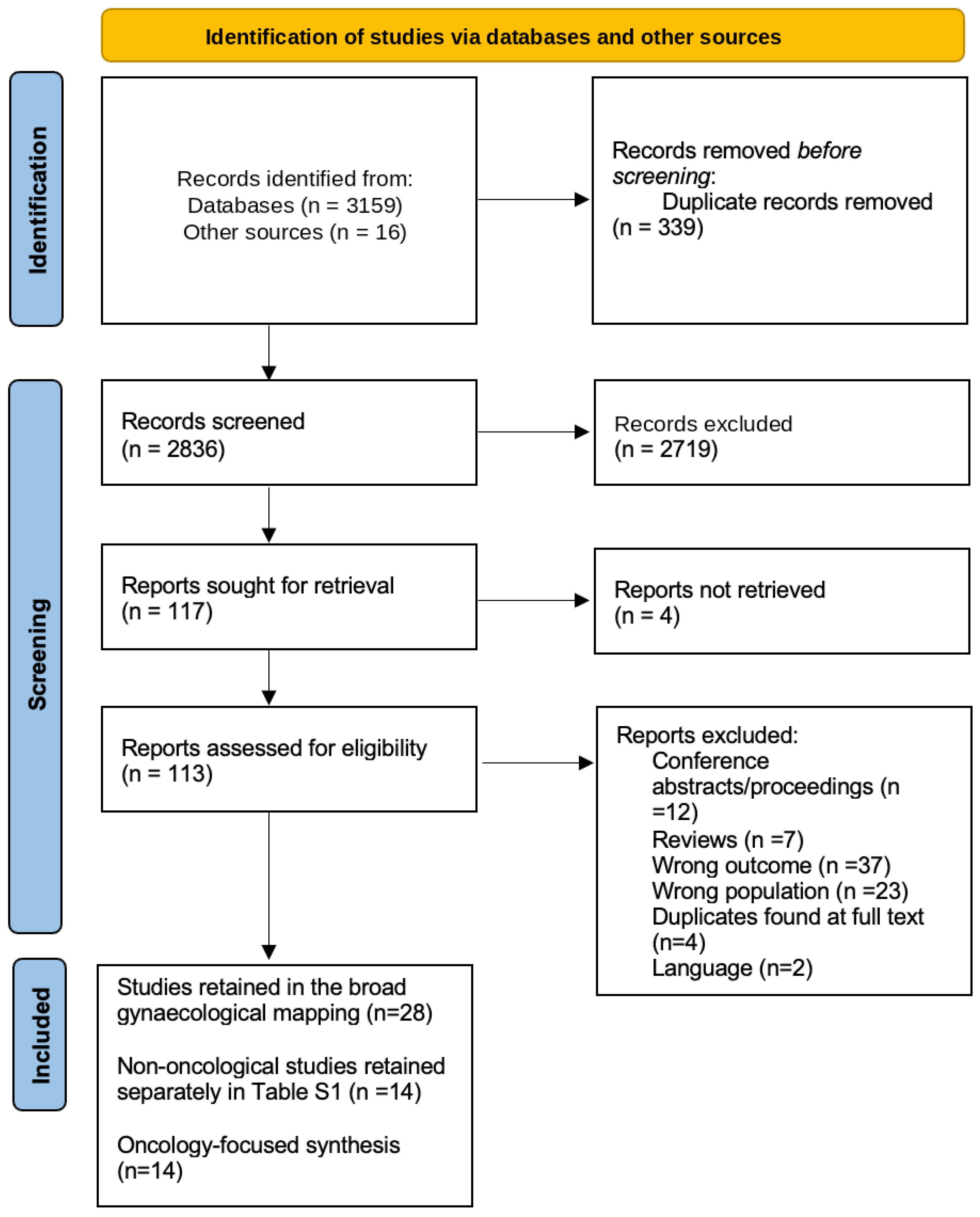
This scoping review was conducted according to the Joanna Briggs Institute (JBI) Reviewer’s Manual [22], and is reported in accordance with the PRISMA extension for Scoping Reviews (PRISMA-ScR) [23]. An internal protocol was developed before searching; this was not registered. The original scope covered gynaecological surgery; the oncology-focused synthesis was defined after searching. The protocol summary and full search strategies are provided in Supplementary File S1. The completed PRISMA-ScR checklist is provided in Supplementary File S2, and the study-selection flow diagram is presented as Figure 1.
Figure 1.
PRISMA flow diagram of study selection. Reports not retrieved are distinguished from reports excluded after eligibility assessment; reasons for exclusion are shown in the diagram.
For the oncology-focused synthesis, the Population–Concept–Context framework comprised the following: women with gynaecological malignancies; patient-specific virtual reconstructions or 3D-printed models; and anatomical assessment, planning or decision-making relevant to gynaecological oncology surgery.
2.2. Information Sources and Search Strategy
PubMed, Scopus and Embase (via Embase.com) were searched, with the final searches of all three databases being conducted on 4 March 2026. The search combined terms relating to 3D printing, reconstruction, models, and imaging with gynaecological and pelvic terms. No publication-year restrictions were applied. Searches were limited to English- and Spanish-language publications, and Scopus was additionally restricted to the medicine subject area. Reference lists of eligible studies were screened for additional publications.
2.3. Eligibility and Study Selection
Initial screening and study selection were conducted during March 2026. Two initially unavailable reports were retrieved and included during manuscript revision. Eligible study designs included case reports, case series, feasibility studies, and comparative observational studies evaluating patient-specific virtual or 3D-printed anatomical models with potential relevance to surgical planning.
Reviews, conference abstracts, proceedings, oral communications, and abstract-only publications were excluded. Generic simulation or training models, education-only applications, and technical or anatomical studies without patient-specific modelling or a stated relevance to surgical planning were excluded. These criteria retained primary evidence on patient-specific surgical applications, including early feasibility work, while excluding secondary summaries and unrelated uses.
After the broad mapping, all 28 retained studies were reassessed using the oncology-focused Population–Concept–Context criteria. Inclusion in the main synthesis required a gynaecological malignancy (or a mixed female pelvic tumour cohort including malignant cases) and a patient-specific 3D model explicitly linked to an anatomical assessment or a defined surgical planning task. Actual intraoperative use or measured perioperative outcomes were not required: feasibility studies and clinician ratings were eligible when they addressed relevant operative anatomy or a proposed surgical decision, such as tumour extent, dissection planes, or relationships with neurovascular or urinary structures. Mixed cohorts were retained but identified as such, and aggregate findings were not interpreted as cancer-specific when malignant-subgroup data were unavailable. The 14 studies limited to non-oncological conditions were retained separately in Table S1 [24,25,26,27,28,29,30,31,32,33,34,35,36,37].
Two reviewers (M.M.P. and S.F.-G.) independently screened titles and abstracts, and assessed full-text eligibility. Disagreements were resolved by discussion or consultation with J.P.S.
2.4. Data Charting and Critical Appraisal
Data were independently charted by M.M.P. and Á.O.C.P. using a predefined extraction form, then compared and cross-checked for accuracy and consistency. Extracted variables included author, publication year, country, study design, sample size, pathology, imaging source, modelling technology, clinical indication, procedure, evaluated outcomes, findings, and limitations. Reported model generation time and direct financial estimates were also recorded when available. For mixed populations, malignancy-specific data were extracted when available; otherwise, the cohort was described as mixed. Imaging used to generate the model was distinguished from imaging obtained for routine clinical assessment where the report permitted this distinction. Funding and material-support disclosures from the included reports are summarised in Table S2.
No formal methodological quality or risk-of-bias appraisal was undertaken, because the objective was to map applications and evidence gaps rather than estimate treatment effectiveness [22]. This limits conclusions about comparative effectiveness.
2.5. Synthesis
In the descriptive qualitative synthesis, studies were grouped into those evaluating virtual models and those incorporating physical printing. Virtual models included screen-based visualisation and augmented-reality displays. Studies combining virtual reconstruction and printing were assigned to the printed-model group to avoid double counting. Interpretation distinguished anatomical feasibility and concordance from perceived usefulness, changes in planning, and perioperative outcomes. No quantitative pooling was undertaken because populations, interventions and outcomes were insufficiently comparable.
During manuscript preparation, ChatGPT OpenAI; including GPT-6, accessed November 2026) was used to assist with drafting and restructuring narrative sections, bibliographic cross-checking, language revision, and manuscript formatting. The authors critically reviewed and edited all AI-assisted output, retained responsibility for study selection, data charting, synthesis, and interpretation, and did not use AI tools to generate primary study data.
3. Results
3.1. Study Selection and Characteristics
The original broad search identified 3159 database records and 16 additional records. After removal of 339 duplicates, 2836 records were screened and 2719 were excluded. Of 117 reports sought for retrieval, four were unavailable and 113 were assessed for eligibility. Eighty-five reports were excluded for the reasons shown in Figure 1, leaving 28 studies in the broad gynaecological mapping. Following reassessment against the oncology-focused criteria, 14 studies contributed to the main synthesis; the remaining 14 addressed non-oncological conditions and were retained separately in Table S1.
Of the 14 oncological studies, nine evaluated virtual 3D models and five incorporated physical printing. Published between 2016 and 2026, these addressed cervical, ovarian, fallopian-tube and endometrial cancers, uterine leiomyosarcoma, and other complex pelvic tumours. One cohort included malignant and non-malignant lesions. Evidence comprised three comparative observational studies, four small feasibility or evaluation studies, and seven single-patient reports. CT and MRI were the main reported imaging sources. Preoperative planning predominated, particularly assessment of tumour extent and relationships with vascular, urinary, neural, and adjacent structures.
Studies evaluated model generation, correspondence with operative or pathological findings, and perceived clinical usefulness. Comparative studies also assessed perioperative outcomes. Study characteristics and applications are summarised in Table 1 and Table 2 for virtual and printed models, respectively.
Table 1.
Patient-specific virtual 3D models in gynaecological oncology.
Table 2.
Patient-specific 3D-printed models in gynaecological oncology.
3.2. Virtual 3D Models
Virtual patient-specific 3D models were investigated for anatomical assessment, preoperative planning, and prediction of disease distribution. Li et al. [38] reconstructed pelvic autonomic nerves in 30 patients with cervical cancer, with agreement between T2-weighted fat-suppressed MRI and MR neurography reconstructions. The models depicted relationships between pelvic autonomic nerves and pelvic organs, with potential applications in nerve-sparing surgical planning. Kahn et al. [42] prospectively evaluated CT-based volumetric rendering through augmented-reality headsets in 15 patients undergoing primary or interval cytoreductive surgery for suspected advanced ovarian cancer. Accuracy for disease localisation ranged from 80% to 100% across anatomical sites, using pathology and intraoperative assessment as the reference; sites without macroscopic disease at surgery were classified as negative.
Comparative evidence was provided by Yanazume et al. [18], who retrospectively evaluated 34 patients undergoing para-aortic lymphadenectomy using preoperative 3D CT and 38 controls. Renal vascular variations and duplicated ureters were identified in 41.2% and 5.9% of the 3D group, respectively, although perioperative outcomes did not differ significantly between groups. Liu et al. [39] compared 31 patients who were assessed using CT-based 3D reconstruction with 30 who were assessed using conventional 2D imaging in a mixed cohort of malignant and non-malignant female pelvic tumours. Reconstruction was successful in all 31 patients. The 3D group had shorter operative times, lower blood losses, shorter hospital stays, and shorter intraoperative consultation times (all p < 0.05). The malignant-case proportion could not be determined reliably from the published report; these aggregate perioperative findings cannot be attributed specifically to patients with malignancy.
Five single-patient reports described anatomically complex oncological procedures. Long et al. [40] used CT-based reconstruction to delineate uterine leiomyosarcoma extending along the right ovarian vein into the retrohepatic inferior vena cava, informing planning for resection under extracorporeal circulation. Gomes da Costa et al. [41] used MRI-based modelling in recurrent fallopian tube carcinoma after adhesions limited laparoscopic assessment. The model depicted colonic and diaphragmatic implants before secondary cytoreduction. Idegami et al. [43] used CT-based vascular measurements relative to the inferior mesenteric artery to guide identification and preservation of two accessory renal arteries during para-aortic lymphadenectomy. Madrazo González et al. [44] mapped an abdominal wall recurrence of endometrial cancer associated with incisional hernias before en bloc resection and abdominal wall reconstruction.
Finally, Ricotta et al. [8] described a 49-year-old woman with pelvic sidewall recurrence of low-grade serous ovarian cancer. Preoperative assessment incorporating virtual 3D reconstruction delineated left iliac vascular involvement and venous thrombosis, involvement of the pelvic ureter, vessels supplying the residual colon and the psoas muscle, and compression of the L5 and S1 nerves. A lateral femoral–femoral bypass was performed three weeks before laterally extended pelvic exenteration. Surgery included left iliac vascular resection, ureteral reconstruction, colon resection, and lumbosacral nerve dissection; complete resection was reported.
3.3. 3D-Printed Models
The five studies incorporating physical printing comprised one comparative study, two small feasibility or evaluation studies, and two case reports. Cai et al. [16] retrospectively evaluated 60 patients with stage III ovarian cancer, with 20 patients per group receiving conventional 2D CT assessment, virtual 3D visualisation, or virtual visualisation combined with printing. Mean operative times in the virtual-only, combined and conventional groups were 393.3, 390.8 and 428.3 min, respectively (overall p = 0.005); corresponding blood losses were 706, 669 and 898 mL (overall p = 0.002). Transfusion volume also differed across groups (p = 0.040). Both 3D groups had shorter times to first flatus and ambulation, as well as shorter drainage durations. No statistically significant differences were detected between the virtual-only and combined virtual-plus-printed groups; however, this does not establish equivalence or exclude a possible incremental benefit of printing. Complication and recurrence rates did not differ significantly across all three groups.
Smaller studies primarily assessed feasibility and perceived utility. Sayed Aluwee et al. [46] generated MRI-based uterine models for five patients with endometrial cancer, using 3D printing and mould casting. Comparison of CT scans of the models with the original MRI data showed mean geometric discrepancies ranging from 1.19 to 2.22 mm across cases. Patient feedback favoured the models for understanding the disease, surgical procedures, and associated risks. Surgeons’ assessments varied across tasks, with the strongest ratings reported for preoperative patient explanation.
Scherer-Quenzer et al. [17] produced life-size, MRI-based models for five patients with International Federation of Gynecology and Obstetrics (FIGO) stage I–III cervical cancer. On five-point scales (higher scores indicated greater usefulness), surgeons rated potential usefulness for surgical planning at 3.3, for use during surgery at 4.0, and for patient communication at 4.7. Radiation oncologists rated potential assistance with radiotherapy planning at 4.3. These assessments reflected clinicians’ perceptions after inspecting the models; changes in treatment delivery or patient outcomes were not measured.
Baek et al. [45] generated a CT-derived printed model of the uterus, cervix and tumour in a 28-year-old woman with cervical adenocarcinoma. The model supported counselling about fertility preservation, planning of radical hysterectomy, and intraoperative delineation of the vaginal resection margin. Pathology showed a 4.5-cm tumour, negative resection margins and two metastatic obturator nodes. Xi et al. [47] used CT-derived virtual and printed models for a patient with bilateral ovarian serous carcinoma (FIGO stage IIA on final pathology). Depiction of tumour vascular supply and sigmoid displacement informed sequential right- then left-sided resection. Complete macroscopic resection without perioperative complications was reported.
Reporting of model generation times and of financial resources was sparse. Sayed Aluwee et al. [46] reported approximately 3–5 days to generate a model, describing the materials used as being of low cost but without providing a numerical estimate. Xi et al. [47] cited an approximate cost of US$400 for 3D reconstruction at their institution; this was not a study-specific estimate of the cost of printing the model. The same report mentioned lower printing cost and a 24-hour processing time as prospective targets, not as observed results. Other studies offered qualitative comments on affordability, but staff time, segmentation, software expenses, and per-patient costs were not reported consistently enough for direct comparison.
4. Discussion
4.1. Summary and Interpretation of the Main Findings
The 14 included studies support the feasibility of patient-specific 3D modelling across several gynaecological oncology procedures. Their most consistent contribution was the representation of anatomy relevant to planning, including tumour distribution and relationships with critical structures. Evidence of improved patient outcomes was less consistent: favourable perioperative associations in Cai et al. [16] and in the mixed malignant and non-malignant cohort of Liu et al. [39] contrasted with anatomical benefits without significant perioperative differences in Yanazume et al. [18]. The case reports extended the range of applications but could not establish comparative effectiveness. These findings support further evaluation in selected complex procedures, with clinical value judged by whether additional anatomical information resolves a specific uncertainty and influences an appropriate treatment decision.
4.2. Findings in the Context of Published Literature
This pattern is consistent with reviews of 3D modelling in other surgical fields, where anatomical visualisation and planning have been studied more extensively than patient outcomes [5,13,14]. Pietrabissa et al. found that 73% of publications on abdominal 3D printing reported fewer than ten clinical cases [48]. Reviews of gynaecological applications similarly describe a broad range of uses, with limited evidence to support routine implementation [15,20,21]. The present synthesis adds an oncological perspective: technical feasibility alone does not establish whether modelling improves decisions concerning resectability, organ preservation or the extent of surgery.
The anatomical problem addressed is likely to influence usefulness. In a distorted or previously operated pelvis, a shared reconstruction may help specialists agree on dissection boundaries, vascular preparation, and reconstructive requirements. The cases reported by Ricotta et al. and Baek et al. extend this rationale to coordination of complex resection and counselling about surgical options [8,45]. Their contribution is to identify decisions that a model can help explain or organise. Successful surgery after model review cannot establish that the same plan or outcome would have been unattainable using conventional imaging and multidisciplinary discussion.
Evidence from other oncological procedures suggests how planning contributions might be evaluated. In the prospective component of their study, Lyuksemburg et al. reported greater concordance between planned and performed operations after virtual-reality review [49]. Cotsoglou et al. found a more accurate assessment of tumour–vascular relationships in a survey of hepatobiliary surgeons when using intraoperative and pathological findings as the reference [4]. These are relevant intermediate outcomes, but concordance requires careful interpretation: the operation performed may itself have been influenced by the model. Future evaluations should assess whether changes in planning are appropriate, rather than treating every change or increase in confidence as beneficial.
Clinical outcome claims require a separate level of evidence. In the randomised partial-nephrectomy trial by Shirk et al., unadjusted perioperative comparisons were not significant, whereas adjusted analyses favoured virtual modelling for selected outcomes [50]. This illustrates how conclusions depend on endpoint definition and analytical approach. Effects observed in renal surgery cannot be assumed to apply to gynaecological oncology. The non-randomised gynaecological comparisons are additionally susceptible to differences in case selection, disease burden, and surgical expertise. Conversely, an unchanged operating time does not exclude useful anticipation of an anatomical hazard. Small cohorts cannot reliably determine whether modelling prevents uncommon vascular, ureteral, or neural injuries.
Virtual reconstruction and physical printing should also be considered separately. Virtual models permit interactive inspection and selective display of structures, whereas printed models provide a tangible representation that can be examined without a display interface [15,48]. In Cai et al., similar outcomes in the virtual-only and combined groups in Cai et al. leave the incremental contribution of printing unresolved [16]. The absence of a statistically significant difference in this small retrospective comparison does not establish equivalence between virtual-only and combined virtual-plus-printed models. Printing should be evaluated against an equivalent virtual reconstruction for a defined task, accounting for production time and material properties, rather than assuming that tactile interaction improves operative performance. Future implementation studies should also consider printing-related material waste, energy use, and disposal requirements when evaluating environmental sustainability alongside clinical benefit and cost.
Anatomical fidelity is essential and hasseveral dimensions. Agreement between a model and its source images assesses reconstruction accuracy; agreement with operative or pathological findings addresses clinical validity. Neither guarantees that all relevant disease has been detected. Segmentation depends on image quality and interpretation, and a convincing surface representation can conceal uncertain tissue boundaries [15,19,48]. In ovarian cancer, the classification of macroscopically normal sites as negative also affects interpretation of reported mapping accuracy [42]. Models should therefore remain linked to the original images, with uncertain boundaries and omitted structures communicated explicitly. Microscopic invasion and functional preservation require separate assessment.
Communication is a plausible clinical application with its own outcomes. Reviews of women’s health and medical education describe opportunities for anatomical explanation and learning [11,20], while the included printed-model studies reported favourable patient or clinician perceptions [17,46]. These observations support evaluating whether models improve understanding of the proposed operation, the alternatives, and any uncertainty. They do not establish that either satisfaction or perceived clarity correspond to better-informed decisions. Patient assessments should distinguish comprehension from reassurance and examine whether explanations remain understandable when anatomy is complex or when modelled boundaries are uncertain.
Implementation also requires resources beyond manufacturing. The 2025 AQuAS assessment described a multidisciplinary workflow involving image processing, clinical validation, production, and quality control, and identified no cost-effectiveness studies within its included evidence [51]. Claims of economic benefit therefore remain unsubstantiated by that assessment. Within the present oncological evidence map, only sparse and heterogeneous information on model-generation time and costs was available [46,47], and qualitative claims of affordability were generally unsupported by detailed accounting. Evaluations should measure staff time, software, model preparation, validation, and delays, alongside operating-room resources and complications. Virtual modelling also requires segmentation and clinical review. A selective service based on a defined planning question may be easier to evaluate than routine production, but its economic advantage must be demonstrated rather than assumed.
4.3. Strengths and Limitations
A strength of this review is the inclusion of both virtual and physical patient-specific models within a common surgical framework. Distinguishing anatomical feasibility, perceived usefulness, and perioperative outcomes avoids treating different forms of evaluation as interchangeable. Retaining non-oncological studies separately also makes the original broad scope visible while maintaining the focus of the main synthesis.
The evidence nevertheless comprises small, heterogeneous studies with substantial variation in pathology, procedures, modelling methods, and outcome definitions. Selective publication of successful cases and novelty-related expectations may favour positive reports. Liu et al. [39] is one of the few comparative reports, but its cohort included patients with malignant and non-malignant pelvic tumours. Neither the proportion of malignant cases nor the outcomes for that subgroup could be reliably extracted. Consequently, the favourable aggregate perioperative findings cannot be interpreted as evidence of benefit in gynaecological oncology, which further limits the strength of oncology-specific comparative conclusions. Differences in source imaging and incomplete reporting of model generation further limit reproducibility. No formal risk-of-bias appraisal was performed, so the review cannot establish the reliability of individual comparative estimates or rank interventions by effectiveness.
The protocol was unregistered, and the oncology-focused synthesis was defined after the broad search. This refinement should be recognised as a potential source of selection bias. The Scopus search was restricted to the medicine subject area, which may have excluded relevant work published in biomedical engineering, medical physics, and computer science journals. The English- and Spanish-language limits and the search terminology may have further restricted retrieval. Four reports remained unavailable after two initially inaccessible reports were obtained during revision. Future scoping reviews should prospectively register or publicly archive their protocols—for example, through the Open Science Framework or Figshare—to document prespecified eligibility criteria and any subsequent changes in scope. These limitations should inform interpretation of the coverage and applicability of the mapped evidence.
4.4. Implications for Clinical Practice and Future Research
Current evidence is insufficient to support routine modelling for every patient undergoing gynaecological cancer surgery. Evaluation should concentrate on cases in which conventional imaging leaves a defined uncertainty about tumour relationships or about operative strategy. Teams should specify the anticipated decision before requesting a model, and document whether its review changes the plan, confirms an existing approach, or supplies no additional useful information. This would help distinguish appropriate selective use from adoption driven primarily by technical availability.
Prospective multicentre studies should record the proposed operation before and after model review, and relate changes to operative findings and patient outcomes. Comparisons should account for anatomical complexity, disease burden, surgeon experience, and institutional resources. Where feasible, standard imaging, virtual reconstruction, and additional printing should be evaluated separately. Consecutive recruitment and reporting of unsuccessful reconstructions, unchanged plans, and delays would provide a more representative assessment than selected technically successful cases.
Investigators should also define a clinically meaningful benefit before selecting sample size or interpreting statistical significance. Reporting effect sizes and confidence intervals would help distinguish an uncertain estimate from evidence that the intervention has little practical effect. Whenever possible, assessment of planning changes and complications should involve evaluators independent of model production. Recording the same anatomical questions in both comparison groups would clarify whether modelling supplies additional information or mainly reformats information already recognised on conventional imaging.
Endpoints should match the intended application. Completeness of cytoreduction, organ injury, major complications, and functional preservation may be more informative than operating time alone. Counselling studies require measures of comprehension and decision quality, while implementation studies should include processing time and costs. Standardised reporting of imaging protocols, segmentation, validation, and the timing of model review would improve reproducibility. Together, these approaches could identify which patients benefit and whether improvements in anatomical understanding justify the additional resources.
5. Conclusions
Patient-specific 3D modelling may support anatomical assessment and surgical planning in selected complex gynaecological oncology procedures. Evidence remains insufficient to establish improvements in perioperative or oncological outcomes. Clinical use should address a defined uncertainty concerning tumour extent, distorted anatomy, or critical structures, with model findings interpreted alongside conventional imaging.
The clinical value of these technologies should be assessed beyond successful model generation or favourable perceptions of usefulness. Virtual reconstruction and physical printing require separate evaluation, as the additional benefit of a printed model remains uncertain. Documenting whether each approach changes surgical decisions would help clarify its contribution to patient care and justify the resources required.
Prospective comparative studies should determine whether changes in planning improve completeness of resection, functional preservation, and safety, while accounting for resource use. Defining appropriate indications is essential before routine implementation can be justified.
Supplementary Materials
The following supporting information can be downloaded at: https://www.mdpi.com/article/10.3390/diagnostics16223872/s1, Supplementary File S1: protocol summary and database search strategies; Table S1: non-oncological studies retained from the original broad mapping; Table S2: funding statements of the included oncological studies. Supplementary File S2: completed PRISMA-ScR checklist with manuscript and supplementary page references.
Author Contributions
Conceptualisation, M.M.P. and Á.O.C.P.; investigation, M.M.P., S.F.-G. and Á.O.C.P.; data curation, M.M.P. and Á.O.C.P.; writing—original draft preparation, M.M.P. and Á.O.C.P.; writing—review and editing, M.M.P., S.F.-G., Á.O.C.P., G.M.O., M.B.O., L.M.C. and J.P.S.; supervision, J.P.S. All authors have read and agreed to the published version of the manuscript.
Funding
This research received no external funding.
Institutional Review Board Statement
Not applicable. This review synthesised published studies and did not recruit participants.
Informed Consent Statement
Not applicable.
Data Availability Statement
The extracted study characteristics are presented in Table 1 and Table 2. The methodological summary, search strategies and additional evidence tables are provided in the Supplementary Materials. The primary sources are identified in the reference list.
Acknowledgments
During the preparation of this manuscript, the authors used ChatGPT (OpenAI, including GPT-6, accessed November 2026) and Codex (OpenAI; accessed September–October 2026) to assist with drafting and restructuring narrative sections, bibliographic cross-checking, language revision and manuscript formatting. The authors have reviewed and edited the AI-assisted output and take full responsibility for the accuracy and content of this publication.
Conflicts of Interest
The authors declare no conflicts of interest. S.F.-G. co-authored one of the included case reports (Madrazo González et al. [44]) and abstained from the eligibility decision regarding that report; the decision was independently reviewed by another reviewer.
References
- Alberti, C. Three-dimensional CT and structure models. Br. J. Radiol. 1980, 53, 261–262. [Google Scholar] [CrossRef] [Scilit] [PubMed]
- Tejo-Otero, A.; Buj-Corral, I.; Fenollosa-Artés, F. 3D Printing in Medicine for Preoperative Surgical Planning: A Review. Ann. Biomed. Eng. 2020, 48, 536–555. [Google Scholar] [CrossRef] [Scilit] [PubMed]
- Cho, H.J.; Lloyd, T.; Zammit, A.; Pattavilakom Sadasivan, A.; Wagels, M.; Sutherland, A. Radiologically derived 3D virtual models for neurosurgical planning. J. Clin. Neurosci. 2024, 123, 23–29. [Google Scholar] [CrossRef] [Scilit] [PubMed]
- Cotsoglou, C.; Granieri, S.; Bassetto, S.; Bagnardi, V.; Pugliese, R.; Grazi, G.L.; Guglielmi, A.; Ruzzenente, A.; Aldrighetti, L.; Ratti, F.; et al. Dynamic surgical anatomy using 3D reconstruction technology in complex hepato-biliary surgery with vascular involvement. Results from an international multicentric survey. HPB 2024, 26, 83–90. [Google Scholar] [CrossRef] [Scilit] [PubMed]
- Robb, H.; Scrimgeour, G.; Boshier, P.; Przedlacka, A.; Balyasnikova, S.; Brown, G.; Bello, F.; Kontovounisios, C. The current and possible future role of 3D modelling within oesophagogastric surgery: A scoping review. Surg. Endosc. 2022, 36, 5907–5920. [Google Scholar] [CrossRef] [Scilit] [PubMed]
- Moglia, A.; Di Franco, G.; Morelli, L. Use of 3D models for planning, simulation, and training in vascular surgery. Updat. Surg. 2019, 71, 185–186. [Google Scholar] [CrossRef] [Scilit] [PubMed]
- Sahnan, K.; Adegbola, S.O.; Tozer, P.J.; Gupta, A.; Baldwin-Cleland, R.; Yassin, N.; Warusavitarne, J.; Faiz, O.D.; Hart, A.L.; Phillips, R.K.S.; et al. Improving the Understanding of Perianal Crohn Fistula Through 3D Modeling. Ann. Surg. 2018, 267, e105–e107. [Google Scholar] [CrossRef] [Scilit] [PubMed]
- Ricotta, G.; Savard, B.; Gauroy, E.; Martinez, A.; Ferron, G. Three-dimensional imaging reconstruction in complex pelvic surgery. Int. J. Gynecol. Cancer 2026, 36, 102768. [Google Scholar] [CrossRef] [Scilit] [PubMed]
- Pandrangi, V.C.; Gaston, B.; Appelbaum, N.P.; Albuquerque, F.C.; Levy, M.M.; Larson, R.A. The Application of Virtual Reality in Patient Education. Ann. Vasc. Surg. 2019, 59, 184–189. [Google Scholar] [CrossRef] [Scilit] [PubMed]
- Bartellas, M.; Ryan, S.; Doucet, G.; Murphy, D.; Turner, J. Three-Dimensional Printing of a Hemorrhagic Cervical Cancer Model for Postgraduate Gynecological Training. Cureus 2017, 9, e950. [Google Scholar] [CrossRef] [Scilit] [PubMed]
- Ardila, C.M.; González-Arroyave, D.; Zuluaga-Gómez, M. Efficacy of three-dimensional models for medical education: A systematic scoping review of randomized clinical trials. Heliyon 2023, 9, e13395. [Google Scholar] [CrossRef] [Scilit] [PubMed]
- Alemayehu, D.G.; Zhang, Z.; Tahir, E.; Gateau, D.; Zhang, D.F.; Ma, X. Preoperative Planning Using 3D Printing Technology in Orthopedic Surgery. BioMed Res. Int. 2021, 2021, 7940242. [Google Scholar] [CrossRef] [Scilit] [PubMed]
- Byrd, C.T.; Lui, N.S.; Guo, H.H. Applications of Three-Dimensional Printing in Surgical Oncology. Surg. Oncol. Clin. N. Am. 2022, 31, 673–684. [Google Scholar] [CrossRef] [Scilit] [PubMed]
- Perica, E.R.; Sun, Z. A Systematic Review of Three-Dimensional Printing in Liver Disease. J. Digit. Imaging 2018, 31, 692–701. [Google Scholar] [CrossRef] [Scilit] [PubMed]
- Flaxman, T.E.; Cooke, C.M.; Miguel, O.X.; Sheikh, A.M.; Singh, S.S. A review and guide to creating patient specific 3D printed anatomical models from MRI for benign gynecologic surgery. 3D Print. Med. 2021, 7, 17. [Google Scholar] [CrossRef] [Scilit] [PubMed]
- Cai, Z.; Zhang, K.; Li, L.; Suo, Y. Application of 3D reconstruction and 3D printing technology in advanced ovarian cancer surgery: A retrospective study. Front. Oncol. 2024, 14, 1432970. [Google Scholar] [CrossRef] [Scilit] [PubMed]
- Scherer-Quenzer, A.C.; Beyers, I.; Kalisz, A.; Sauer, S.T.; Zimmermann, M.; Wöckel, A.; Polat, B.; Schlaiss, T.; Schelbert, S.; Kiesel, M. Evaluating the value of individualized 3D printed models for examination, diagnosis and treatment planning of cervical cancer. 3D Print. Med. 2024, 10, 25. [Google Scholar] [CrossRef] [Scilit] [PubMed]
- Yanazume, S.; Ejima, F.; Kobayashi, Y.; Kozai, A.; Fukuda, M.; Togami, S.; Yoshiura, T.; Kobayashi, H. Potential preoperative three-dimensional computed tomography for para-aortic lymphadenectomy in gynecological malignancies. J. Obstet. Gynaecol. Res. 2025, 51, e16367. [Google Scholar] [CrossRef] [Scilit] [PubMed]
- Bastawrous, S.; Wake, N.; Levin, D.; Ripley, B. Principles of three-dimensional printing and clinical applications within the abdomen and pelvis. Abdom. Radiol. 2018, 43, 2809–2822. [Google Scholar] [CrossRef] [Scilit] [PubMed]
- AlRawi, A.; Basha, T.; Elmeligy, A.O.; Mousa, N.A.; Mohammed, G. The Role of Three-dimensional Printed Models in Women’s Health. Womens Health 2023, 19, 17455057231199040. [Google Scholar] [CrossRef] [Scilit] [PubMed]
- Hu, C.; Zhang, W.; Li, P. 3D Printing and Its Current Status of Application in Obstetrics and Gynecological Diseases. Bioengineering 2023, 10, 299. [Google Scholar] [CrossRef] [Scilit] [PubMed]
- Aromataris, E.; Munn, Z. (Eds.) JBI Reviewer’s Manual; JBI: Adelaide, Australia, 2019. [Google Scholar] [CrossRef] [Scilit]
- Tricco, A.C.; Lillie, E.; Zarin, W.; O’Brien, K.K.; Colquhoun, H.; Levac, D.; Moher, D.; Peters, M.D.J.; Horsley, T.; Weeks, L.; et al. PRISMA Extension for Scoping Reviews (PRISMA-ScR): Checklist and Explanation. Ann. Intern. Med. 2018, 169, 467–473. [Google Scholar] [CrossRef] [Scilit] [PubMed]
- Giannini, A.; Iodice, V.; Picano, E.; Russo, E.; Zampa, V.; Ferrari, V.; Simoncini, T. Magnetic Resonance Imaging–Based Three Dimensional Patient-Specific Reconstruction of Uterine Fibromatosis: Impact on Surgery. J. Gynecol. Surg. 2017, 33, 138–144. [Google Scholar] [CrossRef] [Scilit]
- Bourdel, N.; Collins, T.; Pizarro, D.; Debize, C.; Grémeau, A.-S.; Bartoli, A.; Canis, M. Use of augmented reality in laparoscopic gynecology to visualize myomas. Fertil. Steril. 2017, 107, 737–739. [Google Scholar] [CrossRef] [Scilit] [PubMed]
- Wang, S.; Deng, S.; Zhu, L.; Lu, J.-J.; Wang, Y.; Lang, J.-H. Preoperative Evaluation for Complex Female Genital Tract Malformation Using Three-dimensional Printing Technology. Chin. Med. J. 2017, 130, 2388–2390. [Google Scholar] [CrossRef] [Scilit] [PubMed]
- Ajao, M.O.; Clark, N.V.; Kelil, T.; Cohen, S.L.; Einarsson, J.I. Case Report: Three-Dimensional Printed Model for Deep Infiltrating Endometriosis. J. Minim. Invasive Gynecol. 2017, 24, 1239–1242. [Google Scholar] [CrossRef] [Scilit] [PubMed]
- Tomlin, K.; Barnes, C.; Van Leeuwen, K.; Williamson, A. Three-Dimensional Technology to Diagnose Unilateral Cervical Atresia in Obstructive Hemivagina with Ipsilateral Renal Anomaly: A Case Report and Review of the Literature. J. Pediatr. Adolesc. Gynecol. 2018, 31, 67–70. [Google Scholar] [CrossRef] [Scilit] [PubMed]
- Flaxman, T.; Sheikh, A.; Althobaity, W.; Miguel, O.; Cooke, C.; Singh, S. Optimizing Pre-surgical Planning for a Complex Myomectomy Using a Patient-Specific Three-Dimensional Printed Anatomical Model. J. Obstet. Gynaecol. Can. 2020, 42, 697–699. [Google Scholar] [CrossRef] [Scilit] [PubMed]
- Lee, S.R.; Kim, Y.J.; Kim, K.G. A Fast 3-Dimensional Magnetic Resonance Imaging Reconstruction for Surgical Planning of Uterine Myomectomy. J. Korean Med. Sci. 2018, 33, e12. [Google Scholar] [CrossRef] [Scilit] [PubMed]
- Mackey, A.; Ng, J.I.; Core, J.; Nguyen, L.; Cross, D.; Lim, P.; Woodfield, C.; Pugliese, R.; Ku, B. Three-Dimensional–Printed Uterine Model for Surgical Planning of a Cesarean Delivery Complicated by Multiple Myomas. Obstet. Gynecol. 2019, 133, 720–724. [Google Scholar] [CrossRef] [Scilit] [PubMed]
- Borghese, G.; Coppola, F.; Raimondo, D.; Raffone, A.; Travaglino, A.; Bortolani, B.; Lo Monaco, S.; Cercenelli, L.; Maletta, M.; Cattabriga, A.; et al. 3D Patient-Specific Virtual Models for Presurgical Planning in Patients with Recto-Sigmoid Endometriosis Nodules: A Pilot Study. Medicina 2022, 58, 86. [Google Scholar] [CrossRef] [Scilit] [PubMed]
- Chen, H.; Mao, Y.; Xie, H.; Liu, D.; Zhang, S.; Tian, Y.; Yang, J.; Bai, B. Case report: Cardiac metastatic uterine intravenous leiomyomatosis excision with extracorporeal venous shunt under the guidance of 3-dimensional printing. Front. Cardiovasc. Med. 2023, 10, 1117227. [Google Scholar] [CrossRef] [Scilit] [PubMed]
- Li, Y.H.; Xu, N.; Gou, Y.Q.; Li, M.X.; Li, M. Three-dimensional printing model-assisted single-port laparoscopic multiple myomectomy: A randomized controlled trial. Sci. Rep. 2026, 16, 3667. [Google Scholar] [CrossRef] [Scilit] [PubMed]
- Sinha, R.; Singh, S.; Flaxman, T. Two-Step Parametrial Endometriosis Nodule Excision Using Virtual Reality Technology and 3D Modelling for Surgical Planning. Case Rep. Obstet. Gynecol. 2025, 2025, 5513823. [Google Scholar] [CrossRef] [Scilit] [PubMed]
- Martel, C.; Zhang, C.; Lecointre, L.; Roy, C.; Faller, E.; Boisramé, T.; Soler, L.; Akladios, C. Feasibility and clinical value of virtual reality based on 3D model in colorectal endometriosis for surgical planning. J. Gynecol. Obstet. Hum. Reprod. 2026, 55, 103103. [Google Scholar] [CrossRef] [Scilit] [PubMed]
- Vargova, M.; Kosibova, Z.; Janac, P.; Kristufkova, A.; Korbel, M.; Tvrdon, T.; Vitovic, P.; Niznanska, Z. Application of 3D printing in the evaluation of a clinically significant congenital uterine anomaly. Ginekol. Pol. 2026, 97, 110–115. [Google Scholar] [CrossRef] [Scilit] [PubMed]
- Li, P.; Liu, P.; Chen, C.; Duan, H.; Qiao, W.; Ognami, O.H. The 3D reconstructions of female pelvic autonomic nerves and their related organs based on MRI: A first step towards neuronavigation during nerve-sparing radical hysterectomy. Eur. Radiol. 2018, 28, 4561–4569. [Google Scholar] [CrossRef] [Scilit] [PubMed]
- Liu, Y.; Meng, S.; Wei, M.; Dai, F.; Xiao, L.; Mu, X.; Tang, J. Reconstruction of three-dimensional models for complex female pelvic tumors. Int. J. Gynecol. Obstet. 2022, 157, 747–749. [Google Scholar] [CrossRef] [Scilit] [PubMed]
- Long, L.; Zhou, L.; Ying, D.; Huang, Y.; Yang, J.; Zhou, L.; Li, S.; He, X.; Xie, R. Case Report: A case of uterine leiomyosarcoma metastasized to the vena cava, excised with the aid of preoperative CT three-dimensional imaging. Front. Oncol. 2022, 12, 905857. [Google Scholar] [CrossRef] [Scilit] [PubMed]
- da Costa, A.G.; Albergaria, D.; Almeida, J.; Nave, M.; Oliveira, J.; Catarino, A.; Chi, D.S.; Casanova, J. Application of three-dimensional imaging software to map carcinomatosis in recurrent ovarian cancer. J. Surg. Case Rep. 2024, 2024, rjae188. [Google Scholar] [CrossRef] [Scilit] [PubMed]
- Kahn, R.M.; Murphy, K.; Patel, T.; Yeoshoua, E.; Tian, E.; Finch, L.; Zhou, Q.; Iasonos, A.; Booth, P.; Honeyman, J.N.; et al. Three-dimensional volumetric rendering on augmented reality headsets for ovarian cancer cytoreduction planning: A Memorial Sloan Kettering Cancer Center Team Ovary study. Gynecol. Oncol. 2025, 196, 107–112. [Google Scholar] [CrossRef] [Scilit] [PubMed]
- Idegami, D.; Yoneoka, Y.; Murakami, A.; Yamanaka, H.; Ogawa, C.; Takahashi, A.; Tsuji, S.; Amano, T. Three-Dimensional Imaging-Guided Safe Para-Aortic Lymph Node Dissection Using the Inferior Mesenteric Artery as an Intraoperative Landmark in a Patient with Renal Vascular Variation. Case Rep. Oncol. 2026, 19, 46–51. [Google Scholar] [CrossRef] [Scilit] [PubMed]
- Madrazo González, Z.; Fernandez-Gonzalez, S. Late abdominal wall recurrence in endometrial cancer. Int. J. Gynecol. Cancer 2026, 36, 104490. [Google Scholar] [CrossRef] [Scilit] [PubMed]
- Baek, M.-H.; Kim, D.-Y.; Kim, N.; Rhim, C.C.; Kim, J.-H.; Nam, J.-H. Incorporating a 3-dimensional printer into the management of early-stage cervical cancer. J. Surg. Oncol. 2016, 114, 150–152. [Google Scholar] [CrossRef] [Scilit] [PubMed]
- Sayed Aluwee, S.A.Z.B.; Zhou, X.; Kato, H.; Makino, H.; Muramatsu, C.; Hara, T.; Matsuo, M.; Fujita, H. Evaluation of pre-surgical models for uterine surgery by use of three-dimensional printing and mold casting. Radiol. Phys. Technol. 2017, 10, 279–285. [Google Scholar] [CrossRef] [Scilit] [PubMed]
- Xi, H.; Liu, C.; Tian, N.; Geng, M.; Hu, J.; Li, H.; Pan, W.; Sun, X.; Cai, Z.; Bai, S. Application of 3D printing technology in the surgical management of advanced ovarian serous carcinoma: A case report. Front. Oncol. 2025, 15, 1541919. [Google Scholar] [CrossRef] [Scilit] [PubMed]
- Pietrabissa, A.; Marconi, S.; Negrello, E.; Mauri, V.; Peri, A.; Pugliese, L.; Marone, E.M.; Auricchio, F. An overview on 3D printing for abdominal surgery. Surg. Endosc. 2020, 34, 1–13. [Google Scholar] [CrossRef] [Scilit] [PubMed]
- Lyuksemburg, V.; Abou-Hanna, J.; Marshall, J.S.; Bramlet, M.T.; Waltz, A.L.; Pieta Keller, S.M.; Dwyer, A.; Orcutt, S.T. Virtual Reality for Preoperative Planning in Complex Surgical Oncology: A Single-Center Experience. J. Surg. Res. 2023, 291, 546–556. [Google Scholar] [CrossRef] [Scilit] [PubMed]
- Shirk, J.D.; Thiel, D.D.; Wallen, E.M.; Linehan, J.M.; White, W.M.; Badani, K.K.; Porter, J.R. Effect of 3-Dimensional Virtual Reality Models for Surgical Planning of Robotic-Assisted Partial Nephrectomy on Surgical Outcomes: A Randomized Clinical Trial. JAMA Netw. Open 2019, 2, e1911598. [Google Scholar] [CrossRef] [Scilit] [PubMed]
- Godo Pla, A.; Moreno-Lorente, M.; Barreda Arnau, E.; Estrada Sabadell, M.D.; Vivanco-Hidalgo, R.M. Uso de modelos impresos en 3D personalizados en la planificación quirúrgica: Mapeo sistemático de las indicaciones clínicas y revisión panorámica de aspectos organizativos y legales. In Informes de Evaluación de Tecnologías Sanitarias; Agència de Qualitat i Avaluació Sanitàries de Catalunya (AQuAS): Barcelona, Spain, 2025; p. 212. [Google Scholar] [CrossRef] [PubMed]

Disclaimer/Publisher’s Note: The statements, opinions and data contained in all publications are solely those of the individual author(s) and contributor(s) and not of MDPI and/or the editor(s). MDPI and/or the editor(s) disclaim responsibility for any injury to people or property resulting from any ideas, methods, instructions or products referred to in the content. |
© 2026 by the authors. Licensee MDPI, Basel, Switzerland. This article is an open access article distributed under the terms and conditions of the Creative Commons Attribution (CC BY) license.