


Abstract
Background/Objectives: This study aimed to investigate the clinicoradiological factors associated with retro-orbital extension and clinical course in idiopathic orbital inflammation (IOI), with particular emphasis on quantitative extraocular muscle measurements as potential predictors of retro-orbital extension. Methods: A total of 32 patients with IOI were stratified according to retro-orbital extension (present n = 18 vs. absent n = 14) and clinical course (subacute n = 8 vs. chronic n = 24). Demographic, clinical, laboratory, and magnetic resonance imaging (MRI) findings were evaluated. All patients initially received systemic corticosteroid therapy as first-line treatment. Extraocular muscle width and thickness measurements were obtained. ROC analysis was performed to assess diagnostic performance, and forward stepwise logistic regression was used to identify independent predictors. Results: Patients with retro-orbital extension had significantly worse treatment response (complete response: 55.6% vs. 100%, p = 0.016). MRI revealed higher frequencies of lateral rectus involvement (50.0% vs. 14.3%, p = 0.035), soft tissue involvement (94.4% vs. 57.1%, p = 0.011), and optic disc involvement (33.3% vs. 0.0%, p = 0.017) in the extension group. Lateral rectus thickness was significantly greater in patients with extension (4.46 mm vs. 3.42 mm, p = 0.007). ROC analysis identified lateral rectus thickness ≥ 4.125 mm as the best predictor of retro-orbital extension (AUC = 0.786, p < 0.001; sensitivity 61.1%, specificity 92.9%, PPV 91.7%). In stepwise logistic regression, lateral rectus thickness ≥ 4.125 mm remained the sole independent predictor (OR = 20.39, 95% CI: 1.92–216.40, p = 0.012). For the chronic clinical course, only redness (62.5% vs. 12.5%, p = 0.012) and inferior rectus width showed borderline significance (≤10.22 mm, AUC = 0.719, p = 0.050), with no significant predictors in logistic regression. Conclusions: Lateral rectus thickness may serve as a potential imaging marker of retro-orbital extension in idiopathic orbital inflammation, which was associated with lower complete treatment response rates in our cohort. Accordingly, we recommend that patients with lateral rectus thickness ≥ 4.125 mm be evaluated for retro-orbital extension using advanced imaging and undergo close monitoring of treatment response.
1. Introduction
Idiopathic orbital inflammation (IOI) is a benign, non-infectious, and non-granulomatous inflammatory disorder targeting orbital structures with no identifiable local or systemic etiology [1,2,3]. Representing approximately 5% to 11% of all orbital masses, IOI ranks as the third most common orbital entity, following thyroid eye disease and lymphoproliferative disorders [4,5]. The clinical spectrum of the disease is highly heterogeneous, ranging from localized involvement—such as myositis, dacryoadenitis, or perineuritis—to diffuse inflammatory infiltration encompassing the entire orbit [4,5]. Patients typically present with an acute or subacute onset of orbital pain, proptosis, periorbital edema, erythema, and restriction of extraocular motility [6,7,8]. Because its definitive pathophysiological mechanism remains elusive, IOI stands as a diagnosis of exclusion, requiring clinicians to rigorously rule out orbital infections, malignancies, IgG4-related ophthalmic disease, sarcoidosis, and systemic vasculitides [9,10,11]. Emerging clinical evidence indicates that this therapeutic resistance is strongly correlated with certain anatomical topographies of the inflammation. Specifically, posterior orbital inflammatory involvement, particularly when extending to the orbital apex, may be associated with visual impairment, compressive optic neuropathy, and unfavorable treatment outcomes [9,10,11].
Magnetic resonance imaging (MRI) is indispensable not only for excluding mimicking lesions but also for mapping the structural boundaries of orbital inflammation [3,6]. While qualitative signs of soft-tissue infiltration have been extensively described, contemporary neuro-ophthalmology increasingly emphasizes the need for quantitative imaging biomarkers [3,6]. Objective morphological parameters, such as the exact thickness and width profiles of individual extraocular muscles, provide reproducible metrics that minimize investigator bias and facilitate early prognostic stratification. Despite their clinical potential, there remains a critical gap in the literature regarding which specific extraocular muscle dimensions can independently predict advanced retro-orbital disease or differentiate between distinct clinical courses [12,13,14]. To address this limitation, the present study utilizes robust multi-parametric imaging measurements and advanced multivariable modeling. Therefore, the primary aim of this study was to identify clinicoradiological factors associated with retro-orbital extension in patients with IOI, focusing particularly on quantitative MRI measurements of extraocular muscles. As a secondary objective, we explored clinical and radiological differences between subacute and chronic disease presentations.
2. Materials and Methods
This retrospective observational study was conducted at the Department of Ophthalmology, Cukurova University, a tertiary referral center for orbital diseases. Medical records of all consecutive patients evaluated for orbital inflammatory disease between January 2003 and December 2023 were retrospectively reviewed. The study protocol adhered to the tenets of the Declaration of Helsinki and was approved by the Institutional Ethics Committee of Cukurova University (Approval No. 4.1.2024-140-28). Written informed consent for participation and publication of clinical data and images was obtained from all participants.
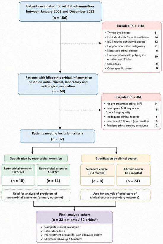
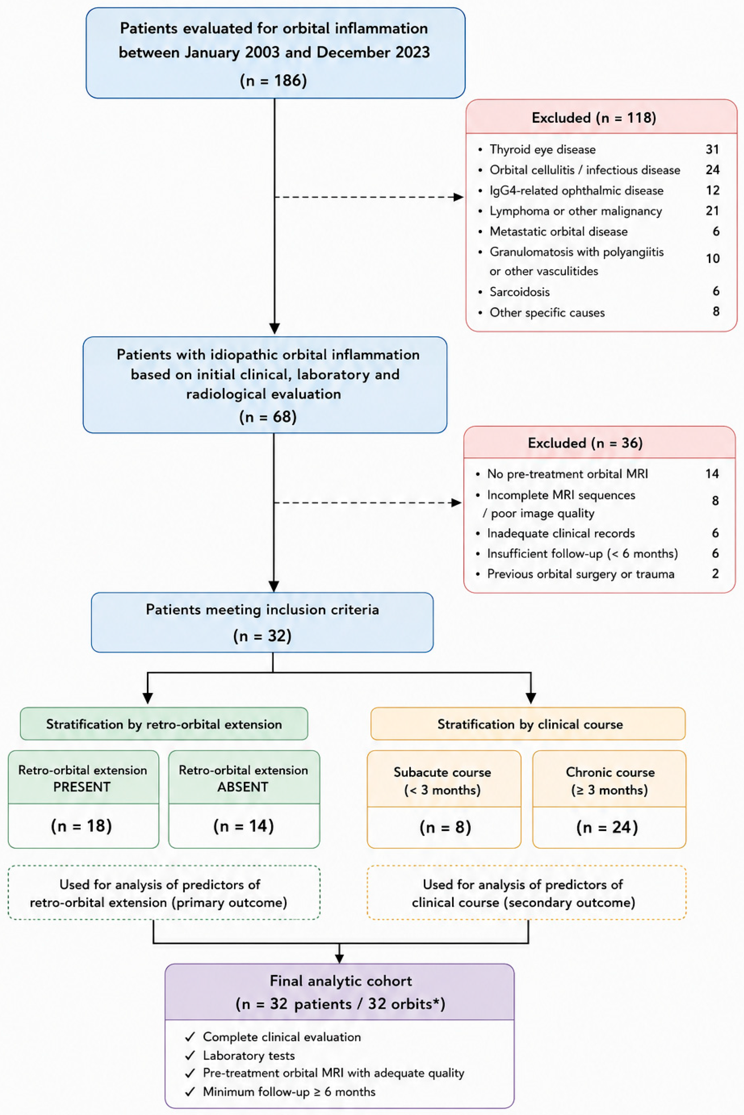
During the study period, all patients initially evaluated for orbital inflammatory disorders were screened. After exclusion of patients with alternative diagnoses, inadequate imaging quality, incomplete clinical records, or insufficient follow-up, 32 patients fulfilled the study criteria and constituted the final study cohort. A detailed flow diagram illustrating patient selection and exclusion is presented in Figure 1.
Figure 1.
Flow diagram of patient selection. * Five patients had bilateral involvement; therefore, 32 patients correspond to 32 orbits in which quantitative measurements were performed.
Patients were included if they had a diagnosis of idiopathic orbital inflammation established by combined clinical, laboratory, and radiological evaluation, underwent orbital magnetic resonance imaging (MRI) before initiation of systemic therapy, had MRI examinations suitable for quantitative radiological measurements, and had complete clinical and follow-up data. Patients with thyroid eye disease, IgG4-related ophthalmic disease, orbital cellulitis, orbital lymphoma or other orbital malignancies, metastatic orbital tumors, granulomatosis with polyangiitis, sarcoidosis, previous orbital surgery or trauma altering orbital anatomy, poor-quality MRI examinations, or incomplete medical records were excluded.
2.1. Diagnostic Evaluation
The diagnosis of idiopathic orbital inflammation was established by an experienced oculoplastic surgeon based on a comprehensive evaluation integrating clinical presentation, ophthalmological examination, laboratory investigations, and orbital MRI findings after exclusion of infectious, neoplastic, thyroid-related, granulomatous, and systemic inflammatory disorders. Histopathological confirmation was reserved for patients with atypical clinical or radiological features, poor response to corticosteroid therapy, progressive disease despite treatment, or when malignancy could not be confidently excluded by non-invasive investigations. In all other patients, the diagnosis was established according to accepted clinicoradiological diagnostic criteria together with treatment response during follow-up.
2.2. Clinical Assessment
Demographic characteristics, including age, sex, laterality, affected eye, duration of symptoms, systemic diseases, and history of previous orbital inflammatory attacks, were retrieved from the medical records. All patients underwent a standardized ophthalmological examination at presentation consisting of best-corrected visual acuity (BCVA), slit-lamp biomicroscopy, Goldmann applanation tonometry, dilated fundus examination, ocular motility assessment using the Kestenbaum limbus test, and external orbital examination. Clinical findings, including eyelid edema, palpable mass, orbital pain, conjunctival hyperemia, proptosis, diplopia, limitation of extraocular movements, decreased visual acuity, and optic nerve dysfunction, were recorded.
All patients initially received systemic corticosteroid therapy as first-line treatment, administered intravenously or orally. Additional immunosuppressive treatments were documented when required. Clinical response was categorized as complete response, partial response, or progressive disease according to improvement in clinical findings during follow-up. Disease recurrence was defined as reappearance of clinical signs or symptoms following complete remission after treatment. For subgroup analyses, patients were classified as having subacute disease if symptom duration at presentation was less than 3 months and chronic disease if symptom duration was 3 months or longer. Clinical evolution during follow-up was also documented.
2.3. Laboratory Evaluation
Laboratory investigations were performed in all patients to exclude secondary causes of orbital inflammation and to evaluate systemic inflammatory activity. Baseline laboratory assessment included complete blood count, erythrocyte sedimentation rate (ESR), C-reactive protein (CRP), thyroid function tests (thyroid-stimulating hormone, free triiodothyronine, and free thyroxine), thyroid autoantibodies, serum immunoglobulin G4 (IgG4), angiotensin-converting enzyme (ACE), proteinase-3 antineutrophil cytoplasmic antibody (PR3-ANCA), and myeloperoxidase antineutrophil cytoplasmic antibody (MPO-ANCA).
The neutrophil-to-lymphocyte ratio (NLR) was calculated from peripheral blood counts and evaluated as a potential systemic inflammatory biomarker. Acute phase reactants were classified as elevated when ESR and/or CRP values exceeded the laboratory reference ranges.
2.4. Magnetic Resonance Imaging and Quantitative Radiological Assessment
Orbital MRI was performed in all patients before initiation of systemic treatment according to the institutional orbital imaging protocol. The MRI protocol included axial and coronal T1-weighted, T2-weighted, fat-suppressed, diffusion-weighted, and contrast-enhanced fat-suppressed T1-weighted sequences. All MRI examinations were independently reviewed by an experienced neuroradiologist who was blinded to patients’ clinical information and treatment outcomes at the time of image assessment. The presence or absence of retro-orbital extension, orbital soft tissue involvement, extraocular muscle involvement, lacrimal gland enlargement, optic nerve or optic disc involvement, and scleral enhancement were systematically evaluated.
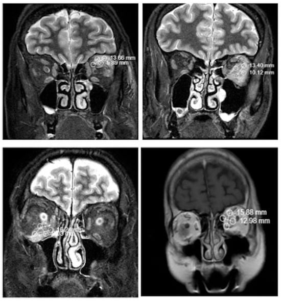
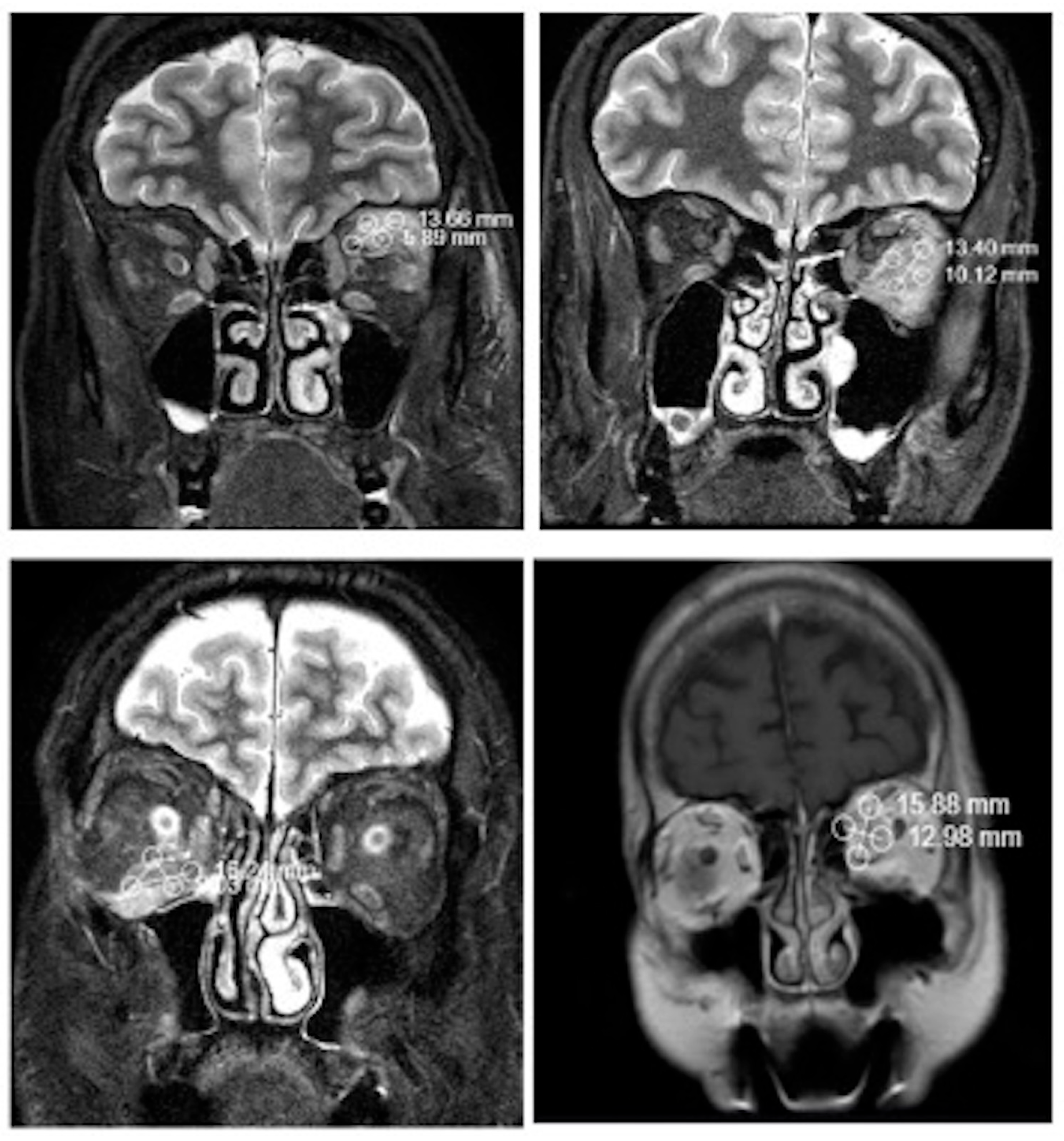
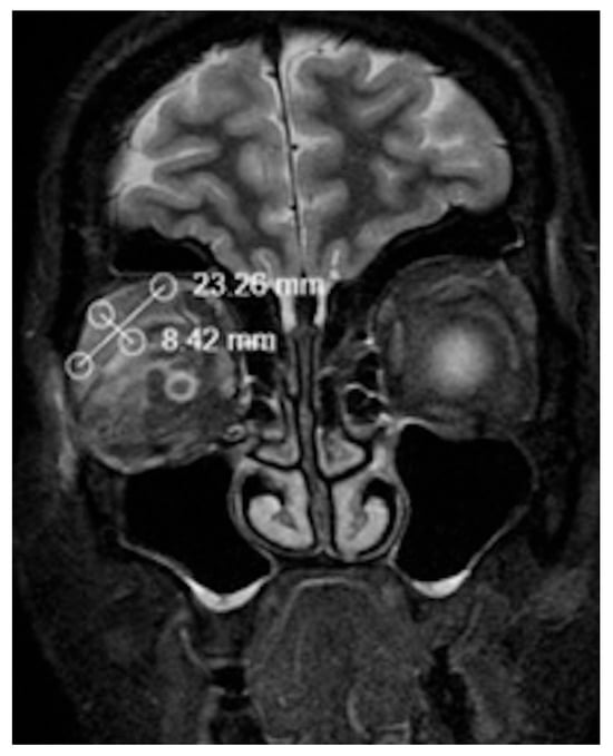
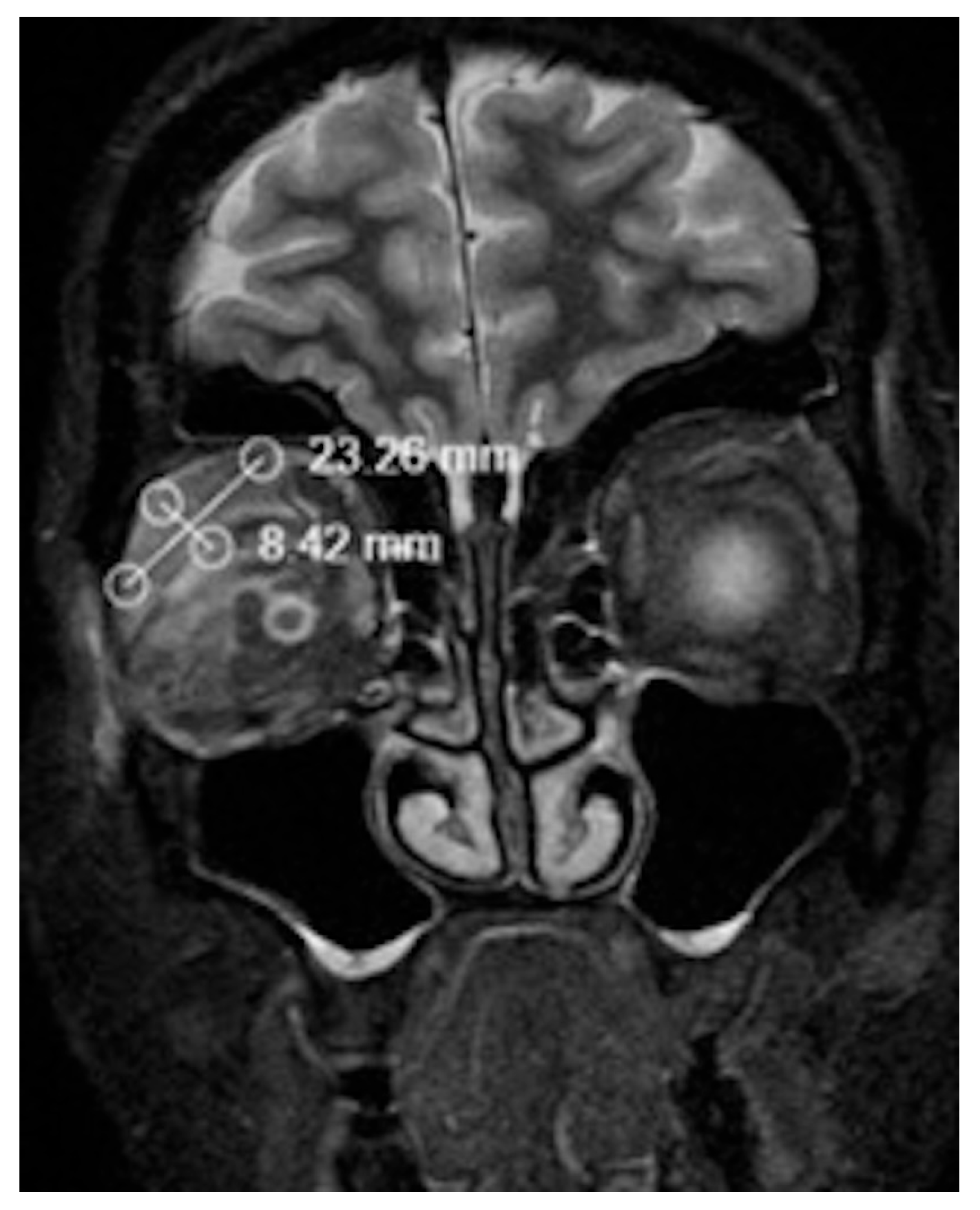
Retro-orbital extension was defined as inflammatory tissue extending into the posterior intraconal fat on contrast-enhanced MRI, regardless of orbital apex involvement. Quantitative MRI measurements were performed using the institutional Picture Archiving and Communication System (PACS). The width and thickness of the superior, inferior, medial, and lateral rectus muscles were measured on coronal MRI images at the level of the globe equator, which served as a standardized anatomical landmark for all measurements. For each patient, the coronal slice corresponding to the globe equator was selected, and the two orthogonal muscle diameters were measured at this level. Long- and short-axis diameters of the lacrimal gland were also measured. In patients with adequate image quality, scleral thickness was additionally evaluated. Representative examples of the quantitative MRI measurements are shown in Figure 2 and Figure 3.
Figure 2.
Examples of measurements on MRI from those with muscle involvement.
Figure 3.
Lacrimal gland involvement observed on a coronal T2W MRI image.
2.5. Outcome Measures
The primary outcome of this study was to identify independent clinicoradiological predictors of retro-orbital extension in patients with idiopathic orbital inflammation, with particular emphasis on quantitative MRI measurements of the extraocular muscles (Figure 4).
Figure 4.
(A) A 55-year-old male patient referred from an external center to our department due to the first presentation of orbital cellulitis; (B) first day of systemic prednol treatment; (C) seventh day of systemic prednol treatment.
Secondary outcomes included identification of predictors associated with chronic disease course, determination of optimal MRI cut-off values for predicting retro-orbital extension using receiver operating characteristic (ROC) analysis, evaluation of treatment response according to radiological characteristics, assessment of the relationship between quantitative MRI measurements and systemic inflammatory biomarkers, and investigation of clinicoradiological factors associated with disease recurrence.
2.6. Statistical Analysis
Statistical analyses were performed using Jamovi 2.6.44. Continuous variables were expressed as median with interquartile range (IQR) due to non-normal distribution, and categorical variables as frequency (percentage). Comparisons between groups (retro-orbital extension present vs. absent; subacute vs. chronic clinical course) were conducted using the Mann–Whitney U test for continuous variables and Fisher’s exact test or Pearson’s chi-square test for categorical variables, as appropriate. ROC curve analysis was performed to evaluate the discriminative ability of extraocular muscle width and thickness measurements for predicting retro-orbital extension and chronic clinical course. The optimal cut-off values were determined using Youden’s index. For each parameter, the area under the curve (AUC) with a 95% confidence interval (CI) was calculated, and sensitivity, specificity, positive predictive value (PPV), negative predictive value (NPV), and accuracy were reported at the optimal cut-off. A forward stepwise multivariable logistic regression analysis was conducted to identify independent predictors of retro-orbital extension. Variables were entered into the model using the likelihood ratio test with entry and removal thresholds set at p < 0.05 and p > 0.10, respectively. Four hierarchical models were constructed: Model 1 (age only), Model 2 (age + lateral rectus thickness dichotomized at the optimal cut-off), Model 3 (age + LR thickness + sex), and Model 4 (full model including age, LR thickness, sex, and systemic disease). Model fit was assessed using deviance, Akaike Information Criterion (AIC), and McFadden’s pseudo R2. Model comparisons were performed using the likelihood ratio test (LRT), with a significant χ2 value (p < 0.05) indicating improved model fit. Multicollinearity was evaluated using the variance inflation factor (VIF), with VIF > 5 considered indicative of significant collinearity. All statistical tests were two-sided, and a p-value < 0.05 was considered statistically significant. Borderline significance was defined as p = 0.050.
3. Results
The study included 32 patients with idiopathic orbital inflammation (mean age 43.97 ± 14.55 years, 43.8% male). Patients with retro-orbital extension (n = 18) differed significantly from those without (n = 14) in treatment response (p = 0.016), with complete response rates of 55.6% vs. 100%. MRI showed higher rates of lateral rectus involvement (50.0% vs. 14.3%, p = 0.035), soft tissue involvement (94.4% vs. 57.1%, p = 0.011), and optic disc involvement (33.3% vs. 0.0%, p = 0.017). Elevated acute phase reactants were also more frequent in the extension group (50.0% vs. 14.3%, p = 0.035). No other variables differed significantly. Between subacute (n = 8) and chronic (n = 24) groups, redness was the only significant difference, being more common in subacute patients (62.5% vs. 12.5%, p = 0.012). Age, sex, treatment response, extraocular muscle involvement, MRI findings, acute phase reactants, attack history, and recurrence were comparable (p > 0.05 for all) (Table 1). Data are presented as median (IQR) or n (%). IQR, interquartile range; MRI, magnetic resonance imaging. Statistically significant p-values (<0.05) are shown in bold.
Table 1.
Demographic, clinical, and radiological characteristics of patients according to retro-orbital extension and clinical course.
Extraocular muscle dimensions were compared between patients with and without retro-orbital extension (Table 2). Lateral rectus thickness was significantly higher in patients with retro-orbital extension compared to those without [4.46 mm (IQR: 3.80–5.34) vs. 3.42 mm (IQR: 2.96–3.79), p = 0.007]. No other statistically significant differences were observed for superior rectus, lateral rectus width, inferior rectus, or medial rectus measurements (p > 0.05 for all). No statistically significant differences were observed between subacute and chronic groups for any extraocular muscle width or thickness measurement (p > 0.05 for all). Although inferior rectus width tended to be higher in the subacute group compared to the chronic group [10.36 mm (IQR: 9.49–10.89) vs. 9.23 mm (IQR: 8.48–9.82)], this difference did not reach statistical significance (p = 0.071).
Table 2.
Comparison of extraocular muscle width and thickness measurements according to retro-orbital extension and clinical course.
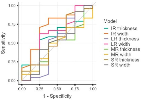
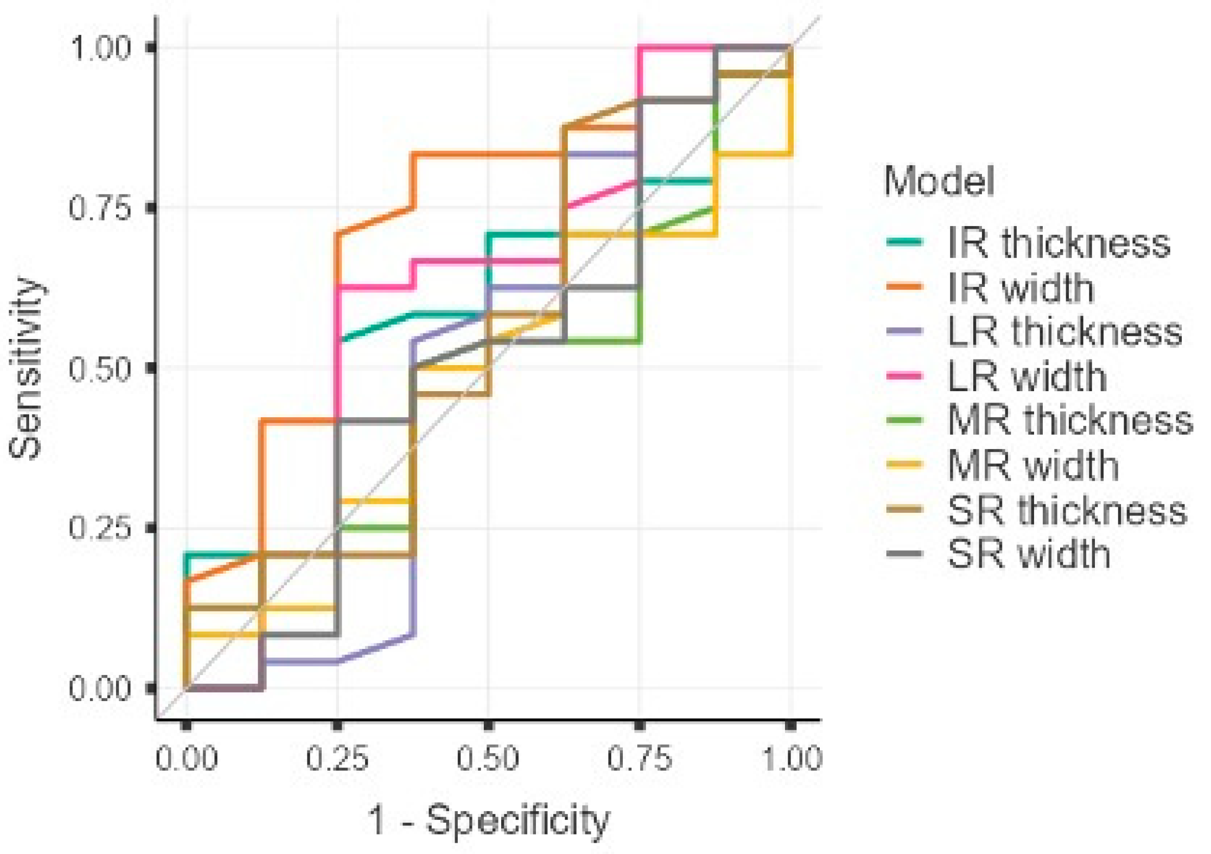
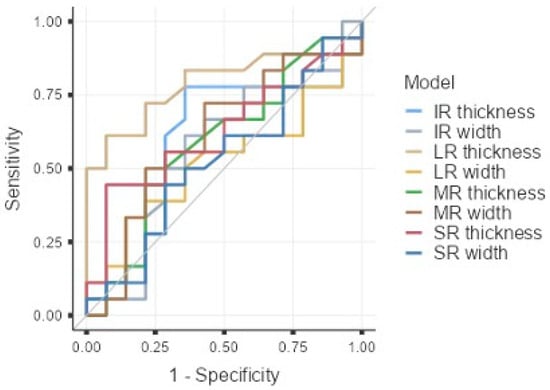
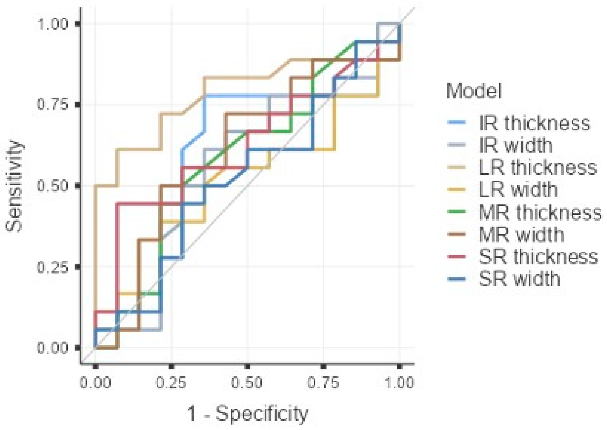
ROC curve analysis was performed to evaluate the discriminative ability of extraocular muscle width and thickness measurements for predicting retro-orbital extension (Table 3). Lateral rectus thickness was the only parameter with statistically significant discriminative ability (AUC = 0.786, 95% CI: 0.618–0.953, p < 0.001). The optimal cut-off value determined by Youden’s index was ≥4.125 mm. At this cut-off, lateral rectus thickness demonstrated a sensitivity of 61.11%, specificity of 92.86%, positive predictive value (PPV) of 91.67%, negative predictive value (NPV) of 65.00%, and overall accuracy of 75.00%. ROC curve analysis was also performed to distinguish the chronic from the subacute clinical course. Inferior rectus width showed borderline significant discriminative ability (AUC = 0.719, 95% CI: 0.500–0.937, p = 0.050). The optimal cut-off value was ≤10.22 mm, yielding a sensitivity of 83.33%, specificity of 62.50%, PPV of 86.96%, NPV of 55.56%, and accuracy of 78.12%. No other parameter demonstrated statistically significant discriminative ability for the chronic clinical course, including superior rectus width (AUC = 0.513, p = 0.923) and thickness (AUC = 0.544, p = 0.741), lateral rectus width (AUC = 0.617, p = 0.383) and thickness (AUC = 0.505, p = 0.973), inferior rectus thickness (AUC = 0.591, p = 0.432), or medial rectus width (AUC = 0.477, p = 0.845) and thickness (AUC = 0.484, p = 0.896) (p > 0.05 for all) (Figure 5 and Figure 6).
Table 3.
ROC curve analysis for prediction of retro-orbital extension and chronic clinical course.
Figure 5.
Combined ROC curves demonstrating the diagnostic performance of extraocular muscle parameters in patients with retro-orbital extension.
Figure 6.
Combined ROC curves demonstrating the diagnostic performance of extraocular muscle parameters in patients with the chronic clinical course.
A forward stepwise logistic regression analysis (entry threshold p < 0.05, removal threshold p > 0.10) was performed to identify independent predictors of retro-orbital extension. Four hierarchical models were constructed: Model 1 (age only), Model 2 (age + lateral rectus thickness ≥ 4.125 mm), Model 3 (age + LR thickness + sex), and Model 4 (full model including age, LR thickness, sex, and systemic disease). Model fit was assessed using deviance, Akaike Information Criterion (AIC), and McFadden’s pseudo R2, with model comparisons based on the likelihood ratio test (LRT). Age alone was not associated with retro-orbital extension (OR = 0.99, 95% CI: 0.93–1.05, p = 0.649). The addition of LR thickness significantly improved model fit (p < 0.001), and LR thickness emerged as a significant predictor (OR = 20.39, 95% CI: 1.92–216.40, p = 0.012). Subsequent addition of sex (p = 0.261) and systemic disease (p = 0.405) did not improve model fit, and neither variable reached statistical significance (sex: OR = 2.57, p = 0.299; systemic disease: OR = 0.45, p = 0.408). In the final model, LR thickness ≥ 4.125 mm remained the only independent predictor of retro-orbital extension (OR = 20.39, 95% CI: 1.92–216.40, p = 0.012), with no evidence of multicollinearity (VIF < 1.10 for all variables). McFadden’s R2 for the final model was 0.299, indicating that LR thickness alone explained approximately 30% of the variance in retro-orbital extension status (Table 4).
Table 4.
Multivariable logistic regression analysis for predictors of retro-orbital extension.
4. Discussion
Idiopathic orbital inflammation (IOI), historically designated as orbital pseudotumor, is a non-neoplastic and non-granulomatous inflammatory disorder accounting for approximately 5% to 11% of all orbital masses [1,2,3]. It represents the third most common orbital pathology after thyroid eye disease and lymphoproliferative disorders. Despite its relative frequency, the clinical management of IOI remains challenging due to its heterogeneous presentation and the historic lack of standardized, objective quantitative markers to predict therapeutic trajectory [4,5]. Our findings suggest that lateral rectus (LR) thickness may serve as a potential imaging marker of retro-orbital extension. Specifically, an LR thickness cut-off of 4.125 mm emerged as the sole independent predictor of extension in our multivariable model, escalating the risk of deep orbital involvement by more than 20-fold (OR = 20.39, 95% CI: 1.92–216.40, p = 0.012). In our study, retro-orbital extension was defined by inflammatory involvement of the posterior intraconal fat, without requiring direct orbital apex involvement. This definition allowed the assessment of posterior orbital inflammatory extension beyond cases with overt orbital apex involvement. Although the medial rectus is traditionally highlighted as a frequently involved muscle in myositis and diffuse IOI, our findings suggest that lateral rectus thickness may have potential value in identifying retro-orbital extension [15,16,17,18]. Normative MRI-based extraocular muscle metrics reported by Rana et al. indicate a mean LR thickness of approximately 3.9 ± 0.7 mm [19]. Our proposed cut-off of 4.125 mm lies just above this normative threshold, offering clinicians a highly specific metric (92.86% specificity, 91.67% PPV) to objectively identify pathologic enlargement indicative of posterior disease propagation. Patients exhibiting retro-orbital extension achieved a significantly lower complete treatment response rate compared to those with disease confined to the anterior orbit (55.6% vs. 100%, p = 0.016). These findings suggest that retro-orbital extension, defined by posterior intraconal fat involvement in our cohort, may be associated with a more extensive inflammatory process and less favorable treatment outcomes. Furthermore, soft tissue involvement (94.4% vs. 57.1%, p = 0.011) and optic disc edema (33.3% vs. 0.0%, p = 0.017) were significantly enriched in the extension cohort, reinforcing the advanced inflammatory burden in these patients.
Our observations closely mirror results from high-impact multicenter studies, such as the French SIOI cohort, which identified orbital fat infiltration—a cardinal component of retro-orbital extension—as a major determinant of corticosteroid failure [1]. Similarly, Kubota and Iwakoshi reported that diffuse intraconal lesions independently predicted steroid-refractory disease (OR = 8.8), leading to long-term visual impairment despite structural remission [20]. Our findings suggest that LR thickness may provide additional quantitative information for identifying retro-orbital extension during the initial radiological assessment. The necessity of early risk stratification transcends adult populations. In pediatric IOI cohorts, such as that reported by Li et al., deep and diffuse disease subtypes were strongly linked to steroid-dependent relapse and secondary ocular morbidity [21]. Li et al. emphasized imaging-based classification to tailor long-term steroid-sparing strategies and avoid the late sequelae of systemic treatment or radiotherapy [21]. Our findings highlight the potential value of quantitative MRI assessment in adult patients with IOI, although further studies are needed to establish its clinical applicability in broader patient populations.
Long-term stability in IOI is heavily compromised by high recurrence rates, reported between 33% and 58%. Anggraini et al. identified non-anterior involvement as a major predictor for relapse, increasing recurrence risk 7.9-fold and demonstrating that corticosteroid monotherapy is largely insufficient in non-anterior subtypes [11]. Our findings are consistent with these observations, suggesting that retro-orbital extension may be associated with less favorable treatment outcomes. Patients with increased LR thickness may benefit from closer clinical monitoring and careful assessment of treatment response, although further studies are required to determine whether this imaging finding should influence therapeutic decisions. In recent years, the search for IOI biomarkers has expanded to tear film cytokines (e.g., IL-8, MCP-1) and peripheral inflammatory ratios [22]. While molecular assays offer pathophysiological insights, their routine clinical integration is hampered by cost, accessibility, and processing delays. In contrast, LR thickness can be assessed using routine MRI and PACS, potentially providing an accessible quantitative parameter for radiological evaluation. As emphasized by Nguyen et al., cost-effective diagnostic algorithms are essential in neuro-ophthalmology to prevent low-yield, expensive diagnostic workups in lower-risk patients [9].
Advanced machine learning and radiomics models have also demonstrated high accuracy (AUCs 0.84–0.95) in differentiating IOI from lymphoproliferative disorders [23,24]. However, these computational pipelines require complex image segmentation and specialized software, rendering them impractical for real-time clinical consultations. The proposed LR thickness threshold may represent a simple quantitative imaging parameter; however, its clinical utility requires further validation in larger patient cohorts. Additionally, the demographic characteristics of our cohort—predominantly middle-aged with a slight female preponderance—align closely with large-scale population data from the AAO IRIS Registry (n = 20,584), strengthening the external validity and generalizability of our findings [4]. Regarding systemic markers, while parameters like the neutrophil-to-lymphocyte ratio (NLR) have been evaluated for distinguishing inflammatory from infectious orbital disease, our study found that local retro-orbital extension was significantly correlated with elevated systemic acute phase reactants (ESR/CRP; 50.0% vs. 14.3%, p = 0.035) [9]. This indicates that deep orbital involvement may represent the local ocular manifestation of a broader systemic inflammatory state, justifying comprehensive systemic evaluation and multidisciplinary care. A multidisciplinary management strategy involving rheumatology is paramount for patients with deep extension. Gulle et al. noted that up to 85.7% of patients managed in joint ocular inflammation clinics required steroid-sparing disease-modifying agents to achieve durable remission [10]. Furthermore, as highlighted in Gerbino et al.’s 10-year surgical experience, interventions targeting the retrobulbar space and orbital apex carry significant risks of nerve injury and vision-threatening complications [16]. Although quantitative MRI parameters such as LR thickness may provide additional information regarding the extent of orbital inflammation, they should be interpreted alongside clinical findings and should not replace established diagnostic evaluation or biopsy when indicated.
Regarding the secondary analysis of clinical course, ocular redness was significantly more frequent in patients with subacute disease than in those with chronic presentations (62.5% vs. 12.5%, p = 0.012). This finding may reflect differences in the clinical expression of inflammatory activity between disease phases. Although inferior rectus width demonstrated borderline discriminative ability for chronic disease (AUC = 0.719, p = 0.050), the corresponding between-group comparison did not reach statistical significance (p = 0.071). Furthermore, none of the quantitative extraocular muscle measurements showed significant differences between the subacute and chronic groups. These findings suggest that, unlike retro-orbital extension, clinical course may not be reliably characterized by individual extraocular muscle dimensions on MRI. However, the small number of patients with subacute disease (n = 8) limits statistical power, and these exploratory findings should be interpreted cautiously.
We acknowledge several limitations in our study. First, its retrospective nature from a single tertiary referral center introduces inherent selection bias, and the sample size (n = 32) is relatively small, albeit typical for single-center cohorts investigating this rare entity. Second, histopathological confirmation was not obtained for all patients, as tissue biopsy in retro-orbital or apical disease is often avoided to prevent iatrogenic neurovascular injury when clinical and radiological features strongly support IOI. Furthermore, the lateral rectus thickness threshold of ≥4.125 mm was derived from a relatively small, single-center cohort without external validation. Future large-scale, prospective multicenter studies are warranted to confirm the diagnostic performance and generalizability of this threshold across diverse patient populations. Additionally, MRI assessments were performed by a single experienced neuroradiologist, and interobserver and intraobserver reproducibility were not evaluated, representing a limitation of the present study. Nevertheless, the standardized imaging assessment and clearly defined inclusion criteria support the methodological consistency of the study, although the findings require confirmation in larger cohorts.
5. Conclusions
In conclusion, lateral rectus thickness may serve as a potential imaging marker for retro-orbital extension in idiopathic orbital inflammation. In our cohort, an LR thickness threshold of ≥4.125 mm demonstrated promising discriminative ability for identifying retro-orbital extension, which was associated with less favorable treatment responses. Incorporating this quantitative measurement into orbital MRI assessment may provide additional information regarding disease extent, although its potential role in clinical decision-making requires further validation.
Author Contributions
Conceptualization, B.U., O.K. and A.A.O.; methodology, B.U., O.K., B.M. and N.T.; formal analysis, N.T., M.G., Z.O.U., R.S.S., H.B. and Y.K.B.; investigation, O.K., A.A.O., B.U. and B.M.; resources, B.U., N.T., M.G., Z.O.U., R.S.S. and H.B.; data curation, A.A.O., H.B. and Y.K.B.; writing—original draft preparation, B.U., A.A.O., O.K., B.M., N.T., M.G., Z.O.U. and Y.K.B.; writing—review and editing, Y.K.B., A.A.O., B.U. and O.K.; supervision, A.A.O. and B.U.; project administration, B.U., O.K. and B.M. All authors have read and agreed to the published version of the manuscript.
Funding
This research received no external funding.
Institutional Review Board Statement
The study was conducted in accordance with the Declaration of Helsinki and approved by the Ethics Committee of the Faculty of Medicine at Cukurova University (Appl. No.: 4.1.2024-140-28; Date: 4 January 2024).
Informed Consent Statement
Written informed consent has been obtained from the patients to publish this paper.
Data Availability Statement
The original contributions presented in this study are included in the article. Further inquiries can be directed to the corresponding author.
Conflicts of Interest
The authors declare no conflicts of interest.
References
- La Rosa, A.; Groh, M.; Martin, A.; Ba, S.T.; Cucherousset, N.; Mouriaux, F.; Sedira, N.; Heron, E.; Galatoire, O.; Saadoun, D.; et al. Cellular pattern and orbital fat involvement are possible risk factors for the failure of corticosteroids in patients with pure idiopathic orbital inflammation syndrome: Lessons from the French prospective SIOI cohort study (part II). BMJ Open Ophthalmol. 2024, 9, e001663. [Google Scholar] [CrossRef] [Scilit] [PubMed]
- Fang, Y.; Shen, B.; Dai, Q.; Xie, Q.; Wu, W.; Wang, M. Orbital inflammatory pseudotumor: New advances in diagnosis, pathogenesis, and treatment. Eur. J. Med. Res. 2023, 28, 395. [Google Scholar] [CrossRef] [Scilit] [PubMed]
- Wu, K.Y.; Kulbay, M.; Daigle, P.; Nguyen, B.H.; Tran, S.D. Nonspecific orbital inflammation: Unraveling the molecular pathogenesis, diagnostic modalities and therapeutic interventions. Int. J. Mol. Sci. 2024, 25, 1553. [Google Scholar] [CrossRef] [Scilit] [PubMed]
- Lu, J.E.; Ross, C.; Ivanov, A.; Hall, E.; Lorch, A.; Miller, J.; Yoon, M.K. Epidemiology of orbital inflammatory disease: An AAO IRIS registry study. Ocul. Immunol. Inflamm. 2024, 32, 2081–2084. [Google Scholar] [CrossRef] [Scilit] [PubMed]
- Yesiltas, Y.S.; Gunduz, A.K. Idıopathic orbital inflammation: Review of literature and new advances. Middle East Afr. J. Ophthalmol. 2018, 25, 71–80. [Google Scholar] [CrossRef] [Scilit] [PubMed]
- Ang, T.; Tong, J.Y.; Patel, S.; Hardy, T.G.; McNab, A.; Selva, D. Magnetic resonance imaging of idiopathic orbital myositis. Ophthalmic Plast. Reconstr. Surg. 2024, 40, 544–551. [Google Scholar] [CrossRef] [Scilit] [PubMed]
- Al-Ghazzawi, K.; Neumann, I.; Knetsch, M.; Chen, Y.; Wilde, B.; Bechrakis, N.E.; Eckstein, A.; Oeverhaus, M. Treatment Outcomes of Patients with Orbital Inflammatory Diseases: Should Steroids Still Be the First Choice? J. Clin. Med. 2024, 13, 3998. [Google Scholar] [CrossRef] [Scilit] [PubMed]
- Wladis, E.J.; Bohnak, C.E.; Law, J.L.; Adam, A.P.; Rothschild, M.I.; Pauze, D.R. Neutrophil-to-lymphocyte ratios distinguish idiopathic orbital inflamamation from orbital infectious disease. Ophthalmic Plast. Reconst. Surg. 2024, 40, 178–180. [Google Scholar] [CrossRef] [Scilit] [PubMed]
- Nguyen, K.; Tobalem, S.; Meer, E.; Hekmatjah, N.; Kao, B.; Ares, S.; Kalin-Hajdu, E.; Kersten, R.C.; Ashraf, D.C. Optimizing cost-effectiveness in the work-up of orbital inflammation: A multicenter, retrospective study. Orbit 2026, 45, 389–395. [Google Scholar] [CrossRef] [Scilit] [PubMed]
- Gulle, S.; Engin, C.D.; Ozizmirliler, D.; Men, S.; Onen, F.; Bajin, M.S. Orbital inflammatory disease: A joint clinical experience in rheumatology and ophthalmology. Int. Ophthalmol. 2025, 45, 48. [Google Scholar] [CrossRef] [Scilit] [PubMed]
- Anggraini, N.; Siregar, N.C.; Sitorus, R.S. Recurrence of idiopathic orbital inflammation: An 11-year retrospective study. Middle East Afr. J. Ophthalmol. 2023, 30, 89–97. [Google Scholar] [CrossRef] [Scilit] [PubMed]
- Landau-Prat, D.; Shemesh, R.; Shalev, D.; Nemet, M.; Ben-Simon, G.J.; Greenberg, G. Magnetic resonance diffusion-weighted imaging in lacrimal gland lymphoma versus inflammation: A comparative study. Indian J. Ophthalmol. 2024, 72, 1448–1452. [Google Scholar] [CrossRef] [Scilit] [PubMed]
- Ulman, E.A.; Palamar, M.; Kupal, B.A.; Yaman, B.; Akalın, T.; Ceylan, N.; Selver, O.B. Clinical, demographic, and outcome differences between isolated and non-isolated lacrimal gland involvement in idiopathic orbital inflammation. Int. Ophthalmol. 2025, 45, 362. [Google Scholar] [CrossRef] [Scilit] [PubMed]
- Rattunde, A.L.; Knecht, V.A.; Bertelmann, E. IgG4-Related Orbital Disease vs. Idiopathic Orbital Inflammation: Clinical Features, Therapy and Outcomes in a Central-European Retrospective Single-Center Cohort. Biomedicines 2025, 13, 2311. [Google Scholar] [CrossRef] [Scilit] [PubMed]
- Huang, S.; Tong, J.Y.; Ang, T.W.; Hardy, T.G.; McNab, A.; Selva, D. Idiopathic apical orbital inflammation: Clinical features and outcomes. Can. J. Ophthalmol. 2025, 60, e436–e442. [Google Scholar] [CrossRef] [Scilit] [PubMed]
- Gerbino, G.; Gugliotta, Y.; Corsico, M.; Ramieri, G. A retrospective analysis of the management and surgical treatment of orbital lesions: Outcomes and rationale. J. Craniomaxillofac. Surg. 2024, 52, 1109–1115. [Google Scholar] [CrossRef] [Scilit] [PubMed]
- McNab, A.A. Orbital myositis: A comprehensive review and reclassification. Ophthalmic Plast. Reconstr. Surg. 2020, 36, 109–117. [Google Scholar] [CrossRef] [Scilit] [PubMed]
- Kang, M.S.; Yang, H.K.; Kim, N.; Hwang, J.M. Clinical features of ocular motility in idiopathic orbital myositis. J. Clin. Med. 2020, 9, 1165. [Google Scholar] [CrossRef] [Scilit] [PubMed]
- Rana, K.; Juniat, V.; Rayan, A.; Patel, S.; Selva, D. Normative measurements of orbital structures by magnetic resonance imaging. Int. Ophthalmol. 2022, 42, 3869–3875. [Google Scholar] [CrossRef] [Scilit] [PubMed]
- Kubota, T.; Iwakoshi, A. Clinical heterogeneity between two subgroups of patients with idiopathic orbital inflammation. BMJ Open Ophthalmol. 2022, 7, e001005. [Google Scholar] [CrossRef] [Scilit] [PubMed]
- Li, Z.; Chen, Y.; Liu, H.; Wu, Q.; Yang, B.; Zhang, J.; Jiang, L.; Chen, F.K. Clinical features and recurrence patterns of pediatric idiopathic orbital inflammatory disease. BMC Ophthalmol. 2025, 25, 649. [Google Scholar] [CrossRef] [Scilit] [PubMed]
- Chen, J.; Xiao, W.; Ye, H.; Xu, Z.; Chen, R.; Yang, H. New clues to the pathogenesis of idiopathic orbital inflammation: Elevated IL-8 and MCP-1 in tear fluid. J. Ophthalmol. 2025, 2025, 4175012. [Google Scholar] [CrossRef] [Scilit] [PubMed]
- Wang, G.R.; Qu, X.; Guo, J.; Luo, Y.; Xian, J. Radiomics and machine learning model can improve the differentiation between ocular adnexal lymphoma and idiopathic orbital inflammation. Chin. Med. J. 2025, 138, 234–236. [Google Scholar] [CrossRef] [Scilit] [PubMed]
- Xie, X.; Yang, L.; Zhao, F.; Wang, D.; Zhang, H.; He, X.; Cao, X.; Yi, H.; He, X.; Hou, Y. A deep learning model combining multimodal radiomics, clinical and imaging features for differentiating ocular adnexal lymphoma from idiopathic orbital inflammation. Eur. Radiol. 2022, 32, 6922–6932. [Google Scholar] [CrossRef] [Scilit] [PubMed]



Disclaimer/Publisher’s Note: The statements, opinions and data contained in all publications are solely those of the individual author(s) and contributor(s) and not of MDPI and/or the editor(s). MDPI and/or the editor(s) disclaim responsibility for any injury to people or property resulting from any ideas, methods, instructions or products referred to in the content. |
© 2026 by the authors. Licensee MDPI, Basel, Switzerland. This article is an open access article distributed under the terms and conditions of the Creative Commons Attribution (CC BY) license.