


Abstract
Background and Clinical Significance: Postoperative lymphorrhea is an uncommon complication of great saphenous vein harvesting for coronary artery bypass grafting (CABG), and accurate localization of the leakage site may be challenging. Case Presentation: We report the case of a 79-year-old man who developed persistent lymphorrhea following great saphenous vein harvesting for CABG. Notably, a Pseudomonas aeruginosa surgical site infection preceded the clinically apparent lymphatic leakage and required surgical wound debridement. Following the onset of lymphorrhea, compression therapy was applied for two weeks, accompanied by frequent dressing changes, without improvement. Due to persistent lymphatic leakage, planar lymphoscintigraphy using 99mTc-nanocolloid was performed, demonstrating active lymphatic leakage. Subsequent hybrid single-photon emission computed tomography/computed tomography (SPECT/CT) localized the suspected region approximately 3.5 cm above the medial femoral condyle, providing anatomical information that guided targeted suture ligation. Conclusions: External lymphorrhea ceased immediately following the intervention and the patient was discharged the following day in good clinical condition, with instructions to wear compression stockings. We also reviewed the relevant English-language literature on this topic.
1. Introduction
Postoperative lymphatic complications following lower-extremity surgery may arise from injury to lymphatic vessels and can manifest as either lymphatic leakage or impaired lymphatic drainage [1]. Lymphatic leakage may present as lymphorrhea, characterized by the external drainage of clear lymphatic fluid through a surgical wound, or as lymphocele formation, involving a localized collection of lymphatic fluid within the surrounding tissues [1]. In contrast, impaired lymphatic drainage may result in lymphatic stasis and subsequent lymphedema. Lymphorrhea has been reported following great saphenous vein harvesting for infrainguinal arterial bypass surgery, particularly in patients with critical limb ischemia [2]. Although lymphatic complications, including lymphorrhea, lymphocele formation, and lymphedema, have also been described following great saphenous vein (GSV) harvesting for coronary artery bypass grafting (CABG), the incidence of persistent lymphorrhea in this setting remains poorly established [3,4,5,6]. A critical challenge in managing this complication lies in selecting an appropriate imaging modality to accurately localize the site of the leak. Although conventional lymphangiography has traditionally been used for this purpose [1], less invasive techniques, such as planar lymphoscintigraphy, can demonstrate abnormal radiotracer accumulation suggestive of lymphatic leakage, but precise anatomical localization may remain challenging. Hybrid single-photon emission computed tomography/computed tomography (SPECT/CT) imaging combines functional lymphatic data with cross-sectional anatomical information, facilitating localization of the leakage site and supporting targeted treatment planning, as illustrated in the present case.
2. Case Presentation
A 79-year-old male patient with a significant medical history of heavy smoking, arterial hypertension, diabetes mellitus, and rheumatoid arthritis underwent triple coronary artery bypass graft (CABG) surgery, which included right great saphenous vein graft harvesting. Postoperatively, the patient developed a deep surgical site infection along the right thigh caused by Pseudomonas aeruginosa. The wound was subsequently opened, evacuated, debrided, and left to heal by secondary intention.
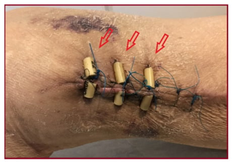
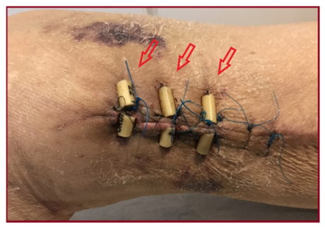
Following debridement, severe lymphorrhea became apparent. Due to the profuse exudation of clear lymphatic fluid, clinical identification of the leakage source was impossible. Application of an elastic compressive bandage for two weeks failed to improve the lymphorrhea. Once the wound infection was clinically controlled, the wound was closed using three heavy sutures (Figure 1). However, the patient still required frequent dressing changes as they became profusely soaked, prompting further diagnostic evaluation via hybrid SPECT/CT to localize the exact source of the leak.
Figure 1.
The wound was closed using three heavy sutures (red arrows) without achieving control of the lymphorrhea.
Initially, lymphoscintigraphy was performed following the intradermal injection of four 0.10 mL aliquots, each containing 7.2 MBq (28.8 MBq in total) of 99mTc-labelled human albumin nanocolloid (Lymphoscan), into the first and second interdigital spaces of both lower extremities. The radiopharmaceutical was prepared according to the manufacturer’s specifications. Following a 2 min massage of the injection sites, sequential planar and SPECT/CT imaging was performed for up to 2 h after injection, following dressing removal. Imaging was performed using a SPECT/CT system (Infinia GE) equipped with low-energy high-resolution (LEHR) collimators. The system was configured in H mode, with the patient lying supine on the examination table. Static images were acquired using a 128 × 128 matrix, an acquisition duration of 3 min per view, and a 10% energy window. SPECT acquisition (360°) was performed with 30 angular sampling steps and 15 s per projection. Reconstruction was performed using ordered-subset expectation maximization (OSEM), with two iterations and 10 subsets. CT was performed with helical acquisition at 140 kVp and 2.5 mA, with a slice thickness of 5 mm.
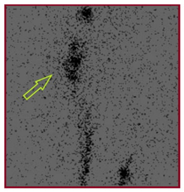
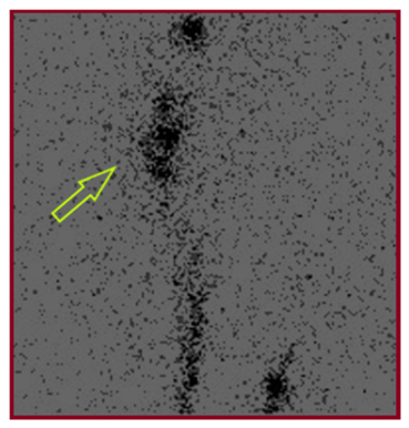
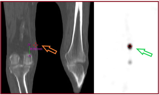
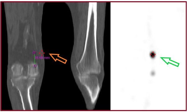
Lymphatic drainage of the left lower extremity was unremarkable. In contrast, the right lower extremity exhibited delayed lymphatic drainage compared to the contralateral side, along with the visualization of collateral lymphatic vessels below the knee. A small, localized area of dermal lymphatic backflow was noted at the mid-portion of the anterior aspect of the right tibia, likely secondary to regional surgical trauma or inflammation. Crucially, foci of increased radiotracer accumulation were detected in the medial region of the right femur distally, which progressively expanded and intensified over time, strongly suggesting an active lymphatic leak (Figure 2). On hybrid SPECT/CT imaging, the dominant focus was localized predominantly within the superficial medial soft tissues, approximately 3.5 cm proximal to the medial femoral condyle and slightly distal to the inferior margin of the surgical scar (Figure 3).
Figure 2.
Lymphoscintigraphy detected foci of increased radiotracer accumulation in the medial region of the right femur, which progressively expanded and intensified over time, strongly suggesting an active lymphatic leak (green arrow).
Figure 3.
Hybrid SPECT/CT imaging showed that focal radiotracer accumulation (green arrow) was precisely localized approximately 3.5 cm above the medial femoral condyle (orange arrow).
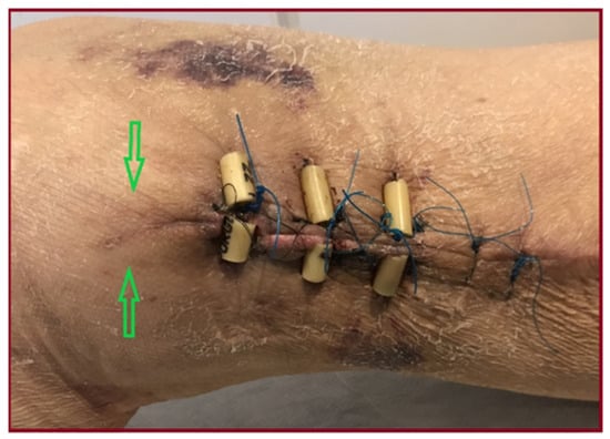
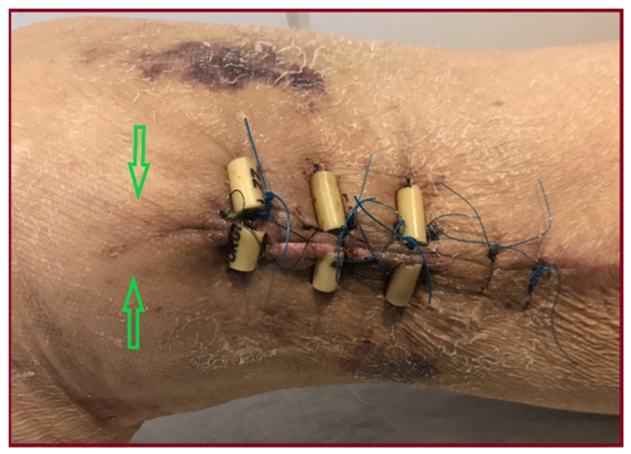
Guided by these imaging findings, a new heavy suture was placed slightly distal to the wound edge to ligate the presumed leaking site within the subcutaneous tissue (Figure 4). Following ligation, the lymphorrhea immediately subsided, and the patient was discharged the following day wearing elastic compression stockings.
Figure 4.
A new heavy suture was placed at the site of presumed lymphatic leakage (green arrows) controlling the lymphorrhea.
Six months postoperatively, the patient underwent percutaneous transluminal tibial angioplasty with stent placement in the right lower extremity due to critical limb ischemia. Four months later, progressive forefoot and heel gangrene necessitated a below-knee amputation. Three years following the initial surgery, the patient succumbed to an acute myocardial infarction.
3. Search Methods
A narrative literature review was conducted to identify published studies on lymphatic complications, particularly lymphatic leakage, following great saphenous vein harvesting for coronary artery bypass grafting (CABG), as well as studies addressing the diagnosis and management of postoperative peripheral lymphatic leakage. PubMed was searched through October 2026 using combinations of the terms “lymphorrhea”, “lymphatic complications”, “lymphatic leakage”, “lymphocele”, “lymphedema”, “vascular surgery”, “great saphenous vein harvesting”, “coronary artery bypass grafting”, “lymphoscintigraphy”, “lymphangiography”, and “SPECT/CT”. Additional relevant publications were obtained through expert recommendations and other sources, including articles available via ScienceDirect and published in Oncotarget. Only English-language publications were considered, including case reports, case series, observational studies, and reviews. Publications were selected based on their relevance to the surgical setting, type of lymphatic complication, diagnostic imaging techniques, and therapeutic management. Recent publications were prioritized, while older studies directly relevant to the present case were retained.
4. Discussion
4.1. Reported Cases
Lymphatic complications following great saphenous vein (GSV) harvesting are recognized postoperative events, although their frequency varies according to the surgical setting and harvesting technique [7]. The relevant cases identified in our literature search can be grouped into three categories according to surgical setting: complications following GSV harvesting for CABG, those following GSV harvesting for other purposes, and peripheral lymphatic complications after other surgical or iatrogenic procedures [3,4,5,6,8,9,10,11,12,13,14,15,16,17,18,19,20,21,22,23,24]. These cases are summarized in Table 1.
Table 1.
Published Cases of Lymphatic Complications Following Saphenous Vein Harvesting and Selected Reports of Peripheral Lymphatic Leakage.
The reported clinical manifestations include external lymphorrhea, lymphocele formation, and lymphedema. Following GSV harvesting, individual reports have described lymphoceles [4,5,8], persistent lymphorrhea [3], and postoperative lymphedema [6]. A recent systematic review and meta-analysis by Razavi et al. [7] reported a significantly higher risk of lymphatic leakage and lymphedema following open vein harvesting (OVH) compared with endoscopic vein harvesting (EVH), whereas no statistically significant difference was observed in lymphocele rates.
Among the reports identified in our literature search, Cha et al. [3] described persistent lymphorrhea following GSV harvesting for CABG, which was localized using pedal lymphangiography and treated with NBCA embolization. In contrast, in the present case, planar lymphoscintigraphy combined with SPECT/CT enabled anatomical localization of the leakage site and guided targeted surgical ligation. Previous reports have also demonstrated the use of lymphoscintigraphy for peripheral lymphatic leakage, as described by Jones et al. [9] following femoral artery injury repair. More recently, Sawamura et al. [12] reported the use of lymphoscintigraphy combined with SPECT/CT to guide treatment of refractory lymphatic leakage following inguinal lymphadenectomy. Together, these reports illustrate different applications of lymphatic imaging in postoperative peripheral leakage, while the present case highlights its role in guiding targeted surgical treatment after GSV harvesting for CABG.
4.2. Pathophysiology
Lymphatic complications following great saphenous vein (GSV) harvesting may result from inadvertent injury to adjacent lymphatic vessels during surgical dissection, owing to their close anatomical relationship with the vein [3,13]. Depending on the extent and consequences of lymphatic disruption, these complications may manifest as external lymphorrhea, characterized by lymphatic fluid drainage through the surgical wound, or as lymphocele formation, involving a localized accumulation of lymphatic fluid. Alternatively, impaired lymphatic drainage may lead to lymphatic stasis and subsequent lymphedema [1,6].
Surgical factors associated with postoperative lymphatic leakage following vascular procedures include lymphatic vessel injury during surgical dissection and reoperation, particularly in the groin region [1,14]. However, the applicability of these findings specifically to GSV harvesting for CABG remains uncertain. Furthermore, diabetes mellitus has been identified as an independent risk factor for surgical site infection following saphenous vein harvesting for CABG [15], although this does not establish a direct association with lymphorrhea. In the present case, lymphatic vessel injury may have occurred during GSV harvesting or subsequent surgical debridement, while the Pseudomonas aeruginosa wound infection may have contributed to local inflammation and impaired wound healing. However, the relative contribution of these factors to the development and persistence of lymphorrhea cannot be established.
If lymphorrhea is not addressed, it may impair wound healing, increase the risk of local wound complications, and prolong postoperative recovery [1,16].
4.3. Diagnosis
When postoperative lymphorrhea is suspected, the diagnostic workup should aim not only to confirm its lymphatic origin but also to identify the disrupted lymphatic pathway and accurately localize the site of leakage. Conventional imaging modalities, including ultrasonography, computed tomography (CT), and magnetic resonance imaging (MRI), can demonstrate postoperative fluid collections and their relationship with adjacent structures. However, they provide limited information regarding lymphatic flow and may therefore be insufficient for identifying the source of an active leak [17,18,19]. Dedicated lymphatic imaging may thus be required, with the choice of modality depending on the anatomical location and whether functional assessment, anatomical localization, or simultaneous therapeutic intervention is intended.
Lymphoscintigraphy is a minimally invasive functional imaging technique based on the intradermal administration of 99mTc-labeled colloids and subsequent assessment of tracer progression through the lymphatic system. This diagnostic modality has been widely utilized for the evaluation of lymphedema [17], including a reported case following GSV harvesting for CABG [6]. However, its diagnostic applications extend beyond lymphedema and include the identification of postoperative lymphatic leakage, as previously described following surgical repair of a femoral artery injury [9]. Compared with conventional lymphangiography, lymphoscintigraphy is less invasive and provides functional information regarding lymphatic drainage. Nevertheless, conventional planar imaging has relatively low spatial resolution and limited anatomical landmarks, which may complicate precise localization of the leakage site. Hybrid SPECT/CT can partially overcome these limitations by combining functional lymphatic information with cross-sectional anatomical imaging, potentially facilitating localization and treatment planning [17,20,21].
Invasive lymphangiographic techniques provide more detailed anatomical visualization and, importantly, may combine diagnosis with treatment. Conventional pedal lymphangiography involves the identification and cannulation of peripheral lymphatic vessels, followed by administration of a radiopaque contrast agent for visualization of the lymphatic system [17,21,22]. Despite providing detailed anatomical information, this technique is invasive, technically demanding, and time-consuming, and entails additional radiation exposure, particularly when visualization of the abdominal lymphatic system is required, and has therefore largely been replaced by less technically demanding access techniques [17,21,22]. Ultrasound- or fluoroscopy-guided intranodal lymphangiography offers an alternative approach to lymphatic access and may facilitate diagnostic evaluation and interventional treatment [17,22].
An important advantage of lymphangiography is its potential to transition directly from diagnosis to treatment, most commonly through embolization using agents such as N-butyl cyanoacrylate (NBCA), as demonstrated in selected cases of postoperative peripheral lymphorrhea [3,10,22]. Nevertheless, these procedures involve additional technical requirements and potential procedure-related complications [22].
While planar lymphoscintigraphy can demonstrate abnormal radiotracer accumulation suggestive of lymphatic leakage, its limited anatomical resolution may restrict precise localization. By integrating functional tracer distribution with anatomical information, SPECT/CT may provide additional guidance for targeted management. In the present case, planar lymphoscintigraphy demonstrated active lymphatic leakage, while hybrid SPECT/CT localized the area of abnormal tracer accumulation approximately 3.5 cm above the medial femoral condyle, providing anatomical information that guided subsequent surgical ligation.
Other approaches are particularly suited to superficial lymphatic leaks requiring operative exploration. Vital dyes such as isosulfan blue and, more recently, near-infrared fluorescence lymphography with indocyanine green (ICG) enable direct intraoperative visualization of leaking superficial lymphatic channels and have been used to identify lymphatic leakage following vascular procedures [13,21,23]. ICG provides real-time, high-resolution imaging without ionizing radiation, although its limited tissue penetration restricts visualization of deeper lymphatics [21,23]. Contrast-enhanced ultrasound (CEUS) lymphography has also been described for localization of lower-extremity lymphatic abnormalities before microsurgical treatment [24]. Magnetic resonance lymphangiography represents another non-ionizing option, providing anatomical assessment of peripheral lymphatic structures, although motion and metallic artifacts, limited visualization of very small lymphatics, and reduced functional information with some protocols remain important limitations [21,23,25].
The main clinical applications, advantages and limitations of the imaging modalities discussed above are summarized in Table 2.
Table 2.
Comparison of imaging modalities used for the diagnosis and localization of lymphatic abnormalities and postoperative lymphatic leakage.
4.4. Comparison
Accordingly, the available lymphatic imaging techniques appear to serve complementary rather than interchangeable roles. Interventional lymphangiography is particularly valuable for postoperative lymphatic leaks when simultaneous embolization is anticipated [10,12,18,22], whereas vital-dye and ICG mapping are particularly suited to real-time intraoperative identification of superficial leaks [13,21,23]. Lymphoscintigraphy primarily provides functional assessment of lymphatic drainage and has been used in lower-extremity lymphatic disorders and after vascular procedures [6,9,17,21]. In the present case, however, planar lymphoscintigraphy demonstrated active lymphatic leakage, while the addition of SPECT/CT provided anatomical localization of the suspected leakage region following GSV harvesting for CABG. This localization was used to guide targeted surgical ligation, resulting in immediate cessation of the external lymphatic leakage. Therefore, the present case illustrates the potential clinical utility of combining functional and anatomical imaging to guide surgical management of persistent peripheral lymphorrhea.
4.5. Management
Management of postoperative lymphorrhea ranges from conservative measures to interventional and surgical approaches, with treatment selection depending on the severity, persistence, and anatomical location of the leakage. Conservative management, including compression or pressure dressings, limb elevation, drainage, and, in selected settings, sclerotherapy, is frequently considered initially. However, persistent or high-output leakage may warrant more invasive treatment [14,26]. Surgical options include direct ligation of the disrupted lymphatic vessel, potentially aided by intraoperative leakage localization techniques or combined with flap coverage in selected cases [27,28]. Other approaches, including negative-pressure wound therapy and lymphaticovenular anastomosis, have been employed in selected refractory cases [8,14,26]. More recently, Tsutsumi et al. [29] described successful treatment of refractory inguinal lymphorrhea using ICG-guided lymphatic ligation combined with negative-pressure wound therapy. Interventional lymphangiography followed by NBCA embolization represents an alternative approach that combines localization with treatment of the leakage [3,10,30]. However, selective embolization is important to minimize potential disruption of normal lymphatic drainage, as highlighted by Tsukada et al. [31]. In the present case, an initial two-week trial of compression therapy failed to control the persistent lymphorrhea. However, precise localization of the disrupted lymphatic pathway by lymphoscintigraphy and SPECT/CT subsequently enabled targeted surgical ligation, resulting in immediate cessation of the leakage. The patient was discharged the following day with compression stockings.
4.6. Limitations
This report has several limitations. First, it describes a single patient, limiting the generalizability of the findings. The severity of lymphorrhea was assessed qualitatively, without quantitative measurement of lymphatic fluid output. Furthermore, although immediate cessation of leakage was observed following surgical ligation, long-term recurrence could not be reliably assessed because the duration of documented follow-up was not clearly established. SPECT/CT localized the region of suspected lymphatic leakage but did not directly confirm the specific injured lymphatic vessel. Additionally, no direct comparison with alternative imaging modalities or treatment strategies was performed. Therefore, this case cannot establish the diagnostic accuracy, comparative superiority, recurrence rates, or cost-effectiveness of SPECT/CT-guided management.
5. Conclusions
Postoperative lymphorrhea following great saphenous vein harvesting for CABG is a rare but potentially challenging complication. This case highlights the potential clinical usefulness of lymphoscintigraphy combined with SPECT/CT for providing anatomical localization of the suspected lymphatic leakage region, particularly when clinical identification is difficult. In the present patient, this anatomical information guided targeted surgical ligation, which was followed by immediate cessation of external lymphorrhea.
Author Contributions
Conceptualization: L.T., C.S., T.S., K.T., C.P. and F.M.; methodology, K.N., M.D., V.A. and Z.P.; writing—original draft preparation, L.T., C.S., T.S., K.T., C.P. and F.M.; writing—review and editing, N.C., S.P., K.N., M.D., V.A. and Z.P.; supervision, F.M., N.C., and S.P. All authors have read and agreed to the published version of the manuscript.
Funding
This research received no external funding.
Institutional Review Board Statement
According to the applicable Greek legislation, including Law 3418/2005 (Code of Medical Ethics), particularly Articles 12 and 13, and the General Data Protection Regulation (EU) 2016/679, patient confidentiality and privacy were strictly respected. As this manuscript describes a single clinical case and does not constitute an interventional clinical study, formal approval by an Ethics Committee was not required, as confirmed by the relevant institutional procedures.
Informed Consent Statement
Written informed consent has been obtained from the patient to publish this paper.
Data Availability Statement
The data presented in this study are available on request from the corresponding author due to patient privacy and confidentiality.
Acknowledgments
We gratefully acknowledge the Department of Vascular Surgery at the University Hospital of Patras and the University of Patras for providing the facilities and resources necessary for this research.
Conflicts of Interest
The authors declare no conflicts of interest.
References
- Lv, S.; Wang, Q.; Zhao, W.; Han, L.; Wang, Q.; Batchu, N.; Ulain, Q.; Zou, J.; Sun, C.; Du, J.; et al. A review of the postoperative lymphatic leakage. Oncotarget 2017, 8, 69062–69075. [Google Scholar] [CrossRef] [Scilit] [PubMed]
- Unno, N.; Yamamoto, N.; Suzuki, M.; Tanaka, H.; Mano, Y.; Sano, M.; Saito, T.; Sugisawa, R.; Konno, H. Intraoperative lymph mapping with preoperative vein mapping to prevent postoperative lymphorrhea following paramalleolar bypass surgery in patients with critical limb ischemia. Surg. Today 2014, 44, 436–442. [Google Scholar] [CrossRef] [Scilit] [PubMed]
- Cha, J.G.; Lee, S.Y.; Hong, J.; Ryeom, H.K.; Kim, G.C.; Do, Y.W. Transpedal lymphatic embolization for lymphorrhea at the graft harvest site after coronary artery bypass grafting. Yeungnam Univ. J. Med. 2021, 38, 74–77. [Google Scholar] [CrossRef] [Scilit] [PubMed] [PubMed Central]
- Deshmukh, N. Treatment of persistent lymphocele following saphenous vein harvesting: A case report. Vasc. Surg. 1986, 20, 116–119. [Google Scholar] [CrossRef] [Scilit]
- Canders, C.P.; Nguyen, P.D.; Festekjian, J.H.; Rudkin, G.H. CASE REPORT Treatment of a Lower Extremity Lymphocele With Intraoperative Lymphatic Mapping. Eplasty 2013, 13, e57. [Google Scholar] [PubMed] [PubMed Central]
- Mirhosseini, S.M.; Soltanipur, M.; Yarmohammadi, H.; Rezaei, M.; Sheikhi, Z. Lymphedema after saphenous harvesting for coronary artery bypass surgery: Case report and literature review. BMC Cardiovasc Disord. 2024, 24, 41. [Google Scholar] [CrossRef] [Scilit] [PubMed] [PubMed Central]
- Razavi, A.; Mahalleh, M.; Vanaki, A.; Fallah, A.; Bahanesteh, A.; Ahmadi, A.; Yarmohammadi, H.; Solati Kooshkqazi, M.; Soltanipur, M. Lymphatic complication in open venous harvesting versus endoscopic venous harvesting: A systematic review and meta-analysis. Gen. Thorac. Cardiovasc Surg. 2025, 73, 297–311. [Google Scholar] [CrossRef] [Scilit] [PubMed]
- Imai, H.; Yoshida, S.; Mese, T.; Roh, S.; Koshima, I. Supermicrosurgical lymphatic venous anastomosis for intractable lymphocele after great saphenous vein harvesting graft. J. Vasc. Surg. Cases Innov. Tech. 2022, 8, 45–47. [Google Scholar] [CrossRef] [Scilit] [PubMed] [PubMed Central]
- Jones, T.R.; Carlisle, M.R.; Hofmann, L.V.; Strauss, H.W.; Olcott, C., 4th. Lymphoscintigraphy in the diagnosis of lymphatic leak after surgical repair of femoral artery injury. Clin. Nucl. Med. 2001, 26, 14–17. [Google Scholar] [CrossRef] [Scilit] [PubMed]
- Dung, P.V.; Cuong, N.N.; Quang, T.D.; Canh, P.H.; Linh, L.T.; Duc, N.M. Combination of Lymph Node Embolization and Musculocutaneous Flap Operation for Managing Groin Lymphorrhea. Ann. Vasc. Dis. 2021, 14, 267–269. [Google Scholar] [CrossRef] [Scilit] [PubMed] [PubMed Central]
- Velanovich, V.; Mallory, P.; Collins, P.S. Lower extremity lymphocele development after saphenous vein harvesting. Mil. Med. 1991, 156, 149–150. [Google Scholar] [CrossRef] [Scilit] [PubMed]
- Sawamura, H.; Kimura, H.; Kou, R.; Nishida, A.; Kawase, K.; Hatsuda, N.; Sato, T.; Shimada, K.; Fujiwara, K.; Kanagaki, M. Direct puncture and NBCA embolization for refractory lymphatic leakage after inguinal lymphadenectomy in the absence of puncturable lymph nodes guided by lymphoscintigraphy. Radiol. Case Rep. 2026, 21, 3430–3435. [Google Scholar] [CrossRef] [Scilit] [PubMed] [PubMed Central]
- Blebea, J.; Choudry, R. Thigh isosulfan blue injection in the treatment of postoperative lymphatic complications. J. Vasc. Surg. 1999, 30, 350–354. [Google Scholar] [CrossRef] [Scilit] [PubMed]
- Nicksic, P.J.; Condit, K.M.; Nayar, H.S.; Michelotti, B.F. Algorithmic approach to the lymphatic leak after vascular reconstruction: A systematic review. Arch. Plast. Surg. 2021, 48, 404–409. [Google Scholar] [CrossRef] [Scilit] [PubMed] [PubMed Central]
- Unosson, H.; Nyman, M.H.; Brynhildsen, K.F.; Friberg, Ö. Risk factors for infection at the saphenous vein harvest site after coronary artery bypass grafting surgery: A retrospective cohort study. J. Cardiothorac. Surg. 2024, 19, 310. [Google Scholar] [CrossRef] [Scilit] [PubMed] [PubMed Central]
- Reifsnyder, T.; Bandyk, D.; Seabrook, G.; Kinney, E.; Towne, J.B. Wound complications of the in situ saphenous vein bypass technique. J. Vasc. Surg. 1992, 15, 843–848; discussion 848–850. [Google Scholar] [CrossRef] [Scilit] [PubMed]
- Schwartz, F.R.; James, O.; Kuo, P.H.; Witte, M.H.; Koweek, L.M.; Pabon-Ramos, W.M. Lymphatic Imaging: Current Noninvasive and Invasive Techniques. Semin Interv. Radiol. 2020, 37, 237–249. [Google Scholar] [CrossRef] [Scilit] [PubMed] [PubMed Central]
- Santos, E.; Moussa, A.M. Chylous Ascites and Lymphoceles: Evaluation and Interventions. Semin Interv. Radiol. 2020, 37, 274–284. [Google Scholar] [CrossRef] [Scilit] [PubMed] [PubMed Central]
- Hur, S.; Jun, H.; Jeong, Y.S. Novel interventional radiological management for lymphatic leakages after gynecologic surgery: Lymphangiography and embolization. Gland Surg. 2021, 10, 1260–1267. [Google Scholar] [CrossRef] [Scilit] [PubMed] [PubMed Central]
- Basavalingu, D.; Elojeimy, S.; Mercer, M.; Itani, M.; Revels, J.W.; Blacklock, L.C.; Zuckier, L.S.; Wang, S.S.; Kolokythas, O.; Behnia, F. Nuclear Medicine Lymphoscintigraphy: Applications and Technical Overview. RadioGraphics 2025, 45, e240207. [Google Scholar] [CrossRef] [Scilit] [PubMed]
- Munn, L.L.; Padera, T.P. Imaging the lymphatic system. Microvasc. Res. 2014, 96, 55–63. [Google Scholar] [CrossRef] [Scilit] [PubMed] [PubMed Central]
- Gómez, F.M.; Baetens, T.R.; Santos, E.; Rocha, B.L.; Horwitz, B.; Lojo-Lendoiro, S.; Vargas, P.; Patel, P.; Beets-Tan, R.; Martínez-Rodrigo, J.J.; et al. Interventional solutions for post-surgical problems: A lymphatic leaks review. CVIR Endovasc. 2024, 7, 61, Erratum in CVIR Endovasc. 2024, 7, 68. https://doi.org/10.1186/s42155-024-00483-1. [Google Scholar] [CrossRef] [Scilit] [PubMed] [PubMed Central]
- Burgard, M.; Cherbanyk, F.; Psathas, E. Indocyanine green lymphangiography for the management of lymphatic complications after vascular surgery. Ann. Vasc. Surg. Brief. Rep. Innov. 2023, 3, 100205. [Google Scholar] [CrossRef] [Scilit]
- Zhu, J.; Yu, X.; Zhang, L.; Liu, Z. Lymphography using microbubbles and contrast-enhanced ultrasound (CEUS) before lymphatic supermicrosurgery for severe lymphorrhea of lower extremity. J. Plast. Reconstr. Aesthet. Surg. 2023, 84, 537–539. [Google Scholar] [CrossRef] [Scilit] [PubMed]
- Pieper, C.C. Nodal and Pedal MR Lymphangiography of the Central Lymphatic System: Techniques and Applications. Semin Interv. Radiol. 2020, 37, 250–262. [Google Scholar] [CrossRef] [Scilit] [PubMed] [PubMed Central]
- Cheong, Y.K.; Jun, H.; Cho, Y.P.; Song, G.W.; Moon, K.M.; Kwon, T.W.; Lee, S.G. Negative pressure wound therapy for inguinal lymphatic complications in critically ill patients. J. Korean Surg. Soc. 2013, 85, 134–138. [Google Scholar] [CrossRef] [Scilit] [PubMed] [PubMed Central]
- Sotsuka, Y.; Wakatsuki, H.; Matsuda, K. Schlieren Phenomenon for Identification of Lymphorrhea Point. Plast. Reconstr. Surg. Glob. Open 2022, 10, e4269. [Google Scholar] [CrossRef] [Scilit] [PubMed] [PubMed Central]
- Shermak, M.A.; Yee, K.; Wong, L.; Jones, C.E.; Wong, J. Surgical management of groin lymphatic complications after arterial bypass surgery. Plast. Reconstr. Surg. 2005, 115, 1954–1962. [Google Scholar] [CrossRef] [Scilit] [PubMed]
- Tsutsumi, A.; Suzuki, Y.; Takayanagi, R.; Takada, K.; Kato, T.; Kishi, K. Surgical Strategy for Intractable Inguinal Lymphorrhea Using Combined Indocyanine Green Imaging and Negative Pressure Wound Therapy. Plast. Reconstr. Surg. Glob. Open 2026, 14, e7938. [Google Scholar] [CrossRef] [Scilit] [PubMed] [PubMed Central]
- Hur, S.; Shin, J.H.; Lee, I.J.; Min, S.K.; Min, S.I.; Ahn, S.; Kim, J.; Kim, S.Y.; Kim, M.; Lee, M.; et al. Early Experience in the Management of Postoperative Lymphatic Leakage Using Lipiodol Lymphangiography and Adjunctive Glue Embolization. J. Vasc. Interv. Radiol. 2016, 27, 1177–1186.e1, Erratum in J. Vasc. Interv. Radiol. 2016, 27, 1627. [Google Scholar] [CrossRef] [Scilit] [PubMed]
- Tsukada, J.; Inoue, M.; Tamura, M.; Torikai, H.; Yoshikawa, H.; Miyabayashi, M.; Tsuzaki, J.; Kogo, T.; Furukawa, Y.; Ogawa, R.; et al. Incidence and Clinical Course of Lower-Limb Lymphedema after Intranodal n-Butyl-2-Cyanoacrylate Embolization for Postoperative Lymphorrhea. J. Vasc. Interv. Radiol. 2026, 37, 108541. [Google Scholar] [CrossRef] [Scilit] [PubMed]

Disclaimer/Publisher’s Note: The statements, opinions and data contained in all publications are solely those of the individual author(s) and contributor(s) and not of MDPI and/or the editor(s). MDPI and/or the editor(s) disclaim responsibility for any injury to people or property resulting from any ideas, methods, instructions or products referred to in the content. |
© 2026 by the authors. Licensee MDPI, Basel, Switzerland. This article is an open access article distributed under the terms and conditions of the Creative Commons Attribution (CC BY) license.