


Abstract
Artificial intelligence (AI) is moving from stand-alone accuracy studies to prospective deployment across breast screening, risk prediction, multimodal diagnosis, treatment response, and workflow support. The goal of this narrative review is to determine which AI applications have demonstrated clinical benefit, which remain technically promising, and what safeguards are required for implementation. We synthesize randomized trials, prospective cohorts, real-world implementations, external validations, systematic reviews, and guidance identified in PubMed/MEDLINE and Scopus through 30 June 2026. In screening, AI-supported mammography can increase cancer detection and reduce reading workload, but effects depend on whether AI is used as concurrent support, an independent reader, triage, or partial autonomy; autonomous workflows may shift work and cost downstream through additional recalls. MASAI met its prespecified noninferiority criterion for interval cancer, but this should not be interpreted as proof of equivalence or mortality benefit. Image-based risk models often discriminate better than density or conventional clinical models, although prospective benefit from AI-guided allocation to supplemental imaging has not yet been demonstrated. Evidence for contrast-enhanced mammography (CEM), MRI, ultrasound, radiomics, and multimodal models is less mature and limited by external validation, domain shift, missing data, operator dependence, and uncertain incremental value beyond clinicopathological factors. Ungrounded large language models and opaque foundation models are not suitable for autonomous clinical advice. Current evidence therefore supports controlled, task-specific integration rather than radiologist replacement; real clinical benefit depends on governance of the whole AI-enabled pathway, including local validation, human factors, downstream resource use, statistical stopping rules, and fallback procedures.
1. Introduction
About 2.3 million women were diagnosed with breast cancer in 2022, and roughly 670,000 died of it [1]. Behind these numbers lies a chain of decisions in which imaging takes part at almost every step: whether and when a woman should be screened, with which test, whether a finding deserves recall or biopsy, how far the disease extends, and whether treatment is working. Organized programs keep this chain manageable by grouping women by age and, at most, a handful of risk factors. The approach is simple and operationally sound, but it treats very different women, and very different tumors, as if they were interchangeable. European and Italian guidance is now moving toward adapting age range, screening interval, and diagnostic work-up to individual risk and to local resources [2,3,4].
Breast imaging is fertile ground for AI because it combines large volumes of highly standardized examinations with rare and difficult decisions. Screening mammography and digital breast tomosynthesis (DBT) pose a demanding signal-detection task: cancers are uncommon, subtle signs hide in a wide range of normal appearances, and a small loss of specificity translates into thousands of additional false-positive recalls. Not surprisingly, the first wave of research focused on lesion detection and malignancy scoring [5,6]. What has changed since is the range of jobs AI is asked to do. It may now flag an abnormality on today’s examination, decide how much human reading an examination receives, estimate future risk from a mammogram that was read as normal, characterize tumor biology, or combine imaging with clinical and molecular data [7,8]. These tasks are related, but each has its own reference standard, time horizon, failure modes, and regulatory claim (Figure 1).
Figure 1.
Artificial intelligence applications across the breast imaging pathway. Shading denotes four evidence-maturity categories: clinical effectiveness, external or operational validation, technical or clinical validity, and feasibility. CEM, contrast-enhanced mammography; DBT, digital breast tomosynthesis; LLM, large language model; MRI, magnetic resonance imaging; pCR, pathological complete response; US, ultrasound.
The evidence has matured from retrospective stand-alone comparisons to randomized trials and national implementations. Retrospective studies establish technical plausibility but cannot determine how radiologists behave when AI is present, whether a selected operating point survives routine practice, or whether additional cancers detected today translate into fewer interval or advanced cancers rather than more overdiagnosis. Prospective studies now report workload, recall, tumor characteristics, and interval cancers, while trials of risk-based screening and supplemental imaging clarify which clinical actions a risk estimate might trigger. This review therefore treats AI as a family of pathway interventions rather than a single technology and evaluates each intended use against its own reference standard, time horizon, benefits, and harms.
Review Aim and Research Questions
The explicit goal of this narrative review is to critically synthesize the maturity, clinical utility, limitations, and implementation requirements of AI across the breast imaging pathway. We address the following research questions (RQs):
RQ1. Across concurrent support, independent-reader, triage, and partially autonomous workflows, what clinical benefits and harms have been demonstrated for AI-supported mammography and DBT?
RQ2. How mature is the evidence for AI-derived risk prediction and AI-guided personalized screening, and how should biological risk be separated from density-related masking?
RQ3. What incremental clinical value and limitations do AI applications in CEM, MRI, ultrasound, multimodal imaging, and radiomics offer beyond expert interpretation and established clinicopathological factors?
RQ4. What safety risks arise from generative AI, foundation models, and human–AI interaction, including automation bias across reader experience levels?
RQ5. Which technical, economic, operational, equity, monitoring, stopping, and fallback requirements are necessary for safe deployment?
RQ6. Does the current evidence support task-specific integration or complete radiologist replacement?
2. Methods: Evidence Identification and Appraisal
2.1. Search Strategy
We searched PubMed/MEDLINE and Scopus from 1 January 2015 through 30 June 2026. Older landmark studies were retained when identified through backward citation tracking from major reviews, guidelines, and pivotal trials. Searches were limited to human studies with English-language full text. No geographic restriction was applied. The search was rerun for the revision using the following database-specific strings and filters.
PubMed/MEDLINE: ((“Breast Neoplasms”[Mesh] OR “breast cancer”[tiab] OR “breast cancers”[tiab] OR “breast neoplasm”[tiab] OR “breast neoplasms”[tiab]) AND (“Mammography”[Mesh] OR mammograph*[tiab] OR “digital breast tomosynthesis”[tiab] OR DBT[tiab] OR “contrast-enhanced mammography”[tiab] OR CEM[tiab] OR “Magnetic Resonance Imaging”[Mesh] OR “breast MRI”[tiab] OR “magnetic resonance imaging”[tiab] OR “Ultrasonography, Mammary”[Mesh] OR ultrasound[tiab] OR ultrasonograph*[tiab]) AND (“Artificial Intelligence”[Mesh] OR “Machine Learning”[Mesh] OR “artificial intelligence”[tiab] OR “machine learning”[tiab] OR “deep learning”[tiab] OR radiomic*[tiab] OR “foundation model”[tiab] OR “foundation models”[tiab] OR “large language model”[tiab] OR “large language models”[tiab] OR “generative AI”[tiab]) AND (screen*[tiab] OR detect*[tiab] OR diagnos*[tiab] OR risk[tiab] OR “interval cancer”[tiab] OR “interval cancers”[tiab] OR staging[tiab] OR “treatment response”[tiab] OR explainab*[tiab] OR implementation[tiab])) AND (“2015/01/01”[Date-Publication]: “2026/06/30”[Date-Publication]) AND English[lang] AND Humans[Mesh].
Scopus: TITLE-ABS-KEY((“breast cancer” OR “breast neoplasm*”) AND (mammograph* OR “digital breast tomosynthesis” OR DBT OR “contrast-enhanced mammography” OR CEM OR “breast MRI” OR “magnetic resonance imaging” OR ultrasound OR ultrasonograph*) AND (“artificial intelligence” OR “machine learning” OR “deep learning” OR radiomic* OR “foundation model*” OR “large language model*” OR “generative AI”) AND (screen* OR detect* OR diagnos* OR risk OR “interval cancer*” OR staging OR “treatment response” OR explainab* OR implementation)) AND PUBYEAR > 2014 AND PUBYEAR < 2027 AND LIMIT-TO(LANGUAGE, “English”) AND (LIMIT-TO(DOCTYPE, “ar”) OR LIMIT-TO(DOCTYPE, “re”)). Because the PUBYEAR field applies a year-level rather than a da y-level restriction, all records assigned to 2026 were exported and records with an online or issue publication date after 30 June 2026 were excluded before screening; this standardized the Scopus window to the PubMed/MEDLINE cut-off.
2.2. Eligibility, Study Selection, and Prioritization
We included human breast-imaging studies evaluating AI, machine learning, deep learning, radiomics, generative AI, or foundation models for screening, risk prediction, diagnosis, staging, treatment response, reporting, workflow, or implementation. Eligible designs comprised randomized trials, prospective interventional or observational cohorts, routine implementations, paired-reader and reader studies, retrospective studies with external validation, systematic reviews or meta-analyses, and consensus, regulatory, or reporting guidance. For emerging modalities, early retrospective studies were retained when they introduced a clinically relevant application or failure mode. We excluded duplicate reports, non-English full texts, conference abstracts or proceedings not subsequently published as a full peer-reviewed journal article, preprints, case reports and very small case series, animal- or phantom-only work, and purely technical studies without a clinically interpretable endpoint. When multiple reports described the same cohort, the most complete and current report was prioritized, while complementary reports were retained for distinct endpoints such as interval cancer or tumor characteristics.
Conference abstracts and proceedings were excluded because this review was designed to prioritize mature clinical evidence: peer-reviewed full reports with sufficiently described populations, workflows, reference standards, follow-up, and patient- or service-relevant outcomes, rather than early proof-of-concept algorithmic benchmarks. We acknowledge that this choice may omit technically influential work first presented at MICCAI, IEEE ISBI, CVPR, SPIE, or similar meetings and may introduce time-lag and publication bias. Conference-originated methods were eligible when subsequently reported in an otherwise eligible full journal article.
Study identification and selection were iterative, combining database searches with backward citation tracking and targeted updating. Because this narrative review was not prospectively conducted as a systematic review, deduplication and screening counts were not logged in a PRISMA-compatible form. We therefore did not retrospectively reconstruct a flow diagram, which could imply a level of completeness and auditability not supported by the review process. Reproducibility is instead supported by the complete database strings, explicit eligibility criteria, evidence hierarchy, and design-specific appraisal; the absence of a PRISMA flow is acknowledged as a methodological limitation.
Evidence was prioritized as follows: randomized and prospective interventional studies; large routine implementations; prospective observational, silent-deployment, or reader studies; independent external validations; systematic reviews and meta-analyses; and consensus or reporting guidance. Older retrospective studies were used selectively for foundational concepts. Study inclusion was guided by relevance to the RQs, design strength, sample size, external validity, completeness of follow-up, and whether the study reported both benefits and harms.
2.3. Evidence Appraisal and Narrative Synthesis
Because this narrative review spans heterogeneous interventions, reference standards, endpoints, and study designs, we did not apply a single formal risk-of-bias instrument or pool estimates. A single score would have obscured design-specific threats rather than made the evidence more comparable. Narrative synthesis bias was mitigated by using the prespecified RQs and evidence hierarchy above; separating randomized interventional studies, prospective routine implementations, silent deployments, simulations, reader studies, and retrospective validations; checking the AI role, operating threshold, denominator, reference standard, follow-up, and comparator in each study; reporting confidence intervals and noninferiority criteria when available; and discussing recalls, false negatives, downstream procedures, overdiagnosis, workload, equity, and technical failure alongside detection performance.
For Figure 1 and Table 1, evidence maturity was assigned to the highest level demonstrated for the intended clinical workflow: clinical effectiveness; external or operational validation; technical or clinical validity; or feasibility. Technical validation means that the model performs on a defined dataset or reader experiment. Clinical effectiveness requires prospective evidence that introducing the model into a specified pathway changes patient- or service-relevant outcomes. These categories are descriptive rather than formal grades of certainty.
Table 1.
Artificial intelligence use cases across the breast imaging pathway.
2.4. Generative AI Use in Manuscript Preparation
During preparation and revision, generative AI tools were used to support language editing, structural reorganization, preparation of Figure 1 and Figure 2, and reference-status checking. Figure 1 and Figure 2 are author-curated conceptual schematics; they do not contain patient images, synthetic clinical data, or generated study results. The authors manually verified all labels, relationships, and evidence categories. No primary data were generated or analyzed by these tools, and no tool made final decisions on study eligibility, evidence extraction, scientific interpretation, or conclusions.
3. AI-Supported Screening: Roles, Prospective Evidence, and Trade-Offs
3.1. AI Roles and Workflow Dependence
Traditional computer-aided detection (CAD) was cleared, reimbursed, and widely adopted in the United States in the 2000s, yet large studies found that it lowered specificity and increased recalls without improving the detection of invasive cancer [9,10]. CAD mostly added marks that readers had to dismiss, and the price was paid in false positives. Deep learning differs from that generation of tools in three practical ways: it learns its own image representations, it improves with more data, and it produces examination-level malignancy scores that can drive workflow decisions as well as marks on an image.
Over the past decade, retrospective work showed that modern systems can match radiologists in selected settings: against more than 100 readers, on international test sets, in multireader studies, and in head-to-head external comparisons of commercial products, while combined human–AI reading often outperformed both [11,12,13,14,15,16,17]. Single-center work, including our own, reported high stand-alone performance on specific findings: on 1986 mammograms, convolutional networks detected microcalcifications with an area under the receiver operating characteristic curve (AUC) of 0.98 and separated benign from malignant groups with AUCs up to 0.94 [18]. In DBT, AI support shortened reading time while maintaining or improving accuracy [19]. These studies established that the image holds a learnable signal. They did not license a universal replacement claim. Performance depends on prevalence, vendor, population, the availability of prior examinations, reading rules, and, above all, the operating threshold chosen for a given workflow.
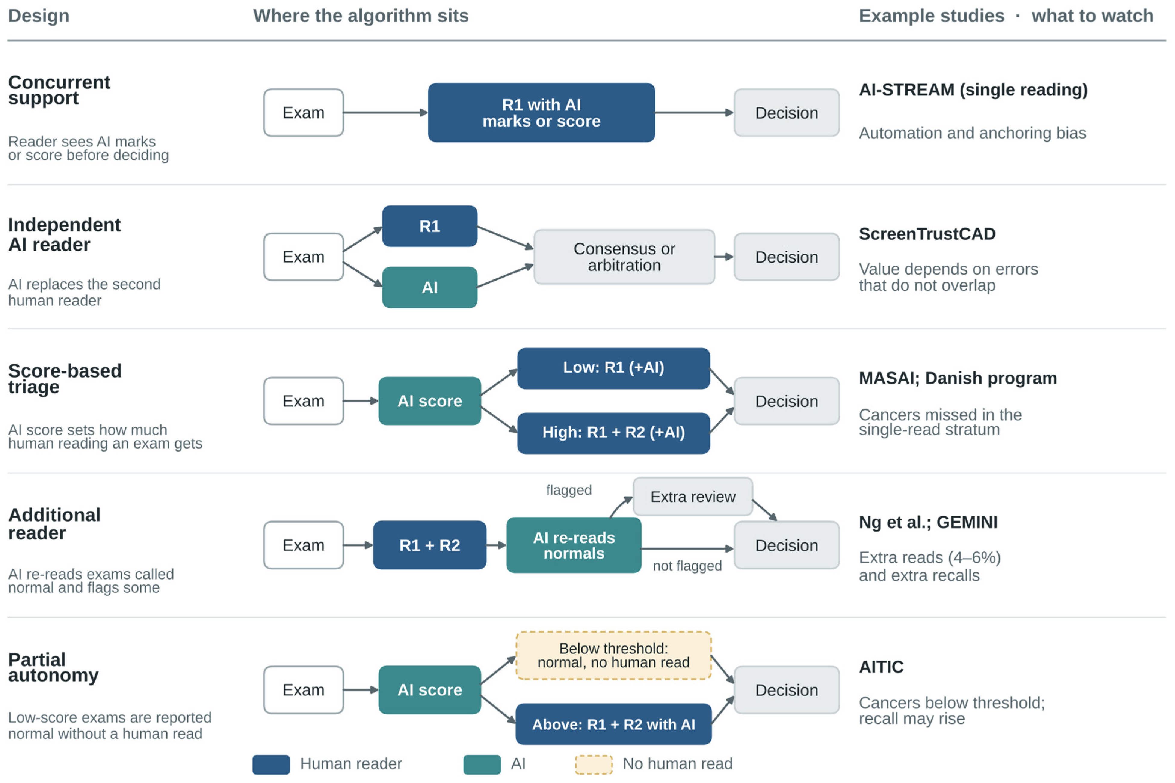
The label “AI-assisted screening” covers quite different interventions, as shown in Figure 2. In concurrent decision support, the radiologist sees marks or a score before deciding. In triage, low-score examinations go to single reading and high-score examinations to double reading or arbitration. In an additional-reader model, AI reviews examinations already read by humans and sends selected cases, typically those both readers called normal, back for review. In partially autonomous screening, a prespecified low-risk fraction is reported as normal without any human reading. Each design changes the number of reads, and each also changes how human and machine errors depend on each other. Two radiologists trained in the same way and limited by the same perceptual blind spots tend to make correlated errors; an AI reader adds value precisely when its errors fall elsewhere. Showing AI output before the reader has formed an opinion can have the opposite effect, aligning human errors with machine errors through automation and anchoring bias.
This taxonomy should be stated explicitly in every study and in every procurement decision. For instance, a system cleared for concurrent support cannot be assumed safe for autonomous triage. A threshold that maximizes the AUC may not meet a program’s noninferiority margin for recall or interval cancer. Nor does an examination-level score say whether the actionable lesion has been correctly localized; when AI is meant to guide human action rather than simply classify an examination, breast- and lesion-level analyses are needed. Table 1 summarizes the main use cases along the pathway, the endpoints by which each should be judged, and their typical failure modes.
Figure 2.
Five ways of putting artificial intelligence (AI) into a screening workflow. The designs differ in how many human reads each examination receives and in how human and machine errors depend on each other. Representative prospective studies are given for each design (AI-STREAM [20]; ScreenTrustCAD [21]; MASAI [22,23] and the Danish program [24]; Ng et al. [25] and GEMINI [26]; AITIC [27]), together with the main point to watch. R1 and R2, first and second human readers.
3.2. Prospective and Real-World Screening Evidence
3.2.1. From the Test Set into the Program
The first prospective studies moved AI out of enriched test sets and into routine populations (Table 2). The initial MASAI randomized safety analysis used AI both to triage examinations to single or double reading and to support detection; cancer detection increased and screen-reading workload fell by about 44%, without a significant excess of false-positive recalls [22]. ScreenTrustCAD was a prospective population-based paired-reader noninferiority study rather than a randomized live-workflow trial; one radiologist plus AI was noninferior to standard double reading for cancer detection [21]. The Hungarian additional-reader study was a prospective routine implementation in which AI re-reviewed examinations called normal by humans, identifying 0.7–1.6 additional cancers per 1000 examinations at the price of 4–6% more reading [25]. The Danish report was an observational routine implementation showing higher detection, fewer false positives, and a one-third reduction in reading workload [24]. These designs answer different questions and should not be treated as interchangeable.
Table 2.
Selected prospective and real-world studies of AI-supported mammography screening.
3.2.2. Scale and Diversity: PRAIM and AI-STREAM
The evidence grew in scale and geographic diversity in 2025. PRAIM, a prospective real-world observational implementation covering more than 460,000 women at 12 German sites, reported a CDR of 6.7 per 1000 with AI-supported reading versus 5.7 per 1000 without AI, a relative increase of 17.6% (95% confidence interval [CI], 5.7–30.8%), with similar recall [29]. AI-STREAM, a prospective multicenter cohort of 24,543 women in South Korea’s single-reading setting, reported CDRs of 5.70 (95% CI, 4.76–6.65) and 5.01 (95% CI, 4.13–5.89) per 1000 with and without AI, respectively, while recall was 4.53% versus 4.48% (p = 0.56) [20]. These results support concurrent assistance in single-reading programs, but they cannot be transferred mechanically to double-reading programs, where the comparator and built-in redundancy differ.
3.2.3. MASAI: More Cancers, Less Reading, and Noninferior Interval-Cancer Rates
The protocol-defined MASAI performance analysis included 105,915 women. AI-supported screening detected 6.4 cancers per 1000 (95% CI, 5.7–7.1) versus 5.0 per 1000 (95% CI, 4.4–5.6) with standard double reading, corresponding to a detection-rate ratio of 1.29 (95% CI, 1.09–1.51); workload fell by 44.2% and false-positive recall did not increase significantly [23]. The interval-cancer analysis reported 1.55 (95% CI, 1.23–1.92) versus 1.76 (95% CI, 1.42–2.15) interval cancers per 1000, with a rate ratio of 0.88 (95% CI, 0.65–1.18), meeting the prespecified 20% noninferiority margin. Sensitivity was 80.5% versus 73.8%, and specificity was 98.5% in both arms [28]. This establishes noninferiority for the stated margin; it does not establish exact equivalence, exclude smaller differences, demonstrate a mortality benefit, or resolve overdiagnosis.
The clinical meaning of additional detection depends on tumor type. Detecting additional small, node-negative invasive cancers may represent a favorable stage shift and could reduce treatment intensity, but this requires confirmation through stage distribution, interval-cancer phenotype, therapy, recurrence, and mortality follow-up. Additional ductal carcinoma in situ (DCIS) has a less certain net effect because DCIS is biologically heterogeneous and is more vulnerable to overdiagnosis and overtreatment. Trials should therefore report invasive cancer and DCIS separately, including invasive size and nodal status and, for DCIS, grade and relevant biological features, rather than treating every additional cancer as equally beneficial.
3.2.4. More Autonomy, More Trade-Offs
More autonomous workflows show why endpoints must be read together. In the AITIC paired noninferiority trial of 31,301 women screened with digital mammography or DBT in Córdoba, examinations below a prespecified AI threshold were reported as normal without radiologist reading, while the remainder were double read with AI support. Workload fell by 63.6% and CDR increased from 6.3 to 7.3 per 1000, a relative increase of 15.2% (95% CI, 6.6–24.4%); however, recall increased by 14.8% (95% CI, 9.0–20.6%), recall noninferiority was not met, and 11 cancers were assigned to the autonomous low-risk group [27]. A high negative predictive value therefore does not remove the possibility of clinically important misses. It can also conceal a transfer of work: each extra recall may generate assessment mammography, targeted ultrasound, CEM or MRI, short-interval follow-up, biopsy, multidisciplinary review, patient travel and anxiety, and delayed capacity for other patients. Autonomous triage should be evaluated on net pathway workload and cost, not on radiologist reads alone.
GEMINI prospectively evaluated 10,889 women in a UK regional program, combining a live additional-review phase with simulation of 17 integration strategies. Its primary modeled workflow increased detection by about one cancer per 1000 (10.4% relative), maintained recall, and saved up to 31% of reading, while other configurations traded these quantities differently [26]. A separate NHS multicenter evaluation combined 115,973 retrospective examinations from five services with a prospective non-interventional silent deployment on 9266 cases at 12 sites. AI was more sensitive than the first human reader (54.1% vs. 43.7%) with noninferior specificity against 39-month follow-up, but the prospective phase revealed a distribution shift that required threshold recalibration [30]. The former estimates alternative workflows; the latter establishes transfer and technical feasibility without measuring live human–AI behavior. Neither design alone proves clinical effectiveness.
The apparent consistency across studies should not obscure important non-comparability. Randomization in MASAI supports causal inference for its specific workflow. Paired-reader studies hold case mix constant but cannot reproduce learning, timing, anchoring, or operational behavior in a live program. Observational implementations may be affected by temporal changes in equipment, staffing, population, and assessment practice, while silent deployments measure data transfer and model stability without testing clinician response. Study populations also differ in age, density, screening prevalence, access to priors, DM/DBT mix, single- versus double-reading rules, vendor, model version, operating threshold, and follow-up. Effect sizes should therefore be interpreted within each design rather than informally pooled.
Implementation should also reflect local expertise. In single-reading programs, AI can function as concurrent support or a virtual second reader, but output shown before an independent search can anchor the sole radiologist. In double-reading programs, replacing one reader can preserve arbitration and reduce workload, although the pattern of correlated errors changes. Services with limited breast-imaging expertise should initially favor conservative decision support, access to remote or referral expertise, and enhanced audit rather than autonomy. High-volume expert programs may evaluate triage or partial autonomy, but only with local interval-cancer follow-up, random audit of low-score examinations, and an immediate fallback to standard reading.
3.2.5. Before Going Live: What Retrospective Program Data Can Tell
Retrospective program analyses still have a place, provided they include complete follow-up and realistic reading rules. The Italian REAI program applied an AI malignancy score retrospectively to consecutive screening examinations, estimated the workload that could be saved at several thresholds, and allowed the cancers that would have fallen below each threshold to be reviewed one by one [31]. This kind of work is valuable before a prospective deployment because it exposes the local score distribution and the likely false negatives. It is a threshold-finding and safety-planning exercise, and it does not replace prospective outcome evaluation.
3.2.6. Adjacent Tasks, Economics, and the Right Endpoint
AI also helps with tasks next to screening interpretation. Automated tumor-size measurement can improve consistency, although agreement with pathology varies with lesion morphology and modality [32]. An AI score is also not a complete description of disease. In a series of 414 women with newly diagnosed breast cancer, an AI system missed 30.7% of the cancers on mammography, mostly small tumors in dense breasts, and readers looking only at diffusion-weighted magnetic resonance imaging (MRI) found about four in five of these misses [33]. When modalities disagree, the disagreement should prompt a second look rather than be averaged away.
Economics are part of clinical effectiveness. DBT can improve detection but increases image volume, reading time, storage, equipment, and assessment requirements. Italian analyses of national DBT implementation show that budget impact and organizational feasibility depend on uptake, replacement cycles, reader productivity, and downstream costs [34,35]. AI creates the same accounting problem. Fewer primary reads generate savings only when released radiologist time can be redeployed and when licensing, integration, cybersecurity, downtime, quality assurance, additional recalls, diagnostic imaging, biopsies, short-interval follow-up, and patient time do not consume the gain. Economic evaluations should therefore report net pathway cost and capacity, not only cost per screen read.
The primary safety endpoint should match the clinical claim. CDR and false-positive recall are reasonable early endpoints for detection support. Reader replacement and triage require interval-cancer follow-up, characteristics of false negatives, and technical routing failures. Risk-adapted screening should emphasize stage II or higher cancers, interval cancers, and cumulative testing burden rather than a short-term rise in detection. Programs should report invasive cancer and DCIS separately and stratify invasive tumors by size, nodal status, and subtype and DCIS by grade. Without next-round follow-up, a gain in CDR may represent threshold-driven overdiagnosis rather than earlier detection of clinically consequential disease.
4. AI-Derived Risk and Personalized Screening
4.1. Detection Is Not Risk
Risk prediction asks a different question: how likely is it that this woman will develop breast cancer over a defined future interval, after a negative or non-actionable examination? A model can be useful for risk without localizing anything, and a detection model can assign a high score because of an occult cancer that is already there rather than because of a stable predisposition. The time horizon must therefore be explicit, and models should ideally be tested separately for cancers diagnosed within a year, interval cancers, and cancers that arise several years later. Modeling in the UK suggests that AI-based risk stratification could improve health outcomes while using fewer screening resources, but the conclusion is sensitive to model cost, interval rules, adherence, and the assumed benefit of earlier diagnosis [36]. Our recent reviews make a related point: risk prediction, lesion detection, triage, and clinical decision support are different tasks and should not be conflated [37,38].
4.2. Image-Based Risk Models and Breast Density
Mammography-based deep learning models can outperform established clinical models for discrimination. In a single-institution cohort, Yala and colleagues showed that an image-only model and a hybrid image–clinical model predicted five-year risk better than the Tyrer–Cuzick model [39]. In Sweden, a deep learning risk score outperformed area-based and percentage density and had a lower false-negative rate for aggressive cancers [40]. Multi-institutional work later showed that image-based risk signals hold across populations and vendors, although calibration and subgroup performance still need local checking [41]. A broader model that combined image features, family and lifestyle factors, and polygenic information identified a small group of women with markedly increased short-term and interval-cancer risk, a natural candidate population for supplemental imaging [42].
Breast density has two clinically distinct meanings that should not be conflated [43]. First, density is an image-masking factor: superimposed fibroglandular tissue can obscure a present lesion, immediately lowering mammographic sensitivity and increasing interval-cancer risk. Second, density is a biological risk factor associated with a higher future incidence of breast cancer over years. The same measurement may contribute to both mechanisms, but the actions and evaluation metrics differ. Masking may justify supplemental imaging or a shorter interval after the current mammogram and should be evaluated through lesion visibility, sensitivity, and interval cancers. Biological risk may influence long-term screening intensity or prevention and should be evaluated through calibrated absolute incidence. AI can standardize density, provide continuous measures, and extract parenchymal patterns beyond density. Deep-LIBRA was independently validated for risk [44], a deep-learning density tool was accepted in routine use [45], and radiomic parenchymal phenotypes carried partly independent information [46,47]. Conventional models such as Tyrer-Cuzick remain essential comparators because clinical thresholds and prevention pathways are built around them [48].
4.3. Turning a Risk Estimate into an Action
The difficult step is translating a continuous risk estimate into an intervention. RIBBS, WISDOM, and MyPeBS evaluate structured risk-based screening strategies [49,50,51,52], but their results should not be presented as direct validation of mammography-derived AI risk scores. WISDOM used clinical, genetic, and established risk information rather than an AI mammographic risk model; among 28,372 randomized women, risk-based screening was noninferior to annual screening for stage IIB or higher cancers and used fewer mammograms, but it did not reduce biopsies [53]. These findings support the feasibility of risk-based screening as a strategy, not the clinical effectiveness of AI-guided allocation. Personalization must be judged by the combined balance of advanced cancers, interval cancers, biopsies, imaging burden, adherence, cost, and equity.
4.4. What Could High Risk Trigger? Supplemental Imaging
Supplemental imaging is the principal action that high biological risk or high masking risk might trigger. DENSE showed that inviting women with extremely dense breasts to supplemental MRI reduced interval cancers, although participation, false positives, and cost remain important [54]. EA1141 showed that abbreviated MRI detected more cancers than DBT in average-risk women with dense breasts [55], and BRAID compared abbreviated MRI, ABUS, CEM, and standard care after a negative dense-breast mammogram, with interim additional-detection rates of 17.4, 4.2, and 19.2 per 1000 for abbreviated MRI, ABUS, and CEM, respectively [56]. These trials validate imaging options and their trade-offs after conventional risk or density selection; they do not show that an AI risk score improves outcomes by selecting women for those examinations. As of the search date, no prospective trial had demonstrated that AI-guided assignment of screening interval or supplemental modality reduces interval cancer, stage II or higher disease, or mortality. That linkage remains proposed and requires prospective validation.
4.5. Thresholds, Evidence Gaps, and Equity
These trials define a menu of possible actions, but an AI risk score must still be calibrated to a prespecified decision rule. A threshold for annual MRI should reflect local MRI capacity, contrast-related considerations, access, and the absolute risk at which benefit outweighs false positives and resource use; different thresholds may be appropriate for abbreviated MRI, CEM, ultrasound, or a shorter mammography interval. Thresholds chosen retrospectively on the evaluation dataset will be optimistically biased. Prospective testing should report calibration-in-the-large, calibration slope, sensitivity and specificity at the chosen threshold, the proportion assigned to each pathway, decision-curve net benefit, adherence, and downstream resource use. A high AUC alone does not establish a clinically useful screening policy.
Equity deserves particular attention. A model can show a similar AUC across groups and still be miscalibrated, assigning systematically different absolute risks. Underrepresentation of younger women, racial and ethnic minorities, women with previous breast surgery, and women imaged on older or lower-end equipment can shift eligibility for supplemental imaging. Because resource-intensive imaging is itself unevenly available, an accurate model can still widen disparities if a high-risk label does not lead to timely access. Reports should therefore include calibration-in-the-large, calibration slope, sensitivity at clinically chosen thresholds, and the share of each subgroup assigned to each pathway.
5. Multimodal AI, Radiomics, and Treatment Response
5.1. Emerging Acquisition Technologies and Domain Shift
AI beyond screening is evolving alongside acquisition technology. Photon-counting detectors may improve dose efficiency and spatial resolution, and an early low-dose photon-counting DBT comparison supports feasibility for cancer detection and characterization [57]. Transfer of a model trained on conventional energy-integrating DBT cannot be assumed. Photon-counting systems change spectral response, pixel pitch, spatial resolution, quantum-noise texture, scatter behavior, dynamic range, reconstruction kernels, slice thickness, detector artifacts, and vendor post-processing. These differences can alter both lesions and background features used by a model, even when images appear acceptable to radiologists. Safe transfer requires paired or representative cross-system validation, scanner- and reconstruction-specific quality control, score-distribution and calibration checks, locked thresholds, and, where used, harmonization or domain adaptation validated on independent photon-counting data. A new detector or reconstruction version should be treated as a potentially material model change rather than a routine hardware substitution.
5.2. Contrast-Enhanced Mammography
CEM is particularly attractive for AI because it combines low-energy morphology with iodine-based recombined functional information, often with lower operational burden than MRI. Emerging applications include lesion detection and segmentation, benign-malignant characterization, radiomic prediction of histotype or receptor-related phenotype, mapping of disease extent and multifocality, analysis of background parenchymal enhancement, and treatment-response assessment. Dual-input models can use low-energy and recombined images, while delayed-phase data may add kinetic information [58,59,60,61,62]. The functional signal may be clinically useful where MRI access is constrained, but current evidence is dominated by retrospective and often single-center cohorts. Generalizability is threatened by differences in vendor-specific dual-energy acquisition, contrast timing and dose, recombination algorithms, delayed-phase protocols, background enhancement, lesion prevalence, and segmentation. Models should be externally validated on both image types and compared with expert CEM interpretation and MRI, with added value demonstrated for a defined decision rather than visual or radiomic separation alone.
5.3. MRI, Ultrasound, and Multimodal Integration
MRI provides multiparametric information on morphology, enhancement kinetics, diffusion, and, in some centers, spectroscopy. Machine learning can characterize mammography-occult cancers, but models should reflect the clinical spectrum rather than artificially balanced classification tasks [63,64]. Routine examinations frequently contain missing or degraded sequences because of motion, incomplete fat suppression, timing errors, contrast interruption, hardware limitations, or protocol variation. A clinically deployable model should first perform explicit sequence-presence and image-quality checks. Robust strategies include training with sequence dropout, missing-modality architectures that accept variable inputs, validated imputation only when uncertainty is retained, out-of-distribution detection, and calibrated abstention. Performance should be reported separately for each missingness pattern and quality stratum. When required information is absent, the system should fall back to a validated reduced-protocol model or human interpretation rather than silently substituting an unvalidated input.
Ultrasound AI covers real-time detection, automated acquisition, classification, segmentation, and radiomics. Semi-automated segmentation can improve reproducibility of ultrasound radiomic feature extraction [65], while nomograms and meta-analyses suggest potential for nodal staging and treatment-response prediction [66,67]. The limiting variation often arises before interpretation: probe pressure and angle, lesion centering, sweep coverage, focal zone, gain, depth, compound imaging, and operator selection of representative frames all change the input. Mitigation strategies include standardized acquisition protocols and presets, automated breast ultrasound where appropriate, probe tracking and coverage maps, real-time AI coaching for lesion centering and sweep completeness, objective quality gates that reject inadequate clips, semi-automated segmentation, operator credentialing, and periodic acquisition audits. Studies should separate acquisition failure from interpretation failure and test whether AI improves less experienced operators rather than evaluating only expert-selected images [68].
The most credible role for multimodal AI is selective integration with visible modality-specific evidence. A mammographic lesion with a high AI score, negative targeted ultrasound, and suspicious diffusion should not be collapsed into a single opaque probability that hides disagreement. Models should state which inputs are present, display uncertainty, and apply prespecified rules for missing or conflicting modalities. Fusion should be compared with the strongest individual modality and with expert multidisciplinary interpretation; otherwise, an apparently strong multimodal model may simply reproduce the dominant input or exploit site- and protocol-specific shortcuts.
5.4. Radiomics, Tumor Biology, Treatment Response, and Prognosis
Radiomics converts images into quantitative descriptors of shape, intensity, texture, enhancement, and spatial heterogeneity; deep radiomics learns such representations directly from the images. Both aim to recover phenotypic information that escapes routine visual reading, including molecular subtype, nodal status, sensitivity to treatment, and recurrence risk [69]. The appeal is easy to see. Imaging samples the whole tumor and its surroundings, repeatedly, whereas a biopsy samples a small region at a single time point. The main methodological risk is just as easy to see: hundreds or thousands of features tested in cohorts with few events produce unstable models unless feature selection, preprocessing, and validation are kept rigorously apart.
Peritumoral tissue can carry stromal, vascular, lymphatic, and immune information. Early DCE-MRI work showed that combined intratumoral and peritumoral features predicted pathological complete response (pCR), with patterns differing by receptor subtype [70]. Multiparametric MRI models have predicted early response and survival [71], and HER2-focused models have associated imaging features with expression level and pathological response [72,73,74]. Most of these studies used retrospective single-center cohorts and internal cross-validation or split-sample testing. Such validation estimates model fit within a source distribution; it is not equivalent to independent external validation across institutions, vendors, populations, and treatment regimens.
AI has also been studied for nodal staging, molecular phenotype, and prognosis. MRI radiomics and deep learning have been applied to axillary metastasis [75], ultrafast MRI has been evaluated prospectively for subtype and histological factors [76], PET/CT radiomics has been applied to pCR [77], and radiomic features have been added to RECIST for recurrence prediction [78]. These findings remain hypothesis-generating because event counts are often small, treatment regimens vary, and independent external validation is uncommon. Prospective data collection does not by itself establish clinical validation if the model is trained, tuned, and tested within the same center or if the decision threshold is selected after seeing the outcomes.
For clinical translation, a model must answer a defined decision question and demonstrate incremental value beyond established variables. A response model should be compared with a baseline containing tumor stage, size, receptor status, molecular subtype, treatment regimen, and early clinical or imaging response; the added imaging-AI model should then report the change in discrimination, calibration, decision-curve net benefit, and clinically relevant reclassification. Predicting pCR may support trial enrollment or escalation, but using the result to de-escalate surgery demands a very low false-negative rate. Predicting non-response after one or two cycles is useful only if it improves a treatment decision beyond subtype and early size change. Temporal validation is essential because new HER2-targeted and immunotherapy regimens can make an otherwise technically stable model clinically obsolete.
Reproducibility requires standardized segmentation, resampling, intensity normalization, feature calculation, and outcome definitions. In a meta-analysis of 42 MRI radiomics studies of neoadjuvant response, the summary AUC was 0.77 with substantial heterogeneity (I2 = 81%), and mixed-vendor studies performed worse than single-vendor studies [79]. Harmonization may reduce scanner effects but can also remove biological signal. External validation should therefore include both technical variation and independent clinical populations, use a locked pipeline and decision threshold, and report failures as well as aggregate performance. Across CEM, MRI, ultrasound, and radiomics, the main limitation is not a shortage of high internal AUCs but the scarcity of evidence that models add value to expert imaging and clinicopathological information in an independent, prospective pathway.
6. Generative AI, Human Factors, and Clinical Governance
6.1. Generative AI, Foundation Models, and Large Language Models
Large language models (LLMs) and foundation models support report drafting, retrieval, education, image-text alignment, and synthetic data, but fluency must not be mistaken for grounded clinical reasoning. Comparative quiz performance varies across systems and question types [80]. An ungrounded LLM can fabricate advice or references, omit contraindications or required clinical context, and present uncertainty as confidence. In breast imaging, such failures could affect contrast use, pregnancy-related decisions, follow-up recommendations, biopsy pathways, or patient counseling. LLM output should not be used for autonomous patient advice or unverified reporting. Safer use requires retrieval from approved and version-controlled local sources, constrained task-specific prompts and outputs, provenance, uncertainty or abstention mechanisms, audit logging, and qualified clinician review and sign-off.
Synthetic mammograms and other generated images may support rare-finding enrichment, subgroup balancing, simulation, training, and privacy-preserving data sharing. They may also reproduce bias, leak identifiable information, generate implausible lesion-context combinations, or inflate performance when synthetic training and test data share latent structure [81]. Visual realism, expert preference, and similarity metrics are insufficient endpoints. Synthetic data should be judged by whether they improve a prespecified downstream clinical task on independent real-world data, with subgroup and device-specific analyses, comparison against real-data baselines, provenance tracking, and empirical tests for memorization and membership inference.
Foundation models may reduce task-specific annotation by learning transferable representations from large image and text corpora [82], but their opacity extends beyond an inability to explain an individual output. Latent representations may encode hidden shortcuts related to site, device, acquisition protocol, prevalence, demographics, or other sensitive attributes, and these biases may reappear after fine-tuning. A clinically deployed derivative therefore needs a locked intended use, documented training-data provenance where available, external subgroup and device validation, a controlled update policy, version-specific release testing, and continuous monitoring. Updates to the foundation model, embedding layer, prompt, retrieval source, or fine-tuning data should be treated as potentially material changes. Near-term use is more defensible for bounded workflow support, retrieval, and feature extraction than for autonomous end-to-end diagnosis.
6.2. Human Factors, Explainability, Fairness, and Patients
AI changes the cognitive environment in which images are read. A score, a heatmap, or a mark can draw attention to a subtle lesion. It can also cut short the reader’s own search, invite premature closure, or pull a radiologist toward a wrong recommendation. Which of these happens depends on when AI is shown, whether the reader commits to an unaided opinion first, how confidence is displayed, and whether uncertainty is communicated at all. Reporting standards such as CLAIM, CONSORT-AI, SPIRIT-AI, DECIDE-AI, TRIPOD+AI, and FUTURE-AI cover the path from model development through early clinical evaluation to prospective trials [83,84,85,86,87,88,89]. Their shared message is simple: the algorithm, the workflow, the comparator, the human interaction, the errors, and the data flow must be described well enough for others to reproduce the intervention.
Clinical impact depends on sociotechnical fit as much as on model performance [90]. Responsible deployment starts with a clinically owned problem definition and continues through data quality, validation, implementation, and monitoring [91]. Fairness cannot be reduced to a subgroup AUC. Imaging models can infer protected attributes such as race from signals invisible to human observers [92]. This does not make bias inevitable, but it does mean that deleting demographic labels from training data is no safeguard. Diverse training data, subgroup calibration, error analysis, and an explicit look at who benefits from the chosen operating point are all needed [93].
Explainability is often offered as the answer to trust, yet many saliency methods are unstable, weakly validated, or only loosely related to what actually drives the model [94,95]. In our systematic review of explainability in mammography, none of the 14 included studies tested explanations in a reader or workflow study, only two assessed interpretabilities on external data, and visually plausible heatmaps were often accepted as sufficient evidence [96]. Explanations should be tested against what they claim to do. A localization map can be checked for overlap with the lesion, a counterfactual for clinical plausibility, and a feature attribution for robustness to irrelevant perturbations. None of these, on its own, shows that a model is safe.
Human-factors experiments show that automation bias varies with reader experience. Incorrect AI BI-RADS suggestions lowered accuracy at every experience level, but inexperienced readers were particularly vulnerable, falling from almost 80% to less than 20% correct in one study [97]. In our simulation, less experienced readers also followed manipulated advice more often; saliency maps reduced but did not eliminate incorrect revisions [98]. Experienced radiologists are not immune, especially under time pressure or when AI output is displayed before an independent search. Mitigation should therefore be stratified by experience and include an unaided first read where feasible, delayed AI display, calibrated confidence and uncertainty, training with deliberately discordant cases, supervision during early deployment, and periodic audits of overrides and human–AI disagreement. Explanations should support verification rather than increase pressure to agree.
Patients set limits too. Reviews of attitudes in breast imaging show a consistent preference for AI as an adjunct rather than a replacement [99]. In our survey of 800 women eligible for screening, 94% wanted a radiologist always to produce the report and 77% favored AI as a second reader; a higher level of education was strongly associated with a positive view of AI [100]. Acceptance, in other words, is shaped by information, cognitive biases, and culture [101]. In a large survey from a symptomatic breast unit, 61% of 1534 respondents accepted combined radiologist–AI reading, whereas 66.9% rejected AI as the sole reader [102]. Consent requirements vary by jurisdiction and intended use, but programs should explain what AI does, whether a radiologist stays involved, how errors are handled, and how data are used. Public engagement should come before autonomous workflows become normal, not after.
A scientifically disciplined approach can accept that some models remain partly opaque, provided that their behavior is testable [103]. The relevant questions are empirical. Is performance stable across sites, devices, densities, and time? Are the errors clinically acceptable? Can failures be detected, and is there a safe fallback? Medical-device regulation in the European Union, the United States, and China is converging on risk classification, data protection, lifecycle management, transparency, and human oversight, even though the pathways differ [104]. Regulatory clearance is the minimum condition for use; it says little about local effectiveness.
6.3. Deployment, Monitoring, and Statistical Stopping Rules
A breast imaging service should start from the clinical claim. “Improve screening” is too vague to be tested. A deployable claim is specific: replace the second reader for a defined screening population; triage the lowest-risk 40% to single reading; provide concurrent marks in DBT; estimate five-year risk after a negative mammogram; predict non-response to neoadjuvant therapy after the first cycle. The claim determines the reference standard, the endpoint, the acceptable error, and the follow-up required.
Local validation should use consecutive examinations that reflect the intended population and technical environment. In screening, negative examinations must be linked to interval cancers and to the next round. Performance should be stratified by age, density, screening round, device, prior surgery, implants, and relevant demographic groups, and the preselected threshold should be expressed in absolute program terms: cancers detected or missed per 1000 screens, recalls per 1000, assessment procedures, biopsies, reader hours, and expected interval cancers. If the threshold is changed locally, the exercise is no longer a confirmation of the vendor’s claim; it is a new configuration and should be documented as such. The same discipline applies to in-house models, whose path to certification as a medical device depends on ethical and regulatory planning that starts long before deployment [105].
Prospective silent deployment is a useful intermediate step for testing data routing, missing images, latency, score distributions, equipment-specific behavior, and threshold stability without affecting care. It cannot establish human–AI performance because clinicians do not act on the output. An interventional pilot should follow with statistical stopping rules fixed before go-live. Thresholds must be tailored to the local baseline and intended claim, but a concrete illustrative specification is: immediate hard stop for any routing failure that leaves an examination unreported or for a sentinel safety event attributable to system failure; automatic fallback if technical failure exceeds 1% in a rolling 500 examinations or occurs in five consecutive examinations; formal review if recall exceeds baseline by more than 2 absolute percentage points or 20% relatively in two consecutive four-week windows, with deployment paused when the one-sided 95% CI crosses the prespecified noninferiority margin; and exact-binomial or cumulative-sum surveillance of false negatives and interval cancers, with pause when the one-sided 95% upper limit exceeds baseline by 0.3 per 1000 screens or sensitivity falls by more than 5 percentage points. The same logic should be applied to prespecified subgroups. Low-volume sites may need longer windows or pooled network surveillance.
Monitoring continues after go-live. A dashboard should track volume, score distribution, CDR, recall, PPV, interval cancers, stage and subtype, reader disagreement, technical failures, and subgroup metrics, with control limits appropriate for small event counts. For example, if a program with a 4.0% baseline recall rate records 6.3% recall in two consecutive monthly windows and the one-sided 95% CI crosses its +2-percentage-point margin, AI-supported routing should be paused and standard reading restored. The safety team should then check model and detector versions, reconstruction changes, case mix, data routing, and threshold calibration; restart should require documented root-cause correction and successful repeat validation. Drift may also follow changes in eligibility, population, treatment, or model version, so the fallback pathway must identify who has authority to suspend and restart the system.
Governance should bring together breast radiologists, medical physicists, radiographers, data scientists, information technology, quality and safety staff, legal and regulatory experts, and patient representatives. Procurement contracts should cover intended use, version control, update notification, performance logs, cybersecurity, data retention, incident investigation, post-market surveillance, and responsibility for downstream harm. The phrase “decision support” should not obscure clinical responsibility: the workflow must specify the final decision-maker, the response to discordance, the stop authority, and the procedure during downtime. Table 3 summarizes minimum requirements.
Table 3.
Minimum requirements for the clinical translation of breast imaging AI.
Research priorities follow from the RQs. Screening trials should report interval cancers, stage, subtype, invasive cancer and DCIS separately, and cumulative false-positive burden over more than one round. AI-derived risk should be tested as a complete strategy that specifies the action attached to each risk stratum. CEM, MRI, ultrasound, multimodal, and radiomic models need independent prospective validation with locked pipelines, explicit handling of missing data and acquisition variability, and demonstration of incremental value beyond expert and clinicopathological baselines. Human-factors studies should analyze erroneous advice by reader experience, timing, and workload. Generative models require provenance, locked intended uses, and validation on real downstream tasks. Implementation studies should report downstream procedures, cost, staff redeployment, equity, patient acceptability, stopping events, and recovery from performance decline.
7. Conclusions
RQ1—Screening effectiveness and trade-offs. The most mature evidence concerns AI-supported mammography screening. Randomized and prospective studies show that defined workflows can increase cancer detection and reduce reading workload, and MASAI met its prespecified noninferiority criterion for interval cancer. The effect is not universal: partial autonomy can increase recall, transfer work to assessment, and place cancers below a triage threshold. Benefits and harms therefore depend on the AI role, threshold, and screening system.
RQ2—Risk prediction and personalized screening. Mammography-based AI risk models often improve discrimination over density or conventional clinical models, but calibration and decision linkage remain limiting. Density-related masking must be separated from biological risk. Conventional risk-based and supplemental-imaging trials show which actions may be effective, but prospective evidence that an AI score improves outcomes by assigning those actions is not yet available.
RQ3—AI beyond mammography. CEM, MRI, ultrasound, multimodal models, and radiomics offer promising signals for characterization, staging, tumor biology, response, and prognosis. Their current clinical contribution is constrained by internal validation, protocol and vendor dependence, missing MRI sequences, ultrasound acquisition variability, domain shift, and limited proof of incremental value beyond expert imaging and clinicopathological factors.
RQ4—Generative AI and human factors. Ungrounded LLMs can fabricate advice, omit contraindications, and conceal uncertainty; synthetic images require downstream validation on independent real data; and opaque foundation models may encode hidden shortcuts and biases. Automation bias affects all readers but is especially marked among less experienced radiologists. These systems require bounded intended uses, grounding, clinician verification, experience-sensitive training, controlled updates, and monitoring.
RQ5—Deployment and governance. Safe implementation requires local consecutive-case validation, interval or next-round follow-up where relevant, technical quality control, assessment of downstream recalls and costs, subgroup calibration, prospective silent and interventional pilots, concrete statistical stopping rules, named stop authority, and a tested fallback to standard reading. Stand-alone algorithmic accuracy is insufficient.
RQ6—Integration versus replacement. Current evidence supports controlled, task-specific integration of AI as support, reader replacement within validated double-reading workflows, or carefully governed triage. It does not support complete radiologist replacement across the breast imaging pathway. The actionable conclusion is that real clinical benefit depends on robust whole-pathway governance—from acquisition and human interaction to assessment capacity, monitoring, stopping, and recovery—rather than on an isolated AUC or cancer-detection score.
Author Contributions
Conceptualization, F.P. and E.C.; methodology, F.P.; investigation, F.P., S.C., L.N., L.A.I., F.A., C.M., V.C., A.L. and S.S.; writing—original draft preparation, F.P.; writing—review and editing, S.C., L.N., F.A., C.M., V.C., A.L., L.A.I., S.S. and E.C.; visualization, F.P.; supervision, E.C. All authors have read and agreed to the published version of the manuscript.
Funding
This work was partially supported by the Italian Ministry of Health with Ricerca Corrente and 5 × 1000 funds.
Institutional Review Board Statement
Not applicable.
Informed Consent Statement
Not applicable.
Data Availability Statement
No new data were created or analyzed in this study. Data sharing is not applicable to this article.
Acknowledgments
During the preparation and revision of this manuscript, the authors used Claude (Anthropic; Opus 5) for language editing and assistance in preparing the author-curated conceptual schematics in Figure 1 and Figure 2, and ChatGPT (GPT-5.6 Sol Pro, OpenAI; accessed on 2 November 2026) for language editing and structural revision. The authors reviewed and edited all outputs, manually verified the figures and the content against the cited sources, and take full responsibility for the content of this publication.
Conflicts of Interest
The authors declare no conflicts of interest.
Abbreviations
The following abbreviations are used in this manuscript:
| ABUS | Automated breast ultrasound |
| AI | Artificial intelligence |
| AUC | Area under the receiver operating characteristic curve |
| BI-RADS | Breast Imaging Reporting and Data System |
| CAD | Computer-aided detection |
| CDR | Cancer detection rate |
| CEM | Contrast-enhanced mammography |
| DBT | Digital breast tomosynthesis |
| DCE | Dynamic contrast-enhanced |
| DM | Digital mammography |
| EUSOBI | European Society of Breast Imaging |
| HER2 | Human epidermal growth factor receptor 2 |
| LLM | Large language model |
| MRI | Magnetic resonance imaging |
| NHS | National Health Service |
| pCR | Pathological complete response |
| PET/CT | Positron emission tomography/computed tomography |
| PPV | Positive predictive value |
| RECIST | Response Evaluation Criteria in Solid Tumors |
| US | Ultrasound |
References
- Bray, F.; Laversanne, M.; Sung, H.; Ferlay, J.; Siegel, R.L.; Soerjomataram, I.; Jemal, A. Global cancer statistics 2022: GLOBOCAN estimates of incidence and mortality worldwide for 36 cancers in 185 countries. CA Cancer J. Clin. 2024, 74, 229–263. [Google Scholar] [CrossRef] [Scilit]
- Pashayan, N.; Antoniou, A.C.; Ivanus, U.; Esserman, L.J.; Easton, D.F.; French, D.; Sroczynski, G.; Hall, P.; Cuzick, J.; Evans, D.G.; et al. Personalized early detection and prevention of breast cancer: ENVISION consensus statement. Nat. Rev. Clin. Oncol. 2020, 17, 687–705, Correction in Nat. Rev. Clin. Oncol. 2020, 17, 716. https://doi.org/10.1038/s41571-020-0412-0. [Google Scholar] [CrossRef] [Scilit]
- Deandrea, S.; Mantellini, P.; Giorgi Rossi, P.; Vecchio, R.; Capobussi, M.; Del Turco, M.R.; Pietribiasi, F.; Bucchi, L.; Senore, C.; Sardanelli, F.; et al. Italian guidelines for age range and test interval in breast cancer screening programmes: GRADE-ADOLOPMENT of the European guidelines. Radiol. Med. 2024, 129, 1778–1789. [Google Scholar] [CrossRef] [Scilit]
- Mantellini, P.; Guida, A.; Del Prete, E.; Battisti, F.; Degrassi, F.; Bonifacino, A.; Brancato, B.; Scatena, C.; Giordano, L.; Costarelli, L.; et al. Italian adaptation of the European guidelines for breast cancer diagnosis, staging, and preoperative planning: A GRADE-ADOLOPMENT approach. Radiol. Med. 2025, 130, 1575–1593. [Google Scholar] [CrossRef] [Scilit]
- Geras, K.J.; Mann, R.M.; Moy, L. Artificial intelligence for mammography and digital breast tomosynthesis: Current concepts and future perspectives. Radiology 2019, 293, 246–259. [Google Scholar] [CrossRef] [Scilit]
- Sechopoulos, I.; Teuwen, J.; Mann, R. Artificial intelligence for breast cancer detection in mammography and digital breast tomosynthesis: State of the art. Semin. Cancer Biol. 2021, 72, 214–225. [Google Scholar] [CrossRef] [Scilit]
- Gastounioti, A.; Desai, S.; Ahluwalia, V.S.; Conant, E.F.; Kontos, D. Artificial intelligence in mammographic phenotyping of breast cancer risk: A narrative review. Breast Cancer Res. 2022, 24, 14. [Google Scholar] [CrossRef] [Scilit]
- Pesapane, F.; Codari, M.; Sardanelli, F. Artificial intelligence in medical imaging: Threat or opportunity? Radiologists again at the forefront of innovation in medicine. Eur. Radiol. Exp. 2018, 2, 35. [Google Scholar] [CrossRef] [Scilit]
- Fenton, J.J.; Taplin, S.H.; Carney, P.A.; Abraham, L.; Sickles, E.A.; D’ORsi, C.; Berns, E.A.; Cutter, G.; Hendrick, R.E.; Barlow, W.E.; et al. Influence of computer-aided detection on performance of screening mammography. N. Engl. J. Med. 2007, 356, 1399–1409. [Google Scholar] [CrossRef] [Scilit]
- Lehman, C.D.; Wellman, R.D.; Buist, D.S.M.; Kerlikowske, K.; Tosteson, A.N.A.; Miglioretti, D.L.; for the Breast Cancer Surveillance Consortium. Diagnostic accuracy of digital screening mammography with and without computer-aided detection. JAMA Intern. Med. 2015, 175, 1828–1837. [Google Scholar] [CrossRef] [Scilit]
- Yoon, J.H.; Strand, F.; Baltzer, P.A.T.; Conant, E.F.; Gilbert, F.J.; Lehman, C.D.; Morris, E.A.; Mullen, L.A.; Nishikawa, R.M.; Sharma, N.; et al. Standalone AI for breast cancer detection at screening digital mammography and digital breast tomosynthesis: A systematic review and meta-analysis. Radiology 2023, 307, e222639. [Google Scholar] [CrossRef] [Scilit]
- Rodriguez-Ruiz, A.; Lång, K.; Gubern-Merida, A.; Broeders, M.; Gennaro, G.; Clauser, P.; Helbich, T.H.; Chevalier, M.; Tan, T.; Mertelmeier, T.; et al. Stand-alone artificial intelligence for breast cancer detection in mammography: Comparison with 101 radiologists. J. Natl. Cancer Inst. 2019, 111, 916–922. [Google Scholar] [CrossRef] [Scilit]
- McKinney, S.M.; Sieniek, M.; Godbole, V.; Godwin, J.; Antropova, N.; Ashrafian, H.; Back, T.; Chesus, M.; Corrado, G.S.; Darzi, A.; et al. International evaluation of an AI system for breast cancer screening. Nature 2020, 577, 89–94. [Google Scholar] [CrossRef] [Scilit]
- Kim, H.E.; Kim, H.H.; Han, B.K.; Kim, K.H.; Han, K.; Nam, H.; Lee, E.H.; Kim, E.-K. Changes in cancer detection and false-positive recall in mammography using artificial intelligence: A retrospective, multireader study. Lancet Digit. Health 2020, 2, e138–e148. [Google Scholar] [CrossRef] [Scilit]
- Salim, M.; Wåhlin, E.; Dembrower, K.; Azavedo, E.; Foukakis, T.; Liu, Y.; Smith, K.; Eklund, M.; Strand, F. External evaluation of 3 commercial artificial intelligence algorithms for independent assessment of screening mammograms. JAMA Oncol. 2020, 6, 1581–1588. [Google Scholar] [CrossRef] [Scilit]
- Schaffter, T.; Buist, D.S.M.; Lee, C.I.; Nikulin, Y.; Ribli, D.; Guan, Y.; Lotter, W.; Jie, Z.; Du, H.; Wang, S.; et al. Evaluation of combined artificial intelligence and radiologist assessment to interpret screening mammograms. JAMA Netw. Open 2020, 3, e200265, Correction in JAMA Netw. Open 2020, 3, e204429. https://doi.org/10.1001/jamanetworkopen.2020.4429. [Google Scholar] [CrossRef] [Scilit]
- Lotter, W.; Diab, A.R.; Haslam, B.; Kim, J.G.; Grisot, G.; Wu, E.; Wu, K.; Onieva, J.O.; Boyer, Y.; Boxerman, J.L.; et al. Robust breast cancer detection in mammography and digital breast tomosynthesis using an annotation-efficient deep learning approach. Nat. Med. 2021, 27, 244–249. [Google Scholar] [CrossRef] [Scilit]
- Pesapane, F.; Trentin, C.; Ferrari, F.; Signorelli, G.; Tantrige, P.; Montesano, M.; Cicala, C.; Virgoli, R.; D’aCquisto, S.; Nicosia, L.; et al. Deep learning performance for detection and classification of microcalcifications on mammography. Eur. Radiol. Exp. 2023, 7, 69. [Google Scholar] [CrossRef] [Scilit]
- van Winkel, S.L.; Rodríguez-Ruiz, A.; Appelman, L.; Gubern-Mérida, A.; Karssemeijer, N.; Teuwen, J.; Wanders, A.J.T.; Sechopoulos, I.; Mann, R.M. Impact of artificial intelligence support on accuracy and reading time in breast tomosynthesis image interpretation: A multi-reader multi-case study. Eur. Radiol. 2021, 31, 8682–8691. [Google Scholar] [CrossRef] [Scilit]
- Chang, Y.W.; Ryu, J.K.; An, J.K.; Choi, N.; Park, Y.M.; Ko, K.H.; Han, K. Artificial intelligence for breast cancer screening in mammography (AI-STREAM): Preliminary analysis of a prospective multicenter cohort study. Nat. Commun. 2025, 16, 2248. [Google Scholar] [CrossRef] [Scilit]
- Dembrower, K.; Crippa, A.; Colón, E.; Eklund, M.; Strand, F. Artificial intelligence for breast cancer detection in screening mammography in Sweden: A prospective, population-based, paired-reader, non-inferiority study. Lancet Digit. Health 2023, 5, e703–e711, Correction in Lancet Digit. Health. 2023, 5, e632–e748. https://doi.org/10.1016/S2589-7500(23)00181-4. [Google Scholar] [CrossRef] [Scilit]
- Lång, K.; Josefsson, V.; Larsson, A.M.; Larsson, S.; Högberg, C.; Sartor, H.; Hofvind, S.; Andersson, I.; Rosso, A. Artificial intelligence-supported screen reading versus standard double reading in the Mammography Screening with Artificial Intelligence trial (MASAI): A clinical safety analysis of a randomised, controlled, non-inferiority, single-blinded, screening accuracy study. Lancet Oncol. 2023, 24, 936–944. [Google Scholar] [CrossRef] [Scilit]
- Hernström, V.; Josefsson, V.; Sartor, H.; Schmidt, D.; Larsson, A.-M.; Hofvind, S.; Andersson, I.; Rosso, A.; Hagberg, O.; Lång, K. Screening performance and characteristics of breast cancer detected in the Mammography Screening with Artificial Intelligence trial (MASAI): A randomised, controlled, parallel-group, non-inferiority, single-blinded, screening accuracy study. Lancet Digit. Health 2025, 7, e175–e183. [Google Scholar] [CrossRef] [Scilit]
- Lauritzen, A.D.; Lillholm, M.; Lynge, E.; Nielsen, M.; Karssemeijer, N.; Vejborg, I. Early indicators of the impact of using AI in mammography screening for breast cancer. Radiology 2024, 311, e232479. [Google Scholar] [CrossRef] [Scilit]
- Ng, A.Y.; Oberije, C.J.G.; Ambrózay, É.; Szabó, E.; Serfőző, O.; Karpati, E.; Fox, G.; Glocker, B.; Morris, E.A.; Forrai, G.; et al. Prospective implementation of AI-assisted screen reading to improve early detection of breast cancer. Nat. Med. 2023, 29, 3044–3049. [Google Scholar] [CrossRef] [Scilit]
- de Vries, C.F.; Lip, G.; Staff, R.T.; Dymiter, J.A.; Tse, B.; Ng, A.; Fox, G.; Oberije, C.; Anderson, L.A. Prospective evaluation of artificial intelligence integration into breast cancer screening in multiple workflow settings: The GEMINI study. Nat. Cancer 2026, 7, 484–493. [Google Scholar] [CrossRef] [Scilit]
- Elías-Cabot, E.; Romero-Martín, S.; Raya-Povedano, J.L.; Rodríguez-Ruiz, A.; Álvarez-Benito, M. AI-based triage and decision support in mammography and digital tomosynthesis for breast cancer screening: A paired, noninferiority trial. Nat. Med. 2026, 32, 1296–1305. [Google Scholar] [CrossRef] [Scilit]
- Gommers, J.; Hernström, V.; Josefsson, V.; Sartor, H.; Schmidt, D.; Hjelmgren, A.; Larsson, A.-M.; Hofvind, S.; Andersson, I.; Rosso, A.; et al. Interval cancer, sensitivity, and specificity comparing AI-supported mammography screening with standard double reading without AI in the MASAI study: A randomised, controlled, non-inferiority, single-blinded, population-based, screening-accuracy trial. Lancet 2026, 407, 505–514. [Google Scholar] [CrossRef] [Scilit]
- Eisemann, N.; Bunk, S.; Mukama, T.; Baltus, H.; Elsner, S.A.; Gomille, T.; Hecht, G.; Heywang-Köbrunner, S.; Rathmann, R.; Siegmann-Luz, K.; et al. Nationwide real-world implementation of AI for cancer detection in population-based mammography screening. Nat. Med. 2025, 31, 917–924. [Google Scholar] [CrossRef] [Scilit]
- Kelly, C.J.; Wilson, M.; Warren, L.M.; Sidebottom, R.; Halling-Brown, M.; Yang, L.; Morigami, M.; Venton, J.; Chopra, R.; Chang, J.; et al. Diagnostic accuracy, fairness and clinical implementation of AI for breast cancer screening: Results of multicenter retrospective and prospective technical feasibility studies. Nat. Cancer 2026, 7, 494–506. [Google Scholar] [CrossRef] [Scilit]
- Nitrosi, A.; Giorgi Rossi, P.; Verzellesi, L.; Creola, M.; Campari, C.; Vacondio, R.; Coriani, C.; Iotti, V.; Pattacini, P.; Besutti, G.; et al. Adding artificial intelligence case malignancy scoring to reduce screen-reading workload in breast screening program: Results of the retrospective REAI program. Radiol. Med. 2026, 131, 406–415. [Google Scholar] [CrossRef] [Scilit]
- Kwon, M.R.; Kim, S.H.; Park, G.E.; Mun, H.S.; Kang, B.J.; Kim, Y.T.; Youn, I. Artificial intelligence-based tumor size measurement on mammography: Agreement with pathology and comparison with human readers’ assessments across multiple imaging modalities. Radiol. Med. 2025, 130, 1339–1351, Correction in Radiol. Med. 2025, 130, 1897. https://doi.org/10.1007/s11547-025-02059-y. [Google Scholar] [CrossRef] [Scilit]
- Kim, J.Y.; Kim, J.J.; Lee, H.J.; Hwangbo, L.; Song, Y.S.; Lee, J.W.; Lee, N.K.; Hong, S.B.; Kim, S. Added value of diffusion-weighted imaging in detecting breast cancer missed by artificial intelligence-based mammography. Radiol. Med. 2026, 131, 607–616. [Google Scholar] [CrossRef] [Scilit]
- Djuric, O.; Deandrea, S.; Mantellini, P.; Sardanelli, F.; Venturelli, F.; Montemezzi, S.; Vecchio, R.; Bucchi, L.; Senore, C.; Giordano, L.; et al. Budget impact analysis of introducing digital breast tomosynthesis in breast cancer screening in Italy. Radiol. Med. 2024, 129, 1288–1302. [Google Scholar] [CrossRef] [Scilit]
- Djuric, O.; Deandrea, S.; Mantellini, P.; Sardanelli, F.; Venturelli, F.; Montemezzi, S.; Vecchio, R.; Bucchi, L.; Senore, C.; Giordano, L.; et al. Organizational impact of systemic implementation of digital breast tomosynthesis as a primary test for breast cancer screening in Italy. Radiol. Med. 2024, 129, 1156–1172. [Google Scholar] [CrossRef] [Scilit]
- Hill, H.; Roadevin, C.; Duffy, S.; Mandrik, O.; Brentnall, A. Cost-effectiveness of AI for risk-stratified breast cancer screening. JAMA Netw. Open 2024, 7, e2431715. [Google Scholar] [CrossRef] [Scilit]
- Pesapane, F.; Nicosia, L.; D’Amelio, L.; Quercioli, G.; Pannarale, M.R.; Priolo, F.; Marinucci, I.; Farina, M.G.; Penco, S.; Dominelli, V.; et al. Artificial intelligence-driven personalization in breast cancer screening: From population models to individualized protocols. Cancers 2025, 17, 2901. [Google Scholar] [CrossRef] [Scilit]
- Pesapane, F.; Caumo, F.; Mantellini, P.; Irmici, G.; Depretto, C.; Penco, S.; Rotili, A.; Trentin, C.; Dominelli, V.; Corso, G.; et al. Artificial intelligence for risk-stratified breast cancer screening: A systematic review of evidence, clinical integration, and ethical implications in risk assessment tools. Radiol. Med. 2026, 131, 1146–1167. [Google Scholar] [CrossRef] [Scilit]
- Yala, A.; Lehman, C.; Schuster, T.; Portnoi, T.; Barzilay, R. A deep learning mammography-based model for improved breast cancer risk prediction. Radiology 2019, 292, 60–66. [Google Scholar] [CrossRef] [Scilit]
- Dembrower, K.; Liu, Y.; Azizpour, H.; Eklund, M.; Smith, K.; Lindholm, P.; Strand, F. Comparison of a deep learning risk score and standard mammographic density score for breast cancer risk prediction. Radiology 2020, 294, 265–272. [Google Scholar] [CrossRef] [Scilit]
- Yala, A.; Mikhael, P.G.; Strand, F.; Lin, G.; Smith, K.; Wan, Y.-L.; Lamb, L.; Hughes, K.; Lehman, C.; Barzilay, R. Toward robust mammography-based models for breast cancer risk. Sci. Transl. Med. 2021, 13, eaba4373. [Google Scholar] [CrossRef] [Scilit]
- Eriksson, M.; Czene, K.; Strand, F.; Zackrisson, S.; Lindholm, P.; Lång, K.; Förnvik, D.; Sartor, H.; Mavaddat, N.; Easton, D.; et al. Identification of women at high risk of breast cancer who need supplemental screening. Radiology 2020, 297, 327–333. [Google Scholar] [CrossRef] [Scilit]
- Boyd, N.F.; Guo, H.; Martin, L.J.; Sun, L.; Stone, J.; Fishell, E.; Jong, R.A.; Hislop, G.; Chiarelli, A.; Minkin, S.; et al. Mammographic density and the risk and detection of breast cancer. N. Engl. J. Med. 2007, 356, 227–236. [Google Scholar] [CrossRef] [Scilit]
- Haji Maghsoudi, O.; Gastounioti, A.; Scott, C.; Pantalone, L.; Wu, F.-F.; Cohen, E.A.; Winham, S.; Conant, E.F.; Vachon, C.; Kontos, D. Deep-LIBRA: An artificial-intelligence method for robust quantification of breast density with independent validation in breast cancer risk assessment. Med. Image Anal. 2021, 73, 102138. [Google Scholar] [CrossRef] [Scilit]
- Lehman, C.D.; Yala, A.; Schuster, T.; Dontchos, B.; Bahl, M.; Swanson, K.; Barzilay, R. Mammographic breast density assessment using deep learning: Clinical implementation. Radiology 2019, 290, 52–58. [Google Scholar] [CrossRef] [Scilit]
- Kontos, D.; Winham, S.J.; Oustimov, A.; Pantalone, L.; Hsieh, M.-K.; Gastounioti, A.; Whaley, D.H.; Hruska, C.B.; Kerlikowske, K.; Brandt, K.; et al. Radiomic phenotypes of mammographic parenchymal complexity: Toward augmenting breast density in breast cancer risk assessment. Radiology 2019, 290, 41–49. [Google Scholar] [CrossRef] [Scilit]
- Kallenberg, M.; Petersen, K.; Nielsen, M.; Ng, A.Y.; Diao, P.; Igel, C.; Vachon, C.M.; Holland, K.; Winkel, R.R.; Karssemeijer, N.; et al. Unsupervised deep learning applied to breast density segmentation and mammographic risk scoring. IEEE Trans. Med. Imaging 2016, 35, 1322–1331. [Google Scholar] [CrossRef] [Scilit]
- Tyrer, J.; Duffy, S.W.; Cuzick, J. A breast cancer prediction model incorporating familial and personal risk factors. Stat. Med. 2004, 23, 1111–1130, Correction in Stat. Med. 2005, 24, 156. https://doi.org/10.1002/sim.1913. [Google Scholar] [CrossRef] [Scilit]
- Gennaro, G.; Bucchi, L.; Ravaioli, A.; Zorzi, M.; Falcini, F.; Russo, F.; Caumo, F. The risk-based breast screening (RIBBS) study protocol: A personalized screening model for young women. Radiol. Med. 2024, 129, 727–736. [Google Scholar] [CrossRef] [Scilit]
- Caumo, F.; Gennaro, G.; Ravaioli, A.; Baldan, E.; Bezzon, E.; Bottin, S.; Carlevaris, P.; Ciampani, L.; Coran, A.; Bosco, C.D.; et al. Personalized screening based on risk and density: Prevalence data from the RIBBS study. Radiol. Med. 2025, 130, 740–752. [Google Scholar] [CrossRef] [Scilit]
- Esserman, L.J.; WISDOM Study and Athena Investigators. The WISDOM Study: Breaking the deadlock in the breast cancer screening debate. npj Breast Cancer 2017, 3, 34. [Google Scholar] [CrossRef] [Scilit]
- Roux, A.; Cholerton, R.; Sicsic, J.; Moumjid, N.; French, D.P.; Rossi, P.G.; Balleyguier, C.; Guindy, M.; Gilbert, F.J.; Burrion, J.-B.; et al. Study protocol comparing the ethical, psychological and socio-economic impact of personalised breast cancer screening to that of standard screening in the “My Personal Breast Screening” (MyPeBS) randomised clinical trial. BMC Cancer 2022, 22, 507. [Google Scholar] [CrossRef] [Scilit]
- Esserman, L.J.; Fiscalini, A.S.; Naeim, A.; Veer, L.J.V.; Kaster, A.; Scheuner, M.T.; LaCroix, A.Z.; Borowsky, A.D.; Anton-Culver, H.; Olopade, O.I.; et al. Risk-based vs annual breast cancer screening: The WISDOM randomized clinical trial. JAMA 2026, 335, 763–774, Correction in JAMA 2026, 335, 1366–1367. https://doi.org/10.1001/jama.2026.3473. [Google Scholar] [CrossRef] [Scilit]
- Bakker, M.F.; de Lange, S.V.; Pijnappel, R.M.; Mann, R.M.; Peeters, P.H.; Monninkhof, E.M.; Emaus, M.J.; Loo, C.E.; Bisschops, R.H.; Lobbes, M.B.; et al. Supplemental MRI screening for women with extremely dense breast tissue. N. Engl. J. Med. 2019, 381, 2091–2102. [Google Scholar] [CrossRef] [Scilit]
- Comstock, C.E.; Gatsonis, C.; Newstead, G.M.; Snyder, B.S.; Gareen, I.F.; Bergin, J.T.; Rahbar, H.; Sung, J.S.; Jacobs, C.; Harvey, J.A.; et al. Comparison of abbreviated breast MRI vs digital breast tomosynthesis for breast cancer detection among women with dense breasts undergoing screening. JAMA 2020, 323, 746–756. [Google Scholar] [CrossRef] [Scilit]
- Gilbert, F.J.; Payne, N.R.; Allajbeu, I.; Yit, L.; Vinnicombe, S.; Lyburn, I.; Sharma, N.; Teh, W.; James, J.; Seth, A.; et al. Comparison of supplemental breast cancer imaging techniques—Interim results from the BRAID randomised controlled trial. Lancet 2025, 405, 1935–1944. [Google Scholar] [CrossRef] [Scilit]
- Venturini, E.; Losio, C.; Rodighiero, M.; Ferrarese, G.; Del Vecchio, A.; Loria, A.; Rehn, J.R.; Panizza, P. Low dose photon-counting digital breast tomosynthesis: Comparison with a commercially available tomosynthesis unit in the detection and characterization of breast carcinomas. Radiol. Med. 2026, 131, 11–20. [Google Scholar] [CrossRef] [Scilit]
- Terzoni, A.; Basile, P.; Gambaro, A.C.; Attanasio, S.; Rampi, A.M.; Brambilla, M.; Carriero, A. Locoregional staging of breast cancer: Contrast-enhanced mammography versus breast magnetic resonance imaging. Radiol. Med. 2024, 129, 558–565. [Google Scholar] [CrossRef] [Scilit]
- Calabrò, N.; Abruzzese, F.; Valentini, E.; Gambaro, A.C.L.; Attanasio, S.; Cannillo, B.; Brambilla, M.; Carriero, A. Evaluating the impact of delayed-phase imaging in contrast-enhanced mammography on breast cancer staging: A comparative study of abbreviated versus complete protocol. Radiol. Med. 2024, 129, 989–998. [Google Scholar] [CrossRef] [Scilit]
- Petrillo, A.; Fusco, R.; Petrosino, T.; Vallone, P.; Granata, V.; Rubulotta, M.R.; Pariante, P.; Raiano, N.; Scognamiglio, G.; Fanizzi, A.; et al. A multicentric study of radiomics and artificial intelligence analysis on contrast-enhanced mammography to identify different histotypes of breast cancer. Radiol. Med. 2024, 129, 864–878. [Google Scholar] [CrossRef] [Scilit]
- Colarieti, A.; Bonetti, A.; Gianfrate, F.; Attanasio, S.; Rampi, A.M.; Gambaro, A.C.L.; Carriero, A. Contrast-enhanced mammography versus breast MRI: A multidimensional cost-effectiveness analysis. Radiol. Med. 2026, 131, 21–30. [Google Scholar] [CrossRef] [Scilit]
- Magni, V.; Cozzi, A.; Muscogiuri, G.; Benedek, A.; Rossini, G.; Fanizza, M.; Di Giulio, G.; Sardanelli, F. Background parenchymal enhancement on contrast-enhanced mammography: Associations with breast density and patient’s characteristics. Radiol. Med. 2024, 129, 1303–1312. [Google Scholar] [CrossRef] [Scilit]
- Yang, W.; Yang, Y.; Zhang, N.; Yin, Q.; Zhang, C.; Han, J.; Zhou, X.; Liu, K. The features associated with mammography-occult MRI-detected newly diagnosed breast cancer analysed by comparing machine learning models with a logistic regression model. Radiol. Med. 2024, 129, 751–766. [Google Scholar] [CrossRef] [Scilit]
- Truhn, D.; Schrading, S.; Haarburger, C.; Schneider, H.; Merhof, D.; Kuhl, C. Radiomic versus convolutional neural networks analysis for classification of contrast-enhancing lesions at multiparametric breast MRI. Radiology 2019, 290, 290–297. [Google Scholar] [CrossRef] [Scilit]
- Bartolotta, T.V.; Militello, C.; Prinzi, F.; Ferraro, F.; Rundo, L.; Zarcaro, C.; Dimarco, M.; Orlando, A.A.M.; Matranga, D.; Vitabile, S. Artificial intelligence-based, semi-automated segmentation for the extraction of ultrasound-derived radiomics features in breast cancer: A prospective multicenter study. Radiol. Med. 2024, 129, 977–988. [Google Scholar] [CrossRef] [Scilit]
- Zhang, W.; Wang, S.; Wang, Y.; Sun, J.; Wei, H.; Xue, W.; Dong, X.; Wang, X. Ultrasound-based radiomics nomogram for predicting axillary lymph node metastasis in early-stage breast cancer. Radiol. Med. 2024, 129, 211–221. [Google Scholar] [CrossRef] [Scilit]
- Li, Z.; Liu, X.; Gao, Y.; Lu, X.; Lei, J. Ultrasound-based radiomics for early predicting response to neoadjuvant chemotherapy in patients with breast cancer: A systematic review with meta-analysis. Radiol. Med. 2024, 129, 934–944. [Google Scholar] [CrossRef] [Scilit]
- Bahl, M.; Chang, J.M.; Mullen, L.A.; Berg, W.A. Artificial intelligence for breast ultrasound: AJR Expert Panel narrative review. AJR Am. J. Roentgenol. 2024, 223, e2330645. [Google Scholar] [CrossRef] [Scilit]
- Pesapane, F.; De Marco, P.; Rapino, A.; Lombardo, E.; Nicosia, L.; Tantrige, P.; Rotili, A.; Bozzini, A.C.; Penco, S.; Dominelli, V.; et al. How radiomics can improve breast cancer diagnosis and treatment. J. Clin. Med. 2023, 12, 1372. [Google Scholar] [CrossRef] [Scilit]
- Braman, N.M.; Etesami, M.; Prasanna, P.; Dubchuk, C.; Gilmore, H.; Tiwari, P.; Plecha, D.; Madabhushi, A. Intratumoral and peritumoral radiomics for the pretreatment prediction of pathological complete response to neoadjuvant chemotherapy based on breast DCE-MRI. Breast Cancer Res. 2017, 19, 57, Correction in Breast Cancer Res. 2017, 19, 80. https://doi.org/10.1186/s13058-017-0862-1. [Google Scholar] [CrossRef] [Scilit]
- Tahmassebi, A.; Wengert, G.J.; Helbich, T.H.; Bago-Horvath, Z.; Alaei, S.; Bartsch, R.; Dubsky, P.; Baltzer, P.; Clauser, P.; Kapetas, P.; et al. Impact of machine learning with multiparametric magnetic resonance imaging of the breast for early prediction of response to neoadjuvant chemotherapy and survival outcomes in breast cancer patients. Investig. Radiol. 2019, 54, 110–117. [Google Scholar] [CrossRef] [Scilit]
- Bitencourt, A.G.V.; Gibbs, P.; Rossi Saccarelli, C.; Daimiel, I.; Gullo, R.L.; Fox, M.J.; Thakur, S.; Pinker, K.; Morris, E.A.; Morrow, M.; et al. MRI-based machine learning radiomics can predict HER2 expression level and pathologic response after neoadjuvant therapy in HER2 overexpressing breast cancer. EBioMedicine 2020, 61, 103042. [Google Scholar] [CrossRef] [Scilit]
- Pesapane, F.; Agazzi, G.M.; Rotili, A.; Ferrari, F.; Cardillo, A.; Penco, S.; Dominelli, V.; D’ECclesiis, O.; Vignati, S.; Raimondi, S.; et al. Prediction of the pathological response to neoadjuvant chemotherapy in breast cancer patients with MRI-radiomics: A systematic review and meta-analysis. Curr. Probl. Cancer 2022, 46, 100883. [Google Scholar] [CrossRef] [Scilit]
- Pesapane, F.; Rotili, A.; Scalco, E.; Pupo, D.; Carriero, S.; Corso, F.; De Marco, P.; Origgi, D.; Nicosia, L.; Ferrari, F.; et al. Predictive value of tumoral and peritumoral radiomic features in neoadjuvant chemotherapy response for breast cancer: A retrospective study. Radiol. Med. 2025, 130, 598–612. [Google Scholar] [CrossRef] [Scilit]
- Fusco, R.; Granata, V.; Mattace Raso, M.; Simonetti, I.; Vallone, P.; Pupo, D.; Tovecci, F.; Iasevoli, M.A.D.; Maio, F.; Gargiulo, P.; et al. Radiomics integrated with machine and deep learning analysis of T2-weighted and arterial-phase T1-weighted magnetic resonance imaging for non-invasive detection of metastatic axillary lymph nodes in breast cancer. Radiol. Med. 2025, 130, 2000–2011. [Google Scholar] [CrossRef] [Scilit]
- Jeong, J.; Ham, S.; Seo, B.K.; Lee, J.T.; Wang, S.; Bae, M.S.; Cho, K.R.; Woo, O.H.; Song, S.E.; Choi, H. Superior performance in classification of breast cancer molecular subtype and histological factors by radiomics based on ultrafast MRI over standard MRI: Evidence from a prospective study. Radiol. Med. 2025, 130, 368–380. [Google Scholar] [CrossRef] [Scilit]
- Urso, L.; Manco, L.; Cittanti, C.; Adamantiadis, S.; Szilagyi, K.E.; Scribano, G.; Mindicini, N.; Carnevale, A.; Bartolomei, M.; Giganti, M. 18F-FDG PET/CT radiomic analysis and artificial intelligence to predict pathological complete response after neoadjuvant chemotherapy in breast cancer patients. Radiol. Med. 2025, 130, 543–554. [Google Scholar] [CrossRef] [Scilit]
- Trombadori, C.M.L.; Boccia, E.; Tran, E.H.; Franco, A.; Orlandi, A.; Franceschini, G.; Carbognin, L.; Di Leone, A.; Masiello, V.; Marazzi, F.; et al. Role of radiomics in predicting early disease recurrence in locally advanced breast cancer patients: Integration of radiomic features and RECIST criteria. Radiol. Med. 2025, 130, 753–765. [Google Scholar] [CrossRef] [Scilit]
- Netti, S.; D’Ecclesiis, O.; Corso, F.; Botta, F.; Origgi, D.; Pesapane, F.; Agazzi, G.M.; Rotili, A.; Gaeta, A.; Scalco, E.; et al. Methodological issues in radiomics: Impact on accuracy of MRI for predicting response to neoadjuvant chemotherapy in breast cancer. Eur. Radiol. 2025, 35, 4325–4334. [Google Scholar] [CrossRef] [Scilit]
- Irmici, G.; Cozzi, A.; Della Pepa, G.; De Berardinis, C.; D’aScoli, E.; Cellina, M.; Cè, M.; Depretto, C.; Scaperrotta, G. How do large language models answer breast cancer quiz questions? A comparative study of GPT-3.5, GPT-4 and Google Gemini. Radiol. Med. 2024, 129, 1463–1467. [Google Scholar] [CrossRef] [Scilit]
- Pesapane, F.; D’Amelio, L.; Nicosia, L.; Mallardi, C.; Bozzini, A.; Meneghetti, L.; Carrafiello, G.; Cassano, E.; Santicchia, S. The role of synthetic data and generative AI in breast imaging: Promise, pitfalls, and pathways forward. Diagnostics 2025, 15, 2996. [Google Scholar] [CrossRef] [Scilit]
- Moor, M.; Banerjee, O.; Abad, Z.S.H.; Krumholz, H.M.; Leskovec, J.; Topol, E.J.; Rajpurkar, P. Foundation models for generalist medical artificial intelligence. Nature 2023, 616, 259–265. [Google Scholar] [CrossRef] [Scilit]
- Mongan, J.; Moy, L.; Kahn, C.E., Jr. Checklist for Artificial Intelligence in Medical Imaging (CLAIM): A guide for authors and reviewers. Radiol. Artif. Intell. 2020, 2, e200029. [Google Scholar] [CrossRef] [Scilit]
- Tejani, A.S.; Klontzas, M.E.; Gatti, A.A.; Mongan, J.T.; Moy, L.; Park, S.H.; Kahn, C.E.; for the CLAIM 2024 Update Panel; Abbara, S.; Afat, S.; et al. Checklist for Artificial Intelligence in Medical Imaging (CLAIM): 2024 update. Radiol. Artif. Intell. 2024, 6, e240300. [Google Scholar] [CrossRef] [Scilit]
- Liu, X.; Cruz Rivera, S.; Moher, D.; Calvert, M.J.; Denniston, A.K. Reporting guidelines for clinical trial reports for interventions involving artificial intelligence: The CONSORT-AI extension. Nat. Med. 2020, 26, 1364–1374. [Google Scholar] [CrossRef] [Scilit]
- Cruz Rivera, S.; Liu, X.; Chan, A.W.; Denniston, A.K.; Calvert, M.J. Guidelines for clinical trial protocols for interventions involving artificial intelligence: The SPIRIT-AI extension. Nat. Med. 2020, 26, 1351–1363. [Google Scholar] [CrossRef] [Scilit]
- Vasey, B.; Nagendran, M.; Campbell, B.; Clifton, D.A.; Collins, G.S.; Denaxas, S.; Denniston, A.K.; Faes, L.; Geerts, B.; Ibrahim, M.; et al. Reporting guideline for the early stage clinical evaluation of decision support systems driven by artificial intelligence: DECIDE-AI. BMJ 2022, 377, e070904. [Google Scholar] [CrossRef] [Scilit]
- Collins, G.S.; Moons, K.G.M.; Dhiman, P.; Riley, R.D.; Beam, A.L.; Van Calster, B.; Ghassemi, M.; Liu, X.; Reitsma, J.B.; van Smeden, M.; et al. TRIPOD+AI statement: Updated guidance for reporting clinical prediction models that use regression or machine learning methods. BMJ 2024, 385, e078378, Correction in BMJ 2024, 385, q902. https://doi.org/10.1136/bmj.q902. [Google Scholar] [CrossRef] [Scilit]
- Lekadir, K.; Frangi, A.F.; Porras, A.R.; Glocker, B.; Cintas, C.; Langlotz, C.P.; Weicken, E.; Asselbergs, F.W.; Prior, F.; Collins, G.S.; et al. FUTURE-AI: International consensus guideline for trustworthy and deployable artificial intelligence in healthcare. BMJ 2025, 388, e081554, Correction in BMJ 2025, 388, r340. https://doi.org/10.1136/bmj.r340. [Google Scholar] [CrossRef] [Scilit]
- Kelly, C.J.; Karthikesalingam, A.; Suleyman, M.; Corrado, G.; King, D. Key challenges for delivering clinical impact with artificial intelligence. BMC Med. 2019, 17, 195. [Google Scholar] [CrossRef] [Scilit]
- Wiens, J.; Saria, S.; Sendak, M.; Ghassemi, M.; Liu, V.X.; Doshi-Velez, F.; Jung, K.; Heller, K.; Kale, D.; Saeed, M.; et al. Do no harm: A roadmap for responsible machine learning for health care. Nat. Med. 2019, 25, 1337–1340, Correction in Nat. Med. 2019, 25, 1627. https://doi.org/10.1038/s41591-019-0609-x. [Google Scholar] [CrossRef] [Scilit]
- Gichoya, J.W.; Banerjee, I.; Bhimireddy, A.R.; Burns, J.L.; Celi, L.A.; Chen, L.-C.; Correa, R.; Dullerud, N.; Ghassemi, M.; Huang, S.-C.; et al. AI recognition of patient race in medical imaging: A modelling study. Lancet Digit. Health 2022, 4, e406–e414. [Google Scholar] [CrossRef] [Scilit]
- Zou, J.; Schiebinger, L. Ensuring that biomedical AI benefits diverse populations. EBioMedicine 2021, 67, 103358. [Google Scholar] [CrossRef] [Scilit]
- Reyes, M.; Meier, R.; Pereira, S.; Silva, C.A.; Dahlweid, F.-M.; von Tengg-Kobligk, H.; Summers, R.M.; Wiest, R. On the interpretability of artificial intelligence in radiology: Challenges and opportunities. Radiol. Artif. Intell. 2020, 2, e190043. [Google Scholar] [CrossRef] [Scilit]
- Ghassemi, M.; Oakden-Rayner, L.; Beam, A.L. The false hope of current approaches to explainable artificial intelligence in health care. Lancet Digit. Health 2021, 3, e745–e750. [Google Scholar] [CrossRef] [Scilit]
- Pesapane, F.; Rotili, A.; Penco, S.; Dominelli, V.; Priolo, F.; Marinucci, I.; Nicosia, L.; Grasso, R.; Pravettoni, G.; Cassano, E. Explainable artificial intelligence in mammography: A systematic review of methods, evaluation practices, and clinical readiness. Diagnostics 2026, 16, 1412. [Google Scholar] [CrossRef] [Scilit]
- Dratsch, T.; Chen, X.; Rezazade Mehrizi, M.; Kloeckner, R.; Mähringer-Kunz, A.; Püsken, M.; Baeßler, B.; Sauer, S.; Maintz, D.; dos Santos, D.P. Automation bias in mammography: The impact of artificial intelligence BI-RADS suggestions on reader performance. Radiology 2023, 307, e222176. [Google Scholar] [CrossRef] [Scilit]
- Pesapane, F.; Latronico, A.; Abbate, F.; Penco, S.; Rotili, A.; Dominelli, V.; Nicosia, L.; Monzani, D.; Grasso, R.; Pravettoni, G.; et al. Evaluating cognitive biases in AI-assisted mammography interpretation: A simulation reader study of explainable AI across radiologist experience levels. Eur. Radiol. 2026, in press. [Google Scholar] [CrossRef] [Scilit]
- Pesapane, F.; Giambersio, E.; Capetti, B.; Monzani, D.; Grasso, R.; Nicosia, L.; Rotili, A.; Sorce, A.; Meneghetti, L.; Carriero, S.; et al. Patients’ perceptions and attitudes to the use of artificial intelligence in breast cancer diagnosis: A narrative review. Life 2024, 14, 454. [Google Scholar] [CrossRef] [Scilit]
- Pesapane, F.; Rotili, A.; Valconi, E.; Agazzi, G.M.; Montesano, M.; Penco, S.; Nicosia, L.; Bozzini, A.; Meneghetti, L.; Latronico, A.; et al. Women’s perceptions and attitudes to the use of AI in breast cancer screening: A survey in a cancer referral centre. Br. J. Radiol. 2023, 96, 20220569. [Google Scholar] [CrossRef] [Scilit]
- Pesapane, F.; Rotili, A.; Cassano, E. Patient perspectives on AI in breast cancer screening: Reflections on educational influence, cognitive bias, and cultural factors. Radiol. Imaging Cancer 2025, 7, e250243. [Google Scholar] [CrossRef] [Scilit]
- Singh, S.; Crean, R.; O’Driscoll, J.; Cahir, C.; Briody, H.; Bambrick, M.; NiMhuircheartaigh, N.; Hambly, N.; Duke, D.; Mullooly, M.; et al. Patient perceptions and attitudes towards the use of artificial intelligence in the symptomatic breast unit. Eur. Radiol. 2026, 36, 4467–4477. [Google Scholar] [CrossRef] [Scilit]
- Pesapane, F.; Sardanelli, F. Keeping AI in medicine and radiology within the framework of scientific method: Measuring to close the epistemic gap. Insights Imaging 2025, 16, 287. [Google Scholar] [CrossRef] [Scilit]
- Pesapane, F.; De Cecco, C.; Wang, H.; Hauglid, M.K.; Sardanelli, F. Artificial intelligence as medical device in radiology in 2025: The regulatory scenario in the EU, USA, and China. Eur. Radiol. 2026, 36, 6246–6257. [Google Scholar] [CrossRef] [Scilit]
- Pesapane, F.; Hauglid, M.K.; Fumagalli, M.; Petersson, L.; Parkar, A.P.; Cassano, E.; Horgan, D. The translation of in-house imaging AI research into a medical device ensuring ethical and regulatory integrity. Eur. J. Radiol. 2025, 182, 111852. [Google Scholar] [CrossRef] [Scilit]
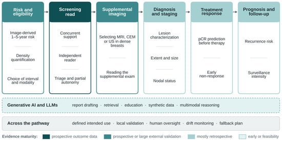
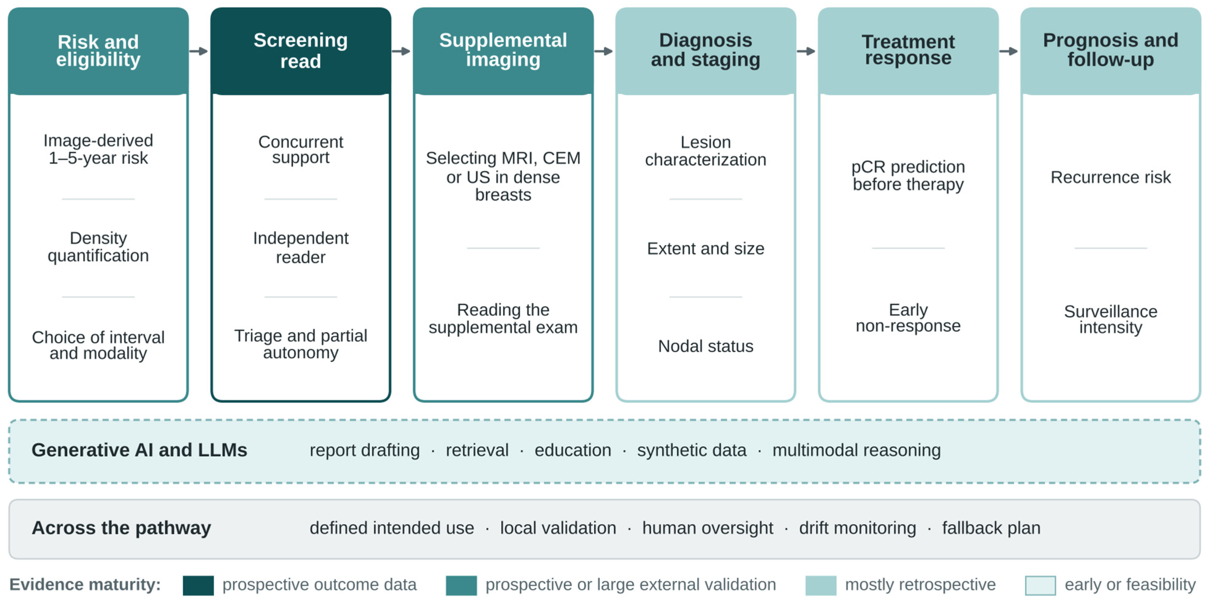

Disclaimer/Publisher’s Note: The statements, opinions and data contained in all publications are solely those of the individual author(s) and contributor(s) and not of MDPI and/or the editor(s). MDPI and/or the editor(s) disclaim responsibility for any injury to people or property resulting from any ideas, methods, instructions or products referred to in the content. |
© 2026 by the authors. Licensee MDPI, Basel, Switzerland. This article is an open access article distributed under the terms and conditions of the Creative Commons Attribution (CC BY) license.