


Abstract
Background/Objectives: Pediatric thyroid nodules carry a higher malignancy risk and more frequent regional cervical lymph-node involvement than adult nodules, making individualized pre-biopsy risk assessment particularly important. We aimed to develop and temporally validate a machine-learning model integrating routinely available thyroid-nodule and cervical lymph-node ultrasonographic features for malignancy-risk estimation in children. Methods: This retrospective single-center study at a tertiary pediatric referral center included consecutive patients aged 18 years or younger undergoing biopsy-based diagnostic evaluation between January 2019 and August 2025. The cohort comprised 195 patients with 221 nodules. Six candidate algorithms were evaluated using patient-grouped nested cross-validation. The selected XGBoost model integrated structured nodule and regional cervical lymph-node ultrasonographic features and was evaluated using discrimination, precision–recall performance, probability accuracy, calibration, and threshold-dependent operating characteristics. ACR TI-RADS served as a secondary standardized comparator. Results: Overall, 151 of 221 nodules (68.3%) were malignant, including 32 of 48 nodules (66.7%) in the temporal validation cohort. The final model achieved an AUROC of 0.973 (95% CI, 0.919–1.000), an AUPRC of 0.986 (95% CI, 0.954–1.000), a Brier score of 0.071 (95% CI, 0.030–0.126), a sensitivity of 84.4%, and a specificity of 87.5%. For comparison, the ACR TI-RADS total score yielded an AUROC of 0.906, while the TR5-positive and TR4/5-positive strategies yielded sensitivities of 84.4% and 93.8% and specificities of 87.5% and 56.2%, respectively. Conclusions: Integrating thyroid-nodule and cervical lymph-node ultrasonographic features enabled individualized malignancy-risk estimation in this selected pediatric biopsy-evaluation cohort. The continuous risk estimate may complement established sonographic assessment as an adjunct to comprehensive diagnostic evaluation. Prospective multicenter external validation is required before broader clinical application.
1. Introduction
Pediatric thyroid cancer is uncommon but clinically important because delayed diagnosis may affect the extent of surgery, lymph-node management, and long-term surveillance [1,2,3]. Thyroid nodules are detected less frequently in children than in adults, yet a nodule identified in a child carries a higher probability of malignancy than one identified in an adult [4]. Initial risk stratification must therefore balance timely evaluation of clinically meaningful disease against avoidable invasive procedures, anxiety, and follow-up burden [3]. Ultrasonography is central to this process because it provides structured information about both nodule morphology and regional cervical lymph nodes before cytologic or surgical confirmation. However, the optimal use of sonographic information in children remains challenging, particularly when biopsy decisions must account for both cancer risk and procedure-related trade-offs [3].
Ultrasound-based risk-stratification systems provide a standardized approach to characterizing thyroid nodules and guiding further evaluation. The 2017 American College of Radiology Thyroid Imaging Reporting and Data System (ACR TI-RADS) was developed primarily for adult practice and combines sonographic risk categories with size-based management thresholds [5]. Although ACR TI-RADS can stratify malignancy risk in children, direct application of adult management thresholds remains uncertain because pediatric nodules have a higher probability of malignancy and pediatric differentiated thyroid carcinoma more commonly involves regional cervical lymph nodes [6,7]. Recent pediatric studies have consequently evaluated modified biopsy thresholds and have generally shown that increasing sensitivity requires accepting additional biopsy recommendations. These findings support pediatric-specific evaluation of sonographic risk and management trade-offs rather than assuming direct transferability of adult action thresholds [6,8,9].
Recent artificial-intelligence research has expanded thyroid imaging assessment from structured-data machine learning to deep-learning, transfer-learning, hybrid image-based, and transformer-based approaches [10,11,12]. However, pediatric validation remains comparatively limited, and algorithmic complexity alone does not address the differences between adult and pediatric disease presentation [13,14,15,16]. Cervical lymph-node assessment is particularly relevant in children because regional nodal disease may contribute clinically meaningful information alongside conventional nodule morphology [7]. Within this context, artificial intelligence-assisted analysis of routinely available structured ultrasonographic information may provide quantitative diagnostic support without requiring a new imaging modality. The potential value of such information therefore lies in integrating routinely assessed regional findings into individualized risk estimation rather than in introducing novel imaging biomarkers or a novel machine-learning algorithm.
We therefore evaluated whether routinely available cervical lymph-node ultrasonographic information, when integrated with conventional nodule features in a machine-learning model, could contribute to individualized malignancy-risk estimation in a biopsy-selected pediatric thyroid nodule population.
2. Materials and Methods
2.1. Study Design and Participants
This retrospective single-center study developed and temporally validated a pediatric ultrasonographic feature-based machine-learning model for estimating the risk of malignant thyroid nodules. The study was approved by the Institutional Review Board of Beijing Children’s Hospital (IRB Approval No. [2025]-E-167-R; approval date: 31 October 2025), with a waiver of informed consent because of the retrospective study design, and was conducted in accordance with the Declaration of Helsinki.
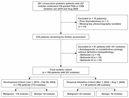
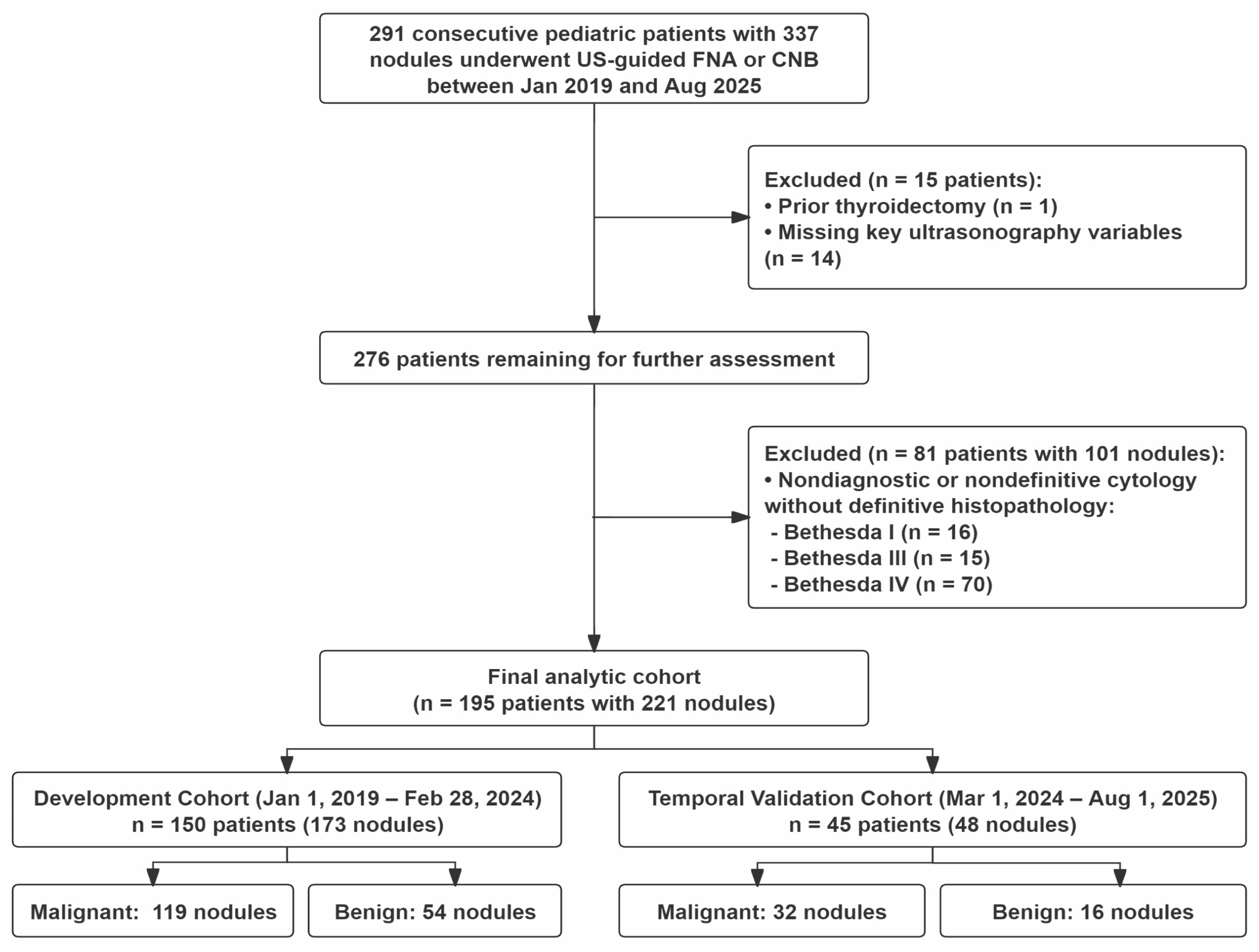
Consecutive pediatric patients aged 18 years or younger with thyroid nodules undergoing biopsy-based diagnostic evaluation between 1 January 2019 and 1 August 2025 were screened for eligibility. Patients were excluded if they had no thyroid nodule, no eligible reference standard, insufficient follow-up, prior nonsurgical treatment that could alter the baseline ultrasonographic phenotype, or unavailable ultrasonographic images or structured reports required for data verification. The dataset was prespecified into a development cohort (1 January 2019 to 28 February 2024) and a temporal validation cohort (1 March 2024 to 1 August 2025) according to examination date (Figure 1).
Figure 1.
Flow diagram of cohort selection and temporal cohort assignment. The final analytic cohort included 195 pediatric patients with 221 thyroid nodules. Patients examined from 1 January 2019 to 28 February 2024 were assigned to the development cohort, and patients examined from 1 March 2024 to 1 August 2025 were assigned to the temporal validation cohort. CNB, core-needle biopsy; FNA, fine-needle aspiration; US, ultrasonography.
2.2. Reference Standard
A predefined hierarchical reference standard incorporating surgical histopathology, cytological findings, and longitudinal clinical and imaging follow-up was used to classify each nodule as benign or malignant. Surgical histopathology was considered the primary reference standard whenever available, with histopathological diagnoses interpreted according to the World Health Organization classification [17]. Nodules were classified as malignant when malignancy was confirmed by surgical histopathology or, when surgical confirmation was unavailable, by Bethesda VI cytology. Nodules were classified as benign on the basis of benign surgical histopathology or Bethesda II cytology accompanied by at least 12 months of follow-up without malignant progression or development of highly suspicious ultrasonographic features [18,19]. Follow-up duration for nodules classified as benign based on Bethesda II cytology was calculated from the date of cytological examination to the last qualifying follow-up assessment. Unresected nodules with Bethesda I, III, IV, or V cytology were excluded from the primary analysis because a reliable binary reference-standard outcome could not be established.
2.3. Ultrasonographic Assessment
Clinical data were obtained from electronic medical records. Ultrasonographic examinations were performed using either a LOGIQ E9 ultrasound system (GE Healthcare, Wauwatosa, WI, USA) equipped with high-resolution linear transducers operating at 6.0–15.0 MHz or a Resona 7 ultrasound system (Shenzhen Mindray Bio-Medical Electronics Co., Ltd., Shenzhen, China) equipped with high-resolution linear transducers operating at 9.0–14.0 MHz. Ultrasound-derived predictors were abstracted exclusively from the most recent thyroid ultrasonography reports before the first biopsy of the index nodule. Cytology and histopathology records were used for reference-standard ascertainment, while follow-up records were used for outcome ascertainment. During predictor abstraction, diagnostic impressions, original TI-RADS categories, and management recommendations were masked, and extractors were blinded to cytologic, histopathologic, and follow-up information. Extracted variables were checked against descriptive report fields, with discrepancies resolved by review. No predictor variables were missing in the analysis-ready dataset.
Predictors included patient age and sex; thyroid background and nodule number; maximum nodule diameter, composition, echogenicity, shape, margin, echogenic foci, and vascularization. Lymph-node variables included regional lymph-node status, maximum lymph-node long-axis diameter (cm), echogenicity, calcification, shape, hilum, vascularity, and sonographically suspected extranodal extension. Regional status was classified as no suspicious lymph nodes identified, central compartment, lateral cervical compartment, both central and lateral compartments, or ultrasound-suspected mediastinal/distant nodal region. This category denoted the suspected nodal region on ultrasound, not pathologically confirmed nodal metastasis or disease stage. The no-suspicious-node category required documented systematic assessment of the cervical nodal compartments; cases with incomplete nodal documentation were excluded. When no suspicious nodes were identified, measurements and other nodal characteristics were obtained from the largest or a representative normal-appearing lymph node described in the report. Shape was classified as round or oval, hilum as obliterated or present, and vascularity as hilar, peripheral, or mixed.
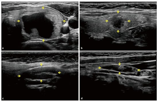
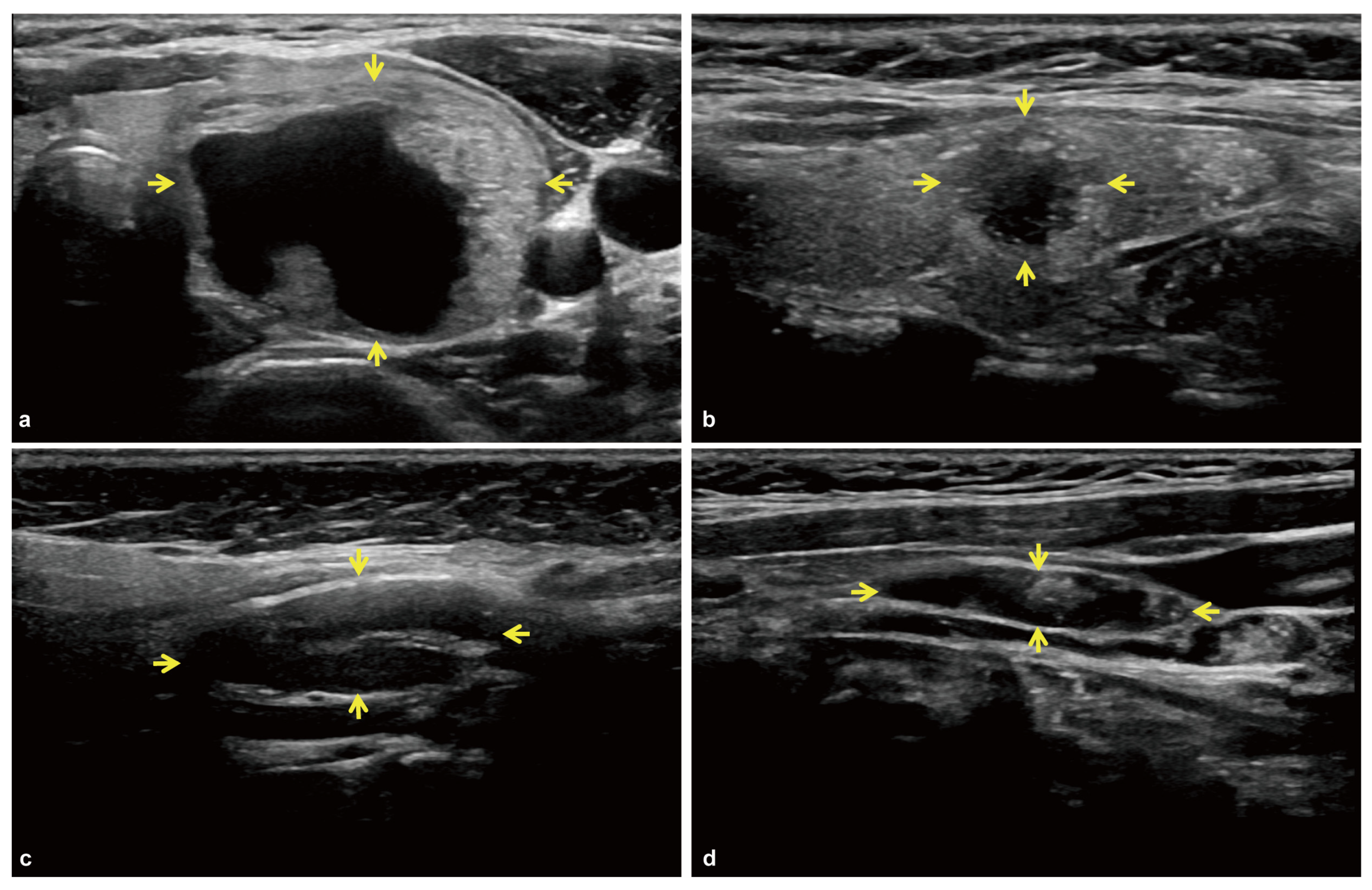
Two physicians with more than 10 years of experience in thyroid ultrasonography jointly reviewed the ultrasound images as an expert panel and assigned ACR TI-RADS feature scores, total scores, categories, and corresponding FNA recommendations. The reviewers were blinded to the original ultrasound reports and diagnostic and outcome information. Differences were resolved through discussion and consensus, and a single consensus assessment was recorded for each nodule. ACR TI-RADS category 5 (TR5-positive) and ACR TI-RADS category 5 (TR4/5-positive) classifications were also evaluated as descriptive comparators. Representative ultrasonographic appearances of pediatric thyroid nodules and cervical lymph nodes are shown in Figure 2.
Figure 2.
Representative ultrasonographic appearances of pediatric thyroid nodules and cervical lymph nodes. (a) Benign thyroid nodule with a smooth margin and no documented cervical lymph-node abnormality. (b) Malignant thyroid nodule with an irregular margin and punctate echogenic foci. (c) Malignant thyroid nodule with cervical lymph-node involvement in the same patient. (d) Cervical lymph node with abnormal morphology. Arrows indicate the corresponding nodules or lymph nodes.
2.4. Model Development and Evaluation
Six candidate algorithms, including penalized logistic regression (PLR), random forest (RF), gradient boosting (GBS), extreme gradient boosting (XGBoost), light gradient boosting machine (LightGBM), and radial-basis-function support vector machine (SVM), were evaluated using patient-grouped nested cross-validation with five outer folds and three inner folds of StratifiedGroupKFold. Hyperparameters were optimized using GridSearchCV with ROC-AUC scoring and algorithm-specific parameter grids (Supplementary Table S1). Numerical predictors were median-imputed, whereas categorical predictors were imputed using the most frequent category and one-hot encoded, with unknown categories ignored. StandardScaler was applied only to PLR and SVM. All preprocessing steps were incorporated into a ColumnTransformer within the modeling Pipeline and fitted within each inner training split. The selected pipeline was subsequently refitted on the complete outer training set and evaluated on the corresponding held-out outer fold. Final model selection was based on an exploratory, clinically informed judgment considering the study objectives and multidimensional development-cohort performance, including area under the receiver operating characteristic curve (AUROC), area under the precision–recall curve (AUPRC), Brier score, calibration intercept and slope, fold-level stability, and model complexity (Supplementary Figure S1). XGBoost was retained and locked before temporal validation, which was reserved exclusively for evaluating the selected model and was not used for model selection, reranking, hyperparameter optimization, or calibration. The development-derived locked statistical cutoff of 0.813 was obtained by maximizing the Youden index in the development cohort and applied unchanged to the temporal validation cohort. A sensitivity analysis restricted to nodules with surgical histopathological confirmation was performed using outer-fold out-of-fold predictions for the development cohort and predictions from the locked final model for the temporal validation cohort.
The final model was evaluated using AUROC, AUPRC, Brier score, calibration intercept and slope. Its AUROC was compared with the ACR TI-RADS total score using paired DeLong testing, and this comparison was interpreted descriptively because of the limited temporal-validation sample size. Decision-curve analysis (DCA) was conducted as an exploratory analysis using predictions from the locked final model in the temporal validation cohort. The DCA visualization covered threshold probabilities from 0.05 to 0.85 (Figure 3D). A vertical dashed reference line was added at 0.813, corresponding to the development-derived locked statistical cutoff obtained by maximizing the Youden index. Model transparency was assessed in the temporal validation cohort using permutation importance and Shapley additive explanations (SHAP). Permutation importance quantified the decrease in AUROC after each feature was permuted, and SHAP analysis described the direction and magnitude of individual feature contributions to malignant-risk prediction.
Figure 3.
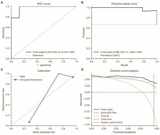
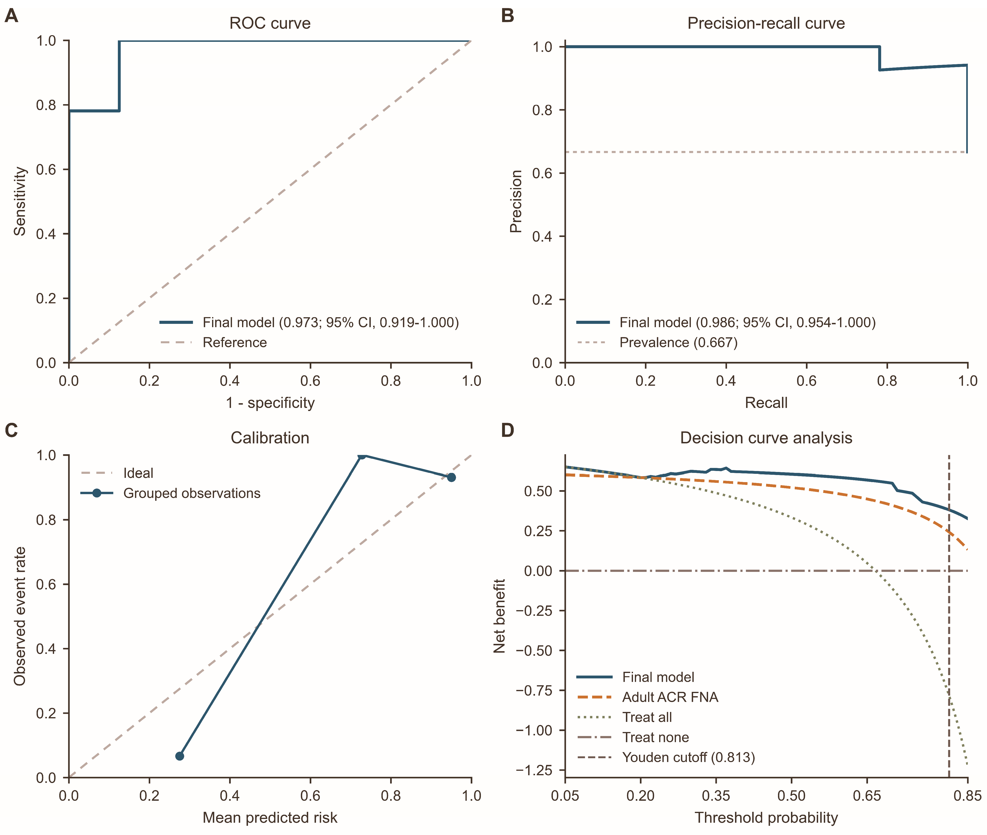
Performance of the final model in the temporal validation cohort. (A) ROC curve for the final model. (B) Precision–recall curves. (C) Calibration plot of the final model, showing observed event rates against mean predicted risk within five equal-width probability bins. The three non-empty bins contained 15, 4, and 29 nodules, respectively. The diagonal dashed line indicates ideal calibration; empty bins are not plotted. (D) Decision-curve analysis in the temporal validation cohort across threshold probabilities ranging from 0.05 to 0.85. The vertical dashed line indicates the development-derived locked statistical cutoff of 0.813. ACR, American College of Radiology; DCA, decision-curve analysis; FNA, fine-needle aspiration.
2.5. Statistical Analysis
Categorical variables were summarized as counts and percentages. Normally distributed continuous variables were presented as mean ± standard deviation, and non-normally distributed variables as median (interquartile range). Baseline differences between the development and temporal validation cohorts were assessed using standardized mean differences (SMDs). Confidence intervals (CI) for model-performance metrics (AUROC, AUPRC, sensitivity, specificity, PPV, and NPV) were estimated using patient-level clustered bootstrap resampling with 2000 iterations. Paired binary classifications between the model and the TR5-positive strategy were compared using a two-sided exact McNemar test, with agreement quantified using Cohen’s κ. AUROC comparisons were performed using clustered bootstrap-adjusted paired DeLong tests to account for within-patient clustering.
All analyses were performed using Python 3.13.5 (Python Software Foundation, Beaverton, OR, USA). The principal software libraries included NumPy 2.4.6, pandas 3.0.3, scikit-learn 1.9.0, XGBoost 3.3.0, LightGBM 4.6.0, SciPy 1.18.0, statsmodels 0.14.6, and Matplotlib 3.11.0. All stochastic processes were fixed with a random seed to ensure reproducibility.
3. Results
3.1. Cohort Characteristics
The study cohort included 195 pediatric patients with 221 thyroid nodules (Figure 1). The development cohort comprised 150 patients with 173 nodules, and the temporal validation cohort comprised 45 patients with 48 nodules. Overall, 151 of 221 nodules (68.3%) were malignant, including 119 of 173 nodules (68.8%) in the development cohort and 32 of 48 nodules (66.7%) in the temporal validation cohort. Surgical histopathology established the final diagnosis in 164 nodules (74.2%), including 31 benign and 133 malignant nodules. Among the remaining 57 nodules without surgical histopathology, 39 benign nodules were classified by Bethesda II cytology with at least 12 months of qualifying follow-up, and 18 malignant nodules were classified by Bethesda VI cytology. All 39 nodules classified as benign based on Bethesda II cytology met the minimum follow-up requirement, with qualifying follow-up available for all nodules. The median follow-up duration was 25.8 months (IQR, 20.4–27.6 months; range, 15.5–34.6 months).
Baseline characteristics are summarized in Table 1. Several nodule and cervical lymph-node characteristics differed between cohorts, with the largest absolute SMDs observed for round lymph-node shape (0.575), mediastinal/distant lymph-node region (0.490), and taller-than-wide nodule shape (0.366), indicating temporal differences in case mix.
Table 1.
Baseline characteristics of the development and temporal validation cohorts.
3.2. Model Performance
Performance of the candidate algorithms is summarized in Table 2. PLR achieved higher AUROC (0.918) and AUPRC (0.955) point estimates than XGBoost (0.907 and 0.936, respectively). Conversely, XGBoost had a lower Brier score (0.102 vs. 0.120) and calibration intercept and slope (−0.079 and 0.941, respectively) closer to their ideal reference values than PLR (0.495 and 2.091, respectively). In development out-of-fold analyses, adding cervical lymph-node features to the nodule-only XGBoost model produced only small and inconsistent changes in overall performance (nodule-only vs. combined: AUROC, 0.899 vs. 0.907; AUPRC, 0.948 vs. 0.936; Brier score, 0.114 vs. 0.102). In the sensitivity analysis restricted to nodules with surgical histopathological confirmation, the AUROC was 0.889 (95% CI, 0.817–0.952) in the development cohort (118 nodules from 110 patients) and 0.984 (95% CI, 0.939–1.000) in the temporal validation cohort (46 nodules from 44 patients).
Table 2.
Predictive performance of candidate models in the development cohort.
In the temporal validation cohort, the final model achieved an AUROC of 0.973 (95% CI, 0.919–1.000), an AUPRC of 0.986 (95% CI, 0.954–1.000), and a Brier score of 0.071 (95% CI, 0.030–0.126) (Figure 3). The ACR TI-RADS total score had an AUROC of 0.906. The paired DeLong comparison was not statistically significant (p = 0.153). The calibration intercept was −0.901 (95% CI, −2.587 to −0.057), and the calibration slope was 1.501 (95% CI, 1.058 to 9.199). Of five prespecified bins, only three were non-empty: 15 nodules were in the 20–40% bin, four in the 60–80% bin, and 29 in the 80–100% bin (mean predicted probability, 95.0%; observed malignancy rate, 93.1%). The 0–20% and 40–60% bins were empty. These estimates carry substantial uncertainty given the limited temporal validation sample; bin-specific calibration estimates are provided in Supplementary Table S2.
At the cutoff of 0.813, sensitivity was 84.4% and specificity was 87.5%, with 5 of 32 malignant nodules (15.6%) classified below the cutoff. The model and TR5-positive strategy yielded identical aggregate sensitivity (84.4%), specificity (87.5%), PPV (93.1%), and NPV (73.7%). However, their case-level classifications were concordant in only 38 of 48 nodules (79.2%), with 10 discordant cases (five positive by XGBoost only and five positive by TR5 only). Cohen’s κ was 0.57 (95% CI, 0.29–0.84), indicating moderate agreement, while the two-sided exact McNemar test showed no difference in marginal positive classification rates (p = 1.000). Detailed paired classification results and individual characteristics of discordant nodules are provided in Supplementary Table S3. Fewer nodules were classified as positive by the model than under the ACR TI-RADS FNA recommendation, whereas the TR4/5-positive strategy achieved higher sensitivity at the cost of more positive classifications (Table 3).
Table 3.
Diagnostic performance of the final model and ACR TI-RADS strategies in the temporal validation cohort.
Decision-curve analysis was visualized across threshold probabilities ranging from 0.05 to 0.85 (Figure 3D). The ACR TI-RADS FNA recommendation was included as an adult-derived reference comparator. In the separate category-based comparisons, the model showed net benefit comparable to the TR5-positive strategy and slightly lower than the TR4/5-positive strategy.
3.3. Model Transparency
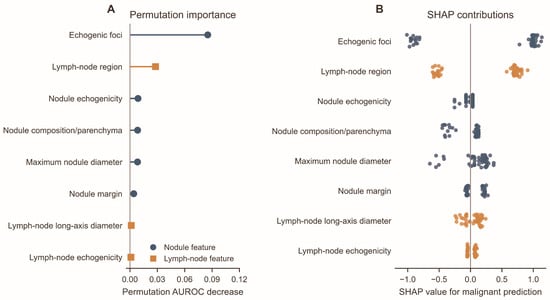
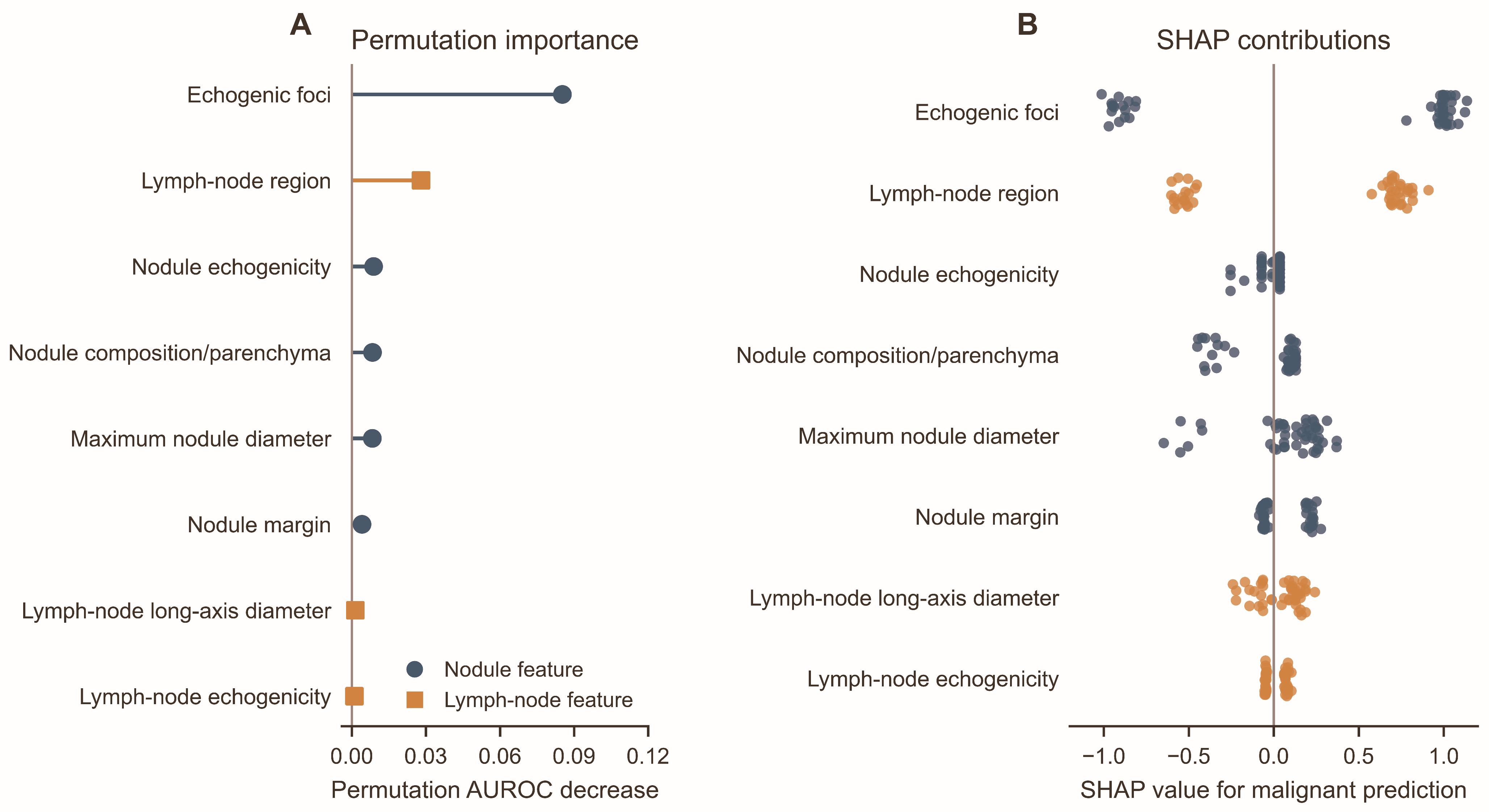
In permutation-importance analysis, echogenic foci showed the largest mean decrease in AUROC when permuted, with a permutation AUROC decrease of 0.085 (2.5th–97.5th percentile range across 100 permutation repeats, 0.024–0.153) (Figure 4). The estimate for the lymph-node region was 0.028 (−0.011 to 0.064). Several other features, including nodule echogenicity, nodule composition, maximum nodule diameter, lymph-node long-axis diameter, and abnormal lymph-node echogenicity, showed smaller importance estimates. SHAP analysis similarly highlighted echogenic foci, lymph-node region, nodule echogenicity, nodule composition, and maximum nodule diameter among the features with relatively prominent model attributions (Figure 4).
Figure 4.
Model transparency analysis of the final XGBoost model. (A) Permutation importance, expressed as the mean decrease in AUROC after feature permutation. The 2.5th–97.5th percentile ranges reported in the Results reflect variability across 100 permutation repeats and are not displayed as error bars; they should not be interpreted as patient-level confidence intervals. (B) SHAP contributions to malignant-risk prediction. Positive SHAP values indicate contributions toward malignant prediction, whereas negative values indicate contributions toward benign prediction. Colors distinguish nodule-level and lymph-node-level features. AUROC, area under the receiver operating characteristic curve; SHAP, Shapley additive explanations.
4. Discussion
In this study, we developed a model integrating routinely documented thyroid-nodule and cervical lymph-node ultrasonographic features to generate continuous malignancy-risk estimates in children referred to a tertiary pediatric center for biopsy or surgical evaluation. The final XGBoost model achieved an AUROC of 0.973, an AUPRC of 0.986, and a Brier score of 0.071; at the cutoff of 0.813, sensitivity and specificity were 84.4% and 87.5%, respectively. The AUROC describes the model’s overall ability to discriminate between malignant and benign nodules in this selected cohort. Its practical contribution lies in synthesizing information already available from comprehensive thyroid and cervical ultrasonography into a continuous malignancy-risk estimate. The model’s pediatric-specific development and continuous risk output provide a structured approach to summarizing sonographic risk alongside conventional assessment.
ACR TI-RADS remains a valuable standardized framework for describing thyroid-nodule morphology and stratifying malignancy risk [5,16]. However, its adult-derived management thresholds may not provide an optimal sensitivity–specificity balance for pediatric management [5,6,8,9,16]. Previous pediatric studies have shown that lowering or modifying biopsy thresholds can improve sensitivity while increasing the number of nodules undergoing biopsy or follow-up [6,8,9]. In this study, the model and the expert TR5-positive strategy showed identical aggregate classification metrics but only moderate case-level agreement (κ = 0.57), indicating that the two approaches are not interchangeable. The AUROC comparison with the ACR TI-RADS total score was not statistically significant (p = 0.153). These findings do not establish the superiority of the model over TR5 or ACR TI-RADS. The model’s pediatric-specific development and continuous probability output may provide a complementary representation of sonographic risk, but whether this format improves clinical decision-making remains to be established.
Regional lymph-node metastases are frequently observed at presentation in pediatric differentiated thyroid carcinoma [7,20]. Sonographic evaluation of cervical lymph nodes therefore contributes to comprehensive pediatric thyroid assessment [19]. Thus, cervical lymph-node assessment was incorporated into the present model to reflect this clinically relevant aspect of routine assessment. In the development out-of-fold analyses, adding cervical lymph-node features was associated with only modest and inconsistent changes in AUROC, AUPRC, and Brier score. Although the present findings did not demonstrate a clear incremental discriminatory benefit over nodule features alone, the relatively limited cohort size may have constrained the ability to fully characterize the contribution of lymph-node features. Further studies involving larger pediatric cohorts, preferably with prospective multicenter designs, are warranted to better clarify their incremental predictive value and potential clinical relevance.
Recent thyroid artificial intelligence research includes structured-data machine learning, deep transfer learning, hybrid image-based methods, and transformer-based approaches [10,11,12,13,14]. Pediatric evidence remains limited, although Ha et al. reported favorable discrimination with an image-based deep-learning model in pediatric cohorts [15]. Unlike image-based approaches, the present model uses routinely documented thyroid-nodule and cervical lymph-node ultrasonographic features. These approaches are not directly comparable because of differences in model inputs, study populations, and validation designs. Their relative performance and clinical utility in pediatric populations remain to be established.
The development-derived locked statistical cutoff of 0.813 was obtained by maximizing the Youden index in the development cohort and applied unchanged to the temporal validation cohort. It was not a prespecified clinical threshold and was not selected to represent a pediatric biopsy decision threshold. In contrast, DCA threshold probabilities represent hypothetical clinical decision thresholds reflecting the relative consequences of missed malignancy and unnecessary intervention. False-negative results remain possible, and the model was evaluated only in tertiary-care pediatric patients being considered for biopsy or resection; spectrum and verification bias limit its generalizability to unselected community populations. The model should therefore be used only as an adjunct to routine sonographic assessment and multidisciplinary clinical judgment. Larger prospective multicenter studies and reader studies are needed to establish clinically actionable thresholds and determine whether model-assisted assessment improves patient management.
This study has several limitations. Its retrospective design, selected case mix, and hierarchical reference standard may introduce selection, spectrum, and differential-verification bias. Temporal validation was conducted within the same center and does not constitute independent external validation; therefore, applicability to other institutions and broader pediatric populations remains to be established. The model was evaluated only in a selected tertiary-care pediatric population undergoing FNA or surgical evaluation and should not be extrapolated to community screening or general pediatric nodule triage. Furthermore, the small temporal validation cohort resulted in substantial uncertainty in calibration estimates, as reflected by wide confidence intervals and only three non-empty probability bins. Although development-cohort out-of-fold calibration estimates informed the exploratory selection of XGBoost, individual predicted probabilities should be interpreted cautiously, and calibration and clinical applicability have not been sufficiently established. Model selection also lacked a prespecified quantitative weighting rule. In addition, ACR TI-RADS assessments were based on joint expert consensus, precluding formal assessment of interobserver agreement. SHAP and permutation-importance analyses were descriptive, and the structured feature set may not capture all image-level characteristics. Prospective multicenter external validation with independent readers is warranted to further evaluate reproducibility, calibration, incremental discriminatory value, and clinical utility.
5. Conclusions
The model generated continuous malignancy-risk probabilities by integrating thyroid-nodule and cervical lymph-node ultrasonographic features in children undergoing biopsy-based evaluation at a tertiary pediatric referral center. This risk output may serve as an adjunct to support pre-biopsy assessment alongside comprehensive thyroid and cervical ultrasound evaluation, rather than independently determining biopsy indications. Prospective multicenter studies are needed to establish the model’s generalizability to other institutions and broader pediatric populations and to determine whether its risk estimates improve biopsy assessment and clinical decision-making.
Supplementary Materials
The following supporting information can be downloaded at https://www.mdpi.com/article/10.3390/diagnostics16223773/s1, Figure S1. Learning curves for six machine learning models. Plots show log-loss as a function of increasing training sample size (60–100% of patients in the outer-fold training cohorts). The blue solid line with circular markers indicates training-set performance; the orange dashed line with square markers indicates held-out outer-fold validation performance. Shaded regions denote 95% confidence intervals. Panels A–F correspond to Penalized Logistic Regression (PLR), Random Forest (RF), Gradient Boosting (GBS), XGBoost, LightGBM, and Support Vector Machine with a radial basis function kernel (SVM-RBF), respectively. Table S1. Full hyperparameter search spaces and optimal values of all evaluated models. Table S2. Bin-specific calibration and overall calibration parameters of the final model in the temporal validation cohort. Table S3. Paired case-level comparison and individual characteristics of discordant nodules.
Author Contributions
Conceptualization, J.D.; methodology, J.D.; validation, J.D.; formal analysis, J.D.; investigation, X.Z., Y.C., S.T., J.W. and Q.L.; data curation, X.Z., Y.C., S.T., J.W. and Q.L.; writing—original draft preparation, J.D.; writing—review and editing, W.S. and X.N.; visualization, J.D.; resources, W.S. and X.N.; supervision, W.S. and X.N.; project administration, W.S. and X.N.; funding acquisition, X.N. All authors have read and agreed to the published version of the manuscript.
Funding
This research was funded by the Beijing Municipal Science & Technology Commission and the Administrative Commission of Zhongguancun Science Park through the Concept Verification Platform Construction Project (grant number 2024030119). The article processing charge (APC) was funded by the same grant. The funders had no role in the design of the study; in the collection, analyses, or interpretation of data; in the writing of the manuscript; or in the decision to publish the results.
Institutional Review Board Statement
The study was conducted in accordance with the Declaration of Helsinki and approved by the Institutional Review Board of Beijing Children’s Hospital (protocol code [2025]-E-167-R; date of approval: 31 October 2025).
Informed Consent Statement
Patient consent was waived by the Institutional Review Board because of the retrospective study design.
Data Availability Statement
The data supporting the findings of this study are available from the corresponding author upon reasonable request due to patient privacy and ethical restrictions. The analysis code is available from the corresponding author upon reasonable request.
Acknowledgments
During the preparation of this manuscript, the authors used ChatGPT (OpenAI; GPT-5.6) solely for text editing, including grammar, structure, spelling, punctuation, and formatting. The authors reviewed and edited the output and take full responsibility for the content of this publication.
Conflicts of Interest
The authors declare no conflicts of interest.
Abbreviations
ACR TI-RADS, American College of Radiology Thyroid Imaging Reporting and Data System; AUPRC, area under the precision–recall curve; AUROC, area under the receiver operating characteristic curve; CI, confidence interval; DCA, decision-curve analysis; FNA, fine-needle aspiration; GBS, gradient boosting; IQR, interquartile range; NPV, negative predictive value; PPV, positive predictive value; PLR, penalized logistic regression; RF, random forest; SD, standard deviation; SHAP, Shapley additive explanations; SVM, support vector machine; SMD, standardized mean difference; XGBoost, extreme gradient boosting.
References
- Qian, Z.J.; Jin, M.C.; Meister, K.D.; Megwalu, U.C. Pediatric thyroid cancer incidence and mortality trends in the United States, 1973–2013. JAMA Otolaryngol. Head Neck Surg. 2019, 145, 617–623. [Google Scholar] [CrossRef] [Scilit] [PubMed]
- Miranda-Filho, A.; Lortet-Tieulent, J.; Bray, F.; Cao, B.; Franceschi, S.; Vaccarella, S.; Dal Maso, L. Thyroid cancer incidence trends by histology in 25 countries: A population-based study. Lancet Diabetes Endocrinol. 2021, 9, 225–234. [Google Scholar] [CrossRef] [Scilit] [PubMed]
- Francis, G.L.; Waguespack, S.G.; Bauer, A.J.; Angelos, P.; Benvenga, S.; Cerutti, J.M.; Dinauer, C.A.; Hamilton, J.; Hay, I.D.; Luster, M.; et al. Management guidelines for children with thyroid nodules and differentiated thyroid cancer. Thyroid 2015, 25, 716–759. [Google Scholar] [CrossRef] [Scilit] [PubMed]
- Buryk, M.A.; Simons, J.P.; Picarsic, J.; Monaco, S.E.; Ozolek, J.A.; Joyce, J.; Gurtunca, N.; Nikiforov, Y.E.; Witchel, S.F. Can malignant thyroid nodules be distinguished from benign thyroid nodules in children and adolescents by clinical characteristics? A review of 89 pediatric patients with thyroid nodules. Thyroid 2015, 25, 392–400. [Google Scholar] [CrossRef] [Scilit] [PubMed]
- Tessler, F.N.; Middleton, W.D.; Grant, E.G.; Hoang, J.K.; Berland, L.L.; Teefey, S.A.; Cronan, J.J.; Beland, M.D.; Desser, T.S.; Frates, M.C.; et al. ACR Thyroid Imaging, Reporting and Data System (TI-RADS): White paper of the ACR TI-RADS Committee. J. Am. Coll. Radiol. 2017, 14, 587–595. [Google Scholar] [CrossRef] [Scilit] [PubMed]
- Lee, D.H.; Cho, Y.J.; Won, J.K.; Lee, S.B.; Choi, Y.H.; Jung, K.C.; Park, Y.J.; Lee, Y.J.; Shin, C.H.; Lee, Y.A. Pediatric thyroid nodules: K-TIRADS/ACR TI-RADS pediatric-specific biopsy cutoff incorporating clinical risk factors. Radiology 2025, 315, e241015. [Google Scholar] [CrossRef] [Scilit] [PubMed]
- Yang, A.T.; Lai, S.T.T.; Laetsch, T.W.; Bhatti, T.; Baloch, Z.; Surrey, L.F.; Franco, A.T.; Ricarte-Filho, J.C.M.; Mostoufi-Moab, S.; Adzick, N.S.; et al. Molecular landscape and therapeutic strategies in pediatric differentiated thyroid carcinoma. Endocr. Rev. 2025, 46, 397–417. [Google Scholar] [CrossRef] [Scilit] [PubMed]
- Marzukie, M.S.; Shapira-Zaltsberg, G.; Martinez-Rios, C. Assessment of the American College of Radiology Thyroid Imaging Reporting and Data System (ACR TI-RADS™) with modification of the management recommendations for pediatric thyroid nodules. Pediatr. Radiol. 2024, 54, 1476–1485. [Google Scholar] [CrossRef] [Scilit] [PubMed]
- Edri, N.; Tsur, N.; Hod, R.; Robenshtok, E.; Bachar, G.; Yaniv, D.; Yehuda, M.; Ritter, A.; Yosefof, E. Diagnostic performance of ACR TI-RADS and sonographic predictors of malignancy in pediatric thyroid nodules. Endocr. Pract. 2026, 32, 1340–1346. [Google Scholar] [CrossRef] [Scilit] [PubMed]
- Hosseini Sarkhosh, S.M.; Shirzad, N.; Taghvaei, M.; Tavangar, S.M.; Farhat, S.; Ebrahiminik, H.; Hemmatabadi, M.; Pourashraf, M.; Chegeni, H. Prediction of thyroid malignancy risk using clinical and ultrasonography features and a machine learning approach. Eur. Radiol. 2025, 35, 5157–5167. [Google Scholar] [CrossRef] [Scilit] [PubMed]
- Li, W.; Zhou, Y.; Luo, Z.; Tan, M.; Yin, R.; Li, J. XGBoost-based machine learning model combining clinical and ultrasound data for personalized prediction of thyroid nodule malignancy. Front. Endocrinol. 2025, 16, 1639639. [Google Scholar] [CrossRef] [Scilit] [PubMed]
- Habchi, Y.; Kheddar, H.; Himeur, Y.; Ghanem, M.C. Machine Learning and Transformers for Thyroid Carcinoma Diagnosis. J. Vis. Commun. Image Represent. 2026, 115, 104668. [Google Scholar] [CrossRef] [Scilit]
- Shen, P.; Yang, Z.; Sun, J.; Wang, Y.; Qiu, C.; Wang, Y.; Ren, Y.; Liu, S.; Cai, W.; Lu, H.; et al. Explainable multimodal deep learning for predicting thyroid cancer lateral lymph node metastasis using ultrasound imaging. Nat. Commun. 2025, 16, 7052. [Google Scholar] [CrossRef] [Scilit] [PubMed]
- Mattay, G.S. Development of a conversational multimodal AI tool for assessing malignancy risk of thyroid nodules. Radiol. Imaging Cancer 2025, 7, e259014. [Google Scholar] [CrossRef] [Scilit] [PubMed]
- Ha, E.J.; Lee, J.H.; Mak, N.; Duh, A.K.; Tong, E.; Yeom, K.W.; Meister, K.D. A deep learning-based artificial intelligence model assisting thyroid nodule diagnosis and management: Pilot results for evaluating thyroid malignancy in pediatric cohorts. Thyroid 2025, 35, 652–661. [Google Scholar] [CrossRef] [Scilit] [PubMed]
- Kim, P.H. Ultrasonographic evaluation of pediatric thyroid nodules: Adult risk stratification systems, 2021 K-TIRADS revision, and future refinements. Korean J. Radiol. 2026, 27, 471–483. [Google Scholar] [CrossRef] [Scilit] [PubMed]
- Baloch, Z.W.; Asa, S.L.; Barletta, J.A.; Ghossein, R.A.; Juhlin, C.C.; Jung, C.K.; LiVolsi, V.A.; Papotti, M.G.; Sobrinho-Simões, M.; Tallini, G.; et al. Overview of the 2022 WHO classification of thyroid neoplasms. Endocr. Pathol. 2022, 33, 27–63. [Google Scholar] [CrossRef] [Scilit] [PubMed]
- Aekka, A.; Zimmerman, D.; Josefson, J.L. An updated review on the use of fine needle aspiration biopsy in pediatric thyroid disease. Endocr. Pract. 2025, 31, 1346–1349. [Google Scholar] [CrossRef] [Scilit] [PubMed]
- Lebbink, C.A.; Links, T.P.; Czarniecka, A.; Dias, R.P.; Elisei, R.; Izatt, L.; Krude, H.; Lorenz, K.; Luster, M.; Newbold, K.; et al. 2022 European Thyroid Association guidelines for the management of pediatric thyroid nodules and differentiated thyroid carcinoma. Eur. Thyroid J. 2022, 11, e220146. [Google Scholar] [CrossRef] [Scilit] [PubMed]
- Navallas, M.; Daneman, A.; Amirabadi, A.; Ngan, B.Y.; Wasserman, J. Utility of sonography for identifying metastatic cervical adenopathy in children with differentiated thyroid carcinoma at presentation. Pediatr. Radiol. 2021, 51, 273–281. [Google Scholar] [CrossRef] [Scilit] [PubMed]

Disclaimer/Publisher’s Note: The statements, opinions and data contained in all publications are solely those of the individual author(s) and contributor(s) and not of MDPI and/or the editor(s). MDPI and/or the editor(s) disclaim responsibility for any injury to people or property resulting from any ideas, methods, instructions or products referred to in the content. |
© 2026 by the authors. Licensee MDPI, Basel, Switzerland. This article is an open access article distributed under the terms and conditions of the Creative Commons Attribution (CC BY) license.