


Abstract
Cervical clear cell carcinoma (CCC) is a rare human papillomavirus (HPV)-independent adenocarcinoma. A 73-year-old woman presented with postmenopausal bleeding. Ultrasonography demonstrated a cervical mass and a 5.0 × 5.0 mm right cornual nodule. On 3.0-T magnetic resonance imaging (MRI), the cornual lesion measured 5.2 × 5.3 mm and, like the cervical tumor, showed high signal intensity on diffusion-weighted imaging (b = 1000 s/mm2). 18F-fluorodeoxyglucose positron emission tomography/computed tomography (18F-FDG PET/CT) demonstrated intense uptake in the cervical mass (30.6 × 29.2 × 41.2 mm; SUVmax 10.4) and in the spatially separate right cornual nodule (5.2 × 5.1 mm; SUVmax 14.6), without FDG-avid nodal or distant metastatic disease. The concordant identification of the cornual lesion by ultrasonography, MRI, and PET/CT prompted multidisciplinary reassessment and radical hysterectomy to obtain definitive pathological characterization of both lesions. Histopathology demonstrated clear cell carcinoma at both sites; however, because molecular, genomic, or clonality analyses were not performed, their biological relationship remains uncertain. All 30 examined lymph nodes and the surgical margins were negative. The cervical primary was staged as pT1b3 pN0 cM0 (FIGO 2018 stage IB3). The patient received four cycles of postoperative chemotherapy with paclitaxel plus cisplatin. At 14 months after surgery, gynecological examination and pelvic MRI showed no evidence of recurrence. This case highlights the complementary roles of ultrasonography, MRI, and 18F-FDG PET/CT in characterizing a minute spatially separate uterine lesion while underscoring the need for cautious interpretation when the relationship between lesions cannot be established.
Figure 1.
Multimodal ultrasonographic and magnetic resonance imaging findings. A 73-year-old woman presented with postmenopausal vaginal bleeding, and human papillomavirus testing was negative. (A) Ultrasonography demonstrates a hypoechoic mass in the uterine cervix (arrowhead). (B) Ultrasonography demonstrates a small hypoechoic nodule in the right uterine cornu (arrowhead), measuring approximately 5.0 × 5.0 mm and spatially separate from the cervical mass. (C) T2-weighted MRI demonstrates the cervical soft-tissue mass with high signal intensity (arrowhead). (D) The cervical mass shows high signal intensity on diffusion-weighted imaging (DWI; b = 1000 s/mm2) (arrowhead). (E) T2-weighted MRI demonstrates the right cornual lesion (arrowhead), measuring approximately 5.2 × 5.3 mm. (F) The right cornual lesion also shows high signal intensity on DWI (b = 1000 s/mm2) (arrowhead). MRI was performed on a 3.0-T system. A large amount of fluid is present within the uterine cavity. Cervical CCC is an uncommon HPV-independent endocervical adenocarcinoma [1]. In a clinical series of 18 patients with cervical clear cell adenocarcinoma, vaginal bleeding was the most frequent presenting symptom, and HPV testing was negative in most tested patients [2]. Ultrasonography initially identified the right cornual nodule, while MRI confirmed its spatial separation from the cervical tumor and provided local soft-tissue characterization. The concordant visualization of the nodule on ultrasonography and MRI supported the presence of a true focal lesion rather than an imaging artifact. MRI is complementary to PET/CT in the imaging assessment of cervical malignancy [3].
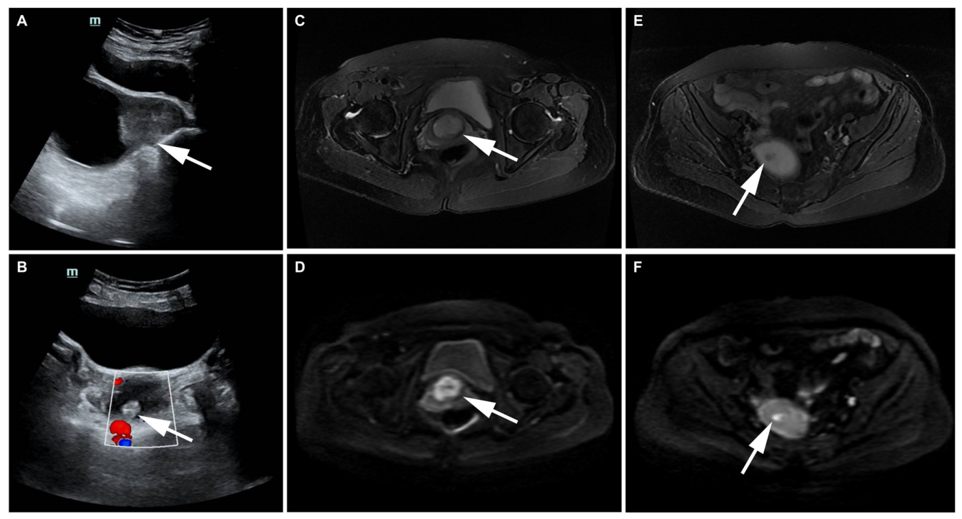
Figure 2.
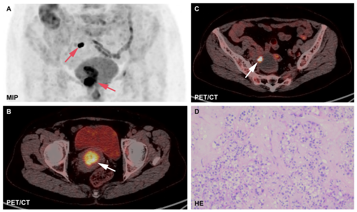
18F-FDG PET/CT and histopathological findings. (A) Maximum-intensity-projection (MIP) 18F-FDG PET image demonstrates abnormal FDG uptake in the pelvis. No FDG-avid nodal or distant metastatic disease was identified on whole-body PET/CT. (B) Fused 18F-FDG PET/CT demonstrates intense FDG uptake in the cervical mass (arrowhead), measuring approximately 30.6 × 29.2 × 41.2 mm, with an SUVmax of 10.4. (C) Fused 18F-FDG PET/CT demonstrates intense focal FDG uptake in the spatially separate right cornual nodule (arrowhead), measuring approximately 5.2 × 5.1 mm, with an SUVmax of 14.6. PET/CT acquisition was performed 60 min after FDG administration using a GE Discovery MI system. PET images were reconstructed with Q. Clear (β = 400) and a reconstructed image spacing of 2 mm. Given the approximately 5 mm size of the cornual lesion, quantitative SUV interpretation is subject to substantial uncertainty from finite spatial resolution and partial-volume effects; SUVmax is also influenced by reconstruction and statistical noise. Accordingly, the reported SUVmax should be interpreted as a semiquantitative descriptor of focal FDG avidity rather than as an accurate estimate of true tracer concentration or a direct measure of biological aggressiveness [4]. (D) Representative hematoxylin and eosin-stained section of the cervical clear cell carcinoma (×400). Ultrasonography initially identified the small right cornual lesion, MRI confirmed its spatial separation and local imaging features, and 18F-FDG PET/CT provided complementary metabolic characterization and whole-body staging rather than serving as the sole modality responsible for lesion detection. The patient had initially been considered for radiotherapy for presumed cervical carcinoma. The concordant identification of a second spatially separate cornual lesion introduced uncertainty about disease extent and the biological relationship between the two lesions. Because no nodal or distant metastatic disease was identified, multidisciplinary reassessment favored radical hysterectomy to achieve local disease control and to obtain definitive histopathological characterization of both lesions. PET/CT is an established complementary imaging modality in cervical cancer staging and treatment planning [3,5]. At surgery, a cervical mass involving the cervical wall and a separate cauliflower-like nodule measuring approximately 5 mm near the right uterine cornu were identified, with no macroscopic continuity between them. The absence of macroscopic continuity does not exclude microscopic spread. Histopathological examination demonstrated cervical clear cell carcinoma with invasion of more than two-thirds of the cervical fibromuscular wall. No lymphovascular or perineural invasion was identified. Immunohistochemistry of the cervical tumor showed ER (+), PR (−), p16 (+), Ki-67 labeling index approximately 30%, p40 (−), CEA (−), CD31 (−), D2-40 (−), wild-type p53 expression, Napsin A (+), and HNF1β (+). The spatially separate right cornual nodule also demonstrated clear cell carcinoma with invasion of less than half of the myometrial thickness and no lymphovascular or perineural invasion; Napsin A and HNF1β were positive, and no broader immunohistochemical panel was performed on the cornual lesion. All 30 examined lymph nodes were negative for metastasis, including 14 left pelvic, 5 right pelvic, 6 right obturator, 1 left common iliac, 2 right common iliac, and 2 para-aortic lymph nodes. Surgical margins were negative, with no carcinoma identified at the parametrial or vaginal wall margins. The cervical primary was staged as pT1b3 pN0 cM0 (FIGO 2018 stage IB3). This stage refers to the cervical primary; the right cornual lesion is described separately because its biological relationship to the cervical tumor could not be definitively established. Uterine corpus/endometrial involvement has previously been described in cervical clear cell carcinoma [2]. Napsin A supports clear cell differentiation, whereas HNF1β is less specific in the differential diagnosis between clear cell and mesonephric carcinomas [6]. The differential interpretation of the cornual lesion includes spread from the cervical primary, multifocal disease, or a synchronous uterine clear cell carcinoma. Although both lesions were diagnosed as clear cell carcinoma and shared Napsin A/HNF1β positivity, the available pathological findings cannot establish a common site of origin or clonal relationship. Because molecular, genomic, or clonality analyses were not performed, the biological relationship remains unresolved; therefore, the cornual lesion should not be regarded as a confirmed metastasis. Following surgery, adjuvant treatment options were reassessed. Pelvic radiotherapy was discussed, but the patient declined radiotherapy because of concerns about radiation-related injury to pelvic organs. After multidisciplinary evaluation, systemic therapy was selected in view of the overall recurrence-risk assessment and concern for occult distant micrometastatic disease. The patient received four cycles of postoperative chemotherapy with paclitaxel plus cisplatin. At 14 months after surgery, gynecological examination and pelvic MRI showed no evidence of recurrent disease. This report has two important limitations: first, the absence of molecular/genomic/clonality analysis prevents definitive determination of the relationship between the two tumors; second, the 14-month follow-up period is relatively short and does not permit conclusions regarding long-term recurrence or survival.
Author Contributions
P.L.: conceptualization, acquisition and interpretation of data, and manuscript preparation. F.C.: clinical data acquisition, interpretation of clinical findings, and manuscript review. L.X.: image analysis, literature review, and critical revision of the manuscript. C.D.: image analysis, literature review, and critical revision of the manuscript. Z.D.: supervision, interpretation of imaging findings, and critical revision of the manuscript. All authors have read and agreed to the published version of the manuscript.
Funding
This research was funded by the College-local Collaborative Innovation Research Project of Jiangsu Medical College (grant number 202490124) and the Specialized Clinical Medicine Research Project of Nantong University (grant number 2024LY010).
Institutional Review Board Statement
Ethical review and approval were waived by the Ethics Committee of Yancheng Third People’s Hospital for this retrospective single-patient case report (Waiver No. 2026-104; 9 January 2026). The case was reported in accordance with the principles of the Declaration of Helsinki.
Informed Consent Statement
Written informed consent was obtained from the patient to publish the clinical information and accompanying images.
Data Availability Statement
The data supporting the findings of this report are available from the corresponding author upon reasonable request. The data are not publicly available because of patient privacy considerations.
Conflicts of Interest
The authors declare no conflicts of interest.
References
- Stolnicu, S.; Karpathiou, G.; Guerra, E.; Mateoiu, C.; Reques, A.; Garcia, A.; Bart, J.; Felix, A.; Fanni, D.; Gama, J.; et al. Clear Cell Carcinoma (CCC) of the Cervix Is a Human Papillomavirus (HPV)-Independent Tumor Associated With Poor Outcome: A Comprehensive Analysis of 58 Cases. Am. J. Surg. Pathol. 2022, 46, 765–773. [Google Scholar] [CrossRef] [Scilit] [PubMed]
- Wang, D.; Zhao, C.; Fu, L.; Liu, Y.; Zhang, W.; Xu, T. Primary Clear Cell Adenocarcinoma of the Cervix: A Clinical Analysis of 18 Cases without Exposure to Diethylstilbestrol. Obstet. Gynecol. Int. 2019, 2019, 9465375. [Google Scholar] [CrossRef] [Scilit] [PubMed]
- Kusmirek, J.; Robbins, J.; Allen, H.; Barroilhet, L.; Anderson, B.; Sadowski, E.A. PET/CT and MRI in the Imaging Assessment of Cervical Cancer. Abdom. Imaging 2015, 40, 2486–2511. [Google Scholar] [CrossRef] [Scilit] [PubMed]
- Soret, M.; Bacharach, S.L.; Buvat, I. Partial-Volume Effect in PET Tumor Imaging. J. Nucl. Med. 2007, 48, 932–945. [Google Scholar] [CrossRef] [Scilit] [PubMed]
- Fischerova, D.; Frühauf, F.; Burgetova, A.; Haldorsen, I.S.; Gatti, E.; Cibula, D. The Role of Imaging in Cervical Cancer Staging: ESGO/ESTRO/ESP Guidelines (Update 2023). Cancers 2024, 16, 775. [Google Scholar] [CrossRef] [Scilit] [PubMed]
- Pors, J.; Segura, S.; Cheng, A.; Ji, J.X.; Tessier-Cloutier, B.; Cochrane, D.; Fix, D.J.; Park, K.; Gilks, B.; Hoang, L. Napsin-A and AMACR Are Superior to HNF-1β in Distinguishing Between Mesonephric Carcinomas and Clear Cell Carcinomas of the Gynecologic Tract. Appl. Immunohistochem. Mol. Morphol. 2020, 28, 593–601. [Google Scholar] [CrossRef] [Scilit] [PubMed]
Disclaimer/Publisher’s Note: The statements, opinions and data contained in all publications are solely those of the individual author(s) and contributor(s) and not of MDPI and/or the editor(s). MDPI and/or the editor(s) disclaim responsibility for any injury to people or property resulting from any ideas, methods, instructions or products referred to in the content. |
© 2026 by the authors. Licensee MDPI, Basel, Switzerland. This article is an open access article distributed under the terms and conditions of the Creative Commons Attribution (CC BY) license.