


Abstract
Background: Transarterial radioembolization (TARE) with Yttrium-90 (90Y) microspheres is an established locoregional treatment for primary and secondary hepatic malignancies. Treatment response is heterogeneous, and accurate pre-procedural prediction of outcome remains a clinical challenge. Radiomics, defined as the high-throughput extraction of quantitative imaging features, may capture subvisual tumour heterogeneity that might be predictive of response, therefore enabling individualised patient selection and treatment planning. Objectives: To systematically review the current evidence on radiomics-based models derived from pre- and post-procedural imaging for predicting treatment response and survival outcomes in patients with primary and secondary hepatic lesions that were treated with TARE, and to evaluate the methodological quality of included studies. Methods: A systematic literature search was conducted across PubMed, Embase, Scopus, and Web of Science. Studies reporting radiomics, texture analysis, dosiomics, or machine learning applied to imaging in patients with hepatic lesions treated with TARE, with at least one reported performance metric, were included. Methodological quality was assessed using the Radiomics Quality Score (RQS) and risk of bias and applicability using the Prediction model Risk Of Bias ASsessment Tool (PROBAST). Due to substantial heterogeneity, a narrative synthesis was performed. Results: Twenty-one studies encompassing 1734 patients were included (up to 73 patients may be counted twice because two cohorts overlap). Studies evaluated patients with hepatocellular carcinoma (HCC; n = 12), intrahepatic cholangiocarcinoma (iCC; n = 2), colorectal cancer liver metastases (CLMs; n = 3), and mixed tumour cohorts (n = 4). Imaging modalities included MRI (n = 9), CT (n = 5), 18F-FDG PET or PET/CT (n = 3), 90Y PET or SPECT (n = 2), cone-beam CT (n = 1), and CT perfusion (n = 1). All studies were retrospective, including two post hoc analyses of randomised trial cohorts, and none used external validation. Eighteen studies reported an area under the curve (AUC). The highest reported AUC ranged from 0.71 to 0.90 in eight studies with no validation, from 0.73 to 1.00 in six studies using internal cross-validation, and from 0.80 to 0.90 in four studies using an internal hold-out set. The median RQS was 2 (interquartile range 1 to 5; maximum 36), and all 21 studies had a high overall risk of bias on PROBAST. Conclusions: Radiomic models for predicting TARE outcomes show moderate to high apparent discrimination, but the evidence comes from small retrospective cohorts, is at high risk of bias, and has not been externally validated. Adding clinical variables to radiomic features produced only small gains. Prospective, multi-centre studies with standardised segmentation, harmonised radiomics pipelines, and robust external validation are needed before routine clinical adoption.
1. Introduction
Hepatocellular carcinoma (HCC) is the sixth most common cancer worldwide and the third leading cause of cancer-related mortality, with rising incidence historically attributable to chronic viral hepatitis, particularly hepatitis B, alongside alcohol-related liver disease [1,2]; more recently, metabolic dysfunction-associated steatotic liver disease (MASLD) has emerged as the fastest-growing cause of HCC, driving much of the increasing incidence in Western countries, while chronic hepatitis B remains the dominant aetiology in Asia and Africa. Hepatitis C-related HCC, by contrast, has declined markedly following the introduction of direct-acting antivirals. Colorectal cancer liver metastases (CLMs) represent the most common cause of death in patients with colorectal cancer [3], while intrahepatic cholangiocarcinoma (iCC) carries a poor prognosis with limited systemic options [4]. For patients with unresectable disease, transarterial radioembolization (TARE), also known as Selective Internal Radiation Therapy (SIRT) or 90Y radioembolization, has emerged as an important locoregional therapy, delivering high-dose internal radiation selectively to hepatic tumours via 90Y-labelled microspheres [5,6].
TARE exploits the predominantly arterial blood supply of hepatic tumours relative to the portal-dominant supply of normal hepatic parenchyma, enabling selective tumour irradiation. It is employed as a bridge or downstaging strategy prior to resection or transplantation, in the palliative setting, and increasingly as a curative-intent radiation segmentectomy in selected patients with HCC [7,8]. Despite demonstrated efficacy in improving overall survival and hepatic progression-free survival, treatment response is highly heterogeneous, reflecting the complex interplay of tumour biology, hepatic reserve, dosimetric factors, and procedural variables [9,10].
Current pre-treatment assessment in HCC relies primarily on the Barcelona Clinic Liver Cancer (BCLC) staging system, Child–Pugh score, alpha-foetoprotein (AFP), and 99mTc-macroaggregated albumin (99mTc-MAA) SPECT/CT dosimetric planning. However, these tools demonstrate only modest ability to predict individual treatment response, highlighting an unmet need for more accurate biomarkers [11,12]. Composite scores such as MAAPE (Model for End-stage Liver Disease score, albumin, alpha-foetoprotein, portal vein involvement, and Eastern Cooperative Oncology Group performance status) have also been proposed to predict survival following radioembolization, although this has not been externally validated [13]. These parameters are specific to HCC arising in the context of underlying cirrhosis and are not directly applicable to CLM or iCC.
Radiomics refers to the high-throughput extraction and analysis of large numbers of quantitative imaging features including shape, first-order statistics, and higher-order textural features from standard medical images [14,15]. These features can capture subvisual tumour heterogeneity reflecting underlying biology, including tumour cellularity, vascularity, and necrosis, information invisible to the human eye on qualitative imaging assessment [16,17]. By coupling radiomics with machine learning (ML), multi-feature predictive models can be constructed that may outperform conventional clinical variables.
This review aims to comprehensively evaluate the current evidence on radiomics-based predictive modelling in TARE across all hepatic tumour types and imaging modalities, as an update to the prior systematic review by Mirza-Aghazadeh-Attari et al. which identified 15 eligible studies through September 2023 [18].
2. Methods
2.1. Study Design and Reporting Guidelines
The methodology employed in this study was similar to that used in our group’s previous radiomics systematic reviews [19,20,21,22], with adaptations to reflect the specific clinical context of TARE. This study is a systematic review conducted in accordance with the Preferred Reporting Items for Systematic Reviews and Meta-Analyses (PRISMA 2020) [23]. The review protocol was registered on PROSPERO (Registration ID: CRD420261381083). The registered protocol specified a meta-analysis, which was not undertaken because of the heterogeneity described in Section 2.6, so the review is reported as a systematic review with narrative synthesis. PROBAST was used in place of the QUADAS-2 tool listed in the protocol because it is designed for studies developing or validating prediction models.
2.2. Search Strategy
A systematic literature search was performed across PubMed (via Medline), Embase, Scopus, and Web of Science, supplemented by a search of ClinicalTrials.gov, with no date restrictions applied. The final search was conducted on 28 May 2026. Full search strings are provided in Supplementary Material S1.
2.3. Inclusion and Exclusion Criteria
Studies were included if they were: (1) retrospective or prospective cohort studies; (2) enrolling adult patients (≥18 years) with hepatic lesions (primary or secondary) undergoing TARE, 90Y, or SIRT; (3) applying radiomics, texture analysis, dosiomics, or ML to any imaging modality [CT, MRI, PET/CT, SPECT/CT, cone-beam CT (CBCT), or combinations]; (4) reporting at least one quantitative performance metric (AUC, sensitivity, specificity, C-index, accuracy); and (5) published in English.
Studies were excluded if they were: reviews, editorials, conference abstracts, or case reports; studies on transarterial chemoembolization only; studies applying dosimetry or dose-volume histogram (DVH) analysis only without explicit radiomic feature extraction from imaging; end-to-end deep learning models without radiomic feature extraction; animal or phantom studies only; or studies reporting no performance metric. Studies representing duplicate patient populations from already-included studies were also excluded.
2.4. Study Selection and Data Extraction
All identified records were imported into Covidence and duplicate entries removed. Two reviewers (RG and HT) independently screened titles and abstracts, then full texts, against the inclusion criteria. Disagreements were resolved by a final decision of the senior author (MK). Data extraction was performed independently by two reviewers (RG and HT) using a standardised form capturing: first author, year, journal, tumour type, imaging modality, timing, segmentation method, radiomics software, number of patients, outcome definition, response criteria, model type, performance metrics, and validation strategy. Disagreements were resolved by consensus. No automation tools were used for screening or extraction. Where a value was not reported, it was recorded as not reported and no assumptions were made. Study authors were not contacted for missing data. For every study, all reported AUC, C-index, sensitivity and specificity values were extracted for each model, and the highest reported AUC of a model containing radiomic features was used in the summary.
2.5. Methodological Quality Assessment
Methodological quality was assessed using the Radiomics Quality Score (RQS) [15], and risk of bias and applicability using the Prediction model Risk Of Bias ASsessment Tool (PROBAST) [24], which is designed for studies developing or validating prediction models. Each study was scored independently by two reviewers (RG and HT), and disagreements were resolved by a final decision of the senior author (MK). The scoring conventions and the results are presented in Supplementary Materials S2 (RQS) and S3 (PROBAST).
2.6. Data Synthesis
Substantial heterogeneity in acquisition parameters, segmentation strategies, radiomics workflows, feature extraction methods, machine learning algorithms, and outcome definitions precluded quantitative meta-analysis. Instead, a structured narrative synthesis was undertaken. Reported performance metrics, including areas under the receiver operating characteristic curve (AUCs) for radiomics-only, clinical-only and combined models, were collated and summarised descriptively. For each study, the highest reported AUC of a model containing radiomic features was tabulated, and AUC ranges were summarised separately by validation strategy (none, internal cross-validation, internal hold-out and external validation), because estimates obtained with different strategies are not comparable. No pooled or median estimates were calculated. Publication bias and the certainty of evidence (for example with GRADE) were not formally assessed because no quantitative synthesis was performed.
3. Results
3.1. Search Results
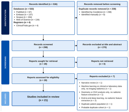
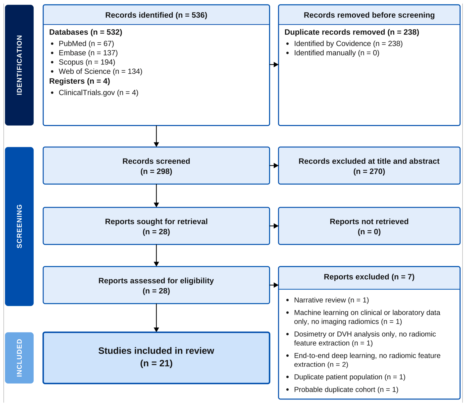
The systematic literature search yielded 536 records: 532 from the four databases and 4 from ClinicalTrials.gov. Following removal of 238 duplicates, 298 studies were screened by title and abstract. After initial screening, 28 full texts were assessed for eligibility. Seven studies were excluded at full-text review: one narrative review [25], one study applying ML to clinical/laboratory parameters without imaging radiomics [26], one study performing DVH/dosimetry analysis only without radiomic feature extraction [27], two end-to-end deep learning approaches without explicit radiomic feature extraction [28,29], one representing a duplicate patient population [30], and one representing a probable duplicate cohort [31]. Ultimately, 21 studies met the inclusion criteria and were included in the final analysis. A PRISMA flow diagram is presented in Figure 1.
Figure 1.
PRISMA 2020 flow diagram.
3.2. Methodological Characteristics and Quality
All 21 included studies were retrospective in design. Most were single-centre; Mosconi et al. [32] was multi-centre. Two studies were post hoc analyses of prospectively collected randomised trial cohorts: Ocal et al. (SIRFLOX, n = 491, of whom 239 received chemotherapy alone) [33] and Saalfeld et al. (SORAMIC sub-study, n = 297, of whom 147 received sorafenib alone) [34]. Because about half of the patients in these two cohorts did not receive TARE, they are described as trial-derived cohorts. Publication years ranged from 2012 to 2026. Table 1 summarises the methodological characteristics of all included studies [32,33,34,35,36,37,38,39,40,41,42,43,44,45,46,47,48,49,50,51,52].
Table 1.
Methodological characteristics of included studies (n = 21).
Methodological quality was low. The median RQS was 2 of a possible 36 (interquartile range 1 to 5; range −5 to 12), and the highest score (Marinelli et al., 12) was 33% of the maximum. No study used external validation, prospective enrolment, decision-curve or other clinical utility analysis; calibration was assessed in only one study (Wei et al.); a phantom study of feature repeatability was performed in only one study (Wei et al.); and inter-observer segmentation agreement was assessed in six studies. Most cohorts were small (fewer than 100 patients). On PROBAST, the overall risk of bias was high in all 21 studies, and the analysis domain was rated high in every study, chiefly because of few outcome events relative to the number of candidate predictors, feature selection or oversampling before validation, absence of calibration, and lack of external validation. Applicability was a high concern for Ocal et al. and Saalfeld et al. because roughly half of their patients did not receive TARE. Detailed results are provided in Supplementary Materials S2 and S3.
3.3. Participant Characteristics
The 21 included studies encompassed 1734 patients as reported (Table 1). Of these, 386 were comparator-arm patients in Ocal et al. and Saalfeld et al. who did not receive TARE, and up to 73 patients in Stocker et al. were also reported in Marinelli et al., so the number of unique TARE-treated patients lies between 1275 and 1348. Individual study populations ranged from 16 to 491 patients. Studies evaluated HCC (n = 12 studies), CLM (n = 3), iCC (n = 2), and mixed tumour cohorts (n = 4; Gensure, Kobe, Reimer and Wei). Table 1 summarises participant and study characteristics.
3.4. Imaging Modalities and Radiomics Workflow
3.4.1. Imaging Modalities
MRI was the most frequently utilised modality (nine studies), used for pre-procedural tumour characterisation, early post-TARE response assessment, and combined baseline/follow-up analysis. Sequences included contrast-enhanced T1-weighted (CE-T1W), T2-weighted (T2W), T2W with fat suppression, diffusion-weighted imaging (ADC maps), and hepatocellular phase imaging. CT was employed in five studies, encompassing portal venous phase, triphasic and quadriphasic acquisitions, and CT perfusion was used in one further study (Reiner et al.) [35]. 18F-FDG PET or PET/CT was used in three studies, and 90Y PET/CT or SPECT/CT in two. Cone-beam CT (CBCT), acquired during the angiographic procedure itself, was used in one study [36]. Post-procedural imaging was employed in four studies [37,38,39,40].
3.4.2. Segmentation
Segmentation approaches varied considerably (Table 2). Contouring was manual or reader-defined in ten studies, semi-automated (threshold, region-growing or interactive tools) in nine, automated with a deep learning model (nnU-Net) in one [33], and manual for the tumour with deep learning for the whole liver in one [40]. Inter-observer reproducibility of segmentation was assessed in six studies (Kobe et al. [36], Stocker et al. [41], Marinelli et al. [42], Reiner et al. [35], Ince et al. [45] and Roll et al. [51]); for example, ICC 0.83 in Marinelli et al. The remaining 15 studies did not assess it, which is a significant methodological gap given the well-established sensitivity of radiomic features to segmentation variability.
Table 2.
Radiomics workflow characteristics across included studies.
3.4.3. Feature Extraction, Selection, and Machine Learning
PyRadiomics was the most commonly used feature extraction package (eight studies), followed by LIFEx (five studies) and Olea Sphere (two studies); CGITA, mint Lesion, an in-house MATLAB toolbox, and 3D Slicer without a stated package were each used in one study, and custom code in two (Table 2). The number of candidate features ranged from 7 to 1128, with feature selection performed using LASSO or elastic-net regularisation, Pearson or Spearman correlation filtering, ICC- or CCC-based reproducibility filtering, XGBoost feature importance, Boruta, or univariable testing. Three studies (Mahmoud, Reimer and Reiner) performed no feature reduction and no correction for multiple testing. Final models incorporated between 2 and 20 selected features. ML algorithms included logistic regression, SVM, random forests, XGBoost, LightGBM, MLP ANN, LASSO Cox regression, and radiomic signature approaches. Table 2 summarises imaging parameters and performance metrics.
3.5. Outcomes and Predictive Performance
Response was assessed using Response Evaluation Criteria in Solid Tumours version 1.1 (RECIST 1.1), modified RECIST (mRECIST), or European Association for the Study of the Liver (EASL) criteria, depending on tumour type and study design. Eighteen studies reported an AUC: seventeen for treatment response and one (Saalfeld et al.) [34] for 1-year survival (Table 3). Because estimates obtained with different validation strategies are not comparable, they were not pooled, and Table 4 gives the range of the highest reported AUC in each validation stratum. Eight studies reported apparent performance with no validation (range 0.71 to 0.90; this includes Roll et al., whose logistic model was fitted on the same validation set on which the AUC is reported), six used internal cross-validation (range 0.73 to 1.00), and four used an internal hold-out set (range 0.80 to 0.90), with small test sets (about 30 to 50 patients or 21 lesions) and wide confidence intervals where reported. No study used external validation. Three studies [33,43,44] reported no AUC or C-index, and one [35] reported an AUC of 0.81 without a confidence interval. Radiomics-containing models had higher AUCs than clinical-only models in the six studies that reported both [41,42,45,46,47,48], whereas adding clinical or dosimetric variables to radiomics gave small increments (0.01 to 0.04) in four of the five studies that reported both models and no gain in one (Topcuoglu et al.) [49]. No single radiomic feature or feature class emerged consistently across studies.
3.5.1. Treatment Response—MRI-Based Studies
Seven studies evaluated pre-procedural MRI radiomics for treatment response prediction [41,42,45,46,47,48,50]. All seven reported an AUC; the highest reported values ranged from 0.74 to 1.00 (0.74 to 0.94 excluding Aujay et al., n = 22, LOOCV), none used external validation, and only Marinelli et al. used a hold-out set. Radiomics-containing models had higher AUCs than clinical-only models in the studies that reported both [41,42,45,46,47,48], but the increment from adding clinical variables to radiomics was small. The largest single-centre MRI study (Stocker et al., n = 154) [41] achieved an AUC of 0.736 with radiomics, clinical parameters and AFP, compared with 0.531 for clinical parameters alone, in repeated internal cross-validation, illustrating that performance is more modest in larger cohorts than in smaller studies. Stocker et al. state that 73 patients were also reported in previous studies, including Marinelli et al. [42], so the two results are not independent. Baseline pre-treatment features were most heavily weighted across models [42].
3.5.2. Treatment Response—CT-Based Studies
Five studies evaluated CT-based radiomics [32,33,34,43,51]. Three reported an AUC: 0.896 for Mosconi et al. (apparent performance, no validation), 0.75 for Roll et al. (95% CI 0.48 to 1.00; the logistic model was fitted on the same validation set, so this is an apparent estimate) and 0.803 for Saalfeld et al. (1-year survival). Texture features reflecting intratumoral heterogeneity, including entropy, energy, and grey-level matrix parameters, were the most consistently predictive across tumour types [32,35,36,51], consistent with the biological rationale that heterogeneity governs differential microsphere uptake. High-risk radiomic profiles correlated with significantly shorter overall survival (OS) in CLM (3.4 vs. 6.4 months, p < 0.001) [51], suggesting prognostic as well as predictive utility. Peri-procedural CBCT radiomics achieved an AUC of 0.85 in a hold-out set of 21 lesions, split at lesion rather than patient level, without dedicated pre-treatment imaging [36]; this is practically attractive but requires validation in independent patients.
3.5.3. Treatment Response and Survival—PET/CT-Based Studies
Three studies evaluated 18F-FDG PET or PET/CT radiomics. Of these three studies, two evaluated HCC [44,52] and one evaluated CLM [49], indicating that the reduced incremental value of clinical features in PET-based models discussed below is not simply attributable to a shift away from HCC, since HCC still comprised the majority of the PET literature. Two reported an AUC: 0.827 for metabolic tumour volume without validation [52] and 0.90 in a 30-patient hold-out set [49]. PET-based studies identified metabolic and textural features as predictors of response and survival, but no PET study was externally validated. In the one PET study that compared them, adding clinical features did not improve on radiomics alone [49] (AUC of 0.88 versus 0.90), so no conclusion can be drawn about differences between modalities. Whole-liver PET radiomics independently predicted progression-free survival (PFS) and OS, suggesting that background liver metabolic texture captures prognostic information beyond the target lesion [44].
3.5.4. Survival Prediction and Body Composition Radiomics
Two studies evaluated radiomics for survival prediction using trial-derived cohorts. Saalfeld et al. reported that CT body composition radiomics predicted 1-year survival in SORAMIC data (n = 297), with test-set AUC of 0.803 (95% CI 0.693 to 0.913) in the SIRT plus sorafenib arm and 0.758 (95% CI 0.638 to 0.878) in the sorafenib arm; the confidence intervals overlap widely, so a SIRT-specific radiomic survival signal is not established [34]. Ocal et al., using SIRFLOX data (n = 491), identified CT radiomic features interacting with treatment allocation that were associated with overall survival benefit from SIRT, with automated nnU-Net segmentation checked against manual expert segmentation in 80 patients; no AUC or C-index was reported and the signature was not validated [33]. Both studies draw on prospectively collected trial data but are retrospective post hoc analyses, about half of their patients (239 of 491 and 147 of 297) did not receive TARE, and both were rated at high risk of bias on PROBAST (Supplementary Material S3); their findings require prospective external validation [33,34].
3.5.5. Post-Procedural and Dosiomics Studies
All four reported an AUC, with highest reported values of 0.71 (Mahmoud et al., no validation, p = 0.07), 0.729 (Wei et al., nested cross-validation), 0.83 (Reimer et al., no validation) and 1.00 (Mansouri et al., nested cross-validation). Results from Mansouri et al. 2025 (AUC of 1.00, n = 17) should be interpreted with extreme caution given the very small cohort size and absence of external validation [40]; excluding this study, the highest reported values were 0.71 to 0.83. Post-procedural radiomics nonetheless offers the conceptually distinct advantage of capturing actual treatment effect rather than predicting it from baseline tumour characteristics. In Wei et al., the model combining post-therapy 90Y PET radiomics with absorbed dose was not significantly better than either alone (AUC of 0.729 versus 0.713 for each; C-index for progression of 0.803 versus 0.754 for dose alone) [39], so complementarity is suggested but not demonstrated. Post-treatment MRI texture analysis performed within a mean of 2.2 days of TARE identified progressive disease earlier than RECIST 1.1 criteria [37], highlighting a potential role for early post-procedural imaging in treatment monitoring. The analysed time point in that study was chosen using the outcome, which may inflate performance. The clinical utility of post-procedural radiomics is, however, constrained by the requirement for early follow-up imaging that is not universally available in routine practice, and by the small cohort sizes in all four studies (range n = 17–37). A summary of performance metrics is presented in Table 3.
Table 3.
Discriminative performance of models with radiomic features, by study and validation type.
Table 4.
Range of highest reported AUC by validation type.
4. Discussion
This systematic review identified 21 studies evaluating the role of radiomics in outcome prediction for TARE, encompassing 1734 patients (up to 73 counted twice) across a broad range of hepatic tumour types, imaging modalities, and clinical contexts. Compared with the prior systematic review by Mirza-Aghazadeh-Attari et al., which identified fifteen eligible studies through September 2023 [18], this update includes nine studies that were not in that review (eight published between 2024 and 2026 and one, Reiner et al. [35], in 2016). Two of the twenty-one studies were post hoc analyses of randomised trial cohorts (Ocal et al., SIRFLOX; Saalfeld et al., SORAMIC) [33,34], although about half of the patients in those cohorts did not receive TARE. Seven studies were excluded at full-text review, most commonly for applying dosimetry or dose-volume histogram analysis without explicit radiomic feature extraction, using end-to-end deep learning without interpretable radiomic features, or representing duplicate patient populations. These exclusions reflect the evolving and inconsistently defined boundaries between radiomics, dosiomics, and deep learning in the interventional oncology literature.
The overarching finding is that radiomic models show moderate to high apparent discrimination but that the evidence is at high risk of bias. Among the 18 studies reporting an AUC, the highest reported values ranged from 0.71 to 0.90 with no validation, 0.73 to 1.00 with internal cross-validation, and 0.80 to 0.90 with an internal hold-out set; no study used external validation. The highest reported AUCs here (0.71 to 1.00) overlap the ranges reported in our group’s earlier radiomics systematic reviews in anal cancer (0.68 to 1.00) [20], ovarian cancer (0.77 to 0.89 in validation sets) [22] and bladder cancer (median 0.75, range 0.70 to 0.84 in validation sets) [21], whereas the sinonasal review reported higher values (median AUC of 0.91 in validation sets) [19]. Those reviews mostly summarised training or validation-set estimates, addressed different outcomes and appraised quality with QUADAS-2, so the comparison is indicative only. Mirza-Aghazadeh-Attari et al. reported pooled sensitivity (83.5%) and specificity (86.7%) and a summary ROC AUC of 0.92 (95% CI 0.89 to 0.94) for response prediction, and a mean RQS of 11.4; because we applied explicit scoring conventions (for example, cross-validation alone scored as no validation; Supplementary Material S2), our median RQS of 2 should not be compared directly with their figure [18]. This should not be interpreted as clinical readiness. The majority of studies were small, retrospective, single-centre analyses without external validation, and AUC inflation by overfitting in small cohorts is well established in the radiomics literature [14,15]. All 21 studies were at high risk of bias on PROBAST.
Radiomics-containing models achieved higher AUCs than clinical-only models in the six studies that reported both [41,42,45,46,47,48], the largest contrast being Stocker et al. (n = 154; AUC of 0.736 with radiomics, clinical parameters and AFP versus 0.531 for clinical parameters alone, in repeated internal cross-validation) [41]. By contrast, adding clinical or dosimetric variables to radiomic features gave only small increments (0.01 to 0.04) in four studies [39,41,47,48] and none in one [49]. The clinical comparators were generally ad hoc models rather than established staging systems, so the incremental value of radiomics over standard care remains unproven. Radiomics may capture subvisual heterogeneity reflecting microarchitecture, vascularity and necrosis, while clinical variables provide systemic context, but the included studies did not test this. In the one PET study that compared them, clinical features added nothing to radiomics [49], and because this is a single study, no difference between modalities can be inferred. Examining findings by tumour type reveals important nuances. HCC was the most studied entity (12 of 21 studies), evaluated across MRI, CT, and PET modalities, with response criteria varying between RECIST 1.1, mRECIST, and EASL. Across HCC studies, pre-procedural MRI-based radiomics most consistently predicted response, with baseline pre-treatment features more heavily weighted than post-treatment delta-features in the majority of models [41,42,45], supporting a pre-procedural patient selection paradigm consistent with the clinical imperative to identify non-responders before rather than after the procedure. For intrahepatic cholangiocarcinoma (two studies), both MRI- and CT-based radiomics demonstrated promising performance, with the multi-centre CT study by Mosconi et al. (AUC of 0.896) particularly notable for its ComBat harmonisation approach to inter-centre imaging variability [32], a methodological step that should be standard practice in multi-centre radiomics research. Ballı et al. similarly demonstrated the predictive value of dynamic MRI-based radiomics in iCC (AUC of 0.839 for the best single sequence and 0.880 for the best combined model, n = 36, without validation) [48]; both iCC studies were unvalidated, and only one used MRI. For colorectal cancer liver metastases (three studies), trial-derived radiomics data from Ocal et al. (SIRFLOX, n = 491) [33] and PET radiomics data from Topcuoglu et al. (n = 74) [49] add to the evidence, but neither was externally validated and Ocal et al. reported no discrimination metric.
The two largest studies in this review draw on prospectively collected randomised trial data but are retrospective post hoc analyses. Ocal et al. conducted a post hoc radiomic analysis of the SIRFLOX randomised controlled trial (n = 491), identifying CT radiomic features from pre-treatment portal venous phase imaging that interacted with treatment allocation to predict overall survival benefit from SIRT, using automated nnU-Net segmentation validated against manual expert segmentation in 80 patients [33]. This is the first attempt at a radiomics-based predictive biomarker for treatment selection in TARE, a conceptually important distinction from prognostic models, but the signature was not validated and no discrimination metric was reported, so it does not yet establish that SIRT confers a survival advantage in a defined subgroup. Saalfeld et al. conducted a post hoc analysis of the SORAMIC trial (n = 297), demonstrating that body composition radiomics derived from skeletal muscle and adipose tissue segmentation predicted 1-year survival with greater accuracy in the SIRT + sorafenib arm (AUC of 0.803) than sorafenib monotherapy (AUC of 0.758; overlapping confidence intervals), which does not establish that treatment context modifies the prognostic value of body composition radiomics [34]. The involvement of body composition as a radiomic predictor aligns with established literature on sarcopenia as a determinant of TARE outcomes, extending this concept into quantitative imaging-based phenotyping. Both studies are post hoc analyses, about half of their patients did not receive TARE, and their findings require prospective external validation.
The breadth of imaging modalities evaluated in this review reflects the clinical heterogeneity of the TARE patient population and the opportunistic nature of radiomics research. MRI dominated, reflecting its superior soft tissue contrast and clinical primacy for HCC characterisation and treatment response assessment. The use of peri-procedural CBCT by Kobe et al. [36] is particularly noteworthy from a translational perspective: radiomic features extracted from CBCT acquired during the TARE procedure itself, without any requirement for dedicated additional imaging, achieved an AUC of 0.85 for response prediction in a hold-out set of 21 lesions split at lesion rather than patient level. If validated prospectively, intra-procedural CBCT radiomics could provide real-time information to guide microsphere dosing without additional patient burden.
Post-procedural radiomics and dosiomics represent a conceptually distinct dimension of this literature. Four studies evaluated post-procedural imaging, with highest reported AUCs of 0.71 to 1.00 (0.71 to 0.83 excluding the Mansouri et al. 2025 result of an AUC of 1.00 in n = 17, which should be interpreted with extreme caution [40]). Post-procedural radiomics offers the advantage of capturing the actual biological response to treatment rather than predicting it from pre-treatment characteristics alone. The combination of post-therapy 90Y PET radiomics with absorbed dose metrics by Wei et al. was not significantly better than either alone (AUC of 0.729 versus 0.713 for each; C-index for progression of 0.803) [39] so complementarity is suggested but not demonstrated. Reimer et al. reported that post-treatment MRI texture analysis performed within a mean of 2.2 days of TARE could discriminate progressive disease (AUC up to 0.83), although the analysed time point was chosen using the outcome [37]; if confirmed, this could have implications for early treatment monitoring. Interpretation of post-SIRT imaging nonetheless remains inherently complex, given overlapping post-treatment inflammatory and necrotic change; imaging features specifically predictive of local recurrence, as distinct from treatment response in general, would represent a particularly useful clinical application of this approach. In practice, post-treatment dosimetry is already reviewed to guide follow-up, with re-imaging typically performed at three months where tumour uptake and absorbed dose appear adequate; a radiomic signature capable of predicting response in this pre-selected population, who by definition had favourable pre-treatment features and therefore proceeded to treatment, would be of considerable additional clinical value. The dosiomics field in TARE remains nascent, with the only dedicated dosiomics study to date too small to draw firm conclusions (n = 17) [40], but the conceptual framework is compelling and warrants dedicated prospective investigation.
Substantial methodological limitations constrain clinical translation. The majority of included studies were small (most n < 100), retrospective, single-centre analyses with no external validation, a pattern consistent with the broader radiomics literature [14,15]. Extreme AUC values in very small cohorts, specifically AUC 1.00 by both Aujay et al. (n = 22, LOOCV) [50] and Mansouri et al. (n = 17) [40], almost certainly reflect overfitting rather than true predictive performance. Methodological heterogeneity was pervasive across tumour types, imaging protocols, segmentation approaches, feature extraction software, machine learning algorithms, and outcome definitions, precluding meta-analytic synthesis. Publication and small-study bias could not be assessed formally without a meta-analysis, and the predominance of small studies reporting high apparent AUCs makes selective reporting likely. Segmentation reproducibility was assessed in only six studies (for example Marinelli et al., ICC = 0.83) [42] representing a significant methodological gap given the well-established sensitivity of radiomic features to segmentation variability. The adoption of automated segmentation (nnU-Net in Ocal et al.) [33] and standardised feature extraction pipelines (PyRadiomics, used in eight studies) represents progress, but adoption of the Imaging Biomarker Standardisation Initiative framework [53] remains far from routine across the included studies. Finally, the absence of biological validation linking radiomic phenotypes to underlying tumour pathophysiology limits mechanistic interpretability and the prospect of regulatory approval.
Compared with Mirza-Aghazadeh-Attari et al. [18], which identified 15 studies and performed a meta-analysis of AUC values, the current update includes 9 studies that were not in that review. The expanded and more heterogeneous evidence base identified here reinforces that quantitative pooling remains premature, and that a structured narrative synthesis is the most appropriate analytical approach at this stage of the field’s development. That review searched until 1 September 2023. Eight of the nine studies not included in it were published between 2024 and 2026 and one (Reiner et al. [35]) in 2016; the recent studies reflect the accelerating pace of radiomic research in interventional oncology and the growing interest in this area among interventional radiology and nuclear medicine groups internationally.
This review has limitations. Two cohorts overlap, because Stocker et al. state that 73 patients were reported in earlier studies including Marinelli et al., so up to 73 patients may be counted twice. Taking the highest reported AUC per study favours optimism, and the validation strata contain few studies. Scoring with the RQS and PROBAST involves judgement, although each study was scored independently by two reviewers. Only English-language studies were included.
Future Directions
Several priority areas emerge from this synthesis. Prospective multi-centre studies with pre-specified outcome definitions, standardised imaging protocols, and rigorous external validation are most urgently needed. Harmonisation of radiomics workflows, including standardised feature extraction software, image normalisation, voxel resampling, and segmentation protocols, is essential to enable cross-study comparisons and eventual meta-analysis. The Imaging Biomarker Standardisation Initiative provides an established framework for feature standardisation that should be universally adopted in future TARE radiomics studies [53]. Similarly, reporting quality should be assessed using validated tools such as METRICS [54] to facilitate quality appraisal across future studies.
Dosiomics merits dedicated investigation in adequately powered cohorts. The combination of spatial dosimetric information with radiomic tumour characterisation is mechanistically grounded in the biology of TARE, where microsphere distribution is governed by tumour vascularity, itself a key determinant of radiomic texture. Prospective studies pairing pre-procedural radiomic assessment with post-procedural dosimetric verification could provide a comprehensive imaging-based framework integrating TARE planning, delivery, and response assessment.
The identification of biologically interpretable radiomic features linked to underlying tumour pathophysiology will be essential to facilitate mechanistic understanding and clinician adoption. Radiogenomic integration, linking radiomic phenotypes to molecular profiles, represents a particularly promising avenue in HCC, where genomic heterogeneity is well characterised and may drive differential TARE response [16]. Finally, studies demonstrating the incremental clinical value of radiomics over current standard of care, rather than standalone discriminative performance, are needed to define clinical utility. Decision curve analyses and prospective clinical impact studies examining whether radiomics improves patient selection beyond BCLC staging, Child–Pugh score, AFP, and 99mTc-MAA SPECT dosimetry would represent a genuine advance toward personalised locoregional therapy for hepatic malignancies.
5. Conclusions
Radiomics applied to pre- and post-procedural imaging in TARE shows moderate to high apparent discrimination across hepatic tumour types and imaging modalities, but all 21 studies were at high risk of bias, none used external validation, and the median RQS was 2 of 36. Adding clinical variables to radiomic features gave only small gains. The evidence base consists of small, retrospective, mostly single-centre studies. Radiomic analyses of trial-derived cohorts (SIRFLOX, n = 491; SORAMIC, n = 297) and emerging dosiomics work are of interest but remain unvalidated. Prospective, multi-centre validation studies with standardised radiomics pipelines are required before radiomics can be routinely adopted in personalised TARE patient selection and treatment optimisation.
Supplementary Materials
The following supporting information can be downloaded at: https://www.mdpi.com/article/10.3390/diagnostics16203322/s1, Supplementary Material S1: Full search strategies for each database; Supplementary Material S2: Radiomics Quality Score scoring conventions and per-study scores; Supplementary Material S3: PROBAST risk of bias and applicability assessments. References [15,24,32,33,34,35,36,37,38,39,40,41,42,43,44,45,46,47,48,49,50,51,52] are cited in the Supplementary Materials.
Author Contributions
Conceptualization, R.T.G. and H.C.T.; Methodology, R.T.G. and H.C.T.; Validation, M.E.K.; Formal Analysis, R.T.G. and H.C.T.; Investigation, R.T.G. and H.C.T.; Data Curation, R.T.G. and H.C.T.; Writing—Original Draft Preparation, R.T.G.; Writing—Review & Editing, R.T.G., H.C.T., O.A.S., N.J.O., B.M.M.C., K.P.S., I.B., Z.Q.N., T.M., M.E.K., J.S. and H.Y.; Supervision, M.E.K.; Project Administration, R.T.G. and H.C.T. All authors have read and agreed to the published version of the manuscript.
Funding
This research received no external funding.
Institutional Review Board Statement
Not applicable.
Informed Consent Statement
Not applicable.
Data Availability Statement
No new data were created or analysed in this study. Data sharing is not applicable to this article.
Acknowledgments
The authors thank the Joly Leadership Fund, Trinity St James Cancer Institute.
Conflicts of Interest
HY received Research Grant from Guerbet LLC and PleuroDynamics. The other authors declare no conflicts of interest.
References
- Sung, H.; Ferlay, J.; Siegel, R.L.; Laversanne, M.; Soerjomataram, I.; Jemal, A.; Bray, F. Global Cancer Statistics 2020: GLOBOCAN Estimates of Incidence and Mortality Worldwide for 36 Cancers in 185 Countries. CA Cancer J. Clin. 2021, 71, 209–249. [Google Scholar] [CrossRef] [Scilit] [PubMed]
- Huang, D.Q.; Singal, A.G.; Kono, Y.; Tan, D.J.H.; El-Serag, H.B.; Loomba, R. Changing global epidemiology of liver cancer from 2010 to 2019: NASH is the fastest growing cause of liver cancer. Cell Metab. 2022, 34, 969–977.e2. [Google Scholar] [CrossRef] [Scilit] [PubMed]
- Siegel, R.L.; Miller, K.D.; Jemal, A. Cancer statistics, 2020. CA Cancer J. Clin. 2020, 70, 7–30. [Google Scholar] [CrossRef] [Scilit] [PubMed]
- Rizvi, S.; Khan, S.A.; Hallemeier, C.L.; Kelley, R.K.; Gores, G.J. Cholangiocarcinoma—Evolving concepts and therapeutic strategies. Nat. Rev. Clin. Oncol. 2018, 15, 95–111. [Google Scholar] [CrossRef] [Scilit] [PubMed]
- Salem, R.; Thurston, K.G. Radioembolization with yttrium-90 microspheres: A state-of-the-art brachytherapy treatment for primary and secondary liver malignancies. J. Vasc. Interv. Radiol. 2006, 17, 1251–1278. [Google Scholar] [CrossRef] [Scilit] [PubMed]
- Gaba, R.C.; Lewandowski, R.J.; Hickey, R. Transcatheter Therapy for Hepatic Malignancy: Standardization of Terminology and Reporting Criteria. J. Vasc. Interv. Radiol. 2016, 27, 457–473. [Google Scholar] [CrossRef] [Scilit] [PubMed]
- Lewandowski, R.J.; Kulik, L.M.; Riaz, A. A comparative analysis of transarterial downstaging for hepatocellular carcinoma. Am. J. Transplant. 2009, 9, 1920–1928. [Google Scholar] [CrossRef] [Scilit] [PubMed]
- Vouche, M.; Lewandowski, R.J.; Atassi, R. Radiation lobectomy: Time-dependent analysis of future liver remnant volume. J. Hepatol. 2013, 59, 1029–1036. [Google Scholar] [CrossRef] [Scilit] [PubMed]
- Ricke, J.; Klümpen, H.J.; Amthauer, H. Impact of combined selective internal radiation therapy and sorafenib on survival in advanced hepatocellular carcinoma. J. Hepatol. 2019, 71, 1164–1174. [Google Scholar] [CrossRef] [Scilit] [PubMed]
- Van Hazel, G.A.; Heinemann, V.; Sharma, N.K. SIRFLOX: Randomized Phase III Trial Comparing First-Line mFOLFOX6 ± Bevacizumab Versus mFOLFOX6 + Selective Internal Radiation Therapy ± Bevacizumab in Patients with Metastatic Colorectal Cancer. J. Clin. Oncol. 2016, 34, 1723–1731. [Google Scholar] [CrossRef] [Scilit] [PubMed]
- Sieghart, W.; Hucke, F.; Peck-Radosavljevic, M. Transarterial chemoembolization: Modalities, indication, and patient selection. J. Hepatol. 2015, 62, 1187–1195. [Google Scholar] [CrossRef] [Scilit] [PubMed]
- Deipolyi, A.R.; Rho, J.; Khademian, Z.; Oklu, R. Differences in yttrium-90 radioembolization outcomes between hepatocellular carcinoma and intrahepatic cholangiocarcinoma. J. Vasc. Interv. Radiol. 2016, 27, 1137–1142. [Google Scholar]
- Wallace, M.C.; Samuelson, S.; Khoo, T.; Ooi, J.; Tibballs, J.; Ferguson, J.; Preen, D.B.; Knuiman, M.; Garas, G.; MacQuillan, G.; et al. The MAAPE score in intermediate and advanced hepatocellular carcinoma treated with Yttrium-90 resin microsphere radioembolization. J. Gastroenterol. Hepatol. 2020, 35, 1945–1952. [Google Scholar] [CrossRef] [Scilit] [PubMed]
- Gillies, R.J.; Kinahan, P.E.; Hricak, H. Radiomics: Images Are More than Pictures, They Are Data. Radiology 2016, 278, 563–577. [Google Scholar] [CrossRef] [Scilit] [PubMed]
- Lambin, P.; Leijenaar, R.T.H.; Deist, T.M. Radiomics: The bridge between medical imaging and personalized medicine. Nat. Rev. Clin. Oncol. 2017, 14, 749–762. [Google Scholar] [CrossRef] [Scilit] [PubMed]
- Avanzo, M.; Wei, L.; Stancanello, J. Machine and deep learning methods for radiomics. Med. Phys. 2020, 47, e185–e202. [Google Scholar] [CrossRef] [Scilit] [PubMed]
- Guiot, J.; Vaidyanathan, A.; Deprez, L. A review in radiomics: Making personalized medicine a reality via routine imaging. Med. Res. Rev. 2022, 42, 426–440. [Google Scholar] [CrossRef] [Scilit] [PubMed]
- Mirza-Aghazadeh-Attari, M.; Srinivas, T.; Kamireddy, A.; Kim, A.; Weiss, C.R. Radiomics Features Extracted from Pre- and Postprocedural Imaging in Early Prediction of Treatment Response in Patients Undergoing Transarterial Radioembolization of Hepatic Lesions: A Systematic Review, Meta-Analysis, and Quality Appraisal Study. J. Am. Coll. Radiol. 2024, 21, 740–751. [Google Scholar] [CrossRef] [Scilit] [PubMed]
- Waters, C.; Hall, T.; Temperley, H.C.; Jones, H.; O’Sullivan, N.J.; McHugh, A.; Tohidinezhad, F.; Subramaniam, T. Role of Radiomics to Predict Disease Recurrence in Sinonasal Squamous Cell Carcinoma: A Systematic Review and Meta-Analysis. Clin. Otolaryngol. 2026, 51, 7–16. [Google Scholar] [CrossRef] [Scilit] [PubMed]
- Temperley, H.C.; O’Sullivan, N.J.; Waters, C.; Corr, A.; Mehigan, B.J.; O’Kane, G.; McCormick, P.; Gillham, C.; Rausa, E.; Larkin, J.O.; et al. Radiomics; Contemporary Applications in the Management of Anal Cancer; A Systematic Review. Am. Surg. 2024, 90, 445–454. [Google Scholar] [CrossRef] [Scilit] [PubMed]
- O’Sullivan, N.J.; Temperley, H.C.; Corr, A.; Meaney, J.F.M.; Lonergan, P.E.; Kelly, M.E. Current role of radiomics and radiogenomics in predicting oncological outcomes in bladder cancer. Curr. Urol. 2025, 19, 43–48. [Google Scholar] [CrossRef] [Scilit] [PubMed]
- O’Sullivan, N.J.; Temperley, H.C.; Horan, M.T.; Kamran, W.; Corr, A.; O’Gorman, C.; Saadeh, F.; Meaney, J.M.; Kelly, M.E. Role of radiomics as a predictor of disease recurrence in ovarian cancer: A systematic review. Abdom. Radiol. 2024, 49, 3540–3547. [Google Scholar] [CrossRef] [Scilit] [PubMed]
- Page, M.J.; McKenzie, J.E.; Bossuyt, P.M. The PRISMA 2020 statement: An updated guideline for reporting systematic reviews. BMJ 2021, 372, n71. [Google Scholar] [CrossRef] [Scilit] [PubMed]
- Wolff, R.; Moons, K.; Riley, R.; Whiting, P.; Westwood, M.; Collins, G.; Reitsma, J.; Kleijnen, J.; Mallett, S. PROBAST: A tool to assess the risk of bias and applicability of prediction model studies. Ann. Intern. Med. 2019, 170, 51–58. [Google Scholar] [CrossRef] [Scilit] [PubMed]
- Hsieh, C.; Laguna, A.; Ikeda, I.; Maxwell, A.W.P.; Chapiro, J.; Nadolski, G.; Jiao, Z.; Bai, H.X. Using Machine Learning to Predict Response to Image-guided Therapies for Hepatocellular Carcinoma. Radiology 2023, 309, e222891. [Google Scholar] [CrossRef] [Scilit] [PubMed]
- Ingrisch, M.; Schoppe, F.; Paprottka, K.; Fabritius, M.; Strobl, F.F.; De Toni, E.N.; Ilhan, H.; Todica, A.; Michl, M.; Paprottka, P.M. Prediction of 90Y Radioembolization Outcome from Pretherapeutic Factors with Random Survival Forests. J. Nucl. Med. 2018, 59, 769–773. [Google Scholar] [CrossRef] [Scilit] [PubMed]
- Veenstra, E.B.; Ruiter, S.J.S.; de Haas, R.J.; Bokkers, R.P.H.; de Jong, K.P.; Noordzij, W. Post-treatment three-dimensional voxel-based dosimetry after Yttrium-90 resin microsphere radioembolization in HCC. EJNMMI Res. 2022, 12, 9. [Google Scholar] [CrossRef] [Scilit] [PubMed]
- Plachouris, D.; Tzolas, I.; Gatos, I.; Papadimitroulas, P.; Spyridonidis, T.; Apostolopoulos, D.; Papathanasiou, N.; Visvikis, D.; Plachouri, K.M.; Hazle, J.D.; et al. A deep-learning-based prediction model for the biodistribution of 90Y microspheres in liver radioembolization. Med. Phys. 2021, 48, 7427–7438. [Google Scholar] [CrossRef] [Scilit] [PubMed]
- Wagstaff, W.V.; Villalobos, A.; Gichoya, J.; Kokabi, N. Using Deep Learning to Predict Treatment Response in Patients with Hepatocellular Carcinoma Treated with Y90 Radiation Segmentectomy. J. Digit. Imaging 2023, 36, 1180–1188. [Google Scholar] [CrossRef] [Scilit] [PubMed]
- Stuber, A.T.; Coors, S.; Schachtner, B.; Weber, T.; Rugamer, D.; Bender, A.; Mittermeier, A.; Ocal, O.; Seidensticker, M.; Ricke, J.; et al. A Comprehensive Machine Learning Benchmark Study for Radiomics-Based Survival Analysis of CT Imaging Data in Patients With Hepatic Metastases of CRC. Investig. Radiol. 2023, 58, 874–881. [Google Scholar] [CrossRef] [Scilit] [PubMed]
- Mansouri, Z.; Salimi, Y.; Hajianfar, G.; Wolf, N.B.; Knappe, L.; Xhepa, G.; Gleyzolle, A.; Ricoeur, A.; Garibotto, V.; Mainta, I.; et al. The role of biomarkers and dosimetry parameters in overall and progression free survival prediction for patients treated with personalized 90Y glass microspheres SIRT: A preliminary machine learning study. Eur. J. Nucl. Med. Mol. Imaging 2024, 51, 4111–4126. [Google Scholar] [CrossRef] [Scilit] [PubMed]
- Mosconi, C.; Cucchetti, A.; Bruno, A.; Cappelli, A.; Bargellini, I.; De Benedittis, C.; Lorenzoni, G.; Gramenzi, A.; Tarantino, F.P.; Parini, L.; et al. Radiomics of cholangiocarcinoma on pretreatment CT can identify patients who would best respond to radioembolisation. Eur. Radiol. 2020, 30, 4534–4544. [Google Scholar] [CrossRef] [Scilit] [PubMed]
- Öcal, O.; Stüber, A.T.; Abacı, G.; Wildgruber, M.; Mansour, N.; Deniz, S.; Puhr-Westerheide, D.; Fabritius, M.P.; Ricke, J.; Ingrisch, M.; et al. Prognostic and predictive value of radiomics-based imaging features in patients with colorectal liver metastasis receiving radioembolisation in first-line setting. Eur. J. Radiol. Open 2026, 16, 100750. [Google Scholar] [CrossRef] [Scilit] [PubMed]
- Saalfeld, S.; Kreher, R.; Hille, G.; Niemann, U.; Hinnerichs, M.; Öcal, O.; Schütte, K.; Zech, C.J.; Loewe, C.; van Delden, O.; et al. Prognostic role of radiomics-based body composition analysis for the 1-year survival for hepatocellular carcinoma patients. J. Cachexia Sarcopenia Muscle 2023, 14, 2301–2309. [Google Scholar] [CrossRef] [Scilit] [PubMed]
- Reiner, C.S.; Gordic, S.; Puippe, G.; Morsbach, F.; Wurnig, M.; Schaefer, N.; Veit-Haibach, P.; Pfammatter, T.; Alkadhi, H. Histogram Analysis of CT Perfusion of Hepatocellular Carcinoma for Predicting Response to Transarterial Radioembolization: Value of Tumor Heterogeneity Assessment. Cardiovasc. Interv. Radiol. 2016, 39, 400–408. [Google Scholar] [CrossRef] [Scilit] [PubMed]
- Kobe, A.; Zgraggen, J.; Messmer, F.; Puippe, G.; Sartoretti, T.; Alkadhi, H.; Pfammatter, T.; Mannil, M. Prediction of treatment response to transarterial radioembolization of liver metastases: Radiomics analysis of pre-treatment cone-beam CT: A proof of concept study. Eur. J. Radiol. Open 2021, 8, 100375. [Google Scholar] [CrossRef] [Scilit] [PubMed]
- Reimer, R.P.; Reimer, P.; Mahnken, A.H. Assessment of therapy response to transarterial radioembolization for liver metastases by means of post-treatment MRI-based texture analysis. Cardiovasc. Interv. Radiol. 2018, 41, 1545–1556. [Google Scholar] [CrossRef] [Scilit] [PubMed]
- Mahmoud, M.; Lin, K.H.; Lee, R.C.; Liu, C.A. Assessment of Y-90 Radioembolization Treatment Response for Hepatocellular Carcinoma Cases Using MRI Radiomics. Mol. Imaging Radionucl. Ther. 2024, 33, 156–166. [Google Scholar] [CrossRef] [Scilit] [PubMed]
- Wei, L.; Cui, C.; Xu, J.; Kaza, R.; El Naqa, I.; Dewaraja, Y.K. Tumor response prediction in 90Y radioembolization with PET-based radiomics features and absorbed dose metrics. EJNMMI Phys. 2020, 7, 74. [Google Scholar] [CrossRef] [Scilit] [PubMed]
- Mansouri, Z.; Salimi, Y.; Hajianfar, G.; Knappe, L.; Wolf, N.B.; Xhepa, G.; Gleyzolle, A.; Ricoeur, A.; Garibotto, V.; Mainta, I.; et al. Potential of Radiomics, Dosiomics, and Dose Volume Histograms for Tumor Response Prediction in Hepatocellular Carcinoma following 90Y-SIRT. Mol. Imaging Biol. 2025, 27, 201–214. [Google Scholar] [CrossRef] [Scilit] [PubMed]
- Stocker, D.; Hectors, S.; Marinelli, B.; Carbonell, G.; Bane, O.; Hulkower, M.; Kennedy, P.; Ma, W.; Lewis, S.; Kim, E.; et al. Prediction of hepatocellular carcinoma response to radiation segmentectomy using an MRI-based machine learning approach. Abdom. Radiol. 2025, 50, 2000–2011. [Google Scholar] [CrossRef] [Scilit] [PubMed]
- Marinelli, B.; Chen, M.; Stocker, D.; Charles, D.; Radell, J.; Lee, J.; Fauveau, V.; Bello-Martinez, R.; Kim, E.; Taouli, B. Early prediction of response of hepatocellular carcinoma to yttrium-90 radiation segmentectomy using a machine learning MR imaging radiomic approach. J. Vasc. Interv. Radiol. 2023, 34, 1794–1801. [Google Scholar] [CrossRef] [Scilit] [PubMed]
- Gensure, R.; Foran, D.; Lee, V.; Gendel, V.; Jabbour, S.; Carpizo, D.; Nosher, J.; Yang, L. Evaluation of hepatic tumor response to yttrium-90 radioembolization therapy using texture signatures generated from contrast-enhanced CT images. Acad. Radiol. 2012, 19, 1201–1207. [Google Scholar] [CrossRef] [Scilit] [PubMed]
- Blanc-Durand, P.; Van Der Gucht, A.; Jreige, M.; Nicod-Lalonde, M.; Silva-Monteiro, M.; Prior, J.; Denys, A.; Depeursinge, A.; Schaefer, N. Signature of survival: A 18F-FDG PET based whole-liver radiomic analysis predicts survival after 90Y-TARE for hepatocellular carcinoma. Oncotarget 2018, 9, 4549–4558. [Google Scholar] [CrossRef] [Scilit] [PubMed]
- Ince, O.; Önder, H.; Gençtürk, M.; Cebeci, H.; Golzarian, J.; Young, S. Prediction of response of hepatocellular carcinoma to radioembolization: Machine learning using preprocedural clinical factors and MR imaging radiomics. J. Vasc. Interv. Radiol. 2023, 34, 235–243. [Google Scholar] [CrossRef] [Scilit] [PubMed]
- Sarioglu, O.; Canturk, A.; Yarol, R.; Gulmez, H.; Sarioglu, F.; Derebek, E.; Gulcu, A. Machine learning using MR imaging radiomics can predict the response of large hepatocellular carcinoma to transarterial radioembolization. Niger. J. Clin. Pract. 2025, 28, 1111–1120. [Google Scholar] [CrossRef] [Scilit] [PubMed]
- Sozutok, S.; Piskin, F.C.; Balli, H.T.; Yucel, S.P.; Aikimbaev, K. Predicting treatment responses using magnetic resonance imaging-based radiomics in hepatocellular carcinoma patients undergoing transarterial radioembolization. Rev. Assoc. Médica Bras. 2024, 70, e20240721. [Google Scholar] [CrossRef] [Scilit] [PubMed]
- Ballı, H.; Pişkin, F.; Yücel, S.; Sözütok, S.; Özgül, D.; Aikimbaev, K. Predictability of the radiological response to yttrium-90 transarterial radioembolization by dynamic magnetic resonance imaging-based radiomics analysis in patients with intrahepatic cholangiocarcinoma. Diagn. Interv. Radiol. 2024, 30, 193–199. [Google Scholar] [CrossRef] [Scilit] [PubMed]
- Topcuoglu, O.M.; Tuncer, O.; Oral, M.; Gormez, A.; Toklu, T.; Selcuk, N.A. 18F-FDG PET radiomics model for predicting TARE response in patients with colorectal cancer liver metastases. Jpn. J. Radiol. 2026, 44, 1062–1070. [Google Scholar] [CrossRef] [Scilit] [PubMed]
- Aujay, G.; Etchegaray, C.; Blanc, J.; Lapuyade, B.; Papadopoulos, P.; Pey, M.; Bordenave, L.; Trillaud, H.; Saut, O.; Pinaquy, J. Comparison of MRI-based response criteria and radiomics for the prediction of early response to transarterial radioembolization in patients with hepatocellular carcinoma. Diagn. Interv. Imaging 2022, 103, 360–366. [Google Scholar] [CrossRef] [Scilit] [PubMed]
- Roll, W.; Masthoff, M.; Köhler, M.; Rahbar, K.; Stegger, L.; Ventura, D.; Morgül, H.; Trebicka, J.; Schäfers, M.; Heindel, W.; et al. Radiomics-based prediction model for outcome of radioembolization in metastatic colorectal cancer. Cardiovasc. Interv. Radiol. 2024, 47, 462–471. [Google Scholar] [CrossRef] [Scilit] [PubMed]
- Karahan Şen, N.; Alataş, Ö.; Gülcü, A.; Özdoğan, Ö.; Derebek, E.; Çapa Kaya, G. The role of volumetric and textural analysis of pretreatment 18F-fluorodeoxyglucose PET/computerized tomography images in predicting complete response to transarterial radioembolization in hepatocellular cancer. Nucl. Med. Commun. 2022, 43, 807–814. [Google Scholar] [CrossRef] [Scilit] [PubMed]
- Zwanenburg, A.; Vallieres, M.; Abdalah, M.A.; Aerts, H.; Andrearczyk, V.; Apte, A.; Ashrafinia, S.; Bakas, S.; Beukinga, R.J.; Boellaard, R.; et al. The Image Biomarker Standardization Initiative: Standardized Quantitative Radiomics for High-Throughput Image-based Phenotyping. Radiology 2020, 295, 328–338. [Google Scholar] [CrossRef] [Scilit] [PubMed]
- Kocak, B.; Akinci D’Antonoli, T.; Mercaldo, N.; Alberich-Bayarri, A.; Baessler, B.; Ambrosini, I.; Andreychenko, A.E.; Bakas, S.; Beets-Tan, R.G.H.; Bressem, K.; et al. METhodological RadiomICs Score (METRICS): A quality scoring tool for radiomics research endorsed by EuSoMII. Insights Imaging 2024, 15, 8. [Google Scholar] [CrossRef] [Scilit] [PubMed]

Disclaimer/Publisher’s Note: The statements, opinions and data contained in all publications are solely those of the individual author(s) and contributor(s) and not of MDPI and/or the editor(s). MDPI and/or the editor(s) disclaim responsibility for any injury to people or property resulting from any ideas, methods, instructions or products referred to in the content. |
© 2026 by the authors. Licensee MDPI, Basel, Switzerland. This article is an open access article distributed under the terms and conditions of the Creative Commons Attribution (CC BY) license.