Thermography and Infrared Spectroscopy in the Detection of Periodontal Inflammation In Vivo: A Systematic Review



 and
and
Abstract
1. Introduction
2. Materials and Methods
2.1. Search Strategy
2.2. Study Detection
Inclusion and Exclusion Criteria
3. Results
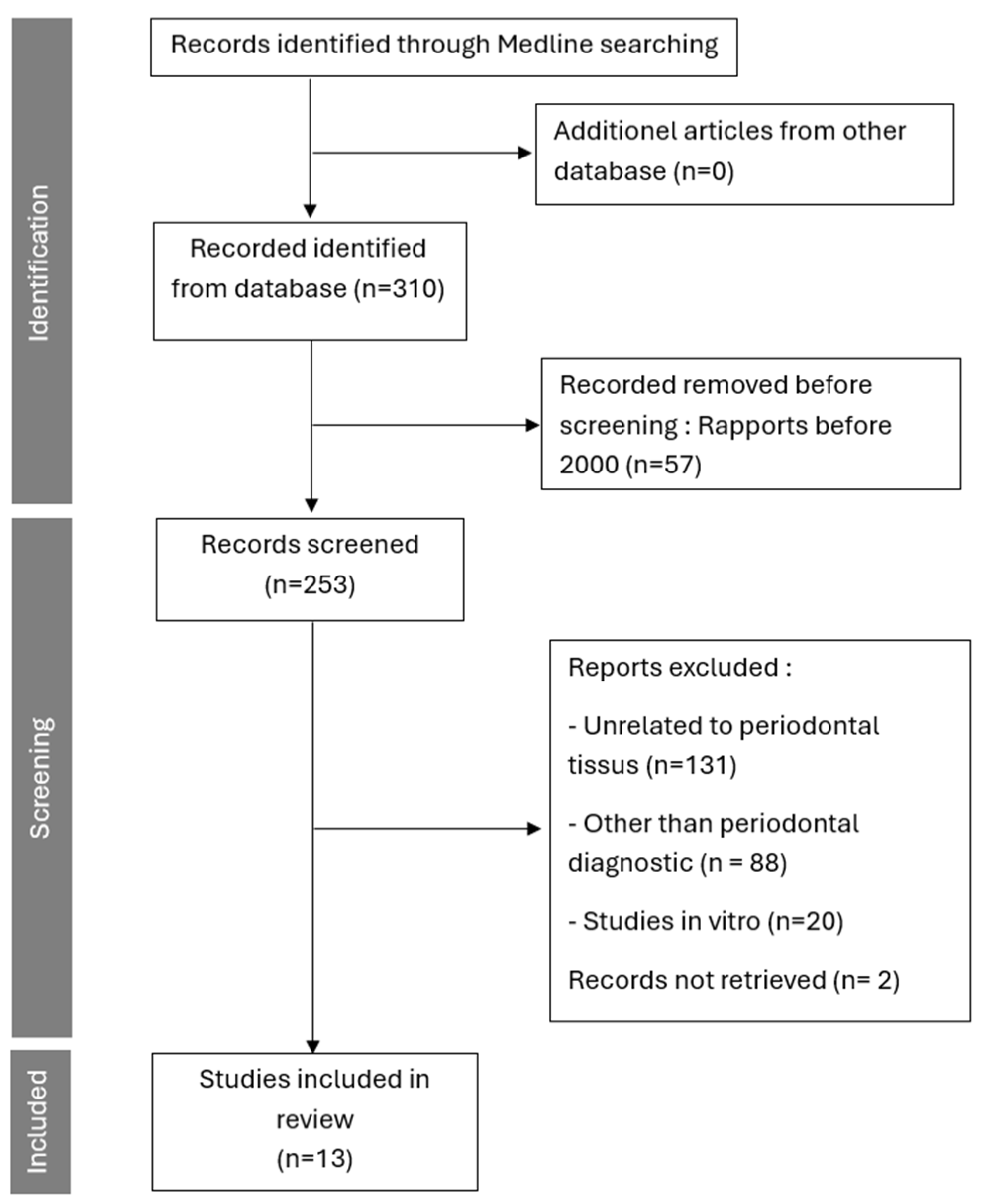
3.1. Study Selection
3.1.1. Presentation of IR Thermography Studies
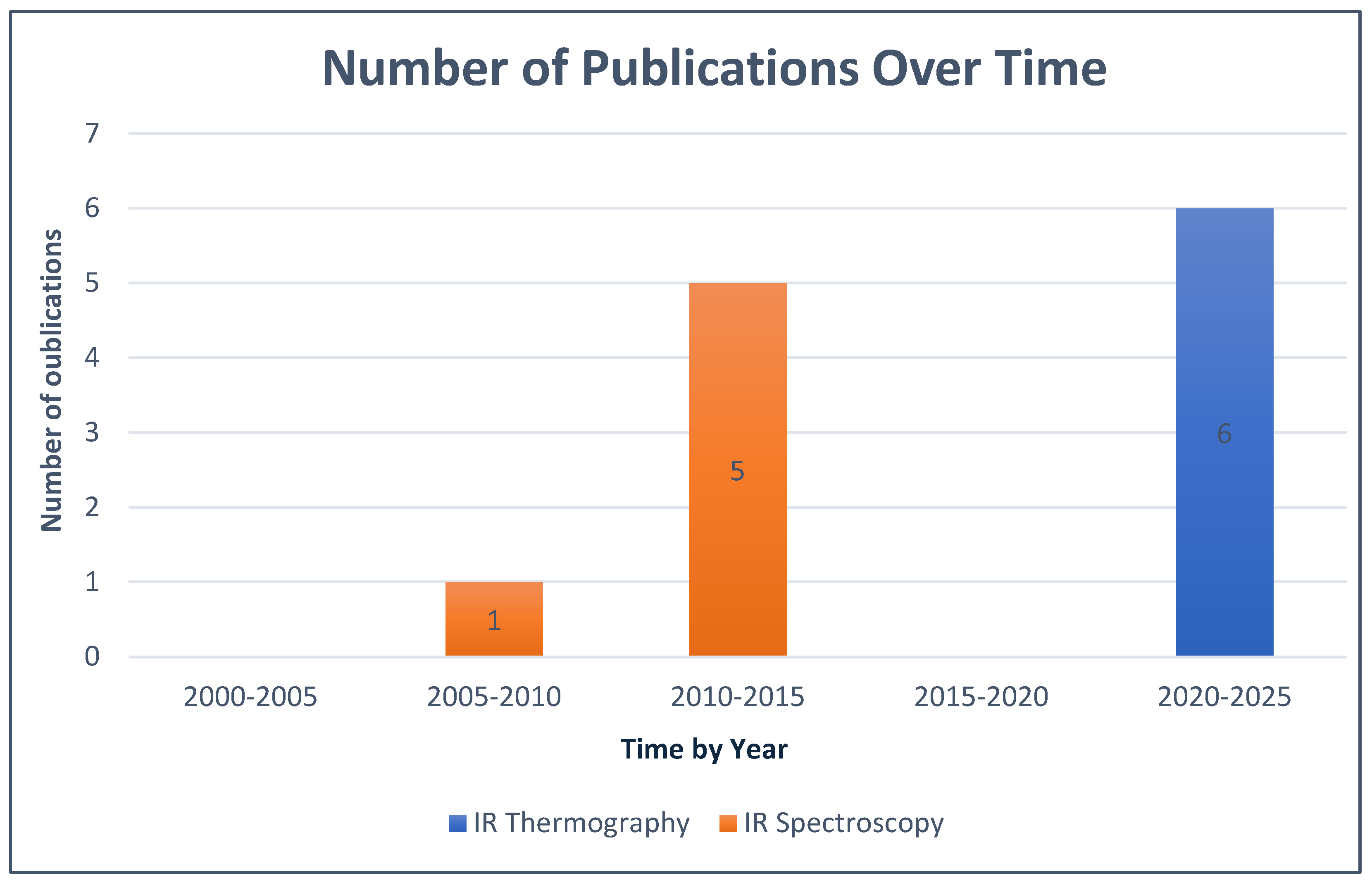
3.1.2. Evolution of the Studies
3.1.3. Presentation of Cameras Used for IR Thermography
3.1.4. Overview of Devices Used for IR Spectroscopy
4. Discussion
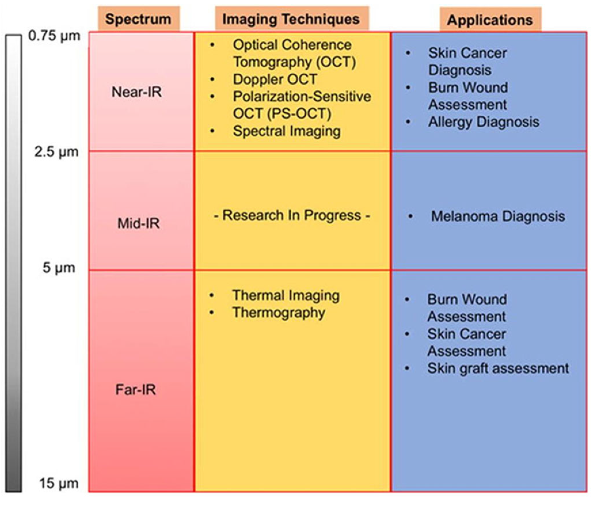
5. Perspectives
6. Conclusions
Supplementary Materials
Author Contributions
Funding
Institutional Review Board Statement
Informed Consent Statement
Data Availability Statement
Conflicts of Interest
References
- Chapple, I.L.C.; Mealey, B.L.; Van Dyke, T.E.; Bartold, P.M.; Dommisch, H.; Eickholz, P.; Geisinger, M.L.; Genco, R.J.; Glogauer, M.; Goldstein, M.; et al. Periodontal health and gingival diseases and conditions on an intact and a reduced periodontium: Consensus report of workgroup 1 of the 2017 World Workshop on the Classification of Periodontal and Peri-Implant Diseases and Conditions. J. Clin. Periodontol. 2018, 45, S68–S77. [Google Scholar]
- Papapanou, P.N.; Sanz, M.; Buduneli, N.; Dietrich, T.; Feres, M.; Fine, D.H.; Flemmig, T.F.; Garcia, R.; Giannobile, W.V.; Graziani, F.; et al. Periodontitis: Consensus report of workgroup 2 of the 2017 World Workshop on the Classification of Periodontal and Peri-Implant Diseases and Conditions. J. Clin. Periodontol. 2018, 45, S162–S170. Available online: https://onlinelibrary.wiley.com/doi/10.1111/jcpe.12946 (accessed on 5 January 2026).
- Herrera, D.; Sanz, M.; Shapira, L.; Brotons, C.; Chapple, I.; Frese, T.; Graziani, F.; Hobbs, F.D.R.; Huck, O.; Hummers, E.; et al. Association between periodontal diseases and cardiovascular diseases, diabetes and respiratory diseases: Consensus report of the Joint Workshop by the European Federation of Periodontology (EFP) and the European arm of the World Organization of Family Doctors (WONCA Europe). J. Clin. Periodontol. 2023, 50, 819–841. [Google Scholar]
- Bui, F.Q.; Almeida-da-Silva, C.L.C.; Huynh, B.; Trinh, A.; Liu, J.; Woodward, J.; Asadi, H.; Ojcius, D.M. Association between periodontal pathogens and systemic disease. Biomed. J. 2019, 42, 27–35. [Google Scholar] [CrossRef] [Scilit] [PubMed]
- Preshaw, P.M.; Alba, A.L.; Herrera, D.; Jepsen, S.; Konstantinidis, A.; Makrilakis, K.; Taylor, R. Periodontitis and diabetes: A two-way relationship. Diabetologia 2012, 55, 21–31. [Google Scholar] [PubMed]
- Sanz, M.; Marco Del Castillo, A.; Jepsen, S.; Gonzalez-Juanatey, J.R.; D’Aiuto, F.; Bouchard, P.; Chapple, I.; Dietrich, T.; Gotsman, I.; Graziani, F.; et al. Periodontitis and cardiovascular diseases: Consensus report. J. Clin. Periodontol. 2020, 47, 268–288. [Google Scholar] [CrossRef] [Scilit]
- Caton, J.G.; Armitage, G.; Berglundh, T.; Chapple, I.L.C.; Jepsen, S.; Kornman, K.S.; Mealey, B.L.; Papapanou, P.N.; Sanz, M.; Tonetti, M.S. A New Classification Scheme for Periodontal and Peri-Implant Diseases and Conditions—Introduction and Key Changes from the 1999 Classification. J. Clin. Periodontol. 2018, 45, S1–S8. Available online: https://onlinelibrary.wiley.com/doi/10.1111/jcpe.12935 (accessed on 5 January 2026).
- Chen, I.D.S.; Yang, C.M.; Chen, M.J.; Chen, M.C.; Weng, R.M.; Yeh, C.H. Deep Learning-Based Recognition of Periodontitis and Dental Caries in Dental X-ray Images. Bioengineering 2023, 10, 911. [Google Scholar]
- Heo, J.S.; Ahn, K.H.; Park, J.S. Radiological screening of maternal periodontitis for predicting adverse pregnancy and neonatal outcomes. Sci. Rep. 2020, 10, 21266. [Google Scholar] [CrossRef] [Scilit] [PubMed]
- Benn, D.K.; Vig, P.S. Estimation of x-ray radiation related cancers in US dental offices: Is it worth the risk? Oral Surg. Oral Med. Oral Pathol. Oral Radiol. 2021, 132, 597–608. [Google Scholar] [CrossRef] [Scilit]
- Benavides, E.; Krecioch, J.R.; Connolly, R.T.; Allareddy, T.; Buchanan, A.; Spelic, D.; O’Brien, K.K.; Keels, M.A.; Mascarenhas, A.K.; Duong, M.L.; et al. Optimizing radiation safety in dentistry. J. Am. Dent. Assoc. 2024, 155, 280–293.e4. [Google Scholar] [CrossRef] [Scilit]
- American Dental Association Council on Scientific Affairs. The use of cone-beam computed tomography in dentistry: An advisory statement from the American Dental Association Council on Scientific Affairs. J. Am. Dent. Assoc. 2012, 143, 899–902. [Google Scholar]
- Tattersall, G.J. Infrared thermography: A non-invasive window into thermal physiology. Comp. Biochem. Physiol. Part A Mol. Integr. Physiol. 2016, 202, 78–98. [Google Scholar] [CrossRef] [Scilit]
- Gurjarpadhye, A.A.; Parekh, M.B.; Dubnika, A.; Rajadas, J.; Inayathullah, M. Infrared Imaging Tools for Diagnostic Applications in Dermatology. SM J. Clin. Med. Imaging 2015, 1, 1–5. [Google Scholar]
- Mapstone, R. Corneal thermal patterns in anterior uveitis. Br. J. Ophthalmol. 1968, 52, 917–921. [Google Scholar] [CrossRef] [Scilit] [PubMed]
- Paterson, J.; Watson, W.S.; Teasdale, E.; Evans, A.L.; Newman, P.; James, W.B.; Pitkeathly, D. Assessment of rheumatoid inflammation in the knee joint. A reappraisal. Ann. Rheum. Dis. 1978, 37, 48–52. [Google Scholar] [CrossRef] [Scilit] [PubMed]
- Bonmarin, M.; Le Gal, F. Chapitre 31—L’imagerie thermique en dermatologie. In L’imagerie en Dermatologie; Academic Press: Amsterdam, The Netherlands, 2016; pp. 437–454. [Google Scholar] [CrossRef] [Scilit]
- Liu, Q.; Li, M.; Wang, W.; Jin, S.; Piao, H.; Jiang, Y.; Li, N.; Yao, H. Infrared thermography in clinical practice: A literature review. Eur. J. Med. Res. 2025, 30, 33. [Google Scholar] [CrossRef] [Scilit] [PubMed]
- Kesztyüs, D.; Brucher, S.; Wilson, C.; Kesztyüs, T. Use of Infrared Thermography in Medical Diagnosis, Screening, and Disease Monitoring: A Scoping Review. Medicina 2023, 59, 2139. [Google Scholar] [CrossRef] [Scilit]
- Claßen, J.; Aupert, F.; Reardon, K.F.; Solle, D.; Scheper, T. Spectroscopic sensors for in-line bioprocess monitoring in research and pharmaceutical industrial application. Anal. Bioanal. Chem. 2017, 409, 651–666. [Google Scholar] [CrossRef] [Scilit]
- Delrue, C.; De Bruyne, S.; Speeckaert, M.M. The Potential Use of Near- and Mid-Infrared Spectroscopy in Kidney Diseases. Int. J. Mol. Sci. 2023, 24, 6740. [Google Scholar] [CrossRef] [Scilit]
- Wu, X.; Dai, Y.; Wang, L.; Peng, Y.; Lu, L.; Zhu, Y.; Shi, Y.; Zhuang, S. Diagnosis of methylglyoxal in blood by using far-infrared spectroscopy and o-phenylenediamine derivation. Biomed. Opt. Express 2020, 11, 960–973. [Google Scholar]
- Sakuma, S.; Inamoto, K.; Higuchi, N.; Ariji, Y.; Nakayama, M.; Izumi, M. Experimental pain in the gingiva and its impact on prefrontal cortical hemodynamics: A functional near-infrared spectroscopy study. Neurosci. Lett. 2014, 575, 74–79. [Google Scholar] [CrossRef] [Scilit]
- Page, M.J.; McKenzie, J.E.; Bossuyt, P.M.; Boutron, I.; Hoffmann, T.C.; Mulrow, C.D.; Shamseer, L.; Tetzlaff, J.M.; Akl, E.A.; Brennan, S.E.; et al. The PRISMA 2020 statement: An updated guideline for reporting systematic reviews. BMJ 2021, 10, n71. [Google Scholar] [CrossRef] [Scilit] [PubMed]
- Wziątek-Kuczmik, D.; Mrowiec, A.; Niedzielska, I.; Stanek, A.; Cholewka, A. Registration of thermal images of dead teeth to identify odontogenic infection foci. Sci. Rep. 2024, 14, 21405. [Google Scholar] [CrossRef] [Scilit]
- Bezerra De Melo, N.; Sobreira Duarte, L.N.; Maia Vieira Pereira, C.; Da Silva Barbosa, J.; Matos Gonçalves Da Silva, A.; De Souza Coelho Soares, R.; Bento, P.M. Thermographic examination of gingival phenotypes: Correlation between morphological and thermal parameters. Clin. Oral Investig. 2023, 27, 7705–7714. [Google Scholar] [CrossRef] [Scilit]
- Wziątek-Kuczmik, D.; Niedzielska, I.; Mrowiec, A.; Bałamut, K.; Handzel, M.; Szurko, A. Is Thermal Imaging a Helpful Tool in Diagnosis of Asymptomatic Odontogenic Infection Foci—A Pilot Study. Int. J. Environ. Res. Public Health 2022, 19, 16325. [Google Scholar] [CrossRef] [Scilit] [PubMed]
- Derruau, S.; Bogard, F.; Exartier-Menard, G.; Mauprivez, C.; Polidori, G. Medical Infrared Thermography in Odontogenic Facial Cellulitis as a Clinical Decision Support Tool. A Technical Note. Diagnostics 2021, 11, 2045. [Google Scholar] [CrossRef] [Scilit]
- Delarue, M.; Derruau, S.; Troyon, P.; Bogard, F.; Polidori, G.; Mauprivez, C. Medical infrared thermography in peri-operative management of peripheral ameloblastoma: A case report. Photodiagn. Photodyn. Ther. 2021, 34, 102167. [Google Scholar] [CrossRef] [Scilit] [PubMed]
- Aboushady, M.A.; Talaat, W.; Hamdoon, Z.; MElshazly, T.; Ragy, N.; Bourauel, C.; Talaat, S. Thermography as a non-ionizing quantitative tool for diagnosing periapical inflammatory lesions. BMC Oral Health 2021, 21, 260. [Google Scholar] [CrossRef] [Scilit]
- Çankaya, Z.T.; Koyuncu, A.; Gürbüz, S. Artificial Intelligence Assisted Thermal Imaging for Gingival Inflammation Assessment: A Novel Approach. J. Esthet. Restor. Dent. 2025. [Google Scholar] [CrossRef] [Scilit]
- Duarte, P.M.; Sowa, M.G.; Xiang, X.; Zhang, C.; Santos, V.R.; Miranda, T.S.; Reis, A.F.; Liu, K. Assessment of the hemodynamic profile in periodontal tissues of diabetic subjects with periodontitis by optical spectroscopy. J. Periodontal Res. 2015, 50, 594–601. [Google Scholar]
- Zhang, C.; Xiang, X.; Xu, M.; Fan, C.; Sowa, M.G.; Liu, K.Z. Assessment of tissue oxygenation of periodontal inflammation in patients with coronary artery diseases using optical spectroscopy. BMC Oral Health 2014, 14, 25. [Google Scholar] [CrossRef] [Scilit]
- Liu, K.; Duarte, P.M.; Santos, V.R.; Xiang, X.; Xu, M.; Miranda, T.S.; Fermiano, D.; Gonçalves, T.E.D.; Sowa, M.G. Assessment of tissue oxygenation of periodontal inflammation in smokers using optical spectroscopy. J. Clin. Periodontol. 2014, 41, 340–347. [Google Scholar] [CrossRef] [Scilit]
- Ge, Z.; Liu, K.Z.; Xiang, X.; Yang, Q.; Hui, J.; Kohlenberg, E.; Sowa, M.G. Assessment of local hemodynamics in periodontal inflammation using optical spectroscopy. J. Periodontol. 2011, 82, 1161–1168. [Google Scholar] [CrossRef] [Scilit]
- Nogueira-Filho, G.; Xiang, X.M.; Shibli, J.A.; Duarte, P.M.; Sowa, M.G.; Ferrari, D.S.; Onuma, T.; de Cardoso, L.A.G.; Liu, K.-Z. On site noninvasive assessment of peri-implant inflammation by optical spectroscopy. J. Periodontal Res. 2011, 46, 382–388. [Google Scholar] [CrossRef] [Scilit]
- Liu, K.Z.; Xiang, X.M.; Man, A.; Sowa, M.G.; Cholakis, A.; Ghiabi, E.; Singer, D.L.; Scott, D.A. In vivo determination of multiple indices of periodontal inflammation by optical spectroscopy. J. Periodontal Res. 2009, 44, 117–124. [Google Scholar] [CrossRef] [Scilit] [PubMed]
- FLIR T1020. Available online: https://www.flir.fr/products/t1020/ (accessed on 5 January 2026).
- Prasanth, C.S.; Betsy, J.; Jayanthi, J.L.; Nisha, U.G.; Prasantila, J.; Subhash, N. In vivo inflammation mapping of periodontal disease based on diffuse reflectance spectral imaging: A clinical study. J. Biomed. Opt. 2013, 18, 26019. [Google Scholar] [CrossRef] [Scilit]
- Bhargava, A.; Chanmugam, A.; Herman, C. Heat transfer model for deep tissue injury: A step towards an early thermographic diagnostic capability. Diagn. Pathol. 2014, 9, 36. [Google Scholar] [CrossRef] [Scilit] [PubMed]
- Xiang, X.; Sowa, M.G.; Iacopino, A.M.; Maev, R.G.; Hewko, M.D.; Man, A.; Liu, K.Z. An Update on Novel Non-Invasive Approaches for Periodontal Diagnosis. J. Periodontol. 2010, 81, 186–198. [Google Scholar] [CrossRef] [Scilit]
- Delrue, C.; De Bruyne, S.; Speeckaert, M.M. Unlocking the Diagnostic Potential of Saliva: A Comprehensive Review of Infrared Spectroscopy and Its Applications in Salivary Analysis. J. Pers. Med. 2023, 13, 907. [Google Scholar] [CrossRef] [Scilit] [PubMed]
- Xiang, X.; Duarte, P.M.; Lima, J.A.; Santos, V.R.; Gonçalves, T.D.; Miranda, T.S.; Liu, K. Diabetes-associated periodontitis molecular features in infrared spectra of gingival crevicular fluid. J Periodontol. 2013, 84, 1792–1800. [Google Scholar] [CrossRef] [Scilit]
- Seredin, P.; Litvinova, T.; Ippolitov, Y.; Goloshchapov, D.; Peshkov, Y.; Kashkarov, V.; Ippolitov, I.; Chae, B. A Study of the Association between Primary Oral Pathologies (Dental Caries and Periodontal Diseases) Using Synchrotron Molecular FTIR Spectroscopy in View of the Patient’s Personalized Clinical Picture (Demographics and Anamnesis). Int. J. Mol. Sci. 2024, 25, 6395. [Google Scholar] [CrossRef] [Scilit]
- Lin, W.S.; Alfaraj, A.; Lippert, F.; Yang, C.C. Performance of the caries diagnosis feature of intraoral scanners and near-infrared imaging technology-A narrative review. J. Prosthodont. 2023, 32, 114–124. [Google Scholar] [PubMed]
- Shmueli, A.; Fux-Noy, A.; Davidovich, E.; Ram, D.; Moskovitz, M. Comparing Images from Near-Infrared Light Reflection and Bitewing Radiography to Detect Proximal Caries in Primary Teeth. Children 2024, 11, 1455. [Google Scholar] [CrossRef] [Scilit] [PubMed]
- Albahri, J.; Allison, H.; Whitehead, K.A.; Muhamadali, H. The role of salivary metabolomics in chronic periodontitis: Bridging oral and systemic diseases. Metabolomics 2025, 21, 24. [Google Scholar] [CrossRef] [Scilit] [PubMed]


| References | Regions of Interest | Number of Patients | Room Temperature | Objectives | Camera Distance | Spectrum | Results |
|---|---|---|---|---|---|---|---|
| Wziątek-Kuczmik D et al. 2024 [25] | Periapical areas of dead teeth | 150 | 23 ± 1 (°C) | Examine the contribution of thermography in detecting asymptomatic infection sites in patients at high risk of systemic infections. | D = 0.4 ± 0.05 m | λ from 7.5 to 14 µm | The results showed a significant temperature difference between each patient group and the healthy group based on the measurement time. |
| Bezerra de Melo N et al. 2023 [26] | Gums of the teeth | 33 | Between 20 and 25 °C (±1 °C) | Analysis of the clinical and thermographic parameters of gingival morphology to detect if there is an exploitable difference, as well as other parameters related to periodontal diagnosis. | D = 0.30 cm | λ from 7.5 to 14 µm | The results show a significant correlation between thermographic features and clinical gingival parameters. |
| Wziątek-Kuczmik et al. 2022 [27] | Periapical regions of the teeth | 8 | 23.0 ± 1 (°C) | Determination of the effectiveness of infrared thermal imaging for detecting the inflammatory response of periapical regions. | NC | λ from 7.5 to 14 µm | The results demonstrate a difference between the temperatures of the periapical regions of suspect teeth and those of the corresponding regions of healthy teeth, with an average temperature increase ranging from 0.51 to 0.39 °C. |
| Derruau S et al. 2021 [28] | Left buccal space | 2 | NC | dentification of cellulitis in 2 patients through thermal imaging to validate its usefulness as a reliable diagnostic tool. | D = 0.4 ± 0.05 m | λ from 7.5 to 14 µm | The thermal results reveal a larger thermally activated area on the affected side compared to the healthy side, with a difference of more than 3 °C for Patient 1 and 2 °C for Patient 2. |
| Delarue M et al. 2021 [29] | Periodontium of the posterior teeth of the mandibular arch | 1 | NC | Evaluation of tumor margins of small masses and/or tumors (peripheral ameloblastoma) not detected by conventional imaging. | NC | λ from 7.5 to 14 µm | The results indicate that the peripheral ameloblastoma region was hotter than the surrounding healthy tissues, with an increase in the thermal gradient from 1.5 to 2.5 °C. |
| Aboushady et al. 2021 [30] | Periapical regions of the teeth | 80 | 20.0 ± 1 (°C) | Evaluation of the validity of thermography for the diagnosis of periapical inflammatory lesions and the temperature ranges of acute pulpitis with apical periodontitis, acute and chronic periapical abscesses. | Indirect Tec: D < 2 m, Direct Tec: D = 20 cm | λ from 7.5 to 13 µm | The results demonstrate a significant difference between the average intra-oral thermal temperatures of the three diagnostic groups, with an increase in temperature in patients with acute pulpitis and apical periodontitis. |
| Turgut Çankaya Z et al. 2025 [31] | Gingival tissues (thermal gingival images annotated and labeled based on bleeding on probing (BoP) and Gingival Index (GI) for inflammation severity) | 40 participants (stratified by periodontal status and breathing pattern: mouth or nasal breathing) | Not specified (performed under standardized imaging conditions) | To detect and classify gingival inflammation severity using AI-supported analysis of thermal gingival images in patients with mouth breathing habits, and to establish specific thermal thresholds for gingival health and disease in this population | Not specified | Not specified (thermal imaging; typically far-infrared (FIR) in the 8–14 µm range for medical thermography, but no explicit details provided) | XGBoost classification achieved an accuracy of 92.74%, precision of 92.95%, sensitivity of 92.74%, and F1 score of 92.78%; cross-validation confirmed reliability with mean test score of 88.28% and validation score of 89.43% |
| Ref. | Number of Patients | Average Age | Objectives | Key Parameters Measured | Spectrum | Results |
|---|---|---|---|---|---|---|
| Duarte PM et al. 2015 [32] | 78 | Between 35 and 66 years | Evaluate optical spectroscopy as a periodontal diagnostic method for patients with type 2 diabetes and chronic periodontitis, while documenting the local hemodynamic profile at the periodontal level in these subjects. | The relative concentration of deoxygenated hemoglobin (Hb) and oxygenated hemoglobin (HbO2), the balance between oxygen supply and utilization in periodontal tissues. | λ from 0.5 to1.1 µm | In diabetic patients, tissue oxygen saturation and HbO2 levels were significantly reduced in periodontitis sites compared to gingivitis sites (p < 0.01). Furthermore, tissue oxygenation in healthy sites was markedly higher in controls than in diabetic subjects (p < 0.01). |
| Zhang C et al. 2014 Maladie [33] | 121 | Between 33 and 71 years | Verify the ability to identify periodontitis in patients with coronary artery disease using spectroscopy, with an instrument previously designed by the research team. | In coronary disease patients, a variation in Hb and HbO2 levels was observed (p < 0.01), and oxygen saturation was reduced in periodontitis sites compared to healthy sites in the diseased patients. In contrast, no difference in saturation was noted between the healthy groups and those with coronary artery disease. | ||
| Liu KZ et al. 2014 [34] | 54 | Between 35 and 65 years | Analyze the effectiveness of spectroscopy in detecting periodontitis in smoking patients, using a device developed by the research team. | In smoking patients, tissue oxygen saturation significantly decreased in gingivitis sites (p = 0.016) and periodontitis sites (p = 0.007) compared to healthy sites. A trend of initial increase followed by a decrease in HbO2 concentration was observed, moving from healthy sites to affected sites. | ||
| Ge Z et al. 2011 [35] | 51 | NC | Analysis of the hemodynamics of periodontal tissues during inflammation using optical spectroscopy. | The results reveal that tissue oxygenation significantly decreases between healthy sites, gingivitis sites, and periodontitis sites. This is explained by a notable increase in deoxyhemoglobin between healthy and gingivitis sites, as well as a significant decrease in oxyhemoglobin between gingivitis and periodontitis sites. | ||
| Nogueira-Filho G et al. 2011 [36] | 64 | NC | Investigation of the diagnostic potential of optical spectroscopy in peri-implant inflammation in vivo. | The results indicate that tissue oxygenation at peri-implant sites was reduced compared to healthy sites (p < 0.05) due to an increase in deoxyhemoglobin and a decrease in oxyhemoglobin. Furthermore, the tissue hydration index, calculated from the optical spectra, was significantly higher in cases of mucositis compared to the other groups (p < 0.05). | ||
| Liu KZ et al. 2009 [37] | 30 | Between 37 and 71 years | Analyze the ability of in vivo optical spectroscopy to simultaneously measure multiple inflammatory indices in periodontal tissues. | Tissue oxygenation, total tissue hemoglobin, deoxyhemoglobin, oxygenated hemoglobin, and tissue edema. | The results highlighted a decrease in oxygenation and an increase in deoxyhemoglobin at periodontitis sites, as well as a variation in the water index associated with electrolytes and temperature between the studied sites. |
| References | Type of IR Camera | Image Resolution | Temperature Resolution | Acquisition Frequency | Image Analysis Software |
|---|---|---|---|---|---|
| Wziątek-Kuczmik D et al. 2024 [25] | FLIR T1020 | 1024 × 768 pixels * | <0.02 °C * | f = 30 Hz * | ThermaCAM Researcher Pro 2.10 |
| Bezerra de Melo N et al. 2023 [26] | FLIR T650 | 640 × 480 pixels | 0.05 °C | NC | FLIR Tools+™ 6.4 |
| Wziątek Kuczmik et al. 2022 [27] | FLIR T1020 | 1024 × 768 pixels * | <0.02 °C * | f = 30 Hz * | ThermaCAM Researcher Pro 2.8 SR-3 |
| Derruau S et al. 2021 [28] | VarioCAM® HD | 1024 × 768 pixels | NC | NC | IRBIS® 3.1, InfraTec |
| Delarue M et al. 2021 [29] | InfraTech VarioCAM HD | 1024 × 768 pixels | 0.03 °C | NC | NC |
| Aboushady et al. 2021 [30] | FLIR E-5 | 120 × 90 pixels | NC | NC | FLIR Thermal Analysis and Reporting |
| References | Device | Probe | Power of the Light Source | Integration Time | Spectral Range | Resolution | Statistical Analysis Software |
|---|---|---|---|---|---|---|---|
| Duarte PM et al. 2015 [32] | Portable spectro-graph PDA512-ISA | Custom bifurcated optical fiber probe for oral use | 5 W | 0.03 s | Between 500 and 1100 nm | 5 nm | Statistica 7.1 |
| Zhang C et al. 2014 [33] | |||||||
| Liu KZ et al. 2014 [34] | |||||||
| Ge Z et al. 2011 [35] | |||||||
| Nogueira-F et al. 2011 [36] | |||||||
| Liu KZ et al. 2009 [37] |
Disclaimer/Publisher’s Note: The statements, opinions and data contained in all publications are solely those of the individual author(s) and contributor(s) and not of MDPI and/or the editor(s). MDPI and/or the editor(s) disclaim responsibility for any injury to people or property resulting from any ideas, methods, instructions or products referred to in the content. |
© 2026 by the authors. Licensee MDPI, Basel, Switzerland. This article is an open access article distributed under the terms and conditions of the Creative Commons Attribution (CC BY) license.
Share and Cite
Guetatlia, H.N.; Gette, M.; Estrade, L.; Rimbaud, V.; Denis, F.; Rochefort, G.Y.; Renaud, M. Thermography and Infrared Spectroscopy in the Detection of Periodontal Inflammation In Vivo: A Systematic Review. Diagnostics 2026, 16, 222. https://doi.org/10.3390/diagnostics16020222
Guetatlia HN, Gette M, Estrade L, Rimbaud V, Denis F, Rochefort GY, Renaud M. Thermography and Infrared Spectroscopy in the Detection of Periodontal Inflammation In Vivo: A Systematic Review. Diagnostics. 2026; 16(2):222. https://doi.org/10.3390/diagnostics16020222
Chicago/Turabian StyleGuetatlia, Heythem Nassim, Mickael Gette, Laurent Estrade, Victor Rimbaud, Frédéric Denis, Gaël Y. Rochefort, and Matthieu Renaud. 2026. "Thermography and Infrared Spectroscopy in the Detection of Periodontal Inflammation In Vivo: A Systematic Review" Diagnostics 16, no. 2: 222. https://doi.org/10.3390/diagnostics16020222
APA StyleGuetatlia, H. N., Gette, M., Estrade, L., Rimbaud, V., Denis, F., Rochefort, G. Y., & Renaud, M. (2026). Thermography and Infrared Spectroscopy in the Detection of Periodontal Inflammation In Vivo: A Systematic Review. Diagnostics, 16(2), 222. https://doi.org/10.3390/diagnostics16020222