


Abstract
Aortopathies in autoimmune rheumatic diseases (ARD) include a spectrum of aortic pathologies—including aortitis, aneurysms, dissections, and insufficiency—primarily caused by systemic inflammation. This comprehensive review investigates the clinical manifestations, pathophysiology, diagnostic modalities, and management strategies across various rheumatic diseases associated with aortopathies such as large vessel vasculitis (e.g., Takayasu arteritis, giant cell arteritis), connective tissue diseases (e.g., systemic lupus erythematosus, rheumatoid arthritis, ankylosing spondylitis, systemic sclerosis) and less common conditions (e.g., relapsing polychondritis, Cogan’s syndrome, Behçet’s disease, IgG4-related disease). Disease-specific pathophysiologic mechanisms of aortic wall inflammation and remodeling, including granulomatous and lymphoplasmacytic patterns and mixed inflammatory infiltrates, are described. Diagnostic imaging modalities—such as CTA, MRI, and PET/CT—are evaluated for their roles in detecting active inflammation, assessing structural complications, and guiding clinical decision-making. Histopathological findings provide insight into disease-specific vascular changes. Management strategies focus on the use of glucocorticoids, disease-modifying antirheumatic drugs (DMARDs), and biologics, including IL-6 and TNF-α inhibitors, with an emphasis on patient-centered approaches, multidisciplinary care, and timely surgical intervention for complications. Evidence gaps include optimal screening intervals and the role of novel biomarkers in risk stratification and in monitoring disease progression, highlighting the need for early recognition, frequent monitoring, and aggressive management of aortic involvement in rheumatic diseases to prevent life-threatening complications.
1. Introduction
Aortopathies in autoimmune rheumatic diseases (ARDs) refer to a spectrum of aortic involvement in various systemic inflammatory conditions. These conditions lead to inflammation of the aortic wall and its surrounding tissues, termed aortitis/periaortitis, at different degrees, rendering the aorta predisposed to dilation, aneurysm, dissection, or rupture [1,2]. Aortopathies are often well-recognized in large vessel vasculitides such as giant cell arteritis (GCA) or temporal arteritis and Takayasu arteritis (TA), according to the 2012 International Chapel Hill Consensus Conference (CHCC) [3]. Noninfectious inflammatory aortopathies have been occasionally reported in polymyalgia rheumatica (PMR), Behçet’s disease (BD), granulomatous with polyangiitis (GPA), Cogan syndrome, immunoglobulin G4-related disease (IgG4-RD), rheumatoid arthritis (RA), systemic lupus erythematosus (SLE), and sarcoidosis [1,2]. Other rheumatic diseases (RD) with aortic involvement are often under-recognized and not well addressed, but have a high mortality risk from complications such as aneurysmal rupture and dissection. Aortic disease may be a recognized manifestation of RD identified by screening using imaging (GCA, TA, occasionally PMR) or identified incidentally on imaging or detected on histology from aortic surgery or an autopsy [4,5,6]. Clinical presentation varies depending on the site of aortic involvement, such as back pain, abdominal pain, or acute aortic insufficiency if the aortic valve is involved. Patients can be asymptomatic or only have constitutional symptoms or present with life-threatening acute aortic syndromes such as aortic dissection, rupture, or intramural hematoma. Constitutional non-specific symptoms, including fever, malaise, and fatigue, are also possible [6,7]. The diagnosis requires a high index of suspicion of a certain rheumatic disease with a vague clinical presentation, especially in chronic or relapsing diseases [7]. Radiological, histopathological, and laboratory findings are necessary to establish the diagnosis, although, practically, tissue pathology is not always feasible in all cases [4,7].
Therefore, this review explores the understanding of aortopathies reported in rheumatic diseases.
Scope and rationale. For clarity, this review focuses on the inflammatory aortopathies that arise in rheumatic diseases, in which primary immune-mediated inflammation of the aortic wall or periaortic tissue (aortitis/periaortitis) predisposes to secondary structural complications such as dilatation, aneurysm, dissection and aortic regurgitation; degenerative and purely atherosclerotic aortopathies are outside its scope. Diseases were included when the published literature documented either (a) direct aortic wall or periaortic inflammation, (b) a reproducible association with clinically relevant secondary structural aortic complications, or (c) disease-specific diagnostic or therapeutic implications when aortic involvement occurs. The disorders are grouped as primary large-vessel vasculitides; other primary vasculitides; systemic autoimmune rheumatic diseases; autoinflammatory syndromes; and other immune-mediated fibro-inflammatory or granulomatous disorders. The review is evidence-based rather than exhaustive, and the strength of the underlying evidence varies substantially, ranging from cohort studies and guidelines to small case series and individual case reports.
2. Pathophysiology and Histopathology
The pathogenesis of aortic inflammation in elastic arteries varies across rheumatic conditions, resulting in distinct histopathological changes. Complications following damage to the elastic media include aneurysmal dilation, dissection, thrombosis with or without embolization, and aortic wall thickening leading to stenosis [8]. Inflammatory cell infiltration and subsequent destruction of the aortic tunica media and adventitia result in scarring and fibrosis over time, narrowing the lumen in some RDs, such as TA, to a greater extent than GCA. The destruction of the elastic lamina, inflammation in the vasa vasorum leading to vascular wall necrosis, and extensive inflammation in the media result in the formation of an aortic aneurysm [6]. The progression of an aortic aneurysm depends on the damage and interactions of all aortic constituents (smooth muscle cells, endothelial cells, macrophages, and fibroblasts) [9]. Aortitis secondary to ARDs often produces saccular/non-circumferential and multiple aneurysms [1]. Aortic dissection occurs due to intimal tears that allow blood into the media, separating the elastic lamellae. Rupture occurs when the dissection extends through the adventitia and can be aneurysmal or non-aneurysmal [1,8]. Aortitis/periaortitis is classified based on the pattern of inflammation, which is not attributable to atherosclerosis. In aortitis, inflammation affects the media and/or intima, often extending to the adventitia, and can impact any segment of the aorta, including its major branches or other large arteries [4]. Periaortitis refers to an inflammatory condition originating from the adventitia of the aortic wall, extending into the surrounding periaortic space as seen in IgG4-RD [10]. Although the definitions of both entities are distinct, there can be significant overlaps in their presentation, particularly when aortitis extends into the PA or vice versa. Table 1a categorizes Rheumatic diseases according to the pattern of inflammation. Table 1b summarizes the histopathological findings of aortitis/periarteritis related to rheumatic diseases. Figure 1 illustrates proposed mechanisms of aortitis development and Figure 2 illustrates the histopathology of aortitis in the setting of representative rheumatic diseases.
Table 1.
(a) Summary of inflammatory patterns of aortitis/periaortitis in rheumatic diseases References: entries are compiled from the primary sources cited in the Pathophysiology and Histopathology section [1,2,4,6,8,9,10]. (b) Histopathology of aortic abnormalities in rheumatic disease and prevalence of aortitis.
Figure 1.
Proposed shared mechanisms in the development of aortitis in rheumatic diseases, from immune triggering and cell activation through cytokine-driven aortic wall injury to structural complications.
Figure 2.
(A) In giant cell aortitis, lymphohistiocytic inflammation (arrow) destroys the media, and (B) scattered multinucleated giant cells are usually seen (arrow). (C) In late-stage Takayasu arteritis, the media and adventitia are densely fibrotic, with minimal residual chronic inflammation at the media–adventitia interface (arrow), and only a few lymphocytes are seen (D). (E) In granulomatosis with polyangiitis, granulomatous inflammation destroys the media (arrows) with (F) palisading histiocytes (arrow) surrounding geographic basophilic necrosis (*). (G) In IgG4-related disease, the adventitia and periadventitia show dense sclerotic fibroinflammatory change with lymphoid aggregates (arrows) that (H) are rich in plasma cells displaying an elevated IgG4: IgG ratio (>40%) by immunohistochemistry (insets). (A–H): Hematoxylin and eosin stains; original magnifications 20× (A,C,E,G), 100× (H), 200× ((H) insets), 400× (B,D,F).
3. Aortic Involvement in Rheumatic Diseases
3.1. Takayasu Arteritis (TA)
Takayasu arteritis (TA) is a chronic inflammatory condition affecting large blood vessels, causing stenosis or occlusion in 98% and aneurysms in 27% of individuals’ blood vessels [11], particularly involving the aorta and its major branches [12]. TA is characterized by pathological changes in large blood vessel layers, including arterial wall thickening, stenosis, occlusion, aneurysm formation, and dissection [12]. In some, it may initially present as aortic dissection after childbirth in women in their 30s or as a myocardial infarction [11,13,14]. TA is currently classified into six types, as shown in Table 2.
Table 2.
Current classification of TA by types, its involvement, and the prevalence of each type Reference: classification and prevalence adapted from [11,12,13,14,15].
These classifications are essential for understanding clinical variations and informing the management strategies of affected patients. The pathogenesis of TA is not well understood [15]. The T-cell-mediated pathway is believed to play a major role in the development of TA. Specific T cells, such as vasculogenic T cells, particularly Th1 and Th17, are dominant in the vascular layers, causing vasculitis and organ damage [16]. Their activity is closely associated with the presence of interleukin-6 (IL-6) and interleukin-17 (IL-17) [17]. Activation of natural killer cells, γ/δ T cells, and macrophages with the production of tumor necrosis factor-α (TNF-α), forming granulomas within the blood vessels [18]. The diagnosis of aortic involvement in TA is essentially made with imaging techniques guided by a high degree of clinical suspicion. Imaging studies also help in determining the extent of vascular involvement. There are two key aspects to imaging of the arterial tree in TA and other large-vessel vasculitides: evaluation of the arterial lumen and evaluation of the arterial wall, with the acknowledgment that arterial wall biopsy is seldom performed. To this effect, magnetic resonance angiography (MRA) or computed tomography angiography (CTA) demonstrates smoothly tapered luminal narrowing or occlusion of the affected vessel. Three-dimensional reconstruction protocols are available to examine the vessel wall in greater detail. Positron emission tomography (PET) in combination with computed tomography (CT) or magnetic resonance (MR) is increasingly used to identify areas of increased uptake, or “hot segments,” in the vessel wall (reflecting higher standardized uptake values) to indicate active vasculitis in the right clinical setting [17,19]. The initial change observed is mural thickening, indicating inflammation of the media and adventitia, followed by structural vascular changes detected on CTA [20]. Aneurysms are less common and typically affect the ascending aorta and abdominal aorta [21]. However, among these modalities, CTA is the most favored diagnostic tool due to its ability to assess disease severity, its widespread availability, and its high-quality images for follow-up of the affected vessels, stents, and bypass grafts [17,22]. The goal of treatment is to eliminate or reduce inflammatory activity in the vessel wall using immunosuppressive therapy (IST) and to limit further structural abnormalities in the vessels. Glucocorticoids (GCs) are effective for most patients; however, given the chronic, relapsing nature of the disease in more than half of TA patients, the use of GC-sparing therapy (also called IST in this setting) is imperative at an early stage [11]. In most cases, upfront use of GC with IST provides better control of disease activity than GC alone [11]. Initial options for IST include methotrexate (MTX) and azathioprine (AZA), with alternatives such as mycophenolate mofetil (MMF) or leflunomide. Unfortunately, relapse is common when tapering prednisone to dosages of 15 mg/day or less [23]. In selected or treatment-refractory cases, tumor necrosis factor alpha inhibitors (TNFi) are used. More severe diseases may respond to TNF inhibition or IL-6 inhibition [24]. Those with severe vascular complications may require surgical evaluation and intervention. Revascularization for TA has been shown to be safe and is associated with low morbidity and mortality rates [23]. Conventional bypass grafts typically achieve optimal long-term outcomes. Additionally, percutaneous transluminal angioplasty has yielded favorable results, particularly for short lesions [23] (Figure 3, Figure 4, Figure 5 and Figure 6).
Figure 3.
An example of Takayasu’s disease status post ascending aorta tubular grafting with recurrent vasculitis. Coronal fused and attenuation corrected PET images of F-18 FDG PET/CT studies at 2 different time points before and after immunosuppressive therapy with high-dose glucocorticoids. Pretherapy images (A,B) show intense abnormal radiotracer uptake throughout the aortic root, ascending aorta graft, brachiocephalic artery, and aortic arch (arrows) consistent with active vasculitis. Post-therapy images (C,D) show significantly improved radiotracer uptake involving the same vessels, consistent with decreased inflammation (arrows). In addition, focal tracer activity in the distal right subclavian artery (arrowheads) also improved after initiation of therapy.
Figure 4.
An example of known Takayasu’s disease evaluated for abnormal and asymmetric peripheral pulses. Axial STIR images of chest MR without contrast (A,C) and axial images of chest MRA with contrast (B,D) showing near concentric mural thickening and edema in the proximal left subclavian artery resulting in luminal narrowing (arrows) and to a lesser extent thickening and edema in the aortic arch (open arrow), right brachiocephalic artery (arrowhead) and left common carotid artery (*).
Figure 5.
An example of known Takayasu’s disease status post descending aorta stent placement on remission. Coronal images of F-18 FDG PET/CT (A) fused, (B) AC PET and (C) CT without contrast showing a distal descending aorta stent with residual mild luminal narrowing (arrows). There is normal radiotracer distribution with no pathological aortic wall activity to suggest active inflammation.
Figure 6.
Takayasu’s disease with aortic involvement and thoracic aorta aneurysm off therapy evaluated for disease activity. Axial (A) and sagittal (B) fused images of F-18 FDG PET/CT showing extensive pathological tracer activity throughout a dilated thoracic aorta (arrows) involving predominantly the ascending aorta to a lesser extent arch and descending aorta most consistent with active vasculitis. Axial (C) and sagittal (D) images of CTA of the chest with IV contrast showing aneurysmal dilation to the ascending and to a lesser extent descending aorta associated to diffuse mural thickening (arrowheads). Axial STIR (E) images of chest MR showing diffuse aortic wall edema (open arrow). Sagittal double inversion recovery images after IV gadolinium administration (F) of chest MR showing diffuse thoracic aorta wall hyperenhancement (open arrow) consistent with active inflammation.
3.2. Giant Cell Arteritis (GCA) and Polymyalgia Rheumatica (PMR)
Giant cell arteritis (GCA) is the most common form of systemic large vessel vasculitis (LVV). GCA and PMR are closely related conditions, often considered different clinical manifestations of the same diagnostic and pathogenic spectrum. During their course, 40–60% of patients with GCA will have PMR, whereas 16–21% of patients with PMR will have GCA [25]. PMR large-vessel involvement, consistent with “subclinical GCA,” is present in 30%. The aortitis itself is not attributed to PMR and is typically considered a missed diagnosis of GCA [26]. Figure 7, Figure 8 and Figure 9 demonstrate imaging findings of the aorta in GCA, and Figure 9 demonstrates imaging findings in a case of PMR. Unlike PMR, GCA is the leading cause of aortitis, accounting for 72% of patients [2,3,4,5]. Aortitis can occur at any stage of the disease onset [2,3,4,5], and often presents silently or with minimal constitutional symptoms at the time of diagnosis [18,27]. Its complications include aortic insufficiency, ruptured aortic aneurysm, aortic dissection, stroke, or myocardial infarction [28]. Among these, the most common aortic complication was found to be an aortic aneurysm (up to 20%) [29]. The prevalence of thoracic aortic aneurysms ranges from 2–18% among patients with GCA [30,31]. A previous retrospective study involving 96 GCA patients showed a 17.3-fold increase in the risk of developing a thoracic aortic aneurysm and a 2.4-fold increase in the risk of an isolated abdominal aneurysm [32]. Predictors of mortality among GCA patients with aortitis include the presence of aortitis or any aortic involvement at disease onset, as well as the presence of symptoms [33,34,35]. Diagnostic imaging modalities for detecting GCA aortic involvement include arteriography [32], CTA, MRA, and FDG-PET [36]. Aortitis was detected in up to 65% of cases on CTA screening, and follow-up aortic screening is recommended to detect stenosis and aneurysms [37]. The reported sensitivity and specificity of PET in the diagnosis of GCA are 88.9% and 95.1%, respectively [32], though these values depend on the timing of glucocorticoid initiation [38]. Generally, a PET scan detects the uptake up to 51–65% at the thoracic aorta and 54% at the abdominal aorta in GCA patients, reflecting ongoing inflammation in those with subclinical aortic inflammation [39,40]. However, large-vessel involvement often goes undetected at early disease onset, whether on MRA or PET scans [18]. Due to the increasing body of evidence on the high prevalence of large vessel vasculitis in GCA, the 2021 American College of Rheumatology (ACR)/Vasculitis Foundation guidelines for management of GCA conditionally recommend obtaining noninvasive vascular imaging (MRA or CTA of the neck/chest/abdomen/pelvis) to evaluate large vessel involvement at the time of diagnosis [24]. While there is no consensus on monitoring guidelines for patients with large-vessel vasculitis, proactive and periodic monitoring using vascular imaging may be needed to identify complications [36]. Given the considerable morbidity and mortality associated with aortic aneurysms, the aorta should be evaluated routinely during the follow-up of patients with GCA. The initial treatment for GCA-associated inflammatory aortitis is high-dose GC, dosing 40–60 mg once daily or 0.7 mg/kg/day with gradual tapering [6,41]. Concurrent initiation of GC-sparing therapy is advised, given the anticipated prolonged duration of GC therapy, which predisposes to side effects, and the accumulating body of evidence with interleukin-6 targeted therapy (tocilizumab). Tocilizumab was recently used as a treatment and is now considered a first-line therapy, in combination with glucocorticoids, per the 2021 ACR management guidelines [24,42,43,44]. While tocilizumab is favored, methotrexate can be considered as an alternative option for patients unable to use tocilizumab due to factors such as recurrent infections, history of gastrointestinal perforations or diverticulitis, and cost. Aortic aneurysms may initiate the disease several years after the diagnosis, occasionally after corticosteroid therapy discontinuation. Aortic ectasia and aneurysms should be closely monitored to determine the appropriate timing for surgical intervention [41]. There is no consensus on the optimal imaging modalities or the frequency of sequential imaging. Imaging screening is recommended at regular intervals over the long term because, despite treatment, 56% still had evidence of active vasculitis of the thoracic vessels by PET and 80% by MRI [32,45]. Despite apparent clinical and biomarker remission, up to 63–84% of scans show persistent uptake on PET [30]. However, after 1 year of corticosteroid treatment, contrast enhancement resolved in 93.75% of cases in GCA patients [30]. However, a minor relapse was found during the 2-year follow-up [44] (Figure 7, Figure 8, Figure 9 and Figure 10).
Figure 7.
Patient with known giant cell arteritis. Images of whole-body F-18 FDG PET/CT, PET MIP (A) and axial fused images at multiple levels (B–D) showing extensive abnormal tracer activity involving the aorta (arrows), as well as head and neck vessels, upper and lower extremity arteries and superficial temporal arteries (arrowheads) indicating active vasculitis. In addition, multifocal areas of abnormal tracer accumulation in lower extremities musculature most consistent with active myositis (*).
Figure 8.
An example of a known giant cell arteritis. Coronal images of F-18 FDG PET/CT scan (A) fused, (B) AC PET,(C) CT without contrast showing intense tracer accumulation throughout the ascending aorta and aortic arch (arrows) as well as right brachiocephalic artery, bilateral subclavian arteries and carotid arteries (arrowheads) indicating active vasculitis.
Figure 9.
An example of known giant cell arteritis on therapy with elevated inflammatory markers evaluated for disease activity. Coronal fused MPR image of F-18 FDG PET/CT (A,C), gray scale (B) and color Doppler (D) ultrasound images of the right subclavian artery and coronal MPR images of chest CTA. There is abnormal radiotracer uptake involving the ascending aorta, right brachiocephalic, bilateral subclavian and to a lesser extent bilateral common carotid arteries (arrows). In addition, evidence of concentric mural thickening to the brachiocephalic and bilateral subclavian arteries (arrowheads). In the right subclavian artery, there is moderate luminal narrowing (arrowheads), turbulent flow on color doppler (*).
Figure 10.
Whole body F-18 FDG PET/CT in a patient with known polymyalgia rheumatica evaluated for persistently elevated inflammatory markers. Axial fused (A), axial AC PET (B), axial CT without contrast (C) and whole-body AC PET MIP (D) images showing abnormal tracer uptake throughout the abdominal aorta (arrows) and to a lesser extent bilateral subclavian, axillary, brachial, iliac and femoral arteries (arrowheads) consistent with active vasculitis. In addition, heterogeneous moderate to intense abnormal tracer accumulation involving bilateral shoulders, elbows, wrists, hands, knees, ankles and feet (*) most consistent with active inflammatory polyarthritis.
3.3. Kawasaki Disease
Kawasaki disease is a vasculitis of unknown cause that primarily affects children under the age of 5 years old. It commonly involves inflammation of medium-sized arteries; thus, aortic involvement is rare. A prospective cohort study of 115 patients with Kawasaki disease in Brazil found that 1% had thoracic aorta aneurysms (TAA) [46]. The complications were more common in patients with persistently elevated inflammatory markers and those who received IVIg after the first 10 days of disease onset [46]. Another cohort from Singapore found that despite early treatment with corticosteroids and IVIg, patients who had prolonged CRP elevation for more than 2 weeks, relapsing symptoms, and prolonged admission were associated with aortitis [47]. MRA, in addition to transthoracic echocardiogram (TTE), could be considered for evaluating large-vessel arteritis in these patients [47]. Treatment of Kawasaki disease typically involves a combination of aspirin and high-dose IVIg as soon as possible after diagnosis [48]. Untreated childhood Kawasaki disease could present with an ascending aortic aneurysm in addition to a coronary artery aneurysm later in life [49]. Prompt recognition and early treatment with IVIg could potentially reduce the risk of cardiac and aortic involvement in patients with Kawasaki disease.
3.4. Antineutrophilic-Cytoplasmic Antibodies (ANCA) Associated Vasculitis (AAV)
AAV with aortic involvement is uncommon, and the incidence is unknown. Less than 20 cases of aortic involvement in AAV, which includes granulomatosis with polyangiitis (GPA), microscopic polyangiitis (MPA), and eosinophilic granulomatosis with polyangiitis (EGPA), have been reported [50,51]. ANCA-related aortic involvement can present with aortic aneurysm, aortic dissection or rupture, and aortic insufficiency as a consequence of aortitis [51,52] and can precede the clinical manifestation of AAV [53,54]. Both the thoracic and abdominal aorta may be involved; some present with PA detected on imaging [4]. An observational study by Kaymakci et al. demonstrated that most patients (n = 25, 69%) were diagnosed with large-vessel vasculitis and AAV within a 1-year period. Most (69%) had histopathological confirmation of an AAV diagnosis in a location other than the temporal artery, the aorta, or the periaortic soft tissue. Seventeen patients had TA-AAV, 10 had aorta-AAV, and 9 had PA-AAV [55]. Clinical symptoms, imaging, and immunologic tests may be required for the diagnosis [56]. Documented possible pathogeneses of aortitis in AAV are vasculitis of vasa vasorum in the adventitial layer, progression from initial intimal injury to transmural aortitis, and peri-aortitis, which eventually causes aortic inflammation [51,53,57,58,59]. Some patients remain asymptomatic, but aortitis was detected on FDG-PET/CT [60]. Treatment with corticosteroids and cyclophosphamide or rituximab has been reportedly successful for remission induction. Surgery is indicated for aortic aneurysms or dissections [61].
3.5. Immunoglobulin G4-Related Diseases (IgG4-RD)
IgG4-RD is a systemic fibro-inflammatory condition involving several organs. Cardiovascular involvement frequently presents as aortitis, predominantly involving the abdominal aorta [62]. Patients are usually asymptomatic; the condition is often detected incidentally on imaging or histopathologically after aortic surgery [63]. Clinical symptoms depend on the site of aortic involvement and may include back pain, abdominal pain, and lower-extremity edema; constitutional symptoms such as fever or night sweats are rare, as is a significant increase in CRP [64,65]. Chronic peri-aortitis (CP) is more common and is associated with peripheral tissue involvement and retroperitoneal fibrosis (RPF); features overlapping with aortitis can also occur [64,66]. Aortic involvement is found in approximately 10% of IgG4-RD and is more common in men [67]. Aortitis can be complicated by aneurysm formation and dilatation and, less frequently, dissection [68,69,70]. Table 3 summarizes the features of IgG4-aortitis and IgG4-CP [71]. The pathogenesis of aortic disease is unknown, but it is presumed to be mediated by B cells and cytotoxic CD4 T cells [67,72]. Thoracic aortitis is twice as common as abdominal aortitis but with less vascular wall thickening, while chronic peri-aortitis affects the abdominal aorta more than the thoracic aorta, with infra-renal abdominal aorta being the most common site [66,73,74,75]. Other reported complications of IgG4-related aortitis include aortic regurgitation, aortic stenosis, and aneurysmal rupture (estimated at 42% of cases) [1,76,77]. Mizushima et al. proposed diagnostic criteria for IgG4-related aortitis/peri-aortitis that required abnormal imaging findings consistent with aortitis, histological features, or an elevated IgG4 level, and an organ-specific IgG4-RD [78]. Characteristics of histological findings are described in Table 1b [62,78,79] and illustrated in Figure 2. Infiltration of the tunica media leads to aneurysm dilatation and rupture. No specific tests are available, but elevated sIL-2R, hypocomplementemia, ANA positivity, and rheumatoid factor positivity are often found in association [64,78,80]. Elevated CRP should prompt consideration of alternative diagnoses, including IgG4 [65]. Various imaging modalities, including ultrasound, CTA, MRA, and FDG-PET (Figure 7 and Figure 8), have been used to evaluate IgG4-R aortic disease. The most common CT findings are diffuse arterial wall thickening with homogeneous enhancement and a surrounding non-stenotic mass with irregular margins [74,81,82,83]. Vessel distribution classification is summarized in Table 4. Aneurysmal dilatation is found in only 7% [82], and stenoses/strictures are rare [66,82,83]. The incidence of an aneurysmal rupture is low in cases with severe aortic wall thickening and surrounding adhesions, but this does not apply to saccular aneurysms [84,85]. FDG-PET, in addition to CT or MRI, identifies active inflammation, guides biopsy site selection, and monitors treatment response [86,87,88,89]. Most patients respond well to moderate-dose steroids (0.5–6 mg/kg/day); higher doses are required for severe disease with multiple organ involvement. Caution should be exercised in large aneurysms prone to rupture [64,86] and in peri-aortitis, in which rapid thinning of the aortic wall is observed post-steroid treatment [73]; 20% of patients showed aortic dilatation post-steroid treatment [74]. A combination of steroids with steroid-sparing agents is superior to steroids alone in achieving remission [64,67]. The FDA approved inebilizumab, which targets and depletes CD19+ B-cells based on a recently published randomized controlled trial for the treatment of IgG4-related disease. Inebilizumab has also shown benefit in a phase 3 clinical trial, with an annualized flare rate of 0.14 (95% confidence interval, 0.06–0.31; p < 0.001) [87]. Benefits with MTX, MMF, AZA, leflunomide, CYC, and rituximab have been reported in conjunction with steroids [64,88,89,90,91]. Rituximab can also be utilized as a monotherapy or in steroid-refractory cases [66]. The standard treatment guideline for usual aneurysms dictates surgical intervention. Post-operative steroids may be administered in cases of persistent inflammation [73] (Figure 11 and Figure 12).
Table 3.
Summary of IgG4-aortitis and IgG4-CP features.
Table 4.
Classification of aortic involvement in IgG4-RD based on imaging findings.
Figure 11.
An example of known IgG4 disease evaluated for persistent elevated inflammatory markers. Axial (A,C), coronal (B) and sagittal (D) images of F-18 FDG PET/CT showing tracer avid mural thickening involving the ascending aorta, proximal aortic arch, and upper abdominal aorta (arrows) indicating active vasculitis. There is a subcentimeter tracer avid mediastinal level 7 lymph node (arrowhead) which is likely inflammatory.
Figure 12.
An example of known IgG4 disease with active vasculitis. Imaging before and after immunosuppressive therapy. Pretherapy images of fused axial F-18 FDG PET/CT at the level of the kidneys (A) and just above the bifurcation (D) and CT scan of the abdomen with oral and IV contrast at the same levels (B,E) showing tracer avid mural thickening to the abdominal aorta (arrows) and ostial portion of the left renal artery (arrowhead) associated to tracer avid mesenteric and retroperitoneal perivascular soft tissue mass (*) consistent with active vasculitis and surrounding inflammation. Please also note decreased perfusion to the left kidney (open arrow) secondary to ostial narrowing. Post therapy images of fused axial F-18 FDG PET/CT (C,F) showing near complete resolution of previously noted tracer avid vasculitis with surrounding inflammatory changes (arrows).
3.6. Relapsing Polychondritis (RP)
Cardiovascular complications are found in about 25% of RP cases and are the second-most common cause of death [92]. These include aortic regurgitation secondary to aortic root dilation, mitral regurgitation, atrioventricular block, myocarditis, pericarditis, and aneurysm of arteries, including the aorta [93,94]. In a retrospective study of 172 patients with RP, 11 patients (6%) had aortic involvement, with a median interval of 27 months from disease onset [95]. These included isolated thoracic aortitis, isolated abdominal aortitis, or a combination of both aortic aneurysm and aortic ectasia [95]. In a smaller retrospective cohort of 33 patients with relapsing polychondritis, the prevalence of aortic complications was also at 6% [96]. The major cause of death is a ruptured AAA [95]. Diagnoses were made by aortic CTA, MRA, or FDG-PET/CT (Figure 9) [95,97]. Of all RP cases with aortic involvement, the majority have isolated aortic vessel involvement, with the ascending aorta being the most common site; some have only aortic valve involvement, while others retain both aortic vascular and valve involvement [98]. The treatment goal is to control the inflammatory process and suppress the underlying immune-mediated destruction while minimizing the adverse effects of medications. This includes a stepwise approach of NSAIDs, dapsone, and colchicine in less severe cases, and corticosteroids or steroid-sparing agents (MTX, AZA, CYC, and MMF) in more severe cases [93]. Several biologics may be used in cases that are resistant to conventional therapies, but evidence is very limited [93,95]. In cases of refractory to conventional IST, infliximab, adalimumab, or tocilizumab have been given [95]. Tocilizumab was also reported to be effective in resolving symptoms and normalizing ESR and CRP levels [99,100]. Surgical interventions were required in some patients [95]. Relapse of aortic involvement was more common in patients with aortitis and abdominal aorta involvement [95]. Higher ESR levels and the presence of aortitis were associated with increased mortality [95]. Therefore, early and aggressive IST could prevent the progression of aortic complications [101] (Figure 13).
Figure 13.
An example of known active relapsing polychondritis evaluated for chest pain. Axial (A), coronal (B) and sagittal (C) fused images of F-18 FDG PET/CT showing moderate intensity heterogeneous mural tracer activity throughout the ascending aorta involving the aortic root consistent with active aortitis (arrows). Axial (D), coronal (E) and sagittal (F) images of chest CTA showing concentric mural thickening to the ascending aorta (arrowheads) with surrounding infiltration to the mediastinal fat (*) corresponding to areas of abnormal tracer activity seen on PET/CT.
3.7. Cogan’s Syndrome
Cogan’s syndrome is a rare autoimmune disease characterized by ocular inflammation and vestibulo-auditory abnormalities, which occur predominantly in children and young adults. Systemic vasculitis can be seen in 10–13% of cases, mainly affecting large vessels indistinguishable from TA and medium vessels (polyarteritis nodosa-like) [102,103]. Aortitis occurs in up to 10% of cases and at any time between 2 weeks and 12 years after diagnosis of Cogan’s syndrome [4,104]. The transmural inflammation of the aorta seen on histopathology results in aortic aneurysm and aneurysmal dilatation, leading to rupture--one of the causes of death in Cogan’s syndrome [105,106]. The pathology usually affects the ascending aorta and arch but can be anywhere from the aortic root to the distal part of the aorta with or without aortic insufficiency, aneurysm, dissection, dilation, or stenosis/thrombus [102,103,105,107,108,109,110,111,112]. Multiple imaging modalities, such as ultrasound, CT/CTA, MRI/MRA, and angiography, have been utilized to diagnose the structural abnormalities of the aorta, but PET F18-FDG has gained popularity for aortitis diagnosis as it is also beneficial for evaluating treatment response during follow-ups, similar to its use in TA [113,114,115]. Systemic corticosteroids (1–1.5 mg/kg/day of prednisone) effectively control the disease. Reported immunosuppressive agents, including MTX, AZA, cyclosporin (CSA), and CYC, may be indicated in severe or refractory cases. Surgical management is indicated if symptomatic [102,104].
3.8. Clinically Isolated Aortitis
Clinically isolated aortitis is a poorly understood subgroup of patients with inflammatory changes on histopathology or imaging that involve only the aorta and are not associated with systemic clinical signs or symptoms. Histopathology will resemble that seen in GCA, with granulomatous inflammation and giant cells, though it may also include lymphoplasmacytic or mixed inflammatory phenotypes [116,117]. When these patients are followed, approximately 40–75% develop additional vascular lesions [118]. Thus, in this group, some experts recommend clinical observation rather than immunosuppressive treatment [119].
3.9. Rheumatoid Arthritis
Rheumatoid arthritis (RA) with extra-articular manifestations like aortic involvement is uncommon. Rheumatoid aortitis is associated with a more generalized rheumatoid vasculitis. Involvement can be seen in either the thoracic or abdominal aorta, with inflammation potentially present in all layers of the aortic wall [4]. Patients with RA exhibit heightened aortic inflammation, detectable through imaging modalities such as FDG-PET. This subclinical vasculitis may play a contributory role in the augmented cardiovascular disease risk observed in RA patients [120]. A case–control study by Shovman et al. in 2016 revealed an independent association between the aortic aneurysm and RA after adjustment for different factors, including age, gender, socio-economic status, and smoking status (OR 1.406, 95% CI 1.094–1.789; p = 0.006) [121]. Aortic stiffness is markedly increased in patients with RA compared to healthy controls, indicating that the aorta is particularly susceptible to the inflammatory processes associated with RA. Increased aortic stiffness and accelerated atherosclerosis are closely linked to adverse clinical outcomes, including cardiovascular risks, increased rates of hospitalization, and mortality [122]. Case reports and clinical studies have also documented granulomatous involvement of the aortic valve and the aorta in RA, a condition that can lead to aortic valve insufficiency and may require surgical intervention [123]. The management of aortitis in RA primarily targets the underlying inflammatory process. Corticosteroids are the first-line option for controlling acute inflammation. Long-term management necessitates the use of steroid-sparing agents, such as methotrexate or other disease-modifying antirheumatic drugs (DMARDs), to minimize corticosteroid adverse effects [124]. Anti-TNF-α therapy has been shown to reduce both aortic inflammation and stiffness in patients with RA, highlighting its potential role in managing aortitis. Surgical interventions, such as aortic valve replacement, may be required for significant aortic insufficiency. However, surgical or endovascular procedures should be postponed until the inflammatory disease is well-controlled to reduce the risk of complications [124].
3.10. Behçet’s Disease (BD)
Vascular involvement is a common finding, occurring in up to 50% of patients with BD [125]. Arterial involvement is found in 2–18% of patients, usually involves the aorta and peripheral arteries, and more commonly results in aneurysm formation than stenosis or occlusion [126,127]. Aortitis can be associated with aortic regurgitation [128,129]. In a retrospective cohort study, an arterial aneurysm was found in 18% of patients with BD, all of whom were male [130]. Involvement of the pulmonary artery (44%), abdominal aorta (15%), and ascending aorta (7%) was among the most common, and 44% had multiple aneurysms [130]. In another study, involvement of the ascending aorta, aortic arch, descending aorta, and abdominal aorta was found in 28%, 4%, 12%, and 30% of cases with arterial involvement, respectively [131]. The angiographic features seen in the aorta were aneurysm (46%), dilatation (20%), and stenosis (6%) [131]. Imaging modalities used to detect these abnormalities include PET, CTA, MRA, and invasive angiography (Figure 10) [22]. Compared with TA, patients with BD and arterial involvement had more constitutional symptoms and higher inflammatory marker levels, but fewer vascular symptoms [131]. Aneurysm formation was more common in BD, while stenosis and occlusion were more common in TA [131]. BD was more commonly associated with vascular complications, including aortic regurgitation, arterial dissection, and arterial rupture, and was associated with higher vascular interventions [131]. For aortic involvement, medical treatment with high-dose GCs and CYC is warranted before surgical intervention [132]. For the treatment of aortic pseudoaneurysms requiring intervention, pre- and postoperative immunosuppressive agents, in addition to endovascular stent grafting or surgical correction, resulted in favorable outcomes [133,134]. In a small cohort of patients with severe vascular involvement, including aortitis and aortic dissection, infliximab was shown to be effective in rapidly inducing and maintaining remission of vascular inflammation [135] (Figure 14).
Figure 14.
An example of known Bechets disease presenting with active vasculitis and pericarditis. Axial fused images of F-18 FDG PET/CT (A,C), axial image of CT chest with intravenous contrast (B) and axial image of CT chest without contrast (D) showing tracer avid concentric mural thickening to the ascending aorta (arrows) and to a lesser extent main pulmonary artery (arrowheads) as well as tracer avid pericardial thickening (*).
3.11. Spondyloarthropathy (SpA)
An important cardiovascular manifestation of ankylosing spondylitis (AS) is aortitis, which involves the aortic root and ascending aorta and leads to valvular insufficiency, especially in long-standing disease [6,136]. The majority of patients are asymptomatic. Aortitis gradually causes dilatation of the aorta and slow progression of aortic insufficiency; however, acute aortic insufficiency with rapid worsening of cardiac function is not impossible [136]. The lone aortic regurgitation is characterized by annulus dilatation, thickening, and inward rolling of aortic cusps, in which the dilation can involve the ascending aorta. Aortic root abnormalities (thickening, increased stiffness, and dilatation) may be seen in AS [137]. Diminished aortic elasticity does not correlate with the duration of AS [138]. Thoracic or abdominal aneurysm dilatation has been described as rare [136,139,140]. An echocardiogram has been used to detect early aortic root involvement in AS and reactive arthritis syndrome, while PET/CT has been utilized to evaluate inflammatory activity [141,142,143]. Aortic involvement in other forms of SpA is rare. Psoriatic arthritis/psoriasis is known to be associated with atherosclerotic disease, with increased aortic stiffness [144,145] and has a higher risk of aortic aneurysm [146]. Medical treatment of aortitis in AS is usually effective; Sulfasalazine (SSZ), prednisone, CYC, or TNFα inhibitor use has been previously reported [147,148,149,150], but some patients may require surgical intervention in addition to medication [150,151,152].
3.12. Systemic Lupus Erythematosus (SLE)
SLE-associated aortic disease is rare. Most aortic aneurysms are degenerative and not inflammatory [4]. Aortic aneurysms are more common in SLE than in the general population and occur at a younger age [153,154]. Most cases of lupus-associated aortitis have been reported in association with aortic aneurysms or aortic dissection, often identified post-mortem or during surgical intervention [153,155,156,157]. Over 70% of SLE aortitis cases involve the thoracic aorta. An echocardiogram, either transesophageal or transthoracic, is useful for assessing aneurysm formation, dissection, and signs of inflammation, such as mural thickening [22]. However, CTA and MRA can provide a better comprehensive assessment of the entire aorta [22]. Recently, the FDG-PET has proven to be a valuable diagnostic tool for detecting aortitis in SLE and for monitoring therapeutic response in these patients [158]. The treatment of aortitis in SLE remains inadequately defined due to its rarity. Glucocorticoids are typically the first-line therapy for managing lupus-related aortitis, with moderate-dose glucocorticoids shown to effectively resolve symptoms and laboratory abnormalities, as evidenced by a case report where such treatment led to clinical improvement [158,159]. Aortitis associated with catastrophic antiphospholipid syndrome (CAPS) necessitates a broader immunosuppressive approach and anticoagulation. This may include a combination of anticoagulants, corticosteroids, hydroxychloroquine, intravenous immunoglobulins (IVIg), plasma exchange, and rituximab, as demonstrated in a case of both SLE and CAPS that responded favorably to this multifaceted approach [160].
3.13. Antiphospholipid Syndrome (APS)
APS is often associated with other autoimmune disorders, such as SLE, but it can also be present in isolation [161]. Cardiovascular involvement can be found in primary antiphospholipid syndrome but is more common in APS secondary to other autoimmune disorders, such as SLE [161,162]. These include heart valve vegetation, thickening, and dysfunction [163,164]. Primary APS is rarely associated with chronic PA [165]. It is more common to see aortic involvement, such as aortitis and abdominal aortic aneurysm (AAA), in APS cases associated with SLE, usually in young female patients [160,166,167]. In a study by Duftner et al., the presence of antiphospholipid antibodies was an independent predictor of AAA disease progression in patients with AAA [168]. However, it was not associated with the presence or the volume of intra-aneurysmal thrombus [168]. The main treatment goals of antiphospholipid syndrome are to prevent thrombosis and pregnancy complications. These include the use of vitamin K antagonist, heparin, and/or low-dose aspirin [169].
3.14. Sjogren Syndrome (SS)
A link between Sjögren’s syndrome (SS) and aortic pathology, particularly aortic aneurysm and aortic dissection, has been reported. A nationwide, population-based cohort study in Taiwan found that patients with SS face a significantly more than threefold increased risk of developing aortic aneurysm or dissection compared to the general population. This finding suggests that the systemic inflammation and immune dysregulation inherent to SS may contribute to vascular changes that predispose individuals to aortic complications [170]. Furthermore, SS has been implicated in the functional impairment of the arterial wall, which may facilitate early atherosclerotic changes. This impairment is thought to arise from a combination of chronic inflammation and immune-mediated damage, impacting endothelial function and vascular smooth muscle cells [171]. The elevated cytokine levels and increased expression of adhesion molecules may exacerbate vascular damage and promote the development of atherosclerosis, a risk factor for the formation of aortic aneurysms and dissections [171]. The management of SS with systemic involvement, such as aortic pathology, involves the use of GCs and immunosuppressive agents. GCs are frequently employed for their rapid anti-inflammatory effects, while immunosuppressive drugs, including cyclophosphamide (CYC), AZA, or MMF, may be considered for long-term management to control disease activity and mitigate progression. Rituximab demonstrated potential in addressing systemic manifestations of SS; however, its use remains largely off-label and is based on limited clinical evidence [172].
3.15. Systemic Sclerosis (SSc)
Vascular abnormalities of SSc are notable in the microvascular system. Large vessel involvement in SSc, including the aorta, is uncommon [173]. Aortic involvement is mainly characterized by progressive aortic stiffness due to chronic-inflammation-mediated loss of elasticity and fibrotic remodeling of the aortic wall, [174,175,176,177], and the atherosclerotic process [178]. A few cases reported aneurysm formation or dissection; the aneurysms are presumed to be caused by vasculopathy of the vasa vasorum of the aorta, causing vessel wall weakness [173,179,180,181].
3.16. VEXAS Syndrome
VEXAS (Vacuoles, E1-enzyme, X-linked, autoinflammatory, somatic) syndrome is a recently identified clinical entity first described by Beck et al. in 2020 [182]. It is caused by an acquired mutation in the bone marrow that preferentially affects the myeloid lineage, leading to widespread inflammatory manifestations and hematologic abnormalities, including macrocytosis and cytopenias. The majority of afflicted patients are men because the gene is X-linked, though rare cases of female patients have been reported, with an estimated prevalence in men over the age of 50 at 1 in 4269 [183]. Many patients with this diagnosis had previously been classified with a range of other defined RD’s, including Cogan’s syndrome, BD, and GCA [182]. Current literature suggests that VEXAS syndrome induces a variable vessel vasculitis, with up to 1/5 of patients having vessel inflammation, though the majority of these will have small to medium vessel cutaneous disease [184]. Aortitis and other large-vessel vasculitis are an recognized manifestations. This has been identified in case reports using CTA and MRA [185,186]. Little information is known about the histopathology of VEXAS syndrome, and no characteristic pattern has been established; temporal artery biopsy is usually unrevealing, with a positive biopsy showing focal neutrophilic-predominant inflammation in the intima reported in only 1 of 18 biopsied patients in a recent cohort [184].
3.17. Sarcoidosis
Aortic involvement in sarcoidosis is rare but well recognized. The rare incidence of aortitis can result in aortic aneurysm/dilation, aortic dissection, stenosis, or pseudoaneurysm with aorto-esophageal fistula [187,188,189]. Pathology has reportedly been found in any section of the aorta [187,188,190,191]. Medial necrosis and loss of elastic lamina (loss of elasticity) due to granulomatous inflammation result in an aneurysm, aneurysm expansion, and possibly rupture due to the fragility of the aneurysmal wall [115,192,193,194]. Histopathology of aortic walls shows classic non-caseating granulomas similar to other organs, but non-specific features like atherosclerotic changes and aortic calcification due to chronic inflammation were also identified [115,190,195,196,197]. CTA/MRA and angiogram have been beneficial in visualizing structural abnormalities, and PET F18-FDG has been applied for functional evaluation [188,198,199]. Like sarcoidosis in other organs, the mainstay of treatment is corticosteroids (1–2 mg/kg/day or prednisone equivalent). Other steroid-sparing agents may be indicated depending on organ involvement and clinical response [187,188]. Surgery is indicated for aneurysms and dissections [6]. A summary of Key points for the differential diagnosis of aortitis/periaortitis among rheumatic diseases presented in Table 5.
Table 5.
Key points for the differential diagnosis of aortitis/periaortitis among rheumatic diseases.
4. Investigation, Diagnostic, and Monitoring Modalities
4.1. Imaging Modalities
Ultrasound, CT, MRI, and PET have been widely used to detect aortitis, the inflammatory and structural changes in the aorta, replacing conventional angiography and direct gross tissue and histological examinations [22]. The EULAR 2023 update has recommended ultrasound as the first-line imaging test in all patients with suspected GCA, and axillary arteries as the standard examination. FDG-PET or MRI can be used as alternatives to ultrasound for assessing cranial and extracranial arteries. For TA, MRI is the preferred imaging modality; FDG-PET, CT, or ultrasound are alternatives [200]. Conventional angiography detects only luminal abnormalities, not the vessel wall [200,201,202], and is reserved for cases undiagnosed by non-invasive imaging modalities or those requiring revascularization [6]. Ultrasound, including echocardiography, has been beneficial in detecting aortic root and aortic valve abnormalities, mural thickening, AAA, and dissection [6,201]. The addition of Doppler helps assess impediments to blood flow. Portability and the lack of ionizing radiation are advantages, but diagnostic accuracy is operator-dependent [6,202]. CTA is widely available; it can detect inflammation (mural wall thickening at >2–3 mm), enhancement, stenosis/occlusion, and surrounding structures of the whole aorta, including extent and sequelae of aortitis, with three-dimensional reconstruction [22]. It allows rapid exclusion of aortic pathologies [6]. Perfusion CT is an imaging tool, in addition to morphological CT, for monitoring disease activity in response to immunosuppressive therapy, particularly in settings where inflammatory markers are initially negative [203]. However, among these modalities, CTA is the most favored diagnostic tool because it can quantify disease severity and provide adequate images for follow-up [17]. The main disadvantages of CT are contrast and radiation exposure, temporal resolution, and non-portability. MRA is similar to CTA but can also detect subtle edema with the advantage of no radiation exposure. However, it requires a longer image acquisition time, is not appropriate for unstable patients, and is contraindicated in patients with certain devices or implants [22,201]. PET imaging uses fluorine-18-fluorodeoxyglucose (18F-FDG) to assess inflammation in aortitis [204]. Co-registration of 18F-FDG PET with CT or MRI enhances diagnostic yield [205]. PET-CT has an advantage over other imaging modalities, as it can identify early inflammatory changes with a sensitivity of 76% and a specificity of 93% [206]. However, it may not be able to distinguish atherosclerosis from vasculitic inflammation. High-intensity, diffuse uptake patterns and lesion localization can assist in distinguishing LVV from atherosclerosis [2,206] and require other supportive clinical signs and symptoms to meet the criteria for each rheumatic disease [202]. Atherosclerotic disease also tends to be eccentric rather than concentric and has an increased incidence of calcification [207]. Not as widely available as PET-CT, PET-MRI with a vasculitic activity scoring system (sensitivity 77%, specificity 88%) can distinguish active from inactive LVV [208] (Table 6).
Table 6.
Summary of indications, advantages, and limitations of imaging modalities used to evaluate aortic involvement in rheumatic diseases.
4.2. Laboratory Work-Up
Aortitis is progressive and may be silent or only associated with non-specific symptoms. Localizing symptoms develops only when a complication occurs. Early detection is pivotal for identifying and monitoring disease activity and for directing early therapy [209]. Aortic valve insufficiency, renal or mesenteric occlusion, decreased perfusion to the extremities, or aortic aneurysms without typical risk factors in patients with rheumatic disease should raise concern for aortitis [210]. Therefore, screening for aortitis in the early stage of RD is challenging. Serologic testing can provide clues to an underlying etiology, but it is frequently unable to provide a definitive diagnosis. An IgG4 level of more than 5 times the upper limit of normal may indicate, but is not sufficient for a diagnosis of IgG4-related disease. ADDIN EN.CITE [210] ANCA titers can be obtained to delineate patients with possible ANCA-associated vasculitis. In males > 40 years of age with suspicious hematologic features, genetic testing for UBA1 mutations can establish the diagnosis (e.g., VEXAS syndrome). Unfortunately, the majority of patients with aortitis are diagnosed through clinical features alone, given the logistical difficulties and risk of biopsying aortic tissue, though histopathology can be helpful in the event it is obtained through surgery.
4.3. Conventional and Emerging Laboratory Biomarkers
PA, like aortitis, has been found to be associated with rheumatic disease. ESR, CRP, and leukocyte count are often used for disease monitoring; however, these may not always correlate with disease activity, especially with PA [211,212]. A study by Blanken et al. demonstrated that immunosuppressive treatment in RA significantly reduced ESR, CRP, and inflammation of the abdominal aorta, but not of the thoracic aorta, as detected on FDG-PET after 6 months [203]. The current comprehensive literature includes only those common rheumatic disease-related aortopathies. About 40% of TA patients and 22% of GCA patients had normal acute-phase reactant levels during active disease, necessitating a more sensitive diagnostic tool [213]. IgG4 levels are variable in patients with IgG4-RD, and definitive diagnosis may require histopathology [214,215,216,217]. Acute-phase reactants (ESR, CRP) are nonspecific and do not correlate with disease activity [218,219]. Several biomarkers have been implicated in the activity of TA, including IL-6,IL-17, and pentraxin-3 (PTX3) [17]. IL-6 has been shown to correlate with disease activity in both TA and GCA consistently [220,221,222,223]. Compared with acute-phase reactants, IL-6 is a more sensitive marker of active disease and disease flares on follow-up [220,224,225], though it may be affected by treatment with IL-6 inhibitors such as tocilizumab [226]. FGD-PET uptake correlates with ESR, CRP, and IL-6 [37,227,228]. Other biomarkers, IL-8, IL-18, and MMP-9, have been investigated in TA and GCA with inconclusive results [229,230,231]. Several other serum markers, including GM-CSF, IL-10, IL-12, IL-23, circulating endothelial cells, cellular adhesion molecules, platelet and coagulation activity markers, and smooth muscle myosin, have also been tested and found to be non-beneficial [230,232]. A meta-analysis has suggested that PTX3, an independent inflammatory marker unrelated to ESR and CRP, may be a superior marker for assessing disease activity relative to CRP, with a sensitivity of 78% and specificity of 85% [17]. PTX3 has been suggested as a more specific marker of arterial inflammation, making it an appealing choice for aortic involvement [233]. Its levels in active TA patients are higher not only in healthy patients but also in those with inactive TA disease or infections [234]. Despite these encouraging reports, a follow-up study in a bigger cohort of patients did not find any correlation between TA disease activity and PTX3 levels [235]. PTX3 has had conflicting reports: an early study showed higher specificity and predictive value in TA, but a later, larger study failed to confirm these results [231,233,234]. A recent study by Carmona et al. showed that the HLA class II DRB1 gene is associated with GCA, while the HLA Class I MICA shows an association with TA [236]. Mendelian randomization analysis showed a significant association between BD and AAA [237]. Three hub genes (CD2, CD247, CCR7) were identified by bioinformatics analyses as potential biomarkers for AAA in BD [237]. Another study suggested that higher serum matrix metalloproteinase 2 (MMP-2) levels were associated with vascular involvement, particularly aneurysmal pathology [238] (Table 7).
Table 7.
Conventional and emerging laboratory biomarkers in the assessment of aortic involvement in rheumatic diseases.
4.4. Management
Immunosuppressive therapy is the mainstay of treatment in aortitis secondary to rheumatic disease. A multidisciplinary team, which includes a rheumatologist, cardiologist, and vascular surgeon, should be considered for the care of patients [6]. Systemic glucocorticoids are considered in most of the rheumatic conditions at various initial dosages to achieve clinical remission before tapering [6]. Short-term and long-term side effects of steroids should be monitored. Steroid-sparing agents may be indicated as adjunctive therapy to allow tapering of steroids or in recalcitrant/relapsing cases and as first-line therapy in many conditions [201,239]. The growing role of novel targeted agents, e.g., IL-6 and TNF-α inhibitors, is directing medical treatment towards targeted therapies, such as biological DMARDs. The choice of targeted agent is guided by the underlying disease: interleukin-6 receptor inhibition with tocilizumab is a first-line and steroid-sparing option in giant cell arteritis and is used in relapsing or refractory Takayasu arteritis, whereas tumour necrosis factor-α inhibitors are preferred in Takayasu arteritis, Behçet disease and ankylosing spondylitis; infliximab is effective for severe vascular Behçet disease, and glucocorticoids with cyclophosphamide are used for aortic involvement in systemic vasculitides such as ANCA-associated vasculitis. It is important to note that medical treatment does not alter aneurysmal changes, and chronic inflammation does not predict the progression or rupture of aneurysms [239,240]. Asymptomatic thoracic or abdominal aneurysms should be monitored periodically for any surgical indication [6,31,241]. Preferably, inflammation of the aorta should be suppressed medically pre- and post-operation, and ideally, clinical remission should be achieved before an elective aortic vascular repair [210,242]. For symptomatic aortic occlusive disease secondary to any rheumatic conditions, revascularization and surgical bypass graft are available options.
4.5. Surgical Considerations
Aortitis-related rheumatic disease contains no guideline management. ESC guidelines for the management of aortic disease regarding aneurysmal aortitis did not define the threshold at which surgery is recommended, and the risk of rupture is unknown [243]. Therefore, the diameter criteria for repairing asymptomatic inflammatory aneurysms follow the same guidelines as non-inflammatory aneurysms [244,245]. Although no clear evidence supports surgical intervention at a specific aortic diameter, intervention at a lower aortic diameter can be considered in aortitis because of the high incidence of acute complications [246]. Similarly, indications for endovascular and conventional surgical management of arterial occlusion in RD are assessed in the same way as for non-inflammatory causes [210]. Open surgery remains the standard of treatment in aortic aneurysms associated with aortitis, although endovascular intervention has gained popularity with reported success [6]. The decision should be based on the aneurysm’s location, size, and shape, as well as the patient’s past medical history and the surgeon’s expertise [247]. In elective surgery, administering corticosteroid and immunosuppressive agents preoperatively to suppress inflammation was associated with fewer postoperative complications [248,249]. A multidisciplinary care team should determine the timing of the intervention [250]. For emergent cases requiring immediate intervention, corticosteroids, conventional DMARDs, and biological DMARDs may be necessary peri- and post-operatively, with assistance from a multidisciplinary team, including rheumatologists, to reduce complications and recurrences [124,247]. The recent guideline recommends EVAR for inflammatory abdominal aneurysms with appropriate anatomy as the first-line treatment [244]. EVAR may be more beneficial in patients with active inflammation, as open repair is associated with pseudoaneurysms at the suture line [251,252]. The increased risk of iatrogenic injury during open repair poses a higher risk than a degenerative aneurysm, especially in cases where PA with retroperitoneal fibrosis occurs, e.g., IgG-4-related aortopathy. As a result, the intra- and post-op morbidity and mortality are reportedly higher (6–11%) compared to non-inflammatory abdominal aneurysms (non-IAA) [253]. Therefore, EVAR can be considered in such cases [254]. A study by Caradu et al. reported no differences in 1- and 2-year mortality between open surgery and EVAR in the inflammatory abdominal aorta (not specified to a specific RD), but EVAR was associated with greater post-operative inflammatory progression in IAA [255]. This may indicate that to mitigate post-operative morbidity (second procedure following complications) and mortality, open surgery should be kept at a minimum in these cases, and surgeons should opt for EVAR and continue post-op treatment with immunosuppressants to reduce the progression of inflammation. However, no data is available to compare which modality is preferred [256]. For inflammatory thoracic aorta, not specified to a specific RD, once the site of inflammation is respected, the recurrence is very low [257]. The introduction of TEVAR is safer and more feasible with anti-inflammatory treatment. In GCA, the development of aortic dissection in aortitis is attributed to delayed corticosteroid treatment from symptom onset to diagnosis [258]. Disease progression in the distal aorta is characteristic of GCA (11-fold compared to the control group [118,119,259]), with prior surgical intervention and progressive dilation of the weakened, inflamed distal aortic wall, thereby decreasing the feasibility of EVAR [260]. Post-operative immunosuppressants are recommended if there is evidence of inflammation beyond the resected segment [155]. This can be applied to RD, which causes systemic inflammation, and the degree of aortic inflammation may be more unpredictable. While GCA may require reintervention due to distal aortic progression, TA did not, which may reflect occlusive disease progression [259]. Revascularization, when inflammation has subsided, is highly recommended [261,262]. The rate of restenosis of angioplasty or stenting is higher compared to open surgery in TA [261,263,264,265]. Chronic inflammation caused by stent grafts can also lead to aortic complications. However, it can also lead to aortic wall weakness, as can the inflammation itself. Therefore, controlling inflammation with optimal immunosuppressive agents and doses is of utmost importance. Revascularization for TA, whether surgical or endovascular, has been shown to be safe and is associated with low morbidity and mortality rates [23]. Conventional bypass grafts typically achieve optimal long-term outcomes for occlusive disease [23]. Additionally, percutaneous transluminal angioplasty has yielded favorable results, particularly for short lesions [23]. About 20% of TA patients require surgery secondary to sclerotic vessels, not amenable to PCI, aneurysms with risk of rupture, or severe aortic regurgitation [242]. Open surgery is best performed when inflammation is controlled to prevent valve and graft detachment caused by tissue friability. The hybrid technique (open surgery plus endovascular repair) has been proposed for vascular reconstruction in TA. Open surgery and endovascular treatment are both effective options for treating aortic pseudoaneurysms [249,266]. The aortic valve can be involved, but the long-term durability of the aortic repair remains elusive [267]. Glucocorticoids pre-operatively to achieve remission in TA reduce surgical revision rates and are recommended for large-vessel GCA and aortitis [261]. A study by Caterson et al. demonstrated that most thoracic aortitis surgeries were performed electively, thereby allowing pre-operative medical management [268,269,270]. A Summary of the management algorithm is presented in Figure 15.
Figure 15.
Proposed management algorithm and periodic imaging in rheumatic disease (RD) patients with aortic involvement.
4.6. Novel Therapies
Studies on several novel treatments, including anti-cytotoxic T-lymphocyte antigen 4, Jak inhibitors, anti-IL-1, anti-IL17, anti-IL 12/23, anti-IL-23, anti-IL6, and other monoclonal antibodies, especially for TAK and GCA, are ongoing [2]. TNF-α antagonists and tocilizumab have shown a 93% improvement rate in DMARD-refractory Takayasu arteritis at 12 months, encompassing both complete and partial responders, while 75% of patients achieved a complete response with a favorable safety profile. Furthermore, TNF-α antagonists and tocilizumab appear to possess similar efficacy and tolerability, according to a multicenter retrospective study conducted on a French population by Mekinian and co-authors [18]. Baricitinib is a Janus kinase inhibitor that has the potential for treating vascular BD refractory to conventional and/or biological treatments [271]. In one small study, it was able to induce complete remission in 77% of such cases within 3 months without any serious adverse events reported [271]. Tocilizumab, an IL-6 receptor antagonist, may be another alternative for the treatment of refractory disease [272]. Several novel biologics, including TNF inhibitors, tocilizumab, anakinra, rituximab, abatacept, and tofacitinib, have shown promising results in cases of relapsing polychondritis [273,274]. However, none of these studies included cases with aortic involvement. Prospective randomized controlled trials with long-term monitoring are needed to assess the efficacy and safety of these novel biologics, although feasibility is a concern given the disease’s rarity. Novel approaches to target immunosuppressive therapy have emerged in recent years for the treatment of ARD and to achieve sustained remission. This has the potential to be used in the treatment of AAV and, hopefully, to extend to large-vessel vasculitis. Cellular- and antigen-specific therapies that have gained significant attention in the field of rheumatology include chimeric antigen receptor (CAR) T cells [275]. These are cytotoxic T cells isolated from the individual patient, genetically engineered to express a receptor that targets a specific antigen, expanded in culture, and then infused back into the patient. The most commonly approved CAR T-cell therapy on the market as of 2024 is CD19 CAR T cells. These CAR T cells seek out and destroy CD19-expressing B cells. Another off-the-shelf targeted therapy under consideration in ARDs that has great potential in vasculitis is a bispecific autoantigen T cell engager (BiAATE) [276]. One end of the BiAATE is a traditional anti-CD3 antibody that engages cytotoxic T cells. This portion is connected to a linker that attaches to the autoantigen, or a portion of it, of choice. The premise is that autoreactive B cells that recognize the autoantigen will be linked to cytotoxic T cells and brought into close proximity for cell death and elimination.
4.7. Monitoring, Follow-Up, and Surveillance
There has been no optimal method to screen for aortitis. Monitoring of asymptomatic aortic involvement should be individualized. Assessment of inflammation with ultrasound, CTA, and MRI is beneficial in detecting subclinical aortitis [277,278]. Although imaging is not routinely recommended for follow-up, ultrasound, FDG-PET, or MRI may be used to assess vascular abnormalities in LVV patients with suspected relapses, particularly when laboratory markers of inflammation are unreliable. CTA, MRA, or ultrasound [200]. 18F-FDG-PET-CT or MRI, used not only for diagnosis, but can also be used to assess disease activity and long-term monitoring of structural damage, particularly at sites of preceding vascular inflammation. and monitor response to treatment, especially in large-vessel vasculitis or rheumatic diseases with aortitis [200,204,227,239,279]. Although a routine follow-up with PET or MRI has no proven benefits in BD and RP, it can help detect large-vessel disease during relapses [38,40]. More literature on TA and GCA is more extensive and serves as a reference for other RDs with aortic involvement. Clinical symptoms remain the primary focus of monitoring patients with TA [280]. A comprehensive search, not limited to the presenting signs or symptoms, may be needed, as many cases involve multiple vascular sites [127,281,282]. Unexplained fever or elevated inflammatory markers in RD should trigger a search for occult vascular involvement [250]. Imaging techniques can provide valuable insights into disease progression and help detect subtle inflammation in arterial structures. The use of 18F-FDG imaging is supported by clinical findings from previous research by Karapolat et al., which showed its effectiveness in detecting subtle inflammation within large vessels [280]. In addition, current follow-up protocols for CTA recommend monitoring the aorta and its branches for disease progression at 6- to 12-month intervals [17]. Given the considerable morbidity and mortality associated with aortic aneurysms, the aorta should be evaluated routinely during the follow-up of patients with GCA. Aortic aneurysms may occur in the early disease process but can also occur more often several years after the diagnosis, occasionally after corticosteroid therapy discontinuation. Disease activity is usually resolved after corticosteroid treatment, but residual contrast enhancement from CT may still be observed. In a study by Marie et al., the majority of LVV patients with aortic abnormalities were symptomatic; half of them resolved or improved with vasculitis treatment, while the rest remained stable, and only a minority (3%) deteriorated [283]. Despite apparent clinical remission, up to 63–84% of scans exhibit persistent uptake, according to a study by Camellino and co-authors [30]. However, after 1 year of corticosteroid treatment, contrast enhancement resolved in 93.75% of cases in GCA patients [30]. Close long-term follow-up is recommended regardless of the treatment approach [284]. Imaging studies are crucial during follow-up, including FDG-PET-CT, perfusion CT, MRI, and MRA [284]. Ongoing surveillance with imaging and close follow-up for disease activity monitoring using clinical symptoms, serologies, and imaging is required, as perianeurysmal inflammation does not fully subside postoperatively and may result in complications (periaortic fibrosis, anastomosis-site pseudoaneurysm) [285]. There is a lack of evidence-based guidelines on the indications, frequency, and modalities of screening for aortic involvement [286]. However, it is not irrational to follow the standard guideline for surveillance imaging of aortic aneurysms, or even earlier, given inflammatory pathogenesis, which may accelerate aneurysmal expansion [22].
4.8. Risk Stratification
F-sodium fluoride (F-NaF) in PET radiotracer correlates with aortic degradation [287]. Forsythe et al. (SoFIA3 study) demonstrated markers of aortic degradation in aortopathy to predict aortic expansion. Individuals with the highest uptake of 18F-NaF in most diseased aneurysmal segments also had the fastest rate of aortic growth and rupture or repair [288]. Current guidelines recommend surveillance of the aorta for risk of dilation with the ultrasound in the abdominal aorta and preferably CT or MRI in thoracic aorta involvement. Routine molecular F-FDG PET use for aortitis has been increasing [209]. The European Alliance of Associations for Rheumatology (EULAR) recommends MRA as the first-choice imaging modality to investigate mural inflammation or luminal changes and confirm the diagnosis of TA. FDG-PET/CT and ultrasound are alternative imaging modalities, but the latter is of limited value for the assessment of the thoracic aorta [200]. There is no specific data for risk stratification for each rheumatic disease with aortopathy. However, there have been studies on inflammatory aortic aneurysms (IAA). A multicenter study by Lindblad et al. demonstrated that the risk of rupture of IAA is lower than that of degenerative aneurysms [289].
5. Future Directions and Research Gaps
Validated diagnostic criteria, specific treatment endpoints, and disease activity monitoring for each rheumatic disease with aortic involvement require further investigation [124]. Targeted treatment modalities based on the pathogenesis of each RD, including inflammatory cytokines and antigen-activation pathways, are still being developed; long-term safety and efficacy should be evaluated [124]. The new PET tracers will provide a more specific interrogation of aortic inflammation and thus enable early detection in aortic involvement surveillance [209]. A head-to-head trial comparing open surgery and EVAR for aortic involvement in RD is an area for future investigation.
6. Role of AI in the Identification of Aortopathies
Studies have been developed to assist in the diagnosis of aortopathies such as aortitis, aneurysm, and dissection. A study by Duff et al. utilized radiomic analysis of FDG-PET images to detect active aortitis in suspected cases or patients with systemic inflammatory responses, with good diagnostic performance [290]. Hata et al. performed a study with an AI model that allowed the detection of aortic dissection by CT imaging, with an average sensitivity and specificity of 91.8% and 88.2%, respectively [291]. A study by Huang et al. used a different model to detect aortic dissection, which is classified as type A or type B. They achieved sensitivity and specificity of 95.5% and 98.6% for type A dissection, 79.3% and 94.1% for type B dissection, and 94.5% and 94.1% for no dissection, respectively [292]. Another comprehensive review by Huang et al. concluded several models from various studies for the screening and diagnosis of aortic aneurysms in non-contrast CT scans, surgical planning, and predicting aortic aneurysm progression, and patient prognosis with high accuracy [293]. These potential advancements could be applied to aortopathies in RD, for which no definitive monitoring guidelines have been established, thereby enabling more accurate, personalized care and improved patient outcomes.
7. Conclusions
Aortopathies in rheumatic diseases encompass a wide range of aortic pathologies primarily driven by inflammatory processes, necessitating early recognition and aggressive management to mitigate severe cardiovascular complications. Due to the complexity and potential severity of aortic involvement, management should be personalized and often requires a multidisciplinary approach to improve outcomes. Regular follow-up and imaging are essential for monitoring disease progression and evaluating the efficacy of therapeutic interventions. Continued research and a personalized, patient-centered approach are essential to address the current evidence gap in clinical practice.
Author Contributions
Conceptualization, M.A.; methodology, M.A. and N.C.; data curation, writing—original draft preparation, N.C., C.P., B.T., C.C.-R., R.I. and H.N.P.; writing—review and editing, supervision, M.A., N.E., M.M.S., V.N., B.T.L., F.M., B.D.N., C.A. and R.A. All authors have read and agreed to the published version of the manuscript.
Funding
This research received no external funding.
Institutional Review Board Statement
Not applicable.
Informed Consent Statement
Not applicable.
Data Availability Statement
No new data were created or analyzed in this study. Data sharing is not applicable to this article.
Conflicts of Interest
The authors declare no conflicts of interest.
Abbreviations
The following abbreviations are used in this manuscript:
| AAA | Abdominal aortic aneurysm |
| AAV | ANCA-associated vasculitis |
| ACR | American College of Rheumatology |
| AI | Artificial intelligence |
| ANA | Antinuclear antibody |
| ANCA | Antineutrophil cytoplasmic antibody |
| APS | Antiphospholipid syndrome |
| ARDs | Autoimmune rheumatic diseases |
| AS | Ankylosing spondylitis |
| AZA | Azathioprine |
| BD | Behçet’s disease |
| BiAATE | Bispecific autoantigen T-cell engager |
| CAPS | Catastrophic antiphospholipid syndrome |
| CAR | Chimeric antigen receptor |
| CHCC | Chapel Hill Consensus Conference |
| CI | Confidence interval |
| CP | Chronic periaortitis |
| CRP | C-reactive protein |
| CSA | Cyclosporine |
| CT | Computed tomography |
| CTA | Computed tomography angiography |
| CYC | Cyclophosphamide |
| DMARDs | Disease-modifying antirheumatic drugs |
| EGPA | Eosinophilic granulomatosis with polyangiitis |
| ESR | Erythrocyte sedimentation rate |
| EULAR | European Alliance of Associations for Rheumatology |
| EVAR | Endovascular aneurysm repair |
| FDA | Food and Drug Administration |
| FDG | Fluorodeoxyglucose |
| FDG-PET | Fluorodeoxyglucose positron emission tomography |
| FDG-PET/CT | Fluorodeoxyglucose positron emission tomography/computed tomography |
| GC | Glucocorticoids |
| GCA | Giant cell arteritis |
| GM-CSF | Granulocyte–macrophage colony-stimulating factor |
| GPA | Granulomatosis with polyangiitis |
| HLA | Human leukocyte antigen |
| IAA | Inflammatory abdominal aortic aneurysm |
| IgG4-RD | Immunoglobulin G4-related disease |
| IL | Interleukin |
| IST | Immunosuppressive therapy |
| IVIg | Intravenous immunoglobulin |
| LVV | Large-vessel vasculitis |
| MICA | Major histocompatibility complex class I chain-related gene A |
| MMF | Mycophenolate mofetil |
| MMP | Matrix metalloproteinase |
| MPA | Microscopic polyangiitis |
| MR | Magnetic resonance |
| MRA | Magnetic resonance angiography |
| MRI | Magnetic resonance imaging |
| MTX | Methotrexate |
| NSAIDs | Nonsteroidal anti-inflammatory drugs |
| OR | Odds ratio |
| PA | Periaortitis |
| PCI | Percutaneous coronary intervention |
| PET | Positron emission tomography |
| PET-CT | Positron emission tomography–computed tomography |
| PET-MRI | Positron emission tomography–magnetic resonance imaging |
| PMR | Polymyalgia rheumatica |
| PTX3 | Pentraxin-3 |
| RA | Rheumatoid arthritis |
| RD | Rheumatic disease |
| RP | Relapsing polychondritis |
| RPF | Retroperitoneal fibrosis |
| sIL-2R | Soluble interleukin-2 receptor |
| SLE | Systemic lupus erythematosus |
| SpA | Spondyloarthropathy |
| SS | Sjögren’s syndrome |
| SSc | Systemic sclerosis |
| SSZ | Sulfasalazine |
| TA | Takayasu arteritis |
| TAA | Thoracic aortic aneurysm |
| TEVAR | Thoracic endovascular aortic repair |
| TNF-α | Tumor necrosis factor-alpha |
| TNFi | Tumor necrosis factor inhibitor |
| TTE | Transthoracic echocardiogram |
| UBA1 | Ubiquitin-like modifier activating enzyme 1 |
| VEXAS | Vacuoles, E1 enzyme, X-linked, autoinflammatory, somatic syndrome |
References
- Maleszewski, J.J.; Tazelaar, H.D.; Horcher, H.M.; Hinkamp, T.J.; Conte, J.V.; Porterfield, J.K.; Halushka, M.K. IgG4-related disease of the aortic valve: A report of two cases and review of the literature. Cardiovasc. Pathol. 2015, 24, 56–59. [Google Scholar] [CrossRef] [Scilit] [PubMed]
- Keser, G.; Atagunduz, P.; Soy, M. Recent advances in the diagnosis and therapy of large vessel vasculitis. Pol. Arch. Intern. Med. 2022, 132, 16272. [Google Scholar] [CrossRef] [Scilit] [PubMed]
- Jennette, J.C.; Falk, R.J.; Bacon, P.A.; Basu, N.; Cid, M.C.; Ferrario, F.; Flores-Suarez, L.F.; Gross, W.L.; Guillevin, L.; Hagen, E.C.; et al. 2012 revised International Chapel Hill Consensus Conference Nomenclature of Vasculitides. Arthritis Rheum. 2013, 65, 1–11. [Google Scholar] [CrossRef] [Scilit] [PubMed]
- Stone, J.R.; Bruneval, P.; Angelini, A.; Bartoloni, G.; Basso, C.; Batoroeva, L.; Buja, L.M.; Butany, J.; d’Amati, G.; Fallon, J.T.; et al. Consensus statement on surgical pathology of the aorta from the Society for Cardiovascular Pathology and the Association for European Cardiovascular Pathology: I. Inflammatory diseases. Cardiovasc. Pathol. 2015, 24, 267–278. [Google Scholar] [CrossRef] [Scilit] [PubMed]
- Slobodin, G.; Naschitz, J.E.; Zuckerman, E.; Zisman, D.; Rozenbaum, M.; Boulman, N.; Rosner, I. Aortic involvement in rheumatic diseases. Clin. Exp. Rheumatol. 2006, 24, S41–S47. [Google Scholar] [PubMed]
- Gornik, H.L.; Creager, M.A. Aortitis. Circulation 2008, 117, 3039–3051. [Google Scholar] [CrossRef] [Scilit] [PubMed]
- Loricera, J.; Blanco, R.; Hernández, J.L.; Carril, J.M.; Martínez-Rodríguez, I.; Canga, A.; Peiró, E.; Alonso-Gutiérrez, J.; Calvo-Río, V.; Ortiz-Sanjuán, F.; et al. Non-infectious aortitis: A report of 32 cases from a single tertiary centre in a 4-year period and literature review. Clin. Exp. Rheumatol. 2015, 33, S-19–S-31. [Google Scholar]
- Miller, D.V.; Maleszewski, J.J. The pathology of large-vessel vasculitides. Clin. Exp. Rheumatol. 2011, 29, S92–S98. [Google Scholar] [PubMed]
- Majesky, M.W.; Dong, X.R.; Hoglund, V.J. Parsing aortic aneurysms: More surprises. Circ. Res. 2011, 108, 528–530. [Google Scholar] [CrossRef] [Scilit] [PubMed]
- Akiyama, M.; Kaneko, Y.; Takeuchi, T. Characteristics and prognosis of IgG4-related periaortitis/periarteritis: A systematic literature review. Autoimmun. Rev. 2019, 18, 102354. [Google Scholar] [CrossRef] [Scilit] [PubMed]
- Bilge, N.S.; Kaşifoğlu, T.; Cansu, D.U.; Korkmaz, C. Retrospective evaluation of 22 patients with Takayasu’s arteritis. Rheumatol. Int. 2012, 32, 1155–1159. [Google Scholar] [CrossRef] [Scilit] [PubMed]
- Yamamoto, H.; Taniguchi, Y.; Miura, Y.; Kobayashi, S. Postpartum onset Takayasu’s arteritis presenting with aortic dissection. Oxf. Med. Case Rep. 2024, 2024, omae078. [Google Scholar] [CrossRef] [Scilit] [PubMed]
- Hamdan, A.; Porter, A.; Georghiou, G.P.; Mulad, Y.; Raanani, E.; Hasdai, D.; Battler, A.; Assali, A.R. Unusual presentation of takayasu arteritis. Int. J. Cardiol. 2007, 119, 249–250. [Google Scholar] [CrossRef] [Scilit] [PubMed]
- Petrovic-Rackov, L.; Pejnovic, N.; Jevtic, M.; Damjanov, N. Longitudinal study of 16 patients with Takayasu’s arteritis: Clinical features and therapeutic management. Clin. Rheumatol. 2009, 28, 179–185. [Google Scholar] [CrossRef] [Scilit] [PubMed]
- Sheikhzadeh, A.; Tettenborn, I.; Noohi, F.; Eftekharzadeh, M.; Schnabel, A. Occlusive Thromboaortopathy (Takayasu Disease): Clinical and Angiographic Features and A Brief Review of Literature. Angiology 2002, 53, 29–40. [Google Scholar] [CrossRef] [Scilit] [PubMed]
- Sun, M.; Zhang, X.; Hao, Y.; Yin, P.; Hu, J.; Zheng, M.; Wen, Z.; Liu, X.; Wang, C. AMPK hyperactivity maintains the survival of vasculogenic T cells in patients with Takayasu’s arteritis. Clin. Exp. Rheumatol. 2023, 41, 902–909. [Google Scholar] [CrossRef] [Scilit] [PubMed]
- As, C.; Danda, D. Current Diagnosis and Management of Takayasu Arteritis. Int. Heart J. 2023, 64, 519–534. [Google Scholar] [CrossRef] [Scilit] [PubMed]
- Mekinian, A.; Comarmond, C.; Resche-Rigon, M.; Mirault, T.; Kahn, J.E.; Lambert, M.; Sibilia, J.; Néel, A.; Cohen, P.; Hie, M.; et al. Efficacy of Biological-Targeted Treatments in Takayasu Arteritis. Circulation 2015, 132, 1693–1700. [Google Scholar] [CrossRef] [Scilit] [PubMed]
- Vista, E.G.S.; Santos Estrella, P.V.; Lichauco, J.J.T. Flourine-18 flourodeoxyglucose Positron Emission Tomography as a non-invasive test of disease activity in Takayasu’s arteritis—A report of four cases. Autoimmun. Rev. 2010, 9, 503–506. [Google Scholar] [CrossRef] [Scilit] [PubMed]
- Khandelwal, N.; Kalra, N.; Garg, M.K.; Kang, M.; Lal, A.; Jain, S.; Suri, S. Multidetector CT angiography in Takayasu arteritis. Eur. J. Radiol. 2011, 77, 369–374. [Google Scholar] [CrossRef] [Scilit] [PubMed]
- Zhu, F.P.; Luo, S.; Wang, Z.J.; Jin, Z.Y.; Zhang, L.J.; Lu, G.M. Takayasu arteritis: Imaging spectrum at multidetector CT angiography. Br. J. Radiol. 2012, 85, e1282–e1292. [Google Scholar] [CrossRef] [Scilit] [PubMed]
- Bois, J.P.; Anand, V.; Anavekar, N.S. Detection of Inflammatory Aortopathies Using Multimodality Imaging. Circ. Cardiovasc. Imaging 2019, 12, e008471. [Google Scholar] [CrossRef] [Scilit] [PubMed]
- Liang, P.; Hoffman, G.S. Advances in the medical and surgical treatment of Takayasu arteritis. Curr. Opin. Rheumatol. 2005, 17, 16–24. [Google Scholar] [CrossRef] [Scilit] [PubMed]
- Maz, M.; Chung, S.A.; Abril, A.; Langford, C.A.; Gorelik, M.; Guyatt, G.; Archer, A.M.; Conn, D.L.; Full, K.A.; Grayson, P.C.; et al. 2021 American College of Rheumatology/Vasculitis Foundation Guideline for the Management of Giant Cell Arteritis and Takayasu Arteritis. Arthritis Rheumatol. 2021, 73, 1349–1365. [Google Scholar] [CrossRef] [Scilit] [PubMed]
- Buttgereit, F.; Dejaco, C.; Matteson, E.L.; Dasgupta, B. Polymyalgia Rheumatica and Giant Cell Arteritis: A Systematic Review. JAMA 2016, 315, 2442–2458. [Google Scholar] [CrossRef] [Scilit] [PubMed]
- Hemmig, A.K.; Gozzoli, D.; Werlen, L.; Ewald, H.; Aschwanden, M.; Blockmans, D.; Brouwer, E.; Buchanan, R.R.C.; Camellino, D.; Campochiaro, C.; et al. Subclinical giant cell arteritis in new onset polymyalgia rheumatica A systematic review and meta-analysis of individual patient data. Semin. Arthritis Rheum. 2022, 55, 152017. [Google Scholar] [CrossRef] [Scilit] [PubMed]
- Kesten, F.; Aschwanden, M.; Gubser, P.; Glatz, K.; Daikeler, T.; Hess, C. Giant cell arteritis—A changing entity. Swiss Med. Wkly. 2011, 141, w13272. [Google Scholar] [CrossRef] [Scilit] [PubMed]
- Lie, J.T. Aortic and extracranial large vessel giant cell arteritis: A review of 72 cases with histopathologic documentation. Semin. Arthritis Rheum. 1995, 24, 422–431. [Google Scholar] [CrossRef] [Scilit] [PubMed]
- Yang, R.; Rhee, R. Systemic Manifestations of Giant Cell Arteritis. Int. Ophthalmol. Clin. 2023, 63, 1–12. [Google Scholar] [CrossRef] [Scilit] [PubMed]
- Camellino, D.; Matteson, E.L.; Buttgereit, F.; Dejaco, C. Monitoring and long-term management of giant cell arteritis and polymyalgia rheumatica. Nat. Rev. Rheumatol. 2020, 16, 481–495. [Google Scholar] [CrossRef] [Scilit] [PubMed]
- Nuenninghoff, D.M.; Hunder, G.G.; Christianson, T.J.; McClelland, R.L.; Matteson, E.L. Incidence and predictors of large-artery complication (aortic aneurysm, aortic dissection, and/or large-artery stenosis) in patients with giant cell arteritis: A population-based study over 50 years. Arthritis Rheum. 2003, 48, 3522–3531. [Google Scholar] [CrossRef] [Scilit] [PubMed]
- Bossert, M.; Prati, C.; Balblanc, J.C.; Lohse, A.; Wendling, D. Aortic involvement in giant cell arteritis: Current data. Jt. Bone Spine 2011, 78, 246–251. [Google Scholar] [CrossRef] [Scilit] [PubMed]
- Pak, M.; Ito, S.; Takeda, M.; Watanabe, N.; Sato, H.; Ito, S.; Adachi, T.; Sugamori, T.; Endo, A.; Takahashi, N.; et al. A Case of Ascending Aortic Dissection and Rupture Caused by Giant Cell Arteritis. Int. Heart J. 2014, 55, 555–559. [Google Scholar] [CrossRef] [Scilit] [PubMed]
- Espitia, O.; Néel, A.; Leux, C.; Connault, J.; Espitia-Thibault, A.; Ponge, T.; Dupas, B.; Barrier, J.H.; Hamidou, M.A.; Agard, C. Giant cell arteritis with or without aortitis at diagnosis. A retrospective study of 22 patients with longterm followup. J. Rheumatol. 2012, 39, 2157–2162. [Google Scholar] [CrossRef] [Scilit] [PubMed]
- Espitia, O.; Blonz, G.; Urbanski, G.; Landron, C.; Connault, J.; Lavigne, C.; Roblot, P.; Maillot, F.; Audemard-Verger, A.; Artifoni, M.; et al. Symptomatic aortitis at giant cell arteritis diagnosis: A prognostic factor of aortic event. Arthritis Res. Ther. 2021, 23, 14. [Google Scholar] [CrossRef] [Scilit] [PubMed]
- Narváez, J.; Narváez, J.A.; Nolla, J.M.; Sirvent, E.; Reina, D.; Valverde, J. Giant cell arteritis and polymyalgia rheumatica: Usefulness of vascular magnetic resonance imaging studies in the diagnosis of aortitis. Rheumatology 2005, 44, 479–483. [Google Scholar] [CrossRef] [Scilit] [PubMed]
- Prieto-Gonzalez, S.; Arguis, P.; Garcia-Martinez, A.; Espigol-Frigole, G.; Tavera-Bahillo, I.; Butjosa, M.; Sanchez, M.; Hernandez-Rodriguez, J.; Grau, J.M.; Cid, M.C. Large vessel involvement in biopsy-proven giant cell arteritis: Prospective study in 40 newly diagnosed patients using CT angiography. Ann. Rheum. Dis. 2012, 71, 1170–1176. [Google Scholar] [CrossRef] [Scilit] [PubMed]
- Narvaez, J.; Estrada, P.; Vidal-Montal, P.; Sanchez-Rodriguez, I.; Sabate-Llobera, A.; Nolla, J.M.; Cortes-Romera, M. Impact of previous glucocorticoid therapy on diagnostic accuracy of [18F] FDG PET-CT in giant cell arteritis. Semin. Arthritis Rheum. 2023, 60, 152183. [Google Scholar] [CrossRef] [Scilit] [PubMed]
- Blockmans, D.; de Ceuninck, L.; Vanderschueren, S.; Knockaert, D.; Mortelmans, L.; Bobbaers, H. Repetitive 18F-fluorodeoxyglucose positron emission tomography in giant cell arteritis: A prospective study of 35 patients. Arthritis Rheum. 2006, 55, 131–137. [Google Scholar] [CrossRef] [Scilit] [PubMed]
- Maksimowicz-McKinnon, K.; Clark, T.M.; Hoffman, G.S. Takayasu Arteritis and Giant Cell Arteritis: A Spectrum Within the Same Disease? Medicine 2009, 88, 221–226. [Google Scholar] [CrossRef] [Scilit] [PubMed]
- Enfrein, A.; Espitia, O.; Bonnard, G.; Agard, C. Aortitis in giant cell arteritis: Diagnosis, prognosis and treatment. Presse Med. 2019, 48, 956–967. [Google Scholar] [CrossRef] [Scilit] [PubMed]
- Ashraf, F.A.; Anjum, S.; Hussaini, A.; Fraser, A. Refractory PMR with aortitis: Life-saving treatment with anti-IL6 monoclonal antibody (tocilizumab) and surgical reconstruction of the ascending aorta. BMJ Case Rep. 2013, 2013, bcr2013009523. [Google Scholar] [CrossRef] [Scilit] [PubMed]
- Martín-Gutiérrez, A.; Loricera, J.; Narváez, J.; Aldasoro, V.; Maiz, O.; Vela, P.; Romero-Yuste, S.; de Miguel, E.; Galíndez-Agirregoikoa, E.; Fernández-López, J.C.; et al. Effectiveness of Tocilizumab in Aortitis and Aneurysms Associated with Giant Cell Arteritis. Eur. J. Intern. Med. 2024, 129, 78–86. [Google Scholar] [CrossRef] [Scilit] [PubMed]
- Tomelleri, A.; Campochiaro, C.; Farina, N.; Mariotti, L.; Baldissera, E.; Grayson, P.C.; Matucci-Cerinic, M.; Dagna, L. Effectiveness of a two-year tapered course of tocilizumab in patients with giant cell arteritis: A single-centre prospective study. Semin. Arthritis Rheum. 2023, 59, 152174. [Google Scholar] [CrossRef] [Scilit] [PubMed]
- Rodríguez-Caulo, E.A.; Velázquez, C.J.; García-Borbolla, M.; Barquero, J.M. Mega-Aorta Syndrome Development in Giant Cell Arteritis. A Same Entity? Ann. Vasc. Surg. 2011, 25, e1141–e1143. [Google Scholar] [CrossRef] [Scilit] [PubMed]
- Alves, N.R.; Magalhães, C.M.; Almeida Rde, F.; Santos, R.C.; Gandolfi, L.; Pratesi, R. Prospective study of Kawasaki disease complications: Review of 115 cases. Rev. Assoc. Med. Bras. 2011, 57, 295–300. [Google Scholar] [CrossRef] [Scilit][Green Version]
- Das, L.; Tan, J.H.T.; Arkachaisri, T. THU0508 Large Vessel Vasculitis in a Cohort of Children with Resistant Kawasaki Disease in Singapore. Ann. Rheum. Dis. 2020, 79, 492–493. [Google Scholar] [CrossRef] [Scilit]
- McCrindle, B.W.; Rowley, A.H.; Newburger, J.W.; Burns, J.C.; Bolger, A.F.; Gewitz, M.; Baker, A.L.; Jackson, M.A.; Takahashi, M.; Shah, P.B.; et al. Diagnosis, Treatment, and Long-Term Management of Kawasaki Disease: A Scientific Statement for Health Professionals from the American Heart Association. Circulation 2017, 135, e927–e999. [Google Scholar] [CrossRef] [Scilit] [PubMed]
- Borulu, F.; Koza, Y.; Erkut, B. Successful Surgical Treatment of Coronary Aneurysm, Ascending Aortic Aneurysm, and Bicuspid Aorta for a Kawasaki Disease Patient. Braz. J. Cardiovasc. Surg. 2022, 37, 784–787. [Google Scholar] [CrossRef] [Scilit] [PubMed]
- Hervier, B.; Masseau, A.; Bossard, C.; Agard, C.; Hamidou, M. Vasa-vasoritis of the aorta and fatal myocarditis in fulminant Churg-Strauss syndrome. Rheumatology 2008, 47, 1728–1729. [Google Scholar] [CrossRef] [Scilit] [PubMed]
- Chirinos, J.A.; Tamariz, L.J.; Lopes, G.; Del Carpio, F.; Zhang, X.; Milikowski, C.; Lichtstein, D.M. Large vessel involvement in ANCA-associated vasculitides: Report of a case and review of the literature. Clin. Rheumatol. 2004, 23, 152–159. [Google Scholar] [CrossRef] [Scilit] [PubMed]
- Kim, B.K.; Park, S.Y.; Choi, C.B.; Kim, T.H.; Jun, J.B.; Jang, S.M.; Park, M.H.; Uhm, W.S. A case of microscopic polyangiitis associated with aortic valve insufficiency. Rheumatol. Int. 2013, 33, 1055–1058. [Google Scholar] [CrossRef] [Scilit] [PubMed]
- Carels, T.; Verbeken, E.; Blockmans, D. p-ANCA-associated periaortitis with histological proof of Wegener’s granulomatosis: Case report. Clin. Rheumatol. 2005, 24, 83–86. [Google Scholar] [CrossRef] [Scilit] [PubMed]
- Tariq, E.; Nishanth, K.; Arshid, A.; Miqdad, M.; Cancarevic, I. Aortic Involvement in Antineutrophil Cytoplasmic Antibodies Vasculitis, a Coincidence or a Real Association? Cureus 2020, 12, e9690. [Google Scholar] [CrossRef] [Scilit] [PubMed]
- Kaymakci, M.S.; Elfishawi, M.M.; Langenfeld, H.E.; Hanson, A.C.; Crowson, C.S.; Bois, M.C.; Ghaffar, U.; Koster, M.J.; Specks, U.; Warrington, K.J. Large vessel involvement in antineutrophil cytoplasmic antibody-associated vasculitis. Rheumatology 2024, 63, 1682–1689. [Google Scholar] [CrossRef] [Scilit] [PubMed]
- Wang, J.; Xu, Y.; Wang, G. ANCA-associated vasculitis with abdominal aortic aneurysm: Case report and literature review. Vascular 2023, 31, 1082–1085. [Google Scholar] [CrossRef] [Scilit] [PubMed]
- Blockmans, D.; Baeyens, H.; Van Loon, R.; Lauwers, G.; Bobbaers, H. Periaortitis and aortic dissection due to Wegener’s granulomatosis. Clin. Rheumatol. 2000, 19, 161–164. [Google Scholar] [CrossRef] [Scilit] [PubMed]
- Marvisi, C.; Accorsi Buttini, E.; Vaglio, A. Aortitis and periaortitis: The puzzling spectrum of inflammatory aortic diseases. Presse Med. 2020, 49, 104018. [Google Scholar] [CrossRef] [Scilit] [PubMed]
- Nakabayashi, K.; Kamiya, Y.; Nagasawa, T. Aortitis syndrome associated with positive perinuclear antineutrophil cytoplasmic antibody: Report of three cases. Int. J. Cardiol. 2000, 75, S89–S97. [Google Scholar] [CrossRef] [Scilit] [PubMed]
- Soussan, M. Detection of asymptomatic aortic involvement in ANCA-associated vasculitis FDG PET/CT. Clin. Exp. Rheumatol. 2013, 31, S56–S58. [Google Scholar] [PubMed]
- Ohta, N.; Waki, T.; Fukase, S.; Suzuki, Y.; Kurakami, K.; Aoyagi, M.; Kakehata, S. Aortic aneurysm rupture as a rare complication of granulomatosis with polyangiitis: A case report. J. Med. Case Rep. 2013, 7, 202. [Google Scholar] [CrossRef] [Scilit] [PubMed]
- Kasashima, S.; Zen, Y. IgG4-related inflammatory abdominal aortic aneurysm. Curr. Opin. Rheumatol. 2011, 23, 18–23. [Google Scholar] [CrossRef] [Scilit] [PubMed]
- Culver, E.L.; Bateman, A.C. General principles of IgG4-related disease. Diagn. Histopathol. 2013, 19, 111–118. [Google Scholar] [CrossRef] [Scilit]
- Peng, L.; Zhang, P.; Li, J.; Liu, Z.; Lu, H.; Zhu, L.; Wang, X.; Teng, F.; Li, X.; Guo, H.; et al. IgG4-related aortitis/periaortitis and periarteritis: A distinct spectrum of IgG4-related disease. Arthritis Res. Ther. 2020, 22, 103. [Google Scholar] [CrossRef] [Scilit] [PubMed]
- Yamada, K.; Yamamoto, M.; Saeki, T.; Mizushima, I.; Matsui, S.; Fujisawa, Y.; Hara, S.; Takahashi, H.; Nomura, H.; Kawa, S.; et al. New clues to the nature of immunoglobulin G4-related disease: A retrospective Japanese multicenter study of baseline clinical features of 334 cases. Arthritis Res. Ther. 2017, 19, 262. [Google Scholar] [CrossRef] [Scilit] [PubMed]
- Perugino, C.A.; Wallace, Z.S.; Meyersohn, N.; Oliveira, G.; Stone, J.R.; Stone, J.H. Large vessel involvement by IgG4-related disease. Medicine 2016, 95, e3344. [Google Scholar] [CrossRef] [Scilit] [PubMed]
- Jayachamarajapura Onkaramurthy, N.; Suresh, S.C.; Theetha Kariyanna, P.; Jayarangaiah, A.; Prakash, G.; Raju, B. IgG4 related disease and aortitis: An up-to-date review. Scand. J. Rheumatol. 2023, 52, 306–316. [Google Scholar] [CrossRef] [Scilit] [PubMed]
- Prucha, M.; Sedivy, P.; Stadler, P.; Zdrahal, P.; Prokopova, P.; Voska, L.; Sedlackova, L. Abdominal aortic aneurysm as an IgG4-related disease. Clin. Exp. Immunol. 2019, 197, 361–365. [Google Scholar] [CrossRef] [Scilit] [PubMed]
- Kajander, H.; Paavonen, T.; Valo, T.; Tarkka, M.; Mennander, A.A. Immunoglobulin G4-positive ascending thoracic aortitis may be prone to dissection. J. Thorac. Cardiovasc. Surg. 2013, 146, 1449–1455. [Google Scholar] [CrossRef] [Scilit] [PubMed]
- Stone, J.H.; Khosroshahi, A.; Deshpande, V.; Chan, J.K.; Heathcote, J.G.; Aalberse, R.; Azumi, A.; Bloch, D.B.; Brugge, W.R.; Carruthers, M.N.; et al. Recommendations for the nomenclature of IgG4-related disease and its individual organ system manifestations. Arthritis Rheum. 2012, 64, 3061–3067. [Google Scholar] [CrossRef] [Scilit] [PubMed]
- Nikiphorou, E.; Galloway, J.; Fragoulis, G.E. Overview of IgG4-related aortitis and periaortitis. A decade since their first description. Autoimmun. Rev. 2020, 19, 102694. [Google Scholar] [CrossRef] [Scilit] [PubMed]
- Perugino, C.A.; Stone, J.H. IgG4-related disease: An update on pathophysiology and implications for clinical care. Nat. Rev. Rheumatol. 2020, 16, 702–714. [Google Scholar] [CrossRef] [Scilit] [PubMed]
- Kasashima, F.; Kawakami, K.; Matsumoto, Y.; Endo, M.; Kasashima, S.; Kawashima, A. IgG4-Related Arterial Disease. Ann. Vasc. Dis. 2018, 11, 72–77. [Google Scholar] [CrossRef] [Scilit] [PubMed]
- Ozawa, M.; Fujinaga, Y.; Asano, J.; Nakamura, A.; Watanabe, T.; Ito, T.; Muraki, T.; Hamano, H.; Kawa, S. Clinical features of IgG4-related periaortitis/periarteritis based on the analysis of 179 patients with IgG4-related disease: A case-control study. Arthritis Res. Ther. 2017, 19, 223. [Google Scholar] [CrossRef] [Scilit] [PubMed]
- Stone, J.H.; Khosroshahi, A.; Deshpande, V.; Stone, J.R. IgG4-related systemic disease accounts for a significant proportion of thoracic lymphoplasmacytic aortitis cases. Arthritis Care Res. 2010, 62, 316–322. [Google Scholar] [CrossRef] [Scilit] [PubMed]
- Honig, H.S.; Weintraub, A.M.; Gomes, M.N.; Hufnagel, C.A.; Roberts, W.C. Severe aortic regurgitation secondary to idiopathic aortitis. Am. J. Med. 1977, 63, 623–633. [Google Scholar] [CrossRef] [Scilit] [PubMed]
- Tioni, C.; Antonutti, M.; Di Nora, C.; Proclemer, A. Never forget the aorta: A case report of IgG4-related disease causing aortitis. Eur. Heart J. Case Rep. 2018, 2, yty111. [Google Scholar] [CrossRef] [Scilit] [PubMed]
- Mizushima, I.; Kasashima, S.; Fujinaga, Y.; Notohara, K.; Saeki, T.; Zen, Y.; Inoue, D.; Yamamoto, M.; Kasashima, F.; Matsumoto, Y.; et al. Clinical and Pathological Characteristics of IgG4-Related Periaortitis/Periarteritis and Retroperitoneal Fibrosis Diagnosed Based on Experts’ Diagnosis. Ann. Vasc. Dis. 2019, 12, 460–472. [Google Scholar] [CrossRef] [Scilit] [PubMed]
- Kasashima, S.; Zen, Y. IgG4-related Inflammatory Abdominal Aortic Aneurysm, Spectrum of IgG4-related Chronic Periaortitis. Ann. Vasc. Dis. 2010, 3, 182–189. [Google Scholar] [CrossRef] [Scilit] [PubMed]
- Sasaki, T.; Akiyama, M.; Kaneko, Y.; Mori, T.; Yasuoka, H.; Suzuki, K.; Yamaoka, K.; Okamoto, S.; Takeuchi, T. Distinct features distinguishing IgG4-related disease from multicentric Castleman’s disease. RMD Open 2017, 3, e000432. [Google Scholar] [CrossRef] [Scilit] [PubMed]
- Oyama-Manabe, N.; Manabe, O.; Tsuneta, S.; Ishizaka, N. RadioGraphics Update: IgG4-related Cardiovascular Disease from the Aorta to the Coronary Arteries. Radiographics 2020, 40, E29–E32. [Google Scholar] [CrossRef] [Scilit] [PubMed]
- Inoue, D.; Zen, Y.; Abo, H.; Gabata, T.; Demachi, H.; Yoshikawa, J.; Miyayama, S.; Nakanuma, Y.; Matsui, O. Immunoglobulin G4-related periaortitis and periarteritis: CT findings in 17 patients. Radiology 2011, 261, 625–633. [Google Scholar] [CrossRef] [Scilit] [PubMed]
- O’Shea, A.; Crotty, R.K.; Randhawa, M.K.; Oliveira, G.; Perugino, C.A.; Stone, J.H.; Harisinghani, M.G.; Wallace, Z.S.; Hedgire, S.S. Multimodality Imaging Features of Immunoglobulin G4-related Vessel Involvement. Radiol. Cardiothorac. Imaging 2024, 6, e230105. [Google Scholar] [CrossRef] [Scilit] [PubMed]
- Kasashima, S.; Zen, Y.; Kawashima, A.; Endo, M.; Matsumoto, Y.; Kasashima, F. A new clinicopathological entity of IgG4-related inflammatory abdominal aortic aneurysm. J. Vasc. Surg. 2009, 49, 1264–1271; discussion 1271. [Google Scholar] [CrossRef] [Scilit] [PubMed]
- Qian, Q.; Kashani, K.B.; Miller, D.V. Ruptured abdominal aortic aneurysm related to IgG4 periaortitis. N. Engl. J. Med. 2009, 361, 1121–1123. [Google Scholar] [CrossRef] [Scilit] [PubMed]
- Mizushima, I.; Inoue, D.; Yamamoto, M.; Yamada, K.; Saeki, T.; Ubara, Y.; Matsui, S.; Masaki, Y.; Wada, T.; Kasashima, S.; et al. Clinical course after corticosteroid therapy in IgG4-related aortitis/periaortitis and periarteritis: A retrospective multicenter study. Arthritis Res. Ther. 2014, 16, R156. [Google Scholar] [CrossRef] [Scilit] [PubMed]
- Stone, J.H.; Khosroshahi, A.; Zhang, W.; Della Torre, E.; Okazaki, K.; Tanaka, Y.; Lohr, J.M.; Schleinitz, N.; Dong, L.; Umehara, H.; et al. Inebilizumab for Treatment of IgG4-Related Disease. N. Engl. J. Med. 2025, 392, 1168–1177. [Google Scholar] [CrossRef] [Scilit] [PubMed]
- Yunyun, F.; Yu, C.; Panpan, Z.; Hua, C.; Di, W.; Lidan, Z.; Linyi, P.; Li, W.; Qingjun, W.; Xuan, Z.; et al. Efficacy of Cyclophosphamide treatment for immunoglobulin G4-related disease with addition of glucocorticoids. Sci. Rep. 2017, 7, 6195. [Google Scholar] [CrossRef] [Scilit] [PubMed]
- Yunyun, F.; Yu, P.; Panpan, Z.; Xia, Z.; Linyi, P.; Jiaxin, Z.; Li, Z.; Shangzhu, Z.; Jinjing, L.; Di, W.; et al. Efficacy and safety of low dose Mycophenolate mofetil treatment for immunoglobulin G4-related disease: A randomized clinical trial. Rheumatology 2019, 58, 52–60. [Google Scholar] [CrossRef] [Scilit] [PubMed]
- Della-Torre, E.; Campochiaro, C.; Bozzolo, E.P.; Dagna, L.; Scotti, R.; Nicoletti, R.; Stone, J.H.; Sabbadini, M.G. Methotrexate for maintenance of remission in IgG4-related disease. Rheumatology 2015, 54, 1934–1936. [Google Scholar] [CrossRef] [Scilit] [PubMed]
- Carruthers, M.N.; Topazian, M.D.; Khosroshahi, A.; Witzig, T.E.; Wallace, Z.S.; Hart, P.A.; Deshpande, V.; Smyrk, T.C.; Chari, S.; Stone, J.H. Rituximab for IgG4-related disease: A prospective, open-label trial. Ann. Rheum. Dis. 2015, 74, 1171–1177. [Google Scholar] [CrossRef] [Scilit] [PubMed]
- Michet, C.J., Jr.; McKenna, C.H.; Luthra, H.S.; O’Fallon, W.M. Relapsing polychondritis. Survival and predictive role of early disease manifestations. Ann. Intern. Med. 1986, 104, 74–78. [Google Scholar] [CrossRef] [Scilit] [PubMed]
- Kingdon, J.; Roscamp, J.; Sangle, S.; D’Cruz, D. Relapsing polychondritis: A clinical review for rheumatologists. Rheumatology 2017, 57, 1525–1532. [Google Scholar] [CrossRef] [Scilit] [PubMed]
- Dib, C.; Moustafa, S.E.; Mookadam, M.; Zehr, K.J.; Michet, C.J., Jr.; Mookadam, F. Surgical treatment of the cardiac manifestations of relapsing polychondritis: Overview of 33 patients identified through literature review and the Mayo Clinic records. Mayo Clin. Proc. 2006, 81, 772–776. [Google Scholar] [CrossRef] [Scilit] [PubMed]
- Le Besnerais, M.; Arnaud, L.; Boutémy, J.; Bienvenu, B.; Lévesque, H.; Amoura, Z.; Marie, I. Aortic involvement in relapsing polychondritis. Jt. Bone Spine 2018, 85, 345–351. [Google Scholar] [CrossRef] [Scilit] [PubMed]
- Nakajima, T.; Yoshifuji, H.; Terao, C.; Murakami, K.; Kuramoto, N.; Nakashima, R.; Imura, Y.; Tanaka, M.; Ohmura, K.; Mimori, T. Severe Complications and Immunosuppressive Treatments in 33 Patients with Relapsing Polychondritis. Arthritis Rheumatol. 2016, 68, 1772. [Google Scholar]
- Kubota, K.; Yamashita, H.; Mimori, A. Clinical Value of FDG-PET/CT for the Evaluation of Rheumatic Diseases: Rheumatoid Arthritis, Polymyalgia Rheumatica, and Relapsing Polychondritis. Semin. Nucl. Med. 2017, 47, 408–424. [Google Scholar] [CrossRef] [Scilit] [PubMed]
- Erdogan, M.; Esatoglu, S.N.; Hatemi, G.; Hamuryudan, V. Aortic involvement in relapsing polychondritis: Case-based review. Rheumatol. Int. 2021, 41, 827–837. [Google Scholar] [CrossRef] [Scilit] [PubMed]
- Stael, R.; Smith, V.; Wittoek, R.; Creytens, D.; Mielants, H. Sustained response to tocilizumab in a patient with relapsing polychondritis with aortic involvement: A case based review. Clin. Rheumatol. 2015, 34, 189–193. [Google Scholar] [CrossRef] [Scilit] [PubMed]
- Calvo Zorrilla, I.; Fernández Berrizbeitia, O.; García Vivar, M.L.; Ruiz Lucea, E.; García Llorente, J.F.; Torre Salaberri, I.; Gόmez Arango, C.; Blanco Madrigal, J.M.; Guerrero Basterretxea, E.; Santander Bilbao, A.; et al. SAT0354 Study of 13 Aortitis Cases from A Single University Hospital in The Last 3 Years. Diagnosis, Treatment and Evolution. Ann. Rheum. Dis. 2016, 75, 795. [Google Scholar] [CrossRef] [Scilit]
- Sharma, A.; Mittal, T.; Kumar, S.; Law, A.D.; Wanchu, A.; Mahajan, R.; Bambery, P.; Singh, S. Successful treatment of aortic root dilatation in a patient with relapsing polychondritis. Clin. Rheumatol. 2013, 32, 59–61. [Google Scholar] [CrossRef] [Scilit] [PubMed]
- Grasland, A.; Pouchot, J.; Hachulla, E.; Bletry, O.; Papo, T.; Vinceneux, P. Study Group for Cogan’s, S. Typical and atypical Cogan’s syndrome: 32 cases and review of the literature. Rheumatology 2004, 43, 1007–1015. [Google Scholar] [CrossRef] [Scilit] [PubMed]
- Gluth, M.B.; Baratz, K.H.; Matteson, E.L.; Driscoll, C.L. Cogan syndrome: A retrospective review of 60 patients throughout a half century. Mayo Clin. Proc. 2006, 81, 483–488. [Google Scholar] [CrossRef] [Scilit] [PubMed]
- Vollertsen, R.S.; McDonald, T.J.; Younge, B.R.; Banks, P.M.; Stanson, A.W.; Ilstrup, D.M. Cogan’s syndrome: 18 cases and a review of the literature. Mayo Clin. Proc. 1986, 61, 344–361. [Google Scholar] [CrossRef] [Scilit] [PubMed]
- Bisdas, T.E.; Teebken, O.E.; Wilhelmi, M.; Lotz, J.; Bredt, M.; Haverich, A.; Pichlmaier, M.A. Surgical treatment of a thoracoabdominal aneurysm in Cogan’s syndrome. Ann. Thorac. Surg. 2009, 88, 1668–1670. [Google Scholar] [CrossRef] [Scilit] [PubMed]
- Low, A.H.; Su, J.W.; Sin, K.Y.; Fong, K.Y.; Thumboo, J. Cogan’s syndrome with recurrent carotid and aortic aneurysms: A potentially fatal disorder mimicking Marfan syndrome. Scand. J. Rheumatol. 2007, 36, 71–73. [Google Scholar] [CrossRef] [Scilit] [PubMed]
- Thomas, H.G. Case report: Clinical and radiological features of Cogan’s syndrome—Non-syphilitic interstitial keratitis, audiovestibular symptoms and systemic manifestations. Clin. Radiol. 1992, 45, 418–421. [Google Scholar] [CrossRef] [Scilit] [PubMed]
- Tseng, J.F.; Cambria, R.P.; Aretz, H.T.; Brewster, D.C. Thoracoabdominal aortic aneurysm in Cogan’s syndrome. J. Vasc. Surg. 1999, 30, 565–568. [Google Scholar] [CrossRef] [Scilit] [PubMed][Green Version]
- Angiletta, D.; Wiesel, P.; Pulli, R.; Marinazzo, D.; Bortone, A.S.; Regina, G. Endovascular treatment of multiple aneurysms complicating Cogan syndrome. Ann. Vasc. Surg. 2015, 29, 361.e9–361.e12. [Google Scholar] [CrossRef] [Scilit] [PubMed]
- Sevgi, D.D.; Sobrin, L.; Papaliodis, G.N. Cogan syndrome with severe medium and large vessel vasculitis. Digit. J. Ophthalmol. 2016, 22, 32–34. [Google Scholar] [CrossRef] [Scilit] [PubMed][Green Version]
- Gasparovic, H.; Djuric, Z.; Bosnic, D.; Petricevic, M.; Brida, M.; Dotlic, S.; Biocina, B. Aortic root vasculitis associated with Cogan’s syndrome. Ann. Thorac. Surg. 2011, 92, 340–341. [Google Scholar] [CrossRef] [Scilit] [PubMed]
- Beltagy, A.; Eshak, N.; Abdelnabi, M.H.; Almaghraby, A.; Magdy, S.; Shehata, H. Aortic valve perforation in the setting of Cogan’s syndrome. Echocardiography 2019, 36, 1590–1593. [Google Scholar] [CrossRef] [Scilit] [PubMed]
- Balink, H.; Bruyn, G.A.W. The role of PET/CT in Cogan’s syndrome. Clin. Rheumatol. 2007, 26, 2177–2179. [Google Scholar] [CrossRef] [Scilit] [PubMed]
- Colodetti, R.; Spina, G.; Leal, T.; Oliveira, M., Jr.; Soeiro, A. Cogan’s syndrome—A rare aortitis, difficult to diagnose but with therapeutic potential. Rev. Assoc. Med. Bras. 2017, 63, 1028–1031. [Google Scholar] [CrossRef] [Scilit] [PubMed]
- Weiler, V.; Redtenbacher, S.; Bancher, C.; Fischer, M.B.; Smolen, J.S. Concurrence of sarcoidosis and aortitis: Case report and review of the literature. Ann. Rheum. Dis. 2000, 59, 850–853. [Google Scholar] [CrossRef] [Scilit] [PubMed]
- Kaymakci, M.; Elfishawi, M.; Langenfeld, H.E.; Crowson, C.S.; Weyand, C.M.; Koster, M.J.; Warrington, K.J. The epidemiology of pathologically confirmed clinically isolated aortitis: A North American population-based study. Clin. Exp. Rheumatol. 2023, 41, 956–960. [Google Scholar] [CrossRef] [Scilit] [PubMed]
- Mayer, A.; Sperry, A.; Quimson, L.; Rhee, R.L. Long-Term Clinical and Radiographic Outcomes in Patients with Clinically Isolated Aortitis. ACR Open Rheumatol. 2022, 4, 1013–1020. [Google Scholar] [CrossRef] [Scilit] [PubMed]
- Clifford, A.H.; Arafat, A.; Idrees, J.J.; Roselli, E.E.; Tan, C.D.; Rodriguez, E.R.; Svensson, L.G.; Blackstone, E.; Johnston, D.; Pettersson, G.; et al. Outcomes Among 196 Patients with Noninfectious Proximal Aortitis. Arthritis Rheumatol. 2019, 71, 2112–2120. [Google Scholar] [CrossRef] [Scilit] [PubMed]
- Cinar, I.; Wang, H.; Stone, J.R. Clinically isolated aortitis: Pitfalls, progress, and possibilities. Cardiovasc. Pathol. 2017, 29, 23–32. [Google Scholar] [CrossRef] [Scilit] [PubMed]
- Geraldino-Pardilla, L.; Zartoshti, A.; Bag Ozbek, A.; Giles, J.T.; Weinberg, R.; Kinkhabwala, M.; Bokhari, S.; Bathon, J.M. Arterial Inflammation Detected With (18) F-Fluorodeoxyglucose-Positron Emission Tomography in Rheumatoid Arthritis. Arthritis Rheumatol. 2018, 70, 30–39. [Google Scholar] [CrossRef] [Scilit] [PubMed]
- Shovman, O.; Tiosano, S.; Comaneshter, D.; Cohen, A.D.; Amital, H.; Sherf, M. Aortic aneurysm associated with rheumatoid arthritis: A population-based cross-sectional study. Clin. Rheumatol. 2016, 35, 2657–2661. [Google Scholar] [CrossRef] [Scilit] [PubMed]
- Cioffi, G.; Viapiana, O.; Ognibeni, F.; Dalbeni, A.; Orsolini, G.; Adami, S.; Gatti, D.; Fisicaro, M.; Tarantini, L.; Rossini, M. Clinical profile and outcome of patients with rheumatoid arthritis and abnormally high aortic stiffness. Eur. J. Prev. Cardiol. 2016, 23, 1848–1859. [Google Scholar] [CrossRef] [Scilit] [PubMed]
- Reimer, K.A.; Rodgers, R.F.; Oyasu, R. Rheumatoid arthritis with rheumatoid heart disease and granulomatous aortitis. JAMA 1976, 235, 2510–2512. [Google Scholar] [CrossRef] [Scilit] [PubMed]
- Kadian-Dodov, D.; Seo, P.; Robson, P.M.; Fayad, Z.A.; Olin, J.W. Inflammatory Diseases of the Aorta: JACC Focus Seminar, Part 2. J. Am. Coll. Cardiol. 2022, 80, 832–844. [Google Scholar] [CrossRef] [Scilit] [PubMed]
- Bettiol, A.; Alibaz-Oner, F.; Direskeneli, H.; Hatemi, G.; Saadoun, D.; Seyahi, E.; Prisco, D.; Emmi, G. Vascular Behçet syndrome: From pathogenesis to treatment. Nat. Rev. Rheumatol. 2023, 19, 111–126. [Google Scholar] [CrossRef] [Scilit] [PubMed]
- Saadoun, D.; Vautier, M.; Cacoub, P. Medium- and Large-Vessel Vasculitis. Circulation 2021, 143, 267–282. [Google Scholar] [CrossRef] [Scilit] [PubMed]
- Fei, Y.; Li, X.; Lin, S.; Song, X.; Wu, Q.; Zhu, Y.; Gao, X.; Zhang, W.; Zhao, Y.; Zeng, X.; et al. Major vascular involvement in Behçet’s disease: A retrospective study of 796 patients. Clin. Rheumatol. 2013, 32, 845–852. [Google Scholar] [CrossRef] [Scilit] [PubMed]
- Saadoun, D.; Asli, B.; Wechsler, B.; Houman, H.; Geri, G.; Desseaux, K.; Piette, J.C.; Huong, D.L.T.; Amoura, Z.; Salem, T.B.; et al. Long-term outcome of arterial lesions in Behcet disease: A series of 101 patients. Medicine 2012, 91, 18–24. [Google Scholar] [CrossRef] [Scilit] [PubMed]
- Calamia, K.T.; Schirmer, M.; Melikoglu, M. Major vessel involvement in Behcet’s disease: An update. Curr. Opin. Rheumatol. 2011, 23, 24–31. [Google Scholar] [CrossRef] [Scilit] [PubMed]
- El-Ghobashy, N.; El-Garf, K.; Abdo, M. Arterial aneurysms in Behçet’s disease patients: Frequency, clinical characteristics and long-term outcome. Egypt. Rheumatol. 2019, 41, 309–312. [Google Scholar] [CrossRef] [Scilit]
- Choi, S.J.; Koo, H.J.; Yang, D.H.; Kang, J.-W.; Oh, J.S.; Hong, S.; Kim, Y.-G.; Yoo, B.; Lee, C.-K. Comparison of Clinical, Angiographic Features and Outcome in Takayasu’s Arteritis and Behcet’s Disease with Arterial Involvement. J. Rheum. Dis. 2020, 27, 100–109. [Google Scholar] [CrossRef] [Scilit]
- Hatemi, G.; Christensen, R.; Bang, D.; Bodaghi, B.; Celik, A.F.; Fortune, F.; Gaudric, J.; Gul, A.; Kötter, I.; Leccese, P.; et al. 2018 update of the EULAR recommendations for the management of Behçet’s syndrome. Ann. Rheum. Dis. 2018, 77, 808–818. [Google Scholar] [CrossRef] [Scilit] [PubMed]
- Bouarhroum, A.; Naouli, H.; Jiber, H. Open surgical repair of abdominal aortic aneurysms in Behçe’s disease. JMV-J. Méd. Vasc. 2019, 44, 9–18. [Google Scholar] [CrossRef] [Scilit] [PubMed]
- Balcioglu, O.; Ertugay, S.; Bozkaya, H.; Parildar, M.; Posacioglu, H. Endovascular Repair and Adjunctive Immunosuppressive Therapy of Aortic Involvement in Behçet’s Disease. Eur. J. Vasc. Endovasc. Surg. 2015, 50, 593–598. [Google Scholar] [CrossRef] [Scilit] [PubMed]
- Adler, S.; Baumgartner, I.; Villiger, P.M. Behçet’s disease: Successful treatment with infliximab in 7 patients with severe vascular manifestations. A retrospective analysis. Arthritis Care Res. 2012, 64, 607–611. [Google Scholar] [CrossRef] [Scilit] [PubMed]
- Palazzi, C.; D’Angelo, S.; Lubrano, E.; Olivieri, I. Aortic involvement in ankylosing spondylitis. Clin. Exp. Rheumatol. 2008, 26, S131. [Google Scholar] [CrossRef] [Scilit] [PubMed]
- Roldan, C.A.; Chavez, J.; Wiest, P.W.; Qualls, C.R.; Crawford, M.H. Aortic root disease and valve disease associated with ankylosing spondylitis. J. AM. Coll. Cardiol. 1998, 32, 1397–1404. [Google Scholar] [CrossRef] [Scilit] [PubMed]
- Demiralp, E.; Kardesoglu, E.; Kiralp, M.Z.; Cebeci, B.S.; Keskin, I.; Ozmen, N.; Dursun, H. Aortic elasticity in patients with ankylosing spondylitis. Acta Cardiol. 2004, 59, 630–634. [Google Scholar] [CrossRef] [Scilit] [PubMed]
- Kawasuji, M.; Hetzer, R.; Oelert, H.; Stauch, G.; Borst, H.G. Aortic valve replacement and ascending aorta replacement in ankylosing spondylitis: Report of three surgical cases and review of the literature. Thorac. Cardiovasc. Surg. 1982, 30, 310–314. [Google Scholar] [CrossRef] [Scilit] [PubMed]
- Seo, J.W.; Park, I.A.; Yoon, D.H.; Lee, S.K.; Ahn, H.; Park, Y.B.; Song, Y.W. Thoracic aortic aneurysm associated with aortitis—Case reports and histological review. J. Korean Med. Sci. 1991, 6, 75–82. [Google Scholar] [CrossRef] [Scilit] [PubMed]
- Thomas, D.; Hill, W.; Geddes, R.; Sheppard, M.; Arnold, J.; Fritzsche, J.; Brooks, P.M. Early detection of aortic dilatation in ankylosing spondylitis using echocardiography. Aust. N. Z. J. Med. 1982, 12, 10–13. [Google Scholar] [CrossRef] [Scilit] [PubMed]
- Kaijasilta, J.P.; Kerola, A.M.; Tuompo, R.; Relas, H.; Loimaala, A.; Koivu, H.; Schildt, J.; Kerola, T.; Eklund, K.; Kauppi, M.J.; et al. Adalimumab and sulfasalazine in alleviating sacroiliac and aortic inflammation detected in PET/CT in patients with axial spondyloarthritis: PETSPA. Immun. Inflamm. Dis. 2022, 10, 155–162. [Google Scholar] [CrossRef] [Scilit] [PubMed]
- Tucker, C.R.; Fowles, R.E.; Calin, A.; Popp, R.L. Aortitis in ankylosing spondylitis: Early detection of aortic root abnormalities with two dimensional echocardiography. Am. J. Cardiol. 1982, 49, 680–686. [Google Scholar] [CrossRef] [Scilit] [PubMed]
- Ardic, I.; Kaya, M.G.; Yarlioglues, M.; Karadag, Z.; Dogan, A.; Yildiz, H.; Dogdu, O.; Zencir, C.; Aktas, E.; Ergin, A. Impaired aortic elastic properties in normotensive patients with psoriasis. Blood Press. 2010, 19, 351–358. [Google Scholar] [CrossRef] [Scilit] [PubMed]
- Dey, A.K.; Joshi, A.A.; Chaturvedi, A.; Lerman, J.B.; Aberra, T.M.; Rodante, J.A.; Teague, H.L.; Harrington, C.L.; Rivers, J.P.; Chung, J.H.; et al. Association Between Skin and Aortic Vascular Inflammation in Patients with Psoriasis: A Case-Cohort Study Using Positron Emission Tomography/Computed Tomography. JAMA Cardiol. 2017, 2, 1013–1018. [Google Scholar] [CrossRef] [Scilit] [PubMed]
- Yu, X.; Feng, X.; Xia, L.; Cao, S.; Wei, X. Risk of aortic aneurysm in patients with psoriasis: A systematic review and meta-analysis of cohort studies. Clin. Cardiol. 2020, 43, 1266–1272. [Google Scholar] [CrossRef] [Scilit] [PubMed]
- Looi, J.L.; Pui, K.; Hart, H.; Edwards, C.; Christiansen, J.P. Valvulitis and aortitis associated with ankylosing spondylitis: Early detection and monitoring response to therapy using cardiac magnetic resonance imaging. Int. J. Rheum. Dis. 2011, 14, e56–e58. [Google Scholar] [CrossRef] [Scilit] [PubMed]
- Kim, T.H.; Jung, S.S.; Sohn, S.J.; Park, M.H.; Kim, S.Y. Aneurysmal dilatation of ascending aorta and aortic insufficiency in juvenile spondyloarthropathy. Scand. J. Rheumatol. 1997, 26, 218–221. [Google Scholar] [CrossRef] [Scilit] [PubMed]
- Takagi, H.; Kato, T.; Matsuno, Y.; Umeda, Y.; Fukumoto, Y.; Mori, Y.; Hirose, H. Aortic dissection without Marfan’s syndrome in ankylosing spondylitis. J. Thorac. Cardiovasc. Surg. 2004, 127, 600–602. [Google Scholar] [CrossRef] [Scilit] [PubMed]
- Takagi, H.; Mori, Y.; Umeda, Y.; Fukumoto, Y.; Kato, Y.; Shimokawa, K.; Hirose, H. Abdominal aortic aneurysm with arteritis in ankylosing spondylitis. J. Vasc. Surg. 2003, 38, 613–616. [Google Scholar] [CrossRef] [Scilit] [PubMed][Green Version]
- Vendramin, I.; De Gaspari, M.; Lechiancole, A.; Bortolotti, U.; Livi, U. Unexpected Aortitis Mimicking an Ascending Aorta Intramural Hematoma in Ankylosing Spondylitis. Circ. Cardiovasc. Imaging 2021, 14, e011014. [Google Scholar] [CrossRef] [Scilit] [PubMed]
- Miller, R.J.H.; Moore, R.; Kim, B.; Mosher, D.; Alvarez, N. Inflammatory aortic aneurysm in a young patient with ankylosing spondylitis. J. Vasc. Surg. 2017, 66, 600–604. [Google Scholar] [CrossRef] [Scilit] [PubMed]
- Kurata, A.; Kawakami, T.; Sato, J.; Sakamoto, A.; Muramatsu, T.; Nakabayashi, K. Aortic aneurysms in systemic lupus erythematosus: A meta-analysis of 35 cases in the literature and two different pathogeneses. Cardiovasc. Pathol. 2011, 20, e1–e7. [Google Scholar] [CrossRef] [Scilit] [PubMed]
- Wang, S.-H.; Chang, Y.-S.; Liu, C.-J.; Lai, C.-C.; Chen, T.-J.; Chen, W.-S. Incidence and risk analysis of aortic aneurysm and aortic dissection among patients with systemic lupus erythematosus: A nationwide population-based study in Taiwan. Lupus 2014, 23, 665–671. [Google Scholar] [CrossRef] [Scilit] [PubMed]
- Rojo-Leyva, F.; Ratliff, N.B.; Cosgrove, D.M., 3rd; Hoffman, G.S. Study of 52 patients with idiopathic aortitis from a cohort of 1,204 surgical cases. Arthritis Rheum. 2000, 43, 901–907. [Google Scholar] [CrossRef] [Scilit]
- Pacini, D.; Leone, O.; Turci, S.; Camurri, N.; Giunchi, F.; Martinelli, G.N.; Di Bartolomeo, R. Incidence, etiology, histologic findings, and course of thoracic inflammatory aortopathies. Ann. Thorac. Surg. 2008, 86, 1518–1523. [Google Scholar] [CrossRef] [Scilit] [PubMed]
- Schmidt, J.; Sunesen, K.; Kornum, J.B.; Duhaut, P.; Thomsen, R.W. Predictors for pathologically confirmed aortitis after resection of the ascending aorta: A 12-year Danish nationwide population-based cross-sectional study. Arthritis Res. Ther. 2011, 13, R87. [Google Scholar] [CrossRef] [Scilit] [PubMed]
- Breynaert, C.; Cornelis, T.; Stroobants, S.; Bogaert, J.; Vanhoof, J.; Blockmans, D. Systemic lupus erythematosus complicated with aortitis. Lupus 2008, 17, 72–74. [Google Scholar] [PubMed]
- Akebo, H.; Sada, R.; Matsushita, S.; Ishimaru, H.; Minoda, S.; Miyake, H.; Tsugihashi, Y.; Hatta, K. Lupus Aortitis Successfully Treated with Moderate-dose Glucocorticoids: A Case Report and Review of the Literature. Intern. Med. 2020, 59, 2789–2795. [Google Scholar] [CrossRef] [Scilit] [PubMed]
- Escoda, T.; George, J.; Jarrot, P.-A.; Jean, R.; Mazodier, K.; Sanderson, F.; Poullin, P.; Saby, L.; Jourde-Chiche, N.; Kaplanski, G.; et al. Aortitis is an under-recognized manifestation of antiphospholipid syndrome: A case report and literature review. Lupus 2022, 31, 744–753. [Google Scholar] [CrossRef] [Scilit] [PubMed]
- Vianna, J.L.; Khamashta, M.A.; Ordi-Ros, J.; Font, J.; Cervera, R.; Lopez-Soto, A.; Tolosa, C.; Franz, J.; Selva, A.; Ingelmo, M.; et al. Comparison of the primary and secondary antiphospholipid syndrome: A European Multicenter Study of 114 patients. Am. J. Med. 1994, 96, 3–9. [Google Scholar] [CrossRef] [Scilit] [PubMed]
- Cervera, R.; Piette, J.C.; Font, J.; Khamashta, M.A.; Shoenfeld, Y.; Camps, M.T.; Jacobsen, S.; Lakos, G.; Tincani, A.; Kontopoulou-Griva, I.; et al. Antiphospholipid syndrome: Clinical and immunologic manifestations and patterns of disease expression in a cohort of 1,000 patients. Arthritis Rheum. 2002, 46, 1019–1027. [Google Scholar] [CrossRef] [Scilit] [PubMed]
- Miyakis, S.; Lockshin, M.D.; Atsumi, T.; Branch, D.W.; Brey, R.L.; Cervera, R.; Derksen, R.H.W.M.; De Groot, P.G.; Koike, T.; Meroni, P.L.; et al. International consensus statement on an update of the classification criteria for definite antiphospholipid syndrome (APS). J. Thromb. Haemost. 2006, 4, 295–306. [Google Scholar] [CrossRef] [Scilit] [PubMed]
- Grygiel-Górniak, B.; Oduah, M.-T.; Olagunju, A.; Klokner, M. Disorders of the Aorta and Aortic Valve in Connective Tissue Diseases. Curr. Cardiol. Rep. 2020, 22, 70. [Google Scholar] [CrossRef] [Scilit] [PubMed]
- Kim, H.; Won, J.; Suh, C. Chronic periaortitis with antiphospholipid syndrome. Int. J. Rheum. Dis. 2010, 13, 91–93. [Google Scholar] [CrossRef] [Scilit] [PubMed]
- Santacruz, J.C.; Londoño, J.D.; Panqueva, U.; Cuervo, F. Aortitis: An Unusual Inflammatory Complication of Systemic Lupus Erythematosus. Cureus 2021, 13, e18028. [Google Scholar] [CrossRef] [Scilit] [PubMed]
- Mohanasundaram, S.; Samuel, M.K.; Kurien, A.A. Abdominal Pain in a Female with Lupus—Opening the Pandora’s Box. Indian J. Nephrol. 2024, 34, 181–184. [Google Scholar] [CrossRef] [Scilit] [PubMed]
- Duftner, C.; Seiler, R.; Dejaco, C.; Chemelli-Steingruber, I.; Schennach, H.; Klotz, W.; Rieger, M.; Herold, M.; Falkensammer, J.; Fraedrich, G.; et al. Antiphospholipid antibodies predict progression of abdominal aortic aneurysms. PLoS ONE 2014, 9, e99302. [Google Scholar] [CrossRef] [Scilit] [PubMed]
- Tektonidou, M.G.; Andreoli, L.; Limper, M.; Amoura, Z.; Cervera, R.; Costedoat-Chalumeau, N.; Cuadrado, M.J.; Dörner, T.; Ferrer-Oliveras, R.; Hambly, K.; et al. EULAR recommendations for the management of antiphospholipid syndrome in adults. Ann. Rheum. Dis. 2019, 78, 1296–1304. [Google Scholar] [CrossRef] [Scilit] [PubMed]
- Tsai, Y.D.; Chien, W.C.; Tsai, S.H.; Chung, C.H.; Chu, S.J.; Chen, S.J.; Liao, W.I.; Yang, C.J.; Liao, M.T.; Wang, J.C. Increased risk of aortic aneurysm and dissection in patients with Sjogren’s syndrome: A nationwide population-based cohort study in Taiwan. BMJ Open 2018, 8, e022326. [Google Scholar] [CrossRef] [Scilit] [PubMed]
- Bartoloni, E.; Alunno, A.; Cafaro, G.; Valentini, V.; Bistoni, O.; Bonifacio, A.F.; Gerli, R. Subclinical Atherosclerosis in Primary Sjogren’s Syndrome: Does Inflammation Matter? Front. Immunol. 2019, 10, 817. [Google Scholar] [CrossRef] [Scilit] [PubMed]
- Sada, P.R.; Isenberg, D.; Ciurtin, C. Biologic treatment in Sjogren’s syndrome. Rheumatology 2015, 54, 219–230. [Google Scholar] [CrossRef] [Scilit] [PubMed]
- Butt, S.A.; Jeppesen, J.L.; Torp-Pedersen, C.; Sam, F.; Gislason, G.H.; Jacobsen, S.; Andersson, C. Cardiovascular Manifestations of Systemic Sclerosis: A Danish Nationwide Cohort Study. J. Am. Heart Assoc. 2019, 8, e013405. [Google Scholar] [CrossRef] [Scilit] [PubMed]
- Colaci, M.; Giuggioli, D.; Manfredi, A.; Sebastiani, M.; Coppi, F.; Rossi, R.; Ferri, C. Aortic pulse wave velocity measurement in systemic sclerosis patients. Reumatismo 2012, 64, 360–367. [Google Scholar] [CrossRef] [Scilit] [PubMed]
- Horn, A.; Distler, J.H. Vascular alterations upon activation of TGFbeta signaling in fibroblasts—Implications for systemic sclerosis. Arthritis Res. Ther. 2010, 12, 125. [Google Scholar] [CrossRef] [Scilit] [PubMed][Green Version]
- Liu, J.; Zhang, Y.; Cao, T.S.; Duan, Y.Y.; Yuan, L.J.; Yang, Y.L.; Li, Y.; Yao, L. Preferential macrovasculopathy in systemic sclerosis detected by regional pulse wave velocity from wave intensity analysis: Comparisons of local and regional arterial stiffness parameters in cases and controls. Arthritis Care Res. 2011, 63, 579–587. [Google Scholar] [CrossRef] [Scilit] [PubMed]
- Moyssakis, I.; Gialafos, E.; Vassiliou, V.; Taktikou, E.; Katsiari, C.; Papadopoulos, D.P.; Sfikakis, P.P. Aortic stiffness in systemic sclerosis is increased independently of the extent of skin involvement. Rheumatology 2005, 44, 251–254. [Google Scholar] [CrossRef] [Scilit] [PubMed]
- Timar, O.; Soltesz, P.; Szamosi, S.; Der, H.; Szanto, S.; Szekanecz, Z.; Szuecs, G. Increased arterial stiffness as the marker of vascular involvement in systemic sclerosis. J. Rheumatol. 2008, 35, 1329–1333. [Google Scholar] [PubMed]
- Chintala, B.; Duggal, L.; Jain, N. Ascending aorta aneurysm in scleroderma. J. Scleroderma Relat. Disord. 2021, 6, 322–326. [Google Scholar] [CrossRef] [Scilit] [PubMed]
- Attaran, R.R.; Guarraia, D. Ascending aortic aneurysm in a man with scleroderma. Clin. Rheumatol. 2007, 26, 1027–1028. [Google Scholar] [CrossRef] [Scilit] [PubMed]
- Seo, Y.H.; Lee, S.I.; Yoo, W.H. Thoracoabdominal aortic aneurysm associated with systemic sclerosis. Rheumatology 2005, 44, 1459–1461. [Google Scholar] [CrossRef] [Scilit] [PubMed]
- Beck, D.B.; Ferrada, M.A.; Sikora, K.A.; Ombrello, A.K.; Collins, J.C.; Pei, W.; Balanda, N.; Ross, D.L.; Ospina Cardona, D.; Wu, Z.; et al. Somatic Mutations in UBA1 and Severe Adult-Onset Autoinflammatory Disease. N. Engl. J. Med. 2020, 383, 2628–2638. [Google Scholar] [CrossRef] [Scilit] [PubMed]
- Beck, D.B.; Bodian, D.L.; Shah, V.; Mirshahi, U.L.; Kim, J.; Ding, Y.; Magaziner, S.J.; Strande, N.T.; Cantor, A.; Haley, J.S.; et al. Estimated Prevalence and Clinical Manifestations of UBA1 Variants Associated with VEXAS Syndrome in a Clinical Population. JAMA 2023, 329, 318–324. [Google Scholar] [CrossRef] [Scilit] [PubMed]
- Sullivan, M.M.; Mead-Harvey, C.; Sartori-Valinotti, J.C.; Kalantari, K.; Kusne, Y.N.; Patnaik, M.M.; Mangaonkar, A.A.; Go, R.S.; Montes, D.; Reichard, K.K.; et al. Vasculitis associated with VEXAS syndrome. Rheumatology 2025, 64, 3889–3894. [Google Scholar] [CrossRef] [Scilit] [PubMed]
- Grambow-Velilla, J.; Braun, T.; Pop, G.; Louzoun, A.; Soussan, M. Aortitis PET Imaging in VEXAS Syndrome: A Case Report. Clin. Nucl. Med. 2023, 48, e67–e68. [Google Scholar] [CrossRef] [Scilit] [PubMed]
- Tsuchiya, J.; Nunokawa, T.; Sogabe, S.; Chinen, N. Aortitis in VEXAS Syndrome. Intern. Med. 2025, 64, 2813–2814. [Google Scholar] [CrossRef] [Scilit] [PubMed]
- Gedalia, A.; Khan, T.A.; Shetty, A.K.; Dimitriades, V.R.; Espinoza, L.R. Childhood sarcoidosis: Louisiana experience. Clin. Rheumatol. 2016, 35, 1879–1884. [Google Scholar] [CrossRef] [Scilit] [PubMed]
- Fernandes, S.R.; Singsen, B.H.; Hoffman, G.S. Sarcoidosis and systemic vasculitis. Semin. Arthritis Rheum. 2000, 30, 33–46. [Google Scholar] [CrossRef] [Scilit] [PubMed]
- Gombert, A.; Grommes, J.; Schick, G.; Binnebosel, M.; Klink, C.; Jacobs, M.J.; Kotelis, D. Sarcoidosis-Associated Aortoesophageal Fistula-Multistage Interdisciplinary Surgical Therapy for a Rare and Life-Threatening Condition. Ann. Vasc. Surg. 2017, 39, 287.e15–287.e20. [Google Scholar] [CrossRef] [Scilit] [PubMed]
- Hou, L.; Masuda, M.; Akashi, M.; Kido, S.; Takase, Y.; Kai, K.; Tokunaga, O. Metachronous aortic aneurysms due to sarcoidosis. Pathol. Int. 2012, 62, 335–338. [Google Scholar] [CrossRef] [Scilit] [PubMed]
- Patel, S.S.; Mirzai-Tehrane, M.; Ott, D.A.; Mortazavi, A.; Rashid, H.; Cheong, B.Y. Abdominal aortic aneurysm and sarcoidosis: Rare presentation of an uncommon disease. Tex. Heart Inst. J. 2015, 42, 90–91. [Google Scholar] [CrossRef] [Scilit] [PubMed]
- Uchida, W.; Tokuda, Y.; Mutsuga, M.; Usui, A. Aortic sarcoidosis complicated by a pulmonary artery aneurysm. Eur. J. Cardiothorac. Surg. 2018, 54, 786–788. [Google Scholar] [CrossRef] [Scilit] [PubMed]
- Wang, L.W.; Omari, A.; Emmett, L.; Jansz, P.C.; Huilgol, R.; Rainer, S.; Subbiah, R.N. Granulomatous sarcoid aortitis: A serious complication of a well-known multisystem disease. Lancet 2015, 385, 2014. [Google Scholar] [CrossRef] [Scilit] [PubMed]
- Ardic, I.; Yarlioglues, M.; Dogdu, O.; Buyukoglan, H.; Kanbay, A.; Akpek, M.; Bol, C.; Yuksel, M.; Akkaya, E.; Vuruskan, E.; et al. Assessment of aortic elastic properties in patients with sarcoidosis. Blood Press. 2012, 21, 286–292. [Google Scholar] [CrossRef] [Scilit] [PubMed]
- Hatta, T.; Tanda, S.; Kusaba, T.; Tamagaki, K.; Kameyama, H.; Okigaki, M.; Kanda, K.; Numata, S.; Inoue, T.; Yaku, H.; et al. Sarcoid granulomatous interstitial nephritis and sarcoid abdominal aortic aneurysms. Nephrol. Dial. Transpl. 2005, 20, 1480–1482. [Google Scholar] [CrossRef] [Scilit] [PubMed]
- Numata, S.; Kanda, K.; Hatta, T.; Tanda, S.; Inoue, T.; Doi, K.; Sasaki, S.; Yaku, H. Sarcoidosis with double saccular abdominal aortic aneurysms. J. Vasc. Surg. 2005, 41, 1065. [Google Scholar] [CrossRef] [Scilit] [PubMed][Green Version]
- Maeda, S.; Murao, S.; Sugiyama, T.; Utaka, I.; Okamoto, R. Generalized sarcoidosis with “sarcoid aortitis”. Acta Pathol. Jpn. 1983, 33, 183–188. [Google Scholar] [CrossRef] [Scilit]
- Ceschia, N.; Scheggi, V.; Marchionni, N.; Stefano, P. Case report of a peculiar aneurysm of the ascending aorta: When there is much more beyond an incidental finding. Eur. Heart J. Case Rep. 2021, 5, ytab205. [Google Scholar] [CrossRef] [Scilit] [PubMed]
- Elfishawi, M.M.; Kaymakci, M.S.; Lo, Y.C.; Araoz, P.A.; Koster, M.J.; Warrington, K.J. Clinical Images: Sarcoidosis with concomitant aortitis and coronary arteritis. ACR Open Rheumatol. 2024, 6, 201–202. [Google Scholar] [CrossRef] [Scilit] [PubMed]
- Dejaco, C.; Ramiro, S.; Bond, M.; Bosch, P.; Ponte, C.; Mackie, S.L.; Bley, T.A.; Blockmans, D.; Brolin, S.; Bolek, E.C.; et al. EULAR recommendations for the use of imaging in large vessel vasculitis in clinical practice: 2023 update. Ann. Rheum. Dis. 2024, 83, 741–751. [Google Scholar] [CrossRef] [Scilit] [PubMed]
- Isselbacher, E.M.; Preventza, O.; Hamilton Black, J., 3rd; Augoustides, J.G.; Beck, A.W.; Bolen, M.A.; Braverman, A.C.; Bray, B.E.; Brown-Zimmerman, M.M.; Chen, E.P.; et al. 2022 ACC/AHA Guideline for the Diagnosis and Management of Aortic Disease: A Report of the American Heart Association/American College of Cardiology Joint Committee on Clinical Practice Guidelines. Circulation 2022, 146, e334–e482. [Google Scholar] [CrossRef] [Scilit] [PubMed]
- Keser, G.; Aksu, K. Diagnosis and differential diagnosis of large-vessel vasculitides. Rheumatol. Int. 2019, 39, 169–185. [Google Scholar] [CrossRef] [Scilit] [PubMed]
- Blanken, A.B.; Agca, R.; van Sijl, A.M.; Voskuyl, A.E.; Boellaard, R.; Smulders, Y.M.; van der Laken, C.J.; Nurmohamed, M.T. Arterial wall inflammation in rheumatoid arthritis is reduced by anti-inflammatory treatment. Semin. Arthritis Rheum. 2021, 51, 457–463. [Google Scholar] [CrossRef] [Scilit] [PubMed]
- Hartlage, G.R.; Palios, J.; Barron, B.J.; Stillman, A.E.; Bossone, E.; Clements, S.D.; Lerakis, S. Multimodality imaging of aortitis. JACC Cardiovasc. Imaging 2014, 7, 605–619. [Google Scholar] [CrossRef] [Scilit] [PubMed]
- Litmanovich, D.E.; Yildirim, A.; Bankier, A.A. Insights into imaging of aortitis. Insights Imaging 2012, 3, 545–560. [Google Scholar] [CrossRef] [Scilit] [PubMed]
- Lee, Y.H.; Choi, S.J.; Ji, J.D.; Song, G.G. Diagnostic accuracy of 18F-FDG PET or PET/CT for large vessel vasculitis: A meta-analysis. Z. Rheumatol. 2016, 75, 924–931. [Google Scholar] [CrossRef] [Scilit] [PubMed]
- Nienhuis, P.H.; van Praagh, G.D.; Glaudemans, A.; Brouwer, E.; Slart, R. A Review on the Value of Imaging in Differentiating between Large Vessel Vasculitis and Atherosclerosis. J. Pers. Med. 2021, 11, 236. [Google Scholar] [CrossRef] [Scilit] [PubMed]
- Pugh, D.; Patel, D.; Macnaught, G.; Czopek, A.; Bruce, L.; Donachie, J.; Gallacher, P.J.; Tan, S.; Ahlman, M.; Grayson, P.C.; et al. (18)F-FDG-PET/MR imaging to monitor disease activity in large vessel vasculitis. Nat. Commun. 2024, 15, 7314. [Google Scholar] [CrossRef] [Scilit] [PubMed]
- Syed, M.B.J.; Fletcher, A.J.; Dweck, M.R.; Forsythe, R.; Newby, D.E. Imaging aortic wall inflammation. Trends Cardiovasc. Med. 2019, 29, 440–448. [Google Scholar] [CrossRef] [Scilit] [PubMed]
- Topel, I.; Zorger, N.; Steinbauer, M. Inflammatory diseases of the aorta: Part 1: Non-infectious aortitis. Gefasschirurgie 2016, 21, 80–86. [Google Scholar] [CrossRef] [Scilit] [PubMed]
- Arnaud, L.; Haroche, J.; Malek, Z.; Archambaud, F.; Gambotti, L.; Grimon, G.; Kas, A.; Costedoat-Chalumeau, N.; Cacoub, P.; Toledano, D.; et al. Is (18)F-fluorodeoxyglucose positron emission tomography scanning a reliable way to assess disease activity in Takayasu arteritis? Arthritis Rheum. 2009, 60, 1193–1200. [Google Scholar] [CrossRef] [Scilit] [PubMed]
- Webb, M.; Chambers, A.; Al-Nahhas, A.; Mason, J.C.; Maudlin, L.; Rahman, L.; Frank, J. The role of 18F-FDG PET in characterising disease activity in Takayasu arteritis. Eur. J. Nucl. Med. Mol. Imaging 2004, 31, 627–634. [Google Scholar] [CrossRef] [Scilit] [PubMed]
- Bier, G.; Kurucay, M.; Henes, J.; Xenitidis, T.; Preibsch, H.; Nikolaou, K.; Horger, M. Monitoring Disease Activity in Patients with Aortitis and Chronic Periaortitis Undergoing Immunosuppressive Therapy by Perfusion CT. Acad. Radiol. 2017, 24, 470–477. [Google Scholar] [CrossRef] [Scilit] [PubMed]
- Ebbo, M.; Daniel, L.; Pavic, M.; Seve, P.; Hamidou, M.; Andres, E.; Burtey, S.; Chiche, L.; Serratrice, J.; Longy-Boursier, M.; et al. IgG4-related systemic disease: Features and treatment response in a French cohort: Results of a multicenter registry. Medicine 2012, 91, 49–56. [Google Scholar] [CrossRef] [Scilit] [PubMed]
- Wallace, Z.S.; Deshpande, V.; Mattoo, H.; Mahajan, V.S.; Kulikova, M.; Pillai, S.; Stone, J.H. IgG4-Related Disease: Clinical and Laboratory Features in One Hundred Twenty-Five Patients. Arthritis Rheumatol. 2015, 67, 2466–2475. [Google Scholar] [CrossRef] [Scilit] [PubMed]
- Aydin, S.Z.; Merkel, P.A.; Direskeneli, H. Outcome measures for Takayasu’s arteritis. Curr. Opin. Rheumatol. 2015, 27, 32–37. [Google Scholar] [CrossRef] [Scilit] [PubMed]
- Garcia-Martinez, A.; Hernandez-Rodriguez, J.; Espigol-Frigole, G.; Prieto-Gonzalez, S.; Butjosa, M.; Segarra, M.; Lozano, E.; Cid, M.C. Clinical relevance of persistently elevated circulating cytokines (tumor necrosis factor alpha and interleukin-6) in the long-term followup of patients with giant cell arteritis. Arthritis Care Res. 2010, 62, 835–841. [Google Scholar] [CrossRef] [Scilit] [PubMed]
- Olthof, S.C.; Krumm, P.; Henes, J.; Nikolaou, K.; la Fougere, C.; Pfannenberg, C.; Schwenzer, N. Imaging giant cell arteritis and Aortitis in contrast enhanced 18F-FDG PET/CT: Which imaging score correlates best with laboratory inflammation markers? Eur. J. Radiol. 2018, 99, 94–102. [Google Scholar] [CrossRef] [Scilit] [PubMed]
- Benhuri, B.; Eljack, A.; Kahaleh, B.; Chakravarti, R. Mechanism and biomarkers in aortitis—A review. J. Mol. Med. 2020, 98, 11–23. [Google Scholar] [CrossRef] [Scilit] [PubMed]
- Pulsatelli, L.; Boiardi, L.; Pignotti, E.; Dolzani, P.; Silvestri, T.; Macchioni, P.; Cantini, F.; Salvarani, C.; Facchini, A.; Meliconi, R. Serum interleukin-6 receptor in polymyalgia rheumatica: A potential marker of relapse/recurrence risk. Arthritis Rheum. 2008, 59, 1147–1154. [Google Scholar] [CrossRef] [Scilit] [PubMed]
- Kong, X.; Sun, Y.; Ma, L.; Chen, H.; Wei, L.; Wu, W.; Ji, Z.; Ma, L.; Zhang, Z.; Zhang, Z.; et al. The critical role of IL-6 in the pathogenesis of Takayasu arteritis. Clin. Exp. Rheumatol. 2016, 34, S21–S27. [Google Scholar] [PubMed]
- Goel, R.; Kabeerdoss, J.; Ram, B.; Prakash, J.A.; Babji, S.; Nair, A.; Jeyaseelan, L.; Jeyaseelan, V.; Mathew, J.; Balaji, V.; et al. Serum Cytokine Profile in Asian Indian Patients with Takayasu Arteritis and its Association with Disease Activity. Open Rheumatol. J. 2017, 11, 23–29. [Google Scholar] [CrossRef] [Scilit] [PubMed]
- van der Geest, K.S.; Abdulahad, W.H.; Rutgers, A.; Horst, G.; Bijzet, J.; Arends, S.; Roffel, M.P.; Boots, A.M.; Brouwer, E. Serum markers associated with disease activity in giant cell arteritis and polymyalgia rheumatica. Rheumatology 2015, 54, 1397–1402. [Google Scholar] [CrossRef] [Scilit] [PubMed]
- Salvarani, C.; Cantini, F.; Boiardi, L.; Hunder, G.G. Laboratory investigations useful in giant cell arteritis and Takayasu’s arteritis. Clin. Exp. Rheumatol. 2003, 21, S23–S28. [Google Scholar] [PubMed]
- Weyand, C.M.; Fulbright, J.W.; Hunder, G.G.; Evans, J.M.; Goronzy, J.J. Treatment of giant cell arteritis: Interleukin-6 as a biologic marker of disease activity. Arthritis Rheum. 2000, 43, 1041–1048. [Google Scholar] [CrossRef] [Scilit]
- Gade, A.R.; Alavala, H.; Allam, S.R. Interleukin 6 levels after tocilizumab administration in transplant recipients with COVID-19. Kidney Int. 2020, 98, 1054. [Google Scholar] [CrossRef] [Scilit] [PubMed]
- Walter, M.A.; Melzer, R.A.; Schindler, C.; Muller-Brand, J.; Tyndall, A.; Nitzsche, E.U. The value of [18F]FDG-PET in the diagnosis of large-vessel vasculitis and the assessment of activity and extent of disease. Eur. J. Nucl. Med. Mol. Imaging 2005, 32, 674–681. [Google Scholar] [CrossRef] [Scilit] [PubMed]
- Moosig, F.; Czech, N.; Mehl, C.; Henze, E.; Zeuner, R.A.; Kneba, M.; Schroder, J.O. Correlation between 18-fluorodeoxyglucose accumulation in large vessels and serological markers of inflammation in polymyalgia rheumatica: A quantitative PET study. Ann. Rheum. Dis. 2004, 63, 870–873. [Google Scholar] [CrossRef] [Scilit] [PubMed]
- Park, M.C.; Lee, S.W.; Park, Y.B.; Lee, S.K. Serum cytokine profiles and their correlations with disease activity in Takayasu’s arteritis. Rheumatology 2006, 45, 545–548. [Google Scholar] [CrossRef] [Scilit] [PubMed]
- Tamura, N.; Maejima, Y.; Tezuka, D.; Takamura, C.; Yoshikawa, S.; Ashikaga, T.; Hirao, K.; Isobe, M. Profiles of serum cytokine levels in Takayasu arteritis patients: Potential utility as biomarkers for monitoring disease activity. J. Cardiol. 2017, 70, 278–285. [Google Scholar] [CrossRef] [Scilit] [PubMed]
- Alibaz-Oner, F.; Yentür, S.P.; Saruhan-Direskeneli, G.; Direskeneli, H. Serum cytokine profiles in Takayasu’s arteritis: Search for biomarkers. Clin. Exp. Rheumatol. 2015, 33, 32–35. [Google Scholar]
- Monach, P.A. Biomarkers in vasculitis. Curr. Opin. Rheumatol. 2014, 26, 24–30. [Google Scholar] [CrossRef] [Scilit] [PubMed]
- Ishihara, T.; Haraguchi, G.; Kamiishi, T.; Tezuka, D.; Inagaki, H.; Isobe, M. Sensitive assessment of activity of Takayasu’s arteritis by pentraxin3, a new biomarker. J. Am. Coll. Cardiol. 2011, 57, 1712–1713. [Google Scholar] [CrossRef] [Scilit] [PubMed]
- Dagna, L.; Salvo, F.; Tiraboschi, M.; Bozzolo, E.P.; Franchini, S.; Doglioni, C.; Manfredi, A.A.; Baldissera, E.; Sabbadini, M.G. Pentraxin-3 as a Marker of Disease Activity in Takayasu Arteritis. Ann. Intern. Med. 2011, 155, 425–433. [Google Scholar] [CrossRef] [Scilit] [PubMed]
- Alibaz-Oner, F.; Aksu, K.; Yentur, S.P.; Keser, G.; Saruhan-Direskeneli, G.; Direskeneli, H. Plasma pentraxin-3 levels in patients with Takayasu’s arteritis during routine follow-up. Clin. Exp. Rheumatol. 2016, 34, S73–S76. [Google Scholar] [PubMed]
- Carmona, F.D.; Coit, P.; Saruhan-Direskeneli, G.; Hernandez-Rodriguez, J.; Cid, M.C.; Solans, R.; Castaneda, S.; Vaglio, A.; Direskeneli, H.; Merkel, P.A.; et al. Analysis of the common genetic component of large-vessel vasculitides through a meta-Immunochip strategy. Sci. Rep. 2017, 7, 43953. [Google Scholar] [CrossRef] [Scilit] [PubMed]
- Liu, C.; Wu, H.; Li, K.; Chi, Y.; Wu, Z.; Xing, C. Identification of biomarkers for abdominal aortic aneurysm in Behçet’s disease via mendelian randomization and integrated bioinformatics analyses. J. Cell. Mol. Med. 2024, 28, e18398. [Google Scholar] [CrossRef] [Scilit] [PubMed]
- Pay, S.; Abbasov, T.; Erdem, H.; Musabak, U.; Simsek, I.; Pekel, A.; Akdogan, A.; Sengul, A.; Dinc, A. Serum MMP-2 and MMP-9 in patients with Behçet’s disease: Do their higher levels correlate to vasculo-Behçet’s disease associated with aneurysm formation? Clin. Exp. Rheumatol. 2007, 25, S70–S75. [Google Scholar] [PubMed]
- Topel, I. Noninfectious aortitis: What the cardiologist needs to know. Curr. Opin. Cardiol. 2017, 32, 692–698. [Google Scholar] [CrossRef] [Scilit] [PubMed]
- Jalalzadeh, H.; Indrakusuma, R.; Planken, R.N.; Legemate, D.A.; Koelemay, M.J.; Balm, R. Inflammation as a Predictor of Abdominal Aortic Aneurysm Growth and Rupture: A Systematic Review of Imaging Biomarkers. Eur. J. Vasc. Endovasc. Surg. 2016, 52, 333–342. [Google Scholar] [CrossRef] [Scilit] [PubMed]
- Baril, D.T.; Carroccio, A.; Palchik, E.; Ellozy, S.H.; Jacobs, T.S.; Teodorescu, V.; Marin, M.L. Endovascular treatment of complicated aortic aneurysms in patients with underlying arteriopathies. Ann. Vasc. Surg. 2006, 20, 464–471. [Google Scholar] [CrossRef] [Scilit] [PubMed]
- Tyagi, S.; Safal, S.; Tyagi, D. Aortitis and aortic aneurysm in systemic vasculitis. Indian. J. Thorac. Cardiovasc. Surg. 2019, 35, 47–56. [Google Scholar] [CrossRef] [Scilit] [PubMed]
- Erbel, R.; Aboyans, V.; Boileau, C.; Bossone, E.; Bartolomeo, R.D.; Eggebrecht, H.; Evangelista, A.; Falk, V.; Frank, H.; Gaemperli, O.; et al. 2014 ESC Guidelines on the diagnosis and treatment of aortic diseases: Document covering acute and chronic aortic diseases of the thoracic and abdominal aorta of the adult. The Task Force for the Diagnosis and Treatment of Aortic Diseases of the European Society of Cardiology (ESC). Eur. Heart J. 2014, 35, 2873–2926. [Google Scholar] [CrossRef] [Scilit] [PubMed]
- Wanhainen, A.; Van Herzeele, I.; Bastos Goncalves, F.; Bellmunt Montoya, S.; Berard, X.; Boyle, J.R.; D’Oria, M.; Prendes, C.F.; Karkos, C.D.; Kazimierczak, A.; et al. Editor’s Choice—European Society for Vascular Surgery (ESVS) 2024 Clinical Practice Guidelines on the Management of Abdominal Aorto-Iliac Artery Aneurysms. Eur. J. Vasc. Endovasc. Surg. 2024, 67, 192–331. [Google Scholar] [CrossRef] [Scilit] [PubMed]
- Czerny, M.; Schmidli, J.; Adler, S.; van den Berg, J.C.; Bertoglio, L.; Carrel, T.; Chiesa, R.; Clough, R.E.; Eberle, B.; Etz, C.; et al. Current options and recommendations for the treatment of thoracic aortic pathologies involving the aortic arch: An expert consensus document of the European Association for Cardio-Thoracic surgery (EACTS) and the European Society for Vascular Surgery (ESVS). Eur. J. Cardiothorac. Surg. 2019, 55, 133–162. [Google Scholar] [CrossRef] [Scilit] [PubMed]
- Hiratzka, L.F.; Bakris, G.L.; Beckman, J.A.; Bersin, R.M.; Carr, V.F.; Casey, D.E., Jr.; Eagle, K.A.; Hermann, L.K.; Isselbacher, E.M.; Kazerooni, E.A.; et al. 2010 ACCF/AHA/AATS/ACR/ASA/SCA/SCAI/SIR/STS/SVM guidelines for the diagnosis and management of patients with Thoracic Aortic Disease: A report of the American College of Cardiology Foundation/American Heart Association Task Force on Practice Guidelines, American Association for Thoracic Surgery, American College of Radiology, American Stroke Association, Society of Cardiovascular Anesthesiologists, Society for Cardiovascular Angiography and Interventions, Society of Interventional Radiology, Society of Thoracic Surgeons, and Society for Vascular Medicine. Circulation 2010, 121, e266–e369. [Google Scholar] [CrossRef] [Scilit] [PubMed]
- Nagafuchi, H.; Kikuchi, H.; Ishibash, H.; Maeda, H.; Ogino, H.; Kirino, Y.; Sawada, T.; Saito, K.; Kuwana, M.; Hirohata, S.; et al. Recommendations for the management of the vascular involvement in Behçet’s disease by the Japanese National Research Committee for Behçet’s disease—Secondary publication. Mod. Rheumatol. 2023, 34, 182–193. [Google Scholar] [CrossRef] [Scilit] [PubMed]
- Park, M.C.; Hong, B.K.; Kwon, H.M.; Hong, Y.S. Surgical outcomes and risk factors for postoperative complications in patients with Behcet’s disease. Clin. Rheumatol. 2007, 26, 1475–1480. [Google Scholar] [CrossRef] [Scilit] [PubMed]
- Wu, L.; Li, F.; Sun, X.; Liu, Z.; Zhou, J.; Zheng, W.; Chen, Y.; Zheng, Y. Long Term Outcomes and Potential Risk Factors for Endovascular Repair of Aortic Pseudoaneurysms in Vascular Behçet’s Disease. Eur. J. Vasc. Endovasc. Surg. 2023, 66, 169–177. [Google Scholar] [CrossRef] [Scilit] [PubMed]
- Kone-Paut, I.; Barete, S.; Bodaghi, B.; Deiva, K.; Desbois, A.-C.; Galeotti, C.; Gaudric, J.; Kaplanski, G.; Mahr, A.; Noel, N.; et al. French recommendations for the management of Behçet’s disease. Orphanet J. Rare Dis. 2021, 16, 352. [Google Scholar] [CrossRef] [Scilit] [PubMed]
- Gaudric, J.; Dennery, M.; Jouhannet, C.; Kagan, N.; Saadoun, D.; Chiche, L.; Koskas, F. Aortitis and surgery. Rev. Med. Interne 2016, 37, 284–291. [Google Scholar] [CrossRef] [Scilit] [PubMed]
- Ogino, H. Surgical strategy for refractory aortitis. Gen. Thorac. Cardiovasc. Surg. 2019, 67, 25–31. [Google Scholar] [CrossRef] [Scilit] [PubMed]
- Cvetkovic, S.; Koncar, I.; Ducic, S.; Zlatanovic, P.; Mutavdzic, P.; Maksimovic, D.; Kukic, B.; Markovic, D.; Davidovic, L. Early and long-term results of open repair of inflammatory abdominal aortic aneurysms: Comparison with a propensity score-matched cohort. J. Vasc. Surg. 2020, 72, 910–917. [Google Scholar] [CrossRef] [Scilit] [PubMed]
- Moll, F.L.; Powell, J.T.; Fraedrich, G.; Verzini, F.; Haulon, S.; Waltham, M.; van Herwaarden, J.A.; Holt, P.J.; van Keulen, J.W.; Rantner, B.; et al. Management of abdominal aortic aneurysms clinical practice guidelines of the European society for vascular surgery. Eur. J. Vasc. Endovasc. Surg. 2011, 41, S1–S58. [Google Scholar] [CrossRef] [Scilit] [PubMed]
- Caradu, C.; Ammollo, R.P.; Dari, L.; Wanhainen, A.; Van Herzeele, I.; Bellmunt-Montoya, S.; Ducasse, E.; Berard, X. Management of Inflammatory Aortic Aneurysms—A Scoping Review. Eur. J. Vasc. Endovasc. Surg. 2023, 65, 493–502. [Google Scholar] [CrossRef] [Scilit] [PubMed]
- Capoccia, L.; Riambau, V. Endovascular repair versus open repair for inflammatory abdominal aortic aneurysms. Cochrane Database Syst. Rev. 2015, 2015, CD010313. [Google Scholar] [CrossRef] [Scilit] [PubMed]
- Oishi, K.; Mizuno, T.; Fujiwara, T.; Kuroki, H.; Yashima, M.; Takeshita, M.; Nagaoka, E.; Oi, K.; Arai, H. Surgical strategy for inflammatory thoracic aortic aneurysms in the endovascular surgery era. J. Vasc. Surg. 2022, 75, 74–80 e72. [Google Scholar] [CrossRef] [Scilit] [PubMed]
- Nuenninghoff, D.M.; Hunder, G.G.; Christianson, T.J.; McClelland, R.L.; Matteson, E.L. Mortality of large-artery complication (aortic aneurysm, aortic dissection, and/or large-artery stenosis) in patients with giant cell arteritis: A population-based study over 50 years. Arthritis Rheum. 2003, 48, 3532–3537. [Google Scholar] [CrossRef] [Scilit] [PubMed]
- Svensson, L.G.; Arafat, A.; Roselli, E.E.; Idrees, J.; Clifford, A.; Tan, C.; Hoffman, G.; Eng, C.; Langford, C.; Rodriguez, E.R.; et al. Inflammatory disease of the aorta: Patterns and classification of giant cell aortitis, Takayasu arteritis, and nonsyndromic aortitis. J. Thorac. Cardiovasc. Surg. 2015, 149, S170–S175. [Google Scholar] [CrossRef] [Scilit] [PubMed]
- Arafat, A.A. Surgery for autoimmune aortitis: Unanswered questions. Cardiothorac. Surg. 2019, 27, 4. [Google Scholar] [CrossRef] [Scilit]
- Fields, C.E.; Bower, T.C.; Cooper, L.T.; Hoskin, T.; Noel, A.A.; Panneton, J.M.; Sullivan, T.M.; Gloviczki, P.; Cherry, K.J., Jr. Takayasu’s arteritis: Operative results and influence of disease activity. J. Vasc. Surg. 2006, 43, 64–71. [Google Scholar] [CrossRef] [Scilit] [PubMed]
- Maksimowicz-McKinnon, K.; Clark, T.M.; Hoffman, G.S. Limitations of therapy and a guarded prognosis in an American cohort of Takayasu arteritis patients. Arthritis Rheum. 2007, 56, 1000–1009. [Google Scholar] [CrossRef] [Scilit] [PubMed]
- Lacombe, M.; Ricco, J.B. Surgical revascularization of renal artery after complicated or failed percutaneous transluminal renal angioplasty. J. Vasc. Surg. 2006, 44, 537–544. [Google Scholar] [CrossRef] [Scilit] [PubMed][Green Version]
- Min, P.-K.; Park, S.; Jung, J.-H.; Ko, Y.-G.; Choi, D.; Jang, Y.; Shim, W.-H. Endovascular therapy combined with immunosuppressive treatment for occlusive arterial disease in patients with Takayasu’s arteritis. J. Endovasc. Ther. Off. J. Int. Soc. Endovasc. Spec. 2005, 12, 28–34. [Google Scholar] [CrossRef] [Scilit] [PubMed]
- Sharma, B.; Jain, S.; Bali, H.; Jain, A.; Kumari, S. A follow-up study of balloon angioplasty and de-novo stenting in Takayasu arteritis. Int. J. Cardiol. 2000, 75, S147–S152. [Google Scholar] [CrossRef] [Scilit] [PubMed]
- Kim, S.W.; Lee, D.Y.; Kim, M.D.; Won, J.Y.; Park, S.I.; Yoon, Y.N.; Choi, D.; Ko, Y.G. Outcomes of endovascular treatment for aortic pseudoaneurysm in Behcet’s disease. J. Vasc. Surg. 2014, 59, 608–614. [Google Scholar] [CrossRef] [Scilit] [PubMed]
- Jansen, M.; Saleh, S.; Bolster, M.; O’Donnell, A.; Ahern, T.; Spence, L.; Sheppard, M.N.; Burke, L. Thoracic vasculitis presenting as surgical problems. Virchows Arch. 2010, 456, 91–96. [Google Scholar] [CrossRef] [Scilit] [PubMed]
- Caterson, H.C.; Li, A.; March, L.; Bannon, P.G.; Marshman, D.; Wolfenden, H.D.; Kench, J.; Davidson, T.; Soh, H.C.; Sammel, A.M. Post-operative outcomes of inflammatory thoracic aortitis: A study of 41 patients from a cohort of 1119 surgical cases. Clin. Rheumatol. 2022, 41, 1219–1226. [Google Scholar] [CrossRef] [Scilit] [PubMed]
- Zheng, T.; Zhu, S.; Ou, J.F.; Fang, W.G.; Qiao, Z.Y.; Qi, R.D.; Chen, L.; Chen, L.; Li, C.N.; Pan, L.L.; et al. Treatment with Corticosteroid and/or Immunosuppressive Agents before Surgery can Effectively Improve the Surgical Outcome in Patients with Takayasu’s Arteritis. J. Investig. Surg. 2019, 32, 220–227. [Google Scholar] [CrossRef] [Scilit] [PubMed]
- Erdogan, M. Aortitis: An update. Curr. Opin. Rheumatol. 2021, 33, 34–40. [Google Scholar] [CrossRef] [Scilit] [PubMed]
- Wang, Z.; Wang, X.; Liu, W.; Wang, Y.; Liu, J.; Zhang, L.; Zhang, S.; Tian, X.; Zhao, Y.; Zheng, W. Baricitinib for the treatment of refractory vascular Behçet’s disease. Clin. Immunol. 2023, 250, 109298. [Google Scholar] [CrossRef] [Scilit] [PubMed]
- Ding, Y.; Li, C.; Liu, J.; Yu, X.; Wang, Y.; Shi, J.; Li, L.; Zhou, J.; Wang, L.; Chen, H.; et al. Tocilizumab in the treatment of severe and/or refractory vasculo-Behçet’s disease: A single-centre experience in China. Rheumatology 2018, 57, 2057–2059. [Google Scholar] [CrossRef] [Scilit] [PubMed]
- Moulis, G.; Pugnet, G.; Costedoat-Chalumeau, N.; Mathian, A.; Leroux, G.; Boutémy, J.; Espitia, O.; Bouillet, L.; Berthier, S.; Gaultier, J.-B.; et al. Efficacy and safety of biologics in relapsing polychondritis: A French national multicentre study. Ann. Rheum. Dis. 2018, 77, 1172–1178. [Google Scholar] [CrossRef] [Scilit] [PubMed]
- Meshkov, A.D.; Novikov, P.I.; Zhilyaev, E.V.; Ilevsky, I.D.J.; Moiseev, S.V. Tofacitinib in steroid-dependent relapsing polychondritis. Ann. Rheum. Dis. 2019, 78, e72. [Google Scholar] [CrossRef] [Scilit] [PubMed]
- Muller, F.; Taubmann, J.; Bucci, L.; Wilhelm, A.; Bergmann, C.; Volkl, S.; Aigner, M.; Rothe, T.; Minopoulou, I.; Tur, C.; et al. CD19 CAR T-Cell Therapy in Autoimmune Disease—A Case Series with Follow-up. N. Engl. J. Med. 2024, 390, 687–700. [Google Scholar] [CrossRef] [Scilit] [PubMed]
- Free, M.E. Emerging cell therapies in the vasculitis field. Rheumatology 2025, 64, i11–i14. [Google Scholar] [CrossRef] [Scilit] [PubMed]
- Schafer, V.S.; Jin, L.; Schmidt, W.A. Imaging for Diagnosis, Monitoring, and Outcome Prediction of Large Vessel Vasculitides. Curr. Rheumatol. Rep. 2020, 22, 76. [Google Scholar] [CrossRef] [Scilit] [PubMed]
- Schmidt, W.A.; Nielsen, B.D. Imaging in large-vessel vasculitis. Best Pract. Res. Clin. Rheumatol. 2020, 34, 101589. [Google Scholar] [CrossRef] [Scilit] [PubMed]
- Fuchs, M.; Briel, M.; Daikeler, T.; Walker, U.A.; Rasch, H.; Berg, S.; Ng, Q.K.; Raatz, H.; Jayne, D.; Kotter, I.; et al. The impact of 18F-FDG PET on the management of patients with suspected large vessel vasculitis. Eur. J. Nucl. Med. Mol. Imaging 2012, 39, 344–353. [Google Scholar] [CrossRef] [Scilit] [PubMed]
- Karapolat, I.; Kalfa, M.; Keser, G.; Yalçin, M.; Inal, V.; Kumanlioğlu, K.; Pirildar, T.; Aksu, K. Comparison of F18-FDG PET/CT findings with current clinical disease status in patients with Takayasu’s arteritis. Clin. Exp. Rheumatol. 2013, 31, S15–S21. [Google Scholar] [PubMed]
- Wang, Y.; Li, S.; Tang, S.; Cai, X.; Bai, J.; Sun, Q.; Qiao, J.; Fang, H. Risk factors of cardiovascular involvement in patients with Behcet’s disease. J. Transl. Autoimmun. 2023, 6, 100195. [Google Scholar] [CrossRef] [Scilit] [PubMed]
- Tascilar, K.; Melikoglu, M.; Ugurlu, S.; Sut, N.; Caglar, E.; Yazici, H. Vascular involvement in Behçet’s syndrome: A retrospective analysis of associations and the time course. Rheumatology 2014, 53, 2018–2022. [Google Scholar] [CrossRef] [Scilit] [PubMed]
- Marie, I.; Proux, A.; Duhaut, P.; Primard, E.; Lahaxe, L.; Girszyn, N.; Louvel, J.P.; Levesque, H. Long-term follow-up of aortic involvement in giant cell arteritis: A series of 48 patients. Medicine 2009, 88, 182–192. [Google Scholar] [CrossRef] [Scilit] [PubMed]
- Xu, J.; Bettendorf, B.; D’Oria, M.; Sharafuddin, M.J. Multidisciplinary diagnosis and management of inflammatory aortic aneurysms. J. Vasc. Surg. 2023, 78, 231–242 e232. [Google Scholar] [CrossRef] [Scilit] [PubMed]
- Wieker, C.M.; von Stein, P.; Bianchini Massoni, C.; Rengier, F.; Bockler, D.; Geisbusch, P. Long-term results after open repair of inflammatory infrarenal aortic aneurysms. J. Vasc. Surg. 2019, 69, 440–447. [Google Scholar] [CrossRef] [Scilit] [PubMed]
- Rednic, S.; Damian, L.; Talarico, R.; Scirè, C.A.; Tobias, A.; Costedoat-Chalumeau, N.; Launay, D.; Mathian, A.; Matthews, L.; Ponte, C.; et al. Relapsing polychondritis: State of the art on clinical practice guidelines. RMD Open 2018, 4, e000788. [Google Scholar] [CrossRef] [Scilit] [PubMed]
- Forsythe, R.O. FT20. Microcalcification Predicts Abdominal Aortic Aneurysm Expansion and Repair: The 18F-Sodium Fluoride Imaging in Abdominal Aortic Aneurysms (SoFIA3) Study. J. Vasc. Surg. 2017, 65, 24S–25S. [Google Scholar] [CrossRef] [Scilit]
- Forsythe, R.O.; Dweck, M.R.; McBride, O.M.B.; Vesey, A.T.; Semple, S.I.; Shah, A.S.V.; Adamson, P.D.; Wallace, W.A.; Kaczynski, J.; Ho, W.; et al. (18)F-Sodium Fluoride Uptake in Abdominal Aortic Aneurysms: The SoFIA(3) Study. J. Am. Coll. Cardiol. 2018, 71, 513–523. [Google Scholar] [CrossRef] [Scilit] [PubMed]
- Lindblad, B.; Almgren, B.; Bergqvist, D.; Eriksson, I.; Forsberg, O.; Glimaker, H.; Jivegard, L.; Karlstrom, L.; Lundqvist, B.; Olofsson, P.; et al. Abdominal aortic aneurysm with perianeurysmal fibrosis: Experience from 11 Swedish vascular centers. J. Vasc. Surg. 1991, 13, 231–237; discussion 237-239. [Google Scholar] [CrossRef] [Scilit] [PubMed]
- Duff, L.M.; Scarsbrook, A.F.; Ravikumar, N.; Frood, R.; van Praagh, G.D.; Mackie, S.L.; Bailey, M.A.; Tarkin, J.M.; Mason, J.C.; van der Geest, K.S.M.; et al. An Automated Method for Artifical Intelligence Assisted Diagnosis of Active Aortitis Using Radiomic Analysis of FDG PET-CT Images. Biomolecules 2023, 13, 343. [Google Scholar] [CrossRef] [Scilit] [PubMed]
- Hata, A.; Yanagawa, M.; Yamagata, K.; Suzuki, Y.; Kido, S.; Kawata, A.; Doi, S.; Yoshida, Y.; Miyata, T.; Tsubamoto, M.; et al. Deep learning algorithm for detection of aortic dissection on non-contrast-enhanced CT. Eur. Radiol. 2021, 31, 1151–1159. [Google Scholar] [CrossRef] [Scilit] [PubMed]
- Huang, L.T.; Tsai, Y.S.; Liou, C.F.; Lee, T.H.; Kuo, P.P.; Huang, H.S.; Wang, C.K. Automated Stanford classification of aortic dissection using a 2-step hierarchical neural network at computed tomography angiography. Eur. Radiol. 2022, 32, 2277–2285. [Google Scholar] [CrossRef] [Scilit] [PubMed]
- Huang, L.; Lu, J.; Xiao, Y.; Zhang, X.; Li, C.; Yang, G.; Jiao, X.; Wang, Z. Deep learning techniques for imaging diagnosis and treatment of aortic aneurysm. Front. Cardiovasc. Med. 2024, 11, 1354517. [Google Scholar] [CrossRef] [Scilit] [PubMed]
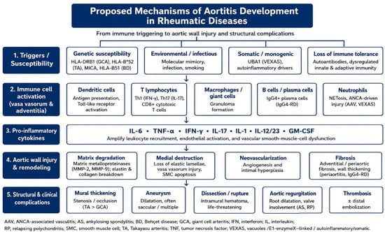
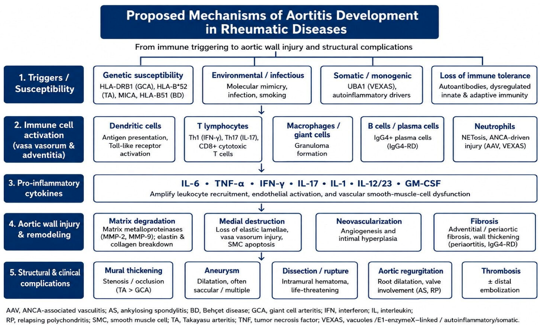
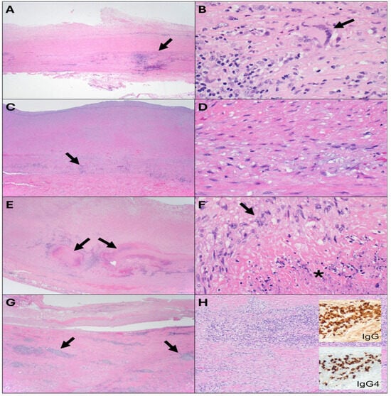
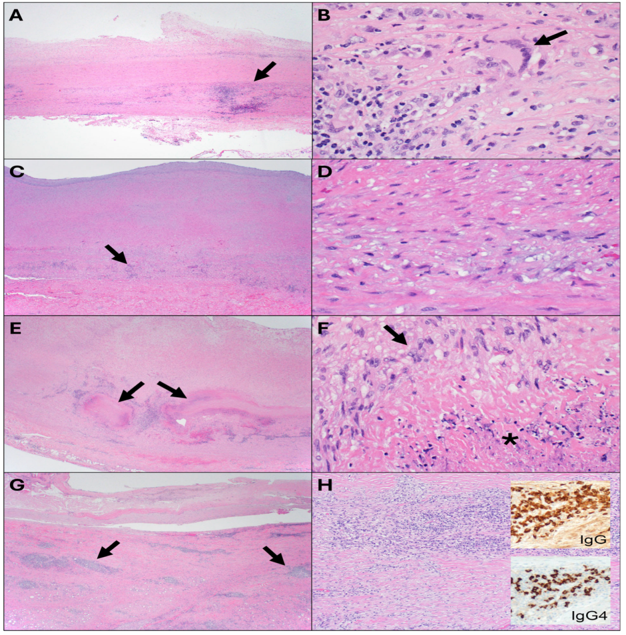
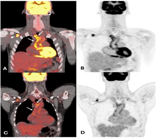
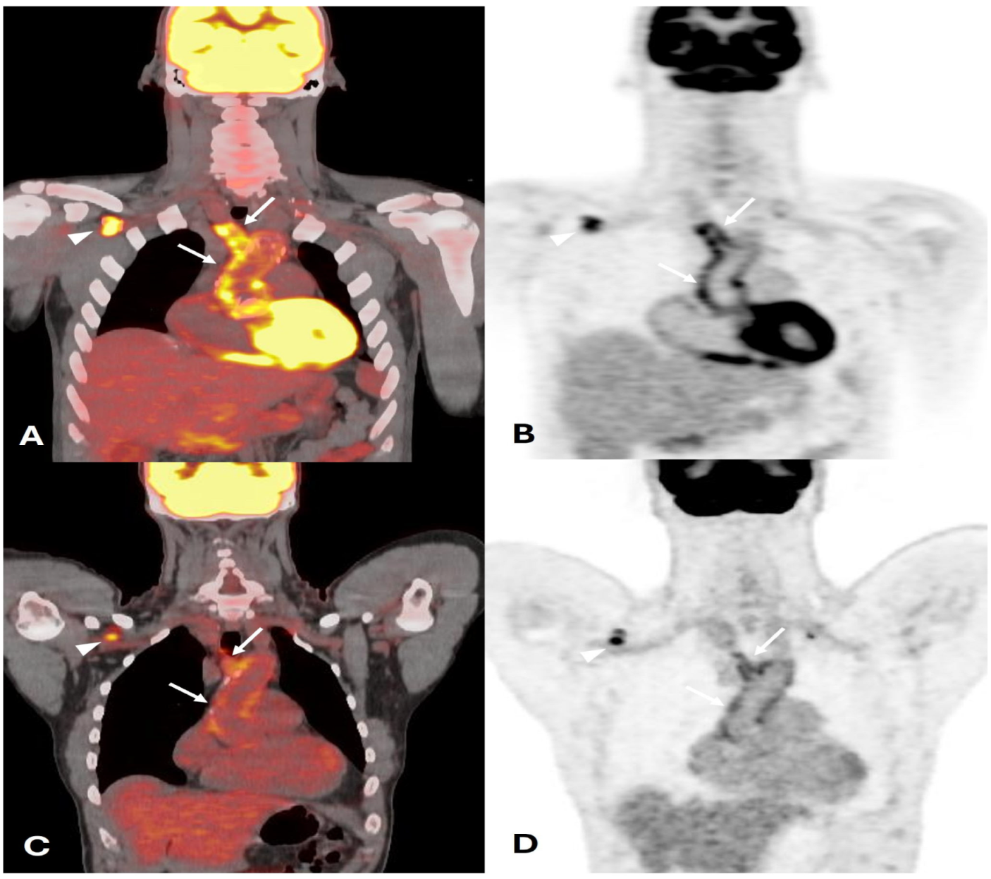
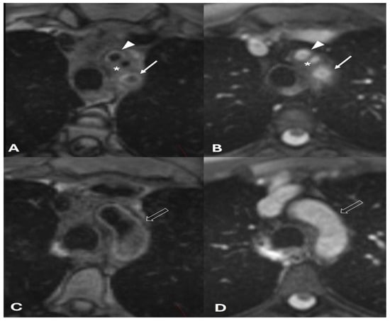
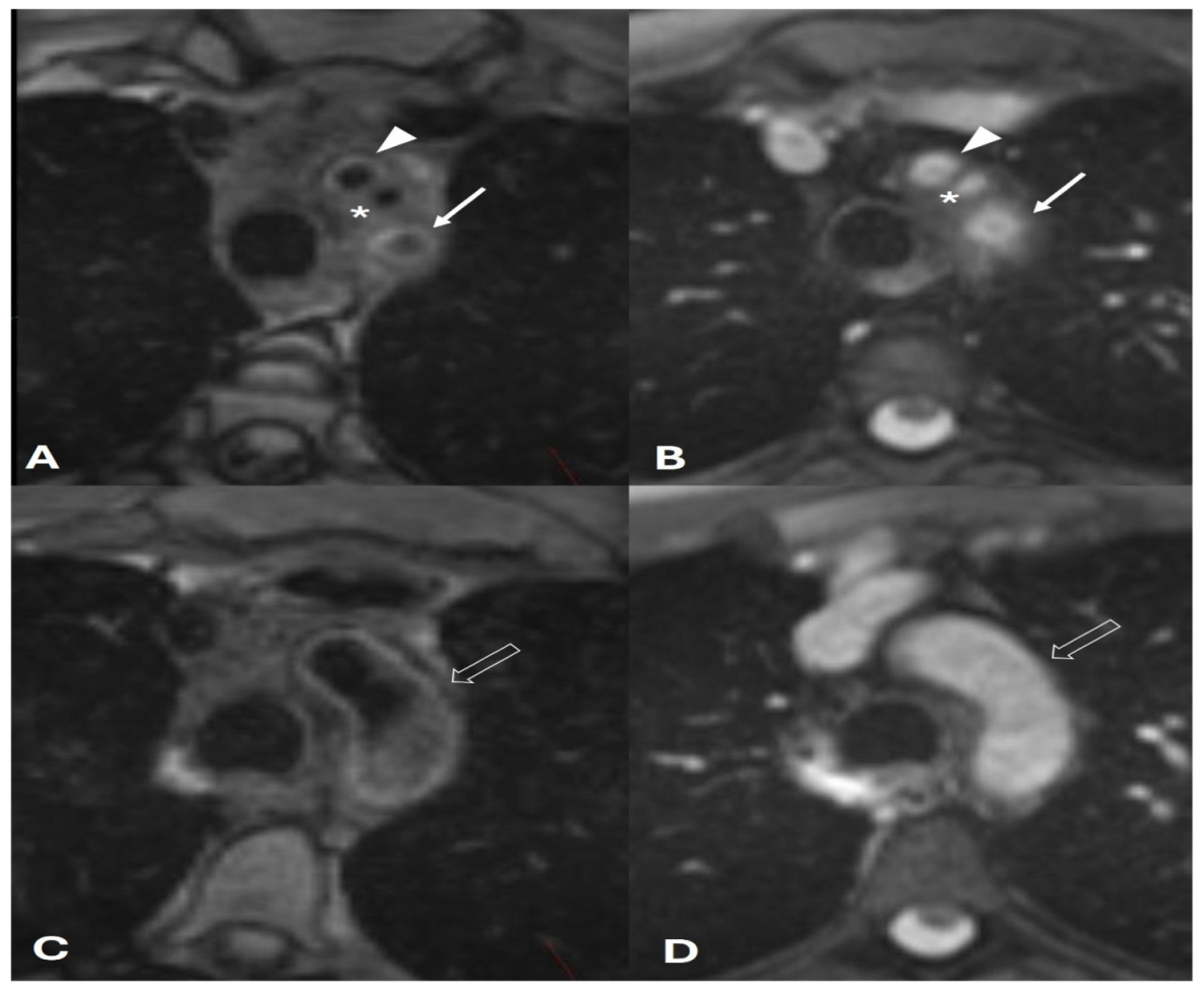
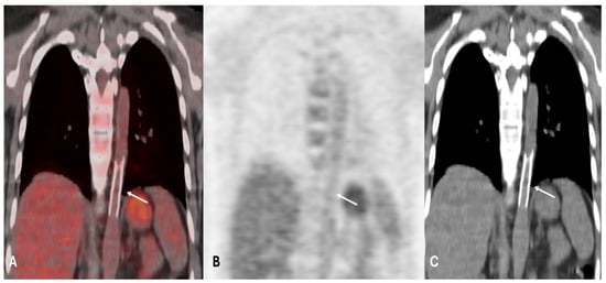
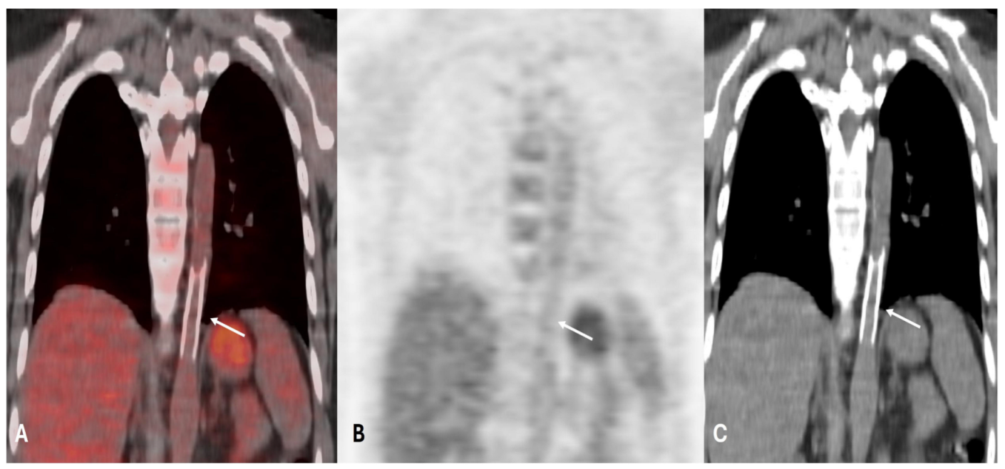
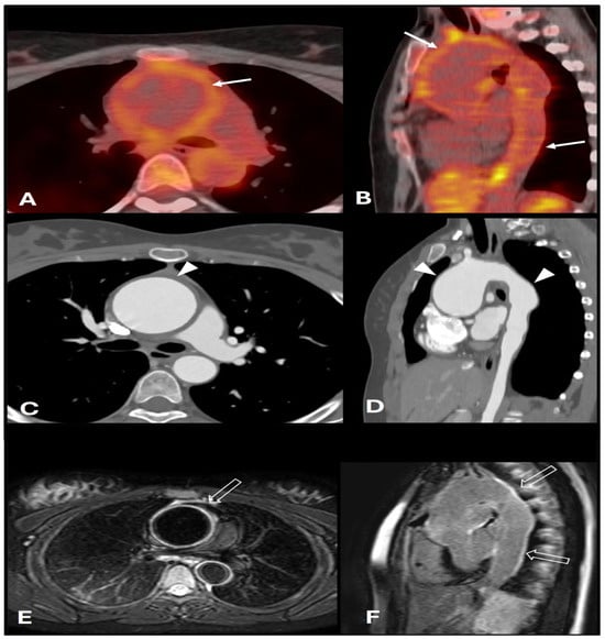
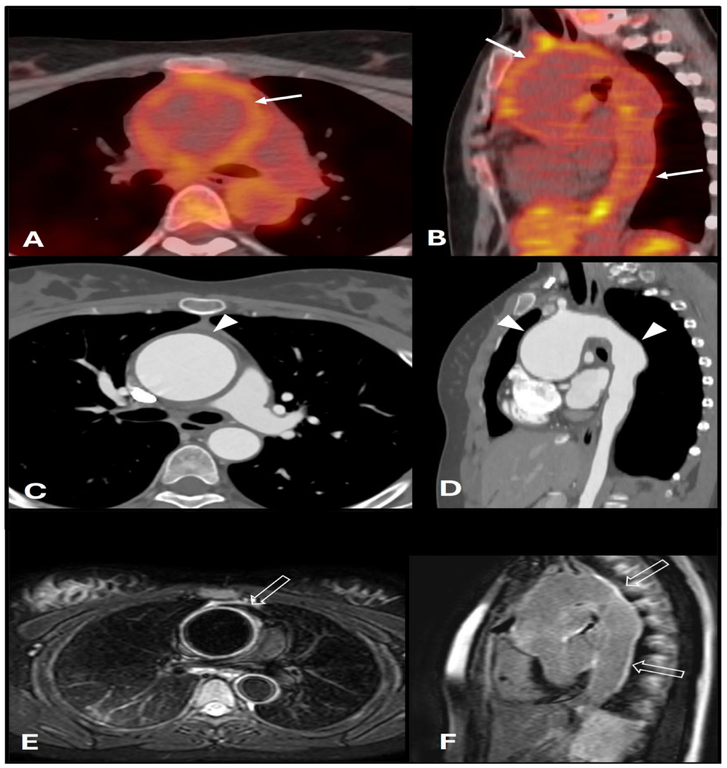
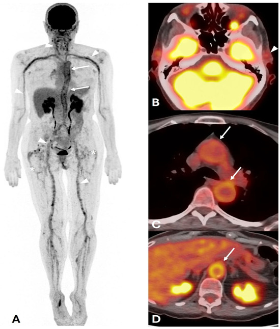
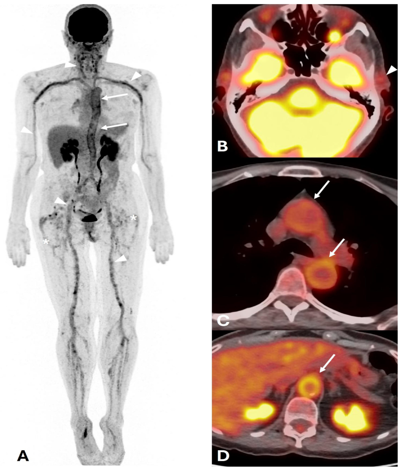
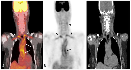
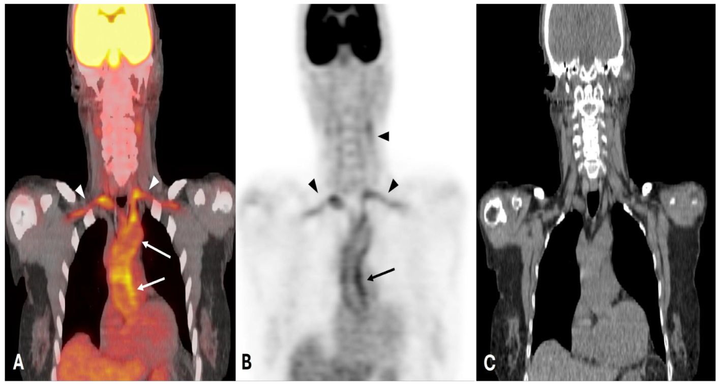
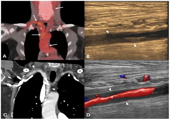
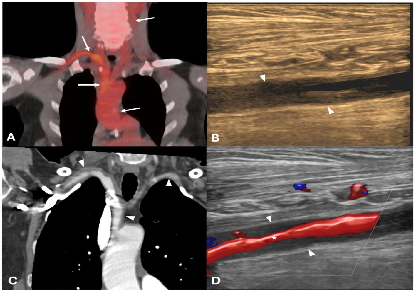
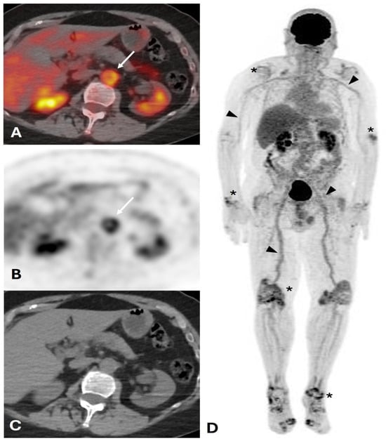
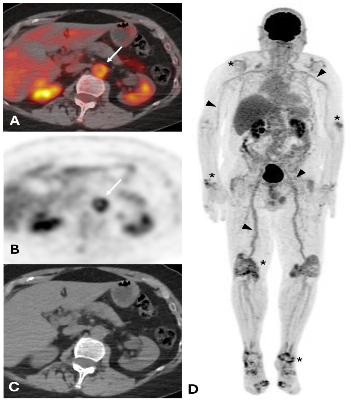
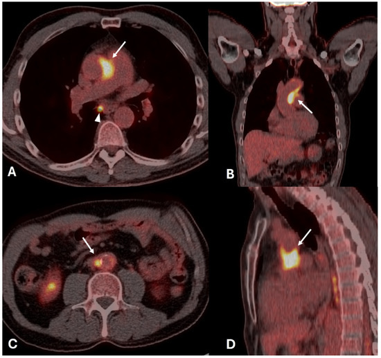
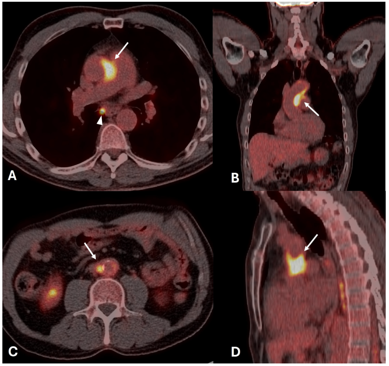
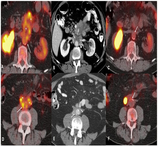
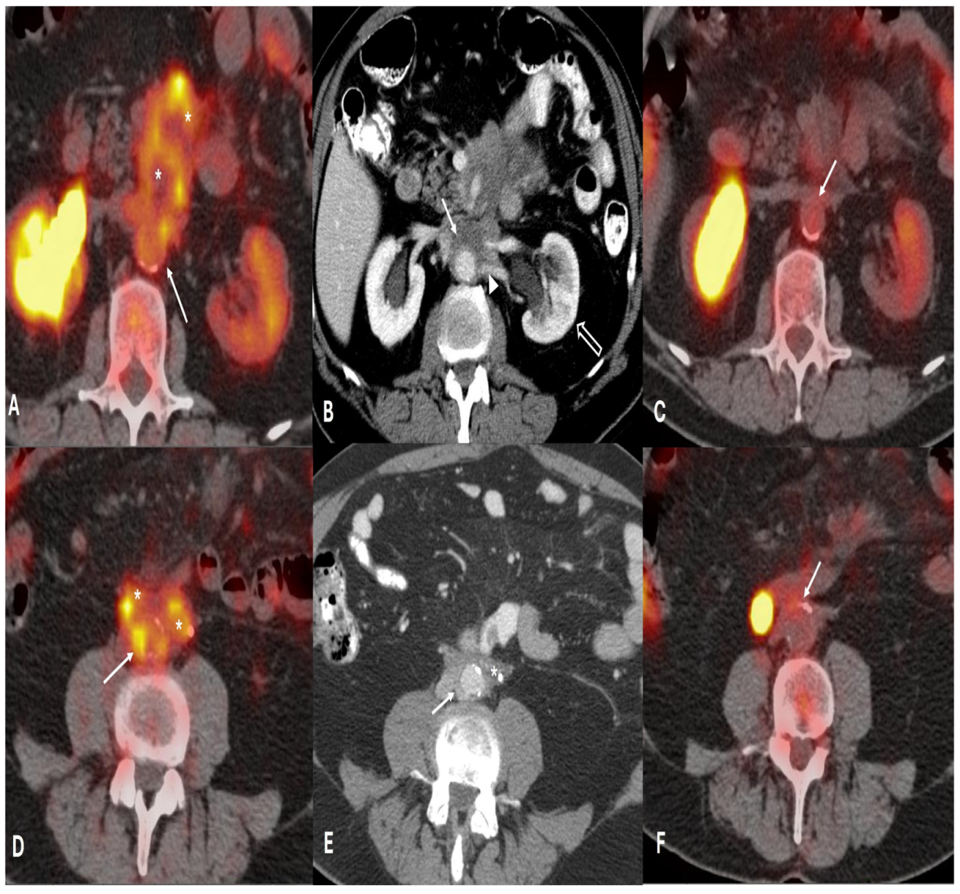
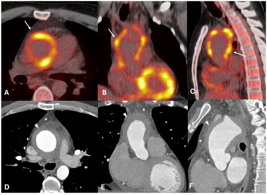
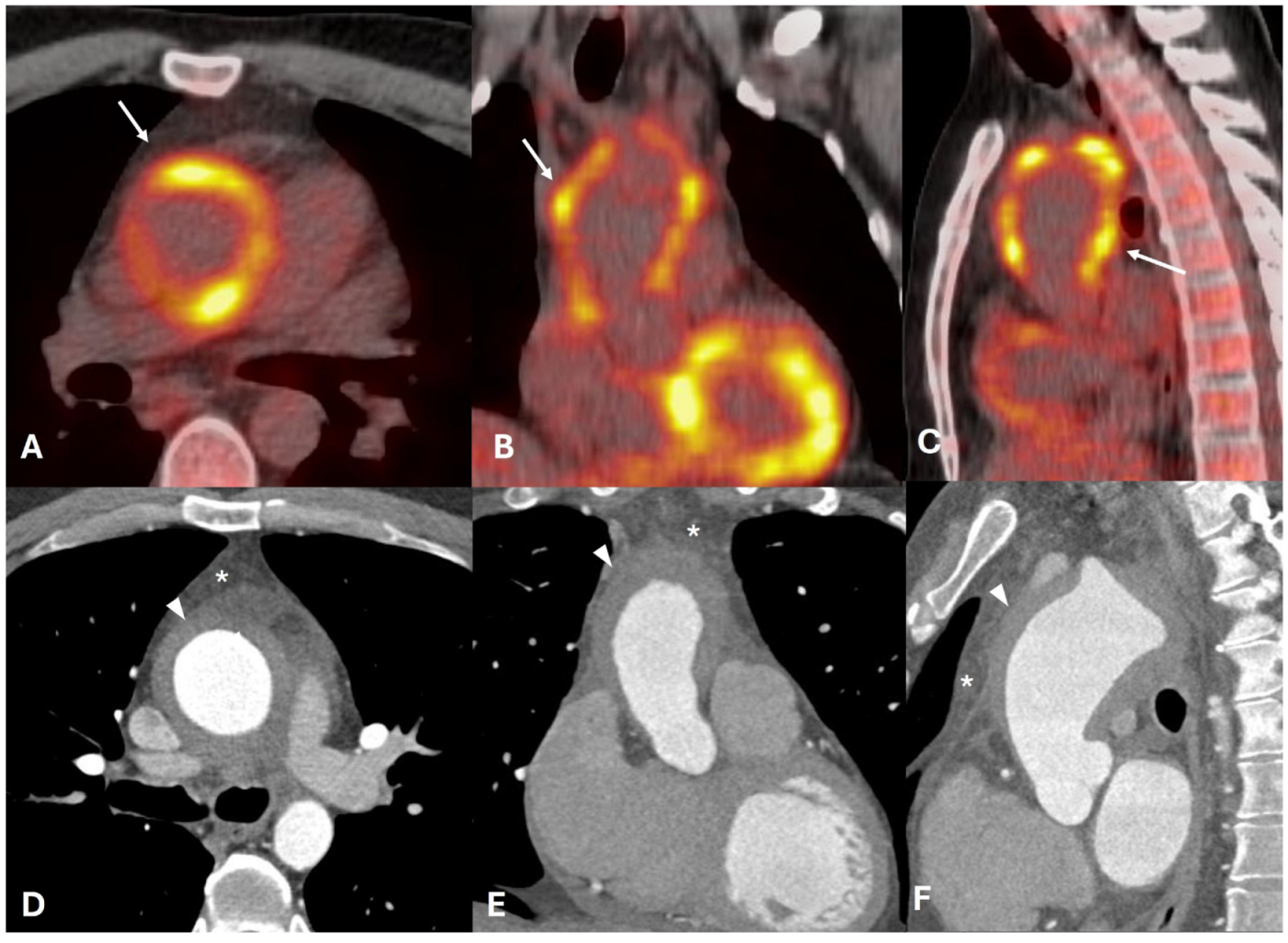
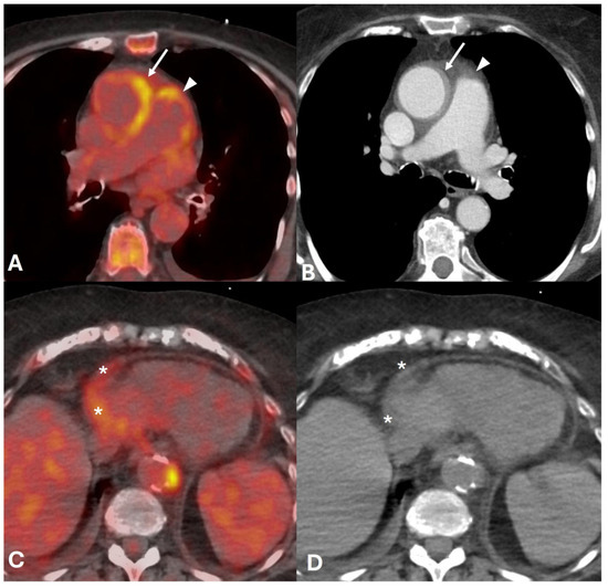
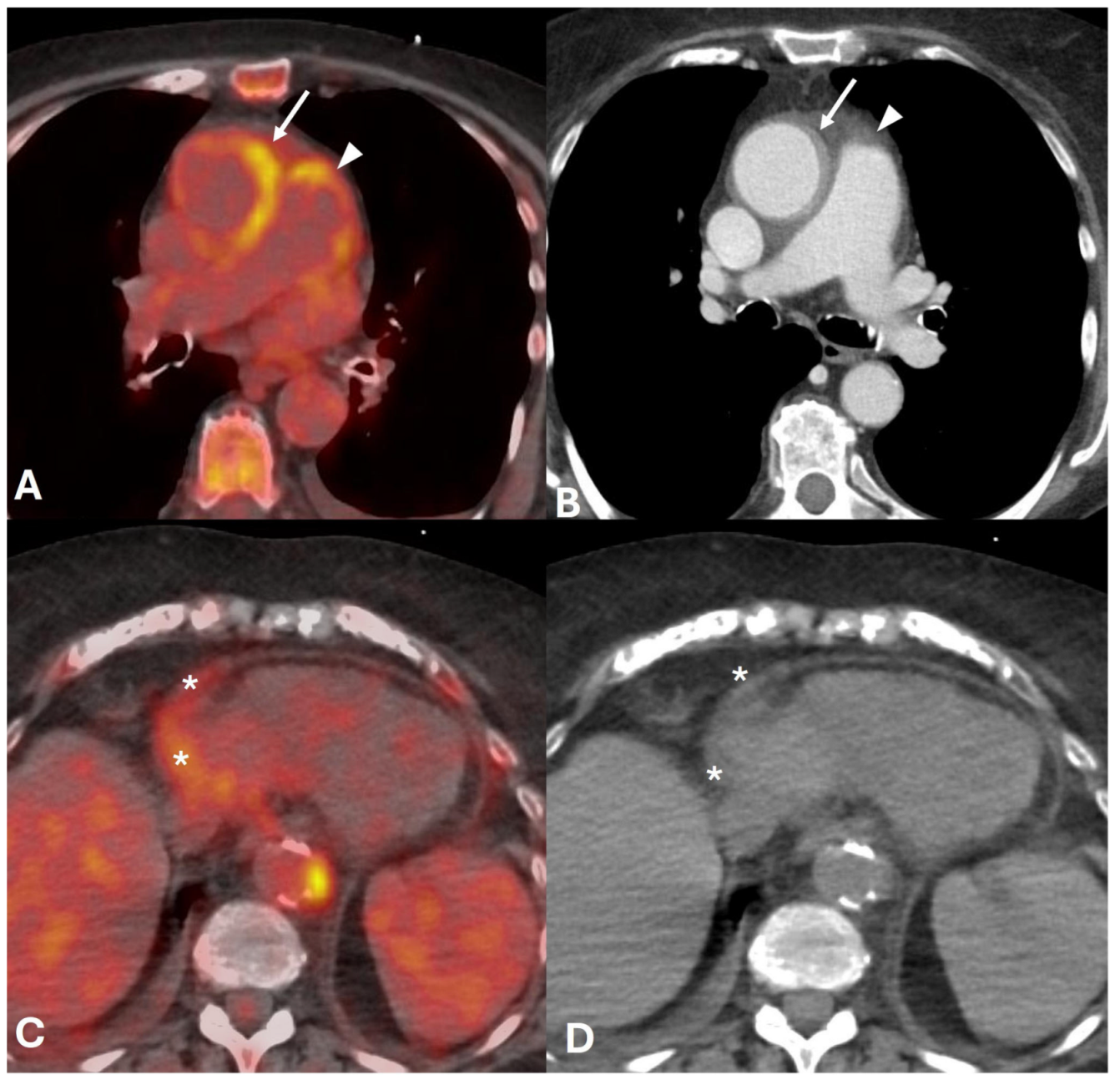
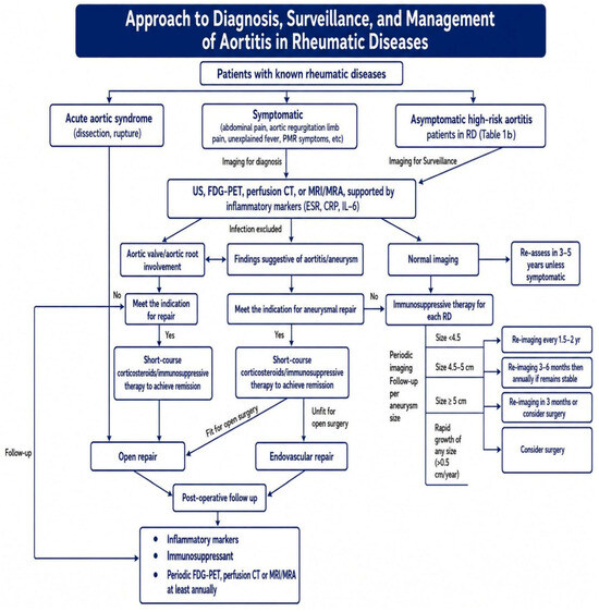
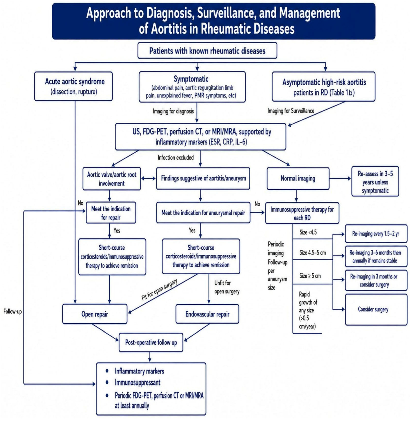

Disclaimer/Publisher’s Note: The statements, opinions and data contained in all publications are solely those of the individual author(s) and contributor(s) and not of MDPI and/or the editor(s). MDPI and/or the editor(s) disclaim responsibility for any injury to people or property resulting from any ideas, methods, instructions or products referred to in the content. |
© 2026 by the authors. Licensee MDPI, Basel, Switzerland. This article is an open access article distributed under the terms and conditions of the Creative Commons Attribution (CC BY) license.