


Abstract
Background: In low-flow, low-gradient aortic stenosis (LFLG AS), restricted aortic valve opening may represent either fixed valvular obstruction or flow-dependent incomplete leaflet opening due to reduced forward flow. Aortic stenosis (AS) and transthyretin cardiac amyloidosis (ATTR-CM) may coexist, making it difficult to distinguish myocardial disease–driven low-flow physiology from clinically relevant valvular obstruction. Case Presentation: An 88-year-old man presented with decompensated heart failure and paradoxical LFLG AS. Dobutamine stress echocardiography (DSE) failed to restore normal flow, and the calculated aortic valve area remained within the severe range despite stress. Computed tomography showed a low aortic valve calcium score (AVCS) of 313 Agatston units, leaving true-severe versus pseudo-severe AS indeterminate. Further evaluation confirmed wild-type ATTR-CM. Because the contribution of AS to heart failure was uncertain, the patient was initially managed with optimized heart failure therapy. Approximately two years later, he was readmitted with recurrent acute decompensated heart failure, severe left ventricular systolic dysfunction, dobutamine dependency, end-organ congestion, and a classical LFLG AS phenotype. Although AS severity remained indeterminate, a clinically relevant valvular afterload component could not be excluded. At that time, tafamidis was not immediately available because of local access limitations. Because left ventricular assist device implantation and heart transplantation were not feasible and surgical aortic valve replacement carried prohibitive risk, transcatheter aortic valve implantation (TAVI) was performed after discussion by the Heart Team. Left ventricular ejection fraction improved early after TAVI, from 19.9% before the procedure to 29.4% at 3 days and 44.1% at 35 days. At 1-year follow-up, left ventricular ejection fraction remained improved at 51%, and more than two years after TAVI, the patient continues regular outpatient follow-up without recurrent heart failure hospitalization. Conclusions: In ATTR-CM with LFLG AS, DSE and AVCS may not definitively determine AS severity. Carefully selected TAVI, combined with ATTR-directed and optimized heart failure therapy, may be associated with early left ventricular functional recovery and sustained clinical improvement.
1. Introduction
In low-flow, low-gradient aortic stenosis (LFLG AS), the aortic valve area (AVA) is in the severe range, whereas the transvalvular gradient remains low because forward flow is reduced. The key diagnostic challenge is to determine whether restricted valve opening reflects fixed severe valvular obstruction requiring aortic valve replacement or flow-dependent incomplete leaflet opening secondary to underlying myocardial disease [1,2,3,4].
The coexistence of transthyretin cardiac amyloidosis (ATTR-CM) further complicates this diagnostic process and is increasingly recognized in older patients with aortic stenosis (AS). Previous studies have estimated that ATTR-CM may be present in up to 15% of patients with AS and in up to 30% of those with LFLG AS [5]. This overlap is clinically important because shared clinical and imaging features—such as concentric left ventricular hypertrophy, heart failure, elevated natriuretic peptides, and low-flow physiology—may obscure the relative contributions of valvular obstruction and myocardial disease.
Dobutamine stress echocardiography (DSE) and computed tomography (CT)–derived aortic valve calcium scoring (AVCS) are commonly used to clarify AS severity in patients with discordant findings [2,4]. However, both modalities have important limitations in patients with an LFLG AS phenotype, particularly when concomitant ATTR-CM or restrictive physiology is present. DSE may remain inconclusive when transvalvular flow fails to normalize despite contractile or flow reserve, a scenario that may be encountered in advanced cardiomyopathy or restrictive physiology [2,6]. Although AVCS provides a flow-independent anatomical marker of calcific valve disease, its interpretation may be less straightforward in LFLG AS and in patients with cardiac amyloidosis [4,7]. Recent studies have shown that AVCS may have limited discrimination between severe and non-severe AS in low-gradient cohorts, and patients with ATTR-CM may exhibit unexpectedly low aortic valve calcium burden despite clinically important valve restriction. This may reflect a greater contribution of leaflet thickening, fibrosis, or amyloid infiltration rather than calcification alone [6,8,9,10].
We report a case of confirmed wild-type ATTR-CM with LFLG AS in which DSE and AVCS did not definitively distinguish true-severe from pseudo-severe AS. Despite persistent diagnostic uncertainty, transcatheter aortic valve implantation (TAVI) was selected after discussion by the Heart Team because a clinically relevant and potentially reversible valvular afterload component could not be excluded in the setting of recurrent heart failure, dobutamine dependency, and limited alternative therapeutic options. This case highlights that, when valvular disease and infiltrative myocardial disease coexist, AS severity may remain difficult to adjudicate despite complementary diagnostic testing. In such cases, treatment decisions may require individualized integration of imaging findings, clinical trajectory, heart failure severity, procedural risk, and therapeutic feasibility.
2. Case Report
2.1. Initial Presentation and Diagnostic Assessment
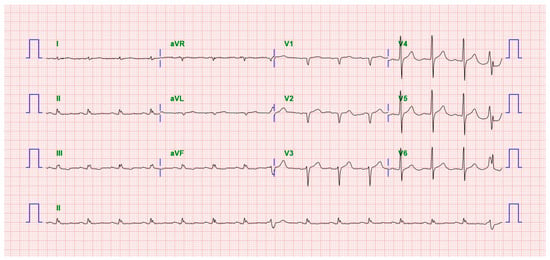
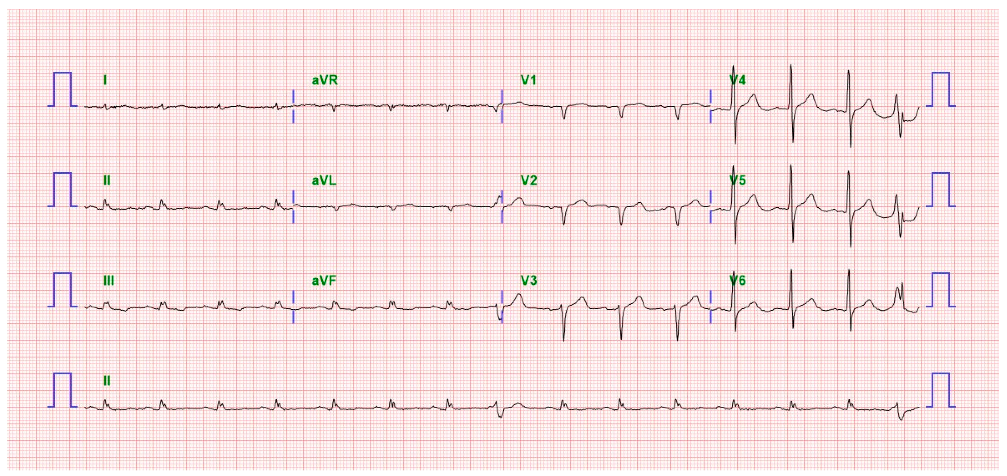
An 88-year-old man was referred for evaluation of progressive dyspnea and bilateral leg edema that had developed over 6 months and worsened during the week before admission. His medical history included multivessel coronary artery disease treated with percutaneous coronary intervention 3 years earlier, diabetes mellitus requiring insulin, hypertension, and dyslipidemia. He was hemodynamically stable on admission. Electrocardiography showed a regular sinus rhythm with low-voltage QRS complexes in the limb leads (Figure 1). Laboratory testing revealed an elevated cardiac troponin T level of 0.120 ng/mL and an NT-proBNP level of 3620 pg/mL, with relatively preserved renal and hepatic function.
Figure 1.
Baseline electrocardiogram at the initial presentation. Twelve-lead electrocardiography showed sinus rhythm with low-voltage QRS complexes in the limb leads.
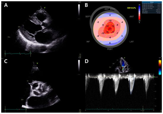
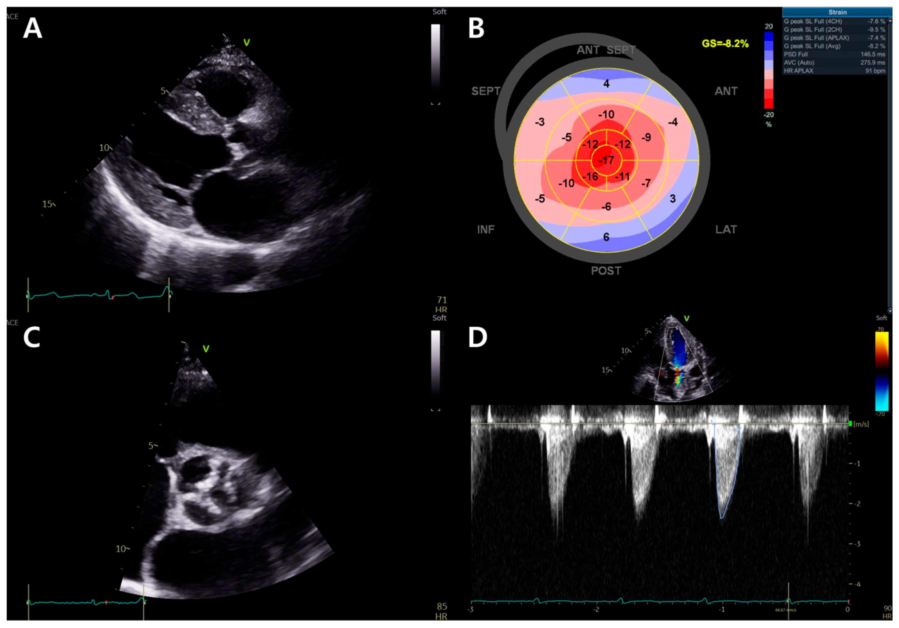
Initial transthoracic echocardiography (TTE) demonstrated concentric left ventricular (LV) hypertrophy with a highly reflective, speckled myocardial texture (Figure 2A), preserved left ventricular ejection fraction (LVEF) of approximately 50%, and markedly reduced global longitudinal strain (GLS) of −8.2% with an apical-sparing pattern (Figure 2B). Diastolic indices showed impaired relaxation, with an E/A ratio of 0.5, septal e′ velocity of 3 cm/s, average E/e′ of 10.6, left atrial volume index (LAVI) of 34.5 mL/m2, and estimated right ventricular systolic pressure of 29 mmHg; these indices were interpreted cautiously because the patient had already received diuretic therapy at the referring hospital before transfer.
Figure 2.
Baseline transthoracic echocardiographic findings at the initial presentation: (A) parasternal long-axis view showing concentric left ventricular hypertrophy with a highly reflective, speckled myocardial texture; (B) bull’s-eye strain map showing markedly reduced global longitudinal strain of −8.2% with a relative apical-sparing pattern; (C) parasternal short-axis view at the aortic valve level showing a sclerotic aortic valve with restricted leaflet opening; (D) representative continuous-wave Doppler tracing across the aortic valve showing low-gradient aortic stenosis. The averaged Doppler measurements are reported in the text.
On the initial TTE, aortic valve assessment showed a sclerotic valve with restricted opening and discordant stenosis parameters (Figure 2C, Supplementary Video S1). The AVA was 0.93 cm2 by the continuity equation, indexed AVA was 0.61 cm2/m2, peak velocity was 2.4 m/s, mean pressure gradient was 13.5 mmHg (Figure 2D), and Doppler velocity index (DVI) was 0.20. In the setting of preserved LVEF and reduced forward flow, with a stroke volume index of 23.9 mL/m2, these findings were interpreted as a paradoxical LFLG AS phenotype.
Taken together, the combination of low-voltage QRS complexes in the limb leads, concentric LV hypertrophy with a highly reflective myocardial texture, impaired diastolic function, and an apical-sparing pattern on the GLS bull’s-eye map raised concern for concomitant cardiac amyloidosis [11], although these findings could not be fully separated from AS-related remodeling at this stage.
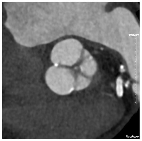
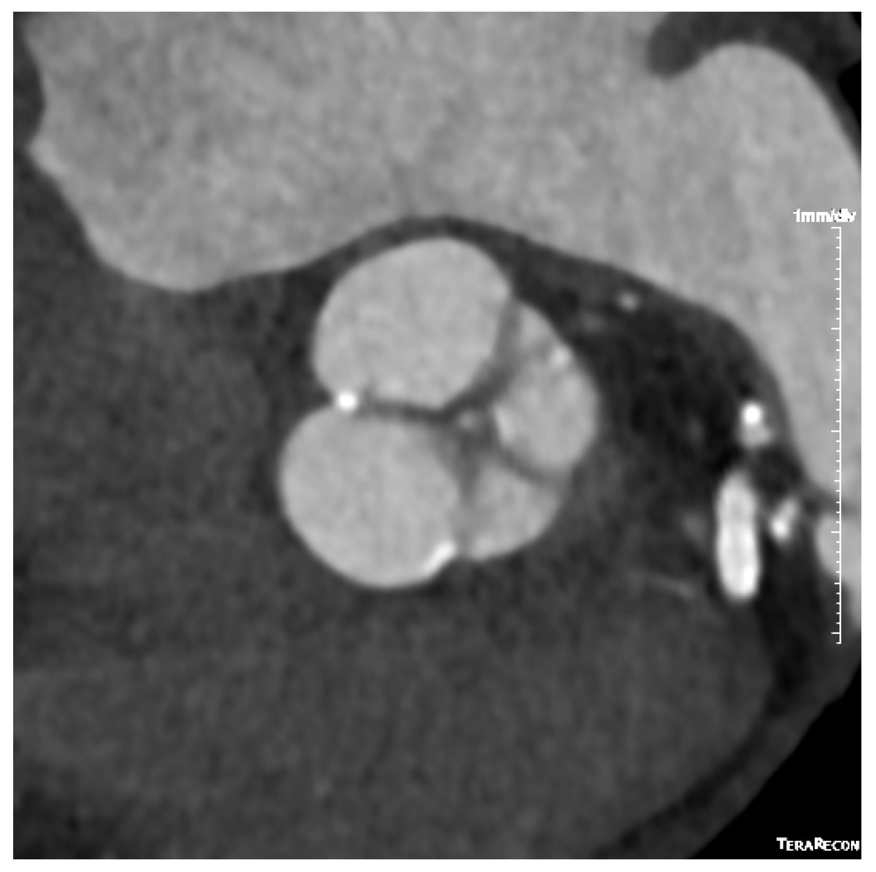
DSE was performed to clarify AS severity (Table 1). With a dobutamine infusion up to 20 μg/kg/min, stroke volume increased by 21%, suggesting flow reserve. However, stroke volume index remained below the normal-flow range, increasing only from 23.9 to 28.9 mL/m2. The calculated AVA remained within the severe range, decreasing from 0.90 to 0.80 cm2. Because continuity equation–derived AVA depends on Doppler-derived LVOT and AV VTI measurements, this decrease during stress was interpreted cautiously as possible measurement variability under low-flow conditions rather than as a physiologic reduction in anatomic valve area. The mean pressure gradient increased only modestly from 13.5 to 20.1 mmHg. Because normal flow was not restored and the peak-stress mean gradient did not reach conventional high-gradient severe AS criteria, DSE was considered inconclusive for distinguishing true-severe from pseudo-severe AS. Cardiac CT showed a low AVCS of 313 Agatston units, which was far below conventional thresholds for severe calcific AS and further contributed to diagnostic uncertainty (Figure 3).
Table 1.
Dobutamine stress echocardiography findings at the initial admission.
Figure 3.
Cardiac computed tomography assessment of aortic valve calcification. Cardiac computed tomography for aortic valve calcium scoring showed only limited focal calcification of the aortic valve, with a total aortic valve calcium score of 313 Agatston units.
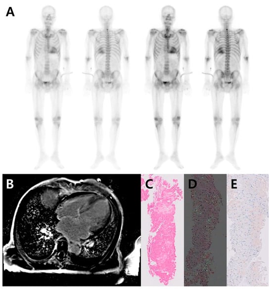
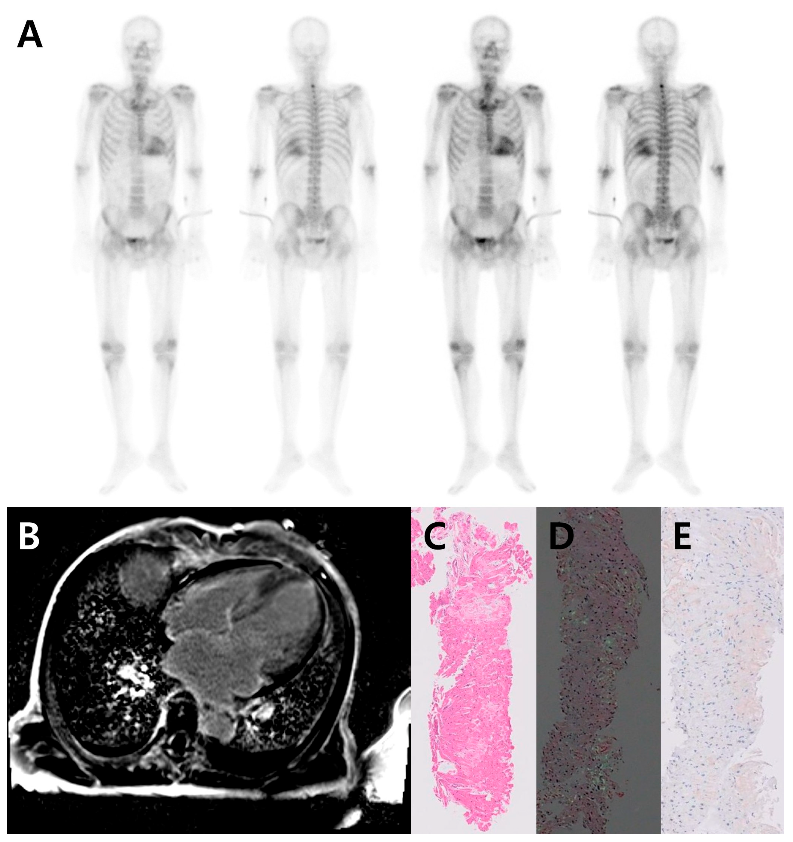
In parallel, further evaluation for cardiac amyloidosis was performed. Tc-99m DPD bone scintigraphy/SPECT demonstrated diffuse myocardial uptake, corresponding to Perugini grade 3 (Figure 4A). Cardiac magnetic resonance imaging showed increased LV wall thickness with a maximal end-diastolic septal thickness of 19 mm; diffuse subendocardial-to-transmural late gadolinium enhancement, involving the basal-to-mid LV, right ventricle (RV), and both atria (Figure 4B); and elevated native T1, T2, and extracellular volume. These findings supported a diagnosis of cardiac amyloidosis. Right ventricular endomyocardial biopsy showed interstitial and pericellular deposition of amorphous pale-pink material (Figure 4C), Congo red–positive amyloid deposits with birefringence under polarized light (Figure 4D), positive immunohistochemical staining for transthyretin (Figure 4E), and negative kappa and lambda light-chain staining. Monoclonal protein testing showed no evidence of plasma cell dyscrasia. A serum-free light-chain assay showed kappa at 26.49 mg/L and lambda at 26.36 mg/L, with a normal kappa/lambda ratio of 1.00. Serum and urine protein electrophoresis showed no monoclonal peak, and serum and urine immunofixation electrophoresis showed no monoclonal immunoglobulin or light-chain component. TTR gene testing showed no mutation. These findings supported a diagnosis of wild-type ATTR-CM.
Figure 4.
Multimodality diagnostic confirmation of transthyretin cardiac amyloidosis: (A) technetium-99m DPD bone scintigraphy demonstrated diffuse myocardial tracer uptake with intensity greater than the adjacent ribs, consistent with Perugini grade 3 uptake; (B) cardiac magnetic resonance imaging with late gadolinium enhancement showed diffuse subendocardial-to-transmural enhancement involving the left ventricle, right ventricle, and both atria; (C) right ventricular endomyocardial biopsy showed interstitial and pericellular deposition of amorphous pale-pink material; (D) Congo red staining demonstrated amyloid deposits with birefringence under polarized light; (E) immunohistochemical staining for transthyretin was positive, supporting transthyretin-type amyloid deposition.
During the first admission, the Heart Team considered the contribution of AS to the acute decompensated heart failure to be uncertain, given the inconclusive DSE, low AVCS, and newly confirmed wild-type ATTR-CM. Therefore, immediate valve intervention was deferred, and the patient was managed with optimization of heart failure therapy, including the addition of a sodium-glucose cotransporter 2 inhibitor, a mineralocorticoid receptor antagonist, and a beta-blocker to his previous medications. At that time, ATTR-CM–directed disease-modifying therapy was constrained by local access. Standard-dose tafamidis was not yet reimbursed locally, and immediate initiation was limited by substantial out-of-pocket cost. The patient was, therefore, enrolled in a free-supply access program and awaited drug availability.
2.2. Recurrent Decompensation and Reassessment of LFLG AS
The patient subsequently developed recurrent decompensated heart failure. Approximately 1 year and 9 months after the first admission, he visited the emergency department for dyspnea and was managed with intensified diuretic therapy. Four months later, he was readmitted with worsening dyspnea and bilateral pleural effusions. Laboratory testing showed a markedly elevated NT-proBNP level of 34,811 pg/mL; an increase in creatinine from 1.17 to 1.85 mg/dL; mild hyperbilirubinemia and liver enzyme elevation consistent with congestive hepatopathy; and an elevated lactate level of 3.8 mmol/L. These findings suggested impaired systemic perfusion in the setting of acute decompensated heart failure.
Repeat TTE demonstrated marked deterioration in myocardial function. LVEF decreased from 50% at the first admission to 19.9% (Supplementary Videos S2 and S3), and GLS was reduced to −5.6%, with diffuse LV hypokinesia. Left atrial enlargement progressed, with LAVI increasing from 34.5 to 71.9 mL/m2. Right-sided remodeling was also evident, with RV enlargement (basal diameter, 50.7 mm), right atrial enlargement (RA area, 25.8 cm2), a plethoric inferior vena cava (IVC), progression of tricuspid regurgitation from trivial to moderate, and an increase in estimated RVSP from 29 to 44 mmHg.
Aortic valve assessment continued to show discordant LFLG physiology, now observed in the setting of severe LV systolic dysfunction. Stroke volume index remained low at 22.3 mL/m2, with an AVA of 0.8 cm2, indexed AVA of 0.5 cm2/m2, mean pressure gradient of 12.1 mmHg, and DVI of 0.20. These findings were consistent with evolution from a paradoxical to a classical LFLG AS phenotype, while true-severe versus pseudo-severe AS remained indeterminate.
Given the new severe LV systolic dysfunction and persistent uncertainty regarding the contribution of AS to clinical deterioration, a repeat DSE examination was performed to reassess flow reserve (Table 2). The resting TTE and the subsequent DSE were performed under different hemodynamic conditions: the resting TTE was obtained before intensive decongestion, and the subsequent DSE assessment was performed four days after the resting TTE, after interval treatment with continuous intravenous diuretic therapy and pleural effusion management. Over this interval, body weight decreased from 57.0 to 48.9 kg. At the time of DSE, the patient was receiving low-dose dobutamine support for advanced heart failure; therefore, the initial DSE stage was obtained with dobutamine at 5 μg/kg/min rather than under drug-free resting conditions. With a dobutamine infusion up to 20 μg/kg/min, LVEF increased from 22.8% to 32.9%, and stroke volume index increased from 12.4 to 21.0 mL/m2, indicating contractile or flow reserve. However, normal flow was not restored, AVA changed minimally from 0.96 to 1.00 cm2, and the mean pressure gradient increased only modestly from 9.5 to 13.5 mmHg. Projected AVA was calculated using the initial and peak DSE stages. Mean transvalvular flow rate increased from 86 to 139 mL/s, and the projected AVA at a standardized flow rate of 250 mL/s was 1.08 cm2. Because the initial DSE stage was obtained with dobutamine at 5 μg/kg/min rather than drug-free resting conditions, and because peak flow remained markedly below the standardized flow rate, this value was interpreted cautiously. Overall, DSE did not support unequivocal true-severe AS but did not eliminate the clinical uncertainty regarding the relevance of the valvular afterload component. Repeat cardiac CT showed no substantial interval change compared with the prior CT and did not clarify AS severity. Coronary assessment on the same CT showed no definite in-stent restenosis in the previously implanted coronary stents and no substantial interval progression of coronary artery disease. Nevertheless, because invasive coronary angiography and formal functional ischemia testing were not performed, a residual ischemic contribution to LV systolic dysfunction could not be completely excluded.
Table 2.
Hemodynamic context and echocardiographic parameters during pre-TAVI reassessment.
2.3. Therapeutic Decision-Making and TAVI
The major therapeutic dilemma was whether the patient’s clinical deterioration was driven predominantly by progression of ATTR-CM or by a potentially reversible valvular afterload component associated with concomitant LFLG AS. The low AVCS was interpreted cautiously in the context of confirmed ATTR-CM because non-calcific leaflet thickening may contribute to valve restriction and may not be fully captured by calcium scoring.
At the time of recurrent decompensation, the patient had features of advanced heart failure, including recurrent hospitalization, severe LV systolic dysfunction, dobutamine dependency, and progressive end-organ congestion despite medical therapy. Given his advanced age, frailty, multiple comorbidities, and overall clinical status, left ventricular assist device implantation and heart transplantation were not considered feasible therapeutic options [12]. Immediate ATTR-CM–directed treatment options were also limited. Tafamidis had not yet become available through the free-supply access program and, even if available, was not expected to provide immediate hemodynamic stabilization.
During the Heart Team discussion, the persistent diagnostic uncertainty was acknowledged: true-severe AS had not been definitively confirmed by DSE or AVCS. Nevertheless, the persistently restricted leaflet opening and low DVI made it difficult to dismiss a clinically relevant and potentially reversible valvular afterload component. The anticipated benefit of valve intervention was, therefore, not based on definitive confirmation of true-severe AS, but on the possibility of reducing a modifiable afterload burden contributing to recurrent hemodynamic deterioration.
Once valve intervention was considered, surgical aortic valve replacement (SAVR) was judged to carry prohibitive risk because of advanced age, frailty, severe LV systolic dysfunction, multiple comorbidities, and a high Society of Thoracic Surgeons score. Therefore, TAVI was favored over SAVR as a less invasive approach if afterload reduction was to be attempted. This potential benefit was weighed against the risks of TAVI, including periprocedural hemodynamic instability and delayed or limited myocardial recovery due to advanced ATTR-CM. After discussion by the Heart Team and shared decision-making with the patient and family, the residual diagnostic uncertainty was accepted, and TAVI was selected as the most immediately actionable strategy to address a potentially reversible valvular afterload component.
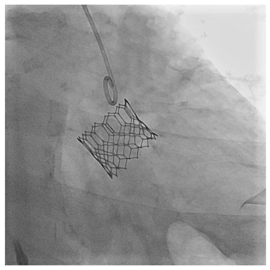
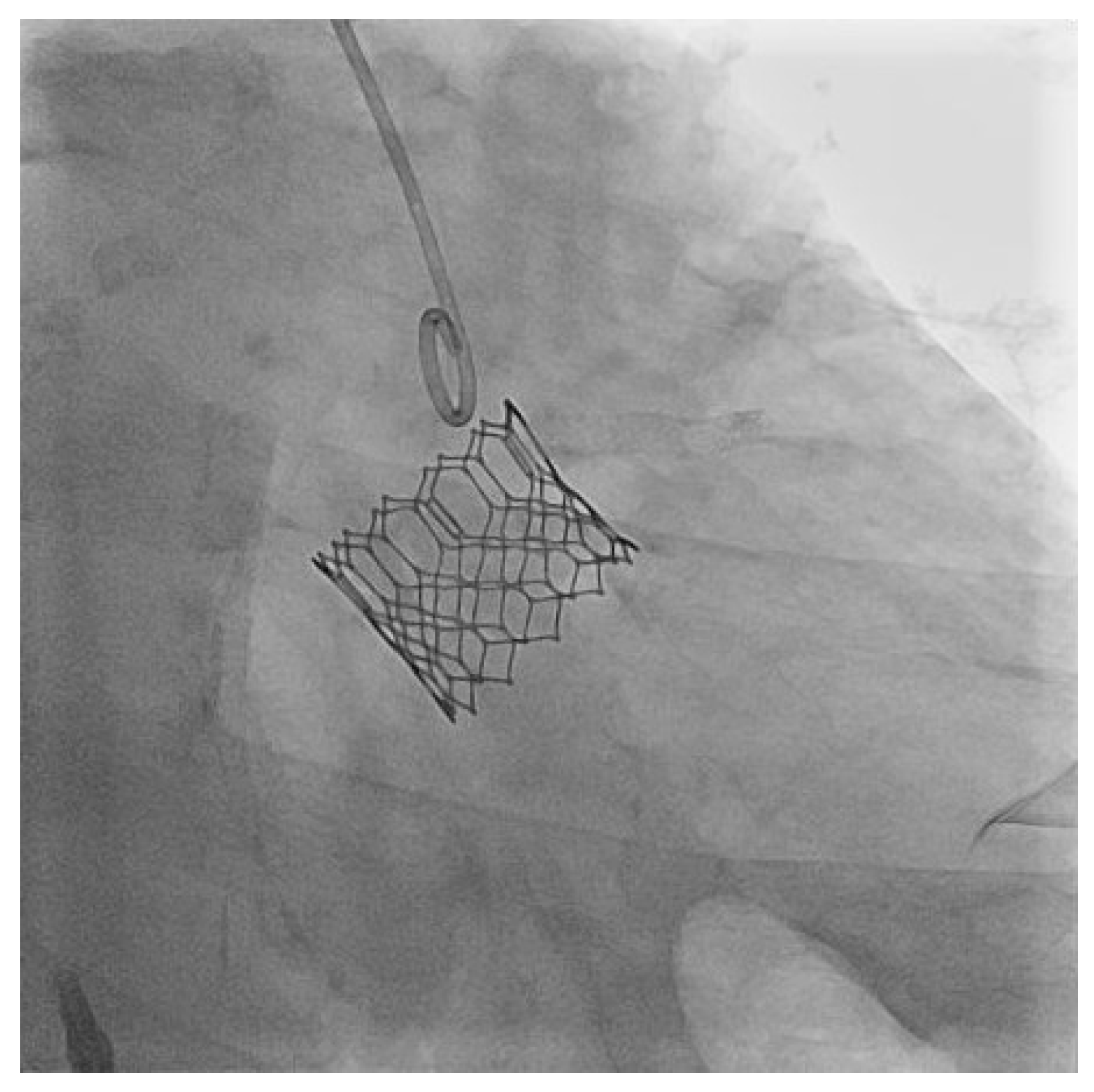
TAVI was performed via the transfemoral approach using a balloon-expandable SAPIEN 3 Ultra 26 mm valve (Figure 5). Immediately after valve deployment, transient severe hypotension and pulseless electrical activity occurred, requiring brief cardiopulmonary resuscitation and epinephrine, with return of spontaneous circulation within approximately 1 min.
Figure 5.
Transcatheter aortic valve implantation. Intra-procedural fluoroscopic image showing implantation of a balloon-expandable SAPIEN 3 Ultra 26 mm transcatheter aortic valve.
2.4. Post-TAVI Course and Follow-Up
After TAVI, serial echocardiography demonstrated early and progressive improvement in LV systolic function during the index hospitalization. LVEF increased from 19.9% before TAVI to 29.4% at 3 days, 39.3% at 26 days, and 44.1% at 35 days after TAVI (Supplementary Videos S4 and S5). The transcatheter valve showed good function, without central aortic regurgitation or paravalvular leak.
Clinical stabilization was more gradual than echocardiographic recovery. The patient remained intermittently dependent on dobutamine after TAVI, with recurrent worsening of renal function, edema, pleural effusion, and NT-proBNP levels during attempts to withdraw inotropic support. Cardiac rehabilitation was initiated, although functional capacity remained limited in the early postprocedural period. Tafamidis became available through the previously arranged free-supply access program and was initiated 7 days after TAVI. Heart failure therapy was further optimized during hospitalization with the addition of vericiguat and sacubitril/valsartan. The hospital course was complicated by pneumonia, and the patient was discharged 36 days after TAVI.
At approximately 1-year follow-up, the improvement in LV systolic function was sustained, with LVEF increasing to 51%. Forward flow also improved, with the stroke volume index increasing to 62 mL/m2. The transcatheter valve continued to function well, with a peak velocity of 2.1 m/s, mean pressure gradient of 9.1 mmHg, DVI of 0.43, and no paravalvular regurgitation. LAVI decreased from 71.9 to 58.0 mL/m2. Right-sided parameters also improved, with normalization of RV dimensions and IVC collapsibility, and reduction in tricuspid regurgitation to trivial. At more than 2 years after TAVI, the patient remains under regular outpatient follow-up and has had no recurrent hospitalization for heart failure.
3. Discussion
This case illustrates the difficulty of determining the relative contributions of restrictive cardiomyopathy and valvular obstruction to heart failure presentation in patients with coexisting ATTR-CM and LFLG AS. In this patient, DSE and AVCS were insufficient for definitive characterization of AS severity. At the initial presentation, he had a paradoxical LFLG AS phenotype, characterized by preserved LVEF, AVA in the severe range, reduced forward flow, and a low transvalvular gradient. During follow-up, the patient’s hemodynamic phenotype evolved from paradoxical to classical LFLG AS, accompanied by severe LV systolic dysfunction, persistent low-flow physiology, and recurrent decompensated heart failure. This trajectory illustrates how progressive amyloid cardiomyopathy and persistent valvular afterload may interact to worsen low-flow physiology and clinical decompensation. The key clinical question was whether a clinically relevant and potentially modifiable valvular afterload component remained despite advanced myocardial disease.
DSE is commonly used to differentiate true-severe from pseudo-severe AS in patients with LFLG AS [1,2]. However, its diagnostic utility depends on sufficient augmentation of transvalvular flow during stress. In patients with cardiac amyloidosis, restrictive ventricular physiology, impaired LV filling, and limited preload reserve may blunt the increase in stroke volume during dobutamine infusion, thereby limiting augmentation of transvalvular flow and resulting in inconclusive assessment of AS severity [5,6]. In the present case, although flow reserve was present, normal flow was not restored, AVA changed minimally, and the increase in transvalvular gradient remained modest. Therefore, DSE was limited in assessing AS severity in this patient with restrictive cardiomyopathy.
AVCS was also limited in assessing AS severity. Although CT-derived AVCS is a flow-independent adjunctive marker in discordant AS grading, the original sex-specific AVCS thresholds were derived from AS cohorts with preserved LVEF and normal-flow reference groups and should therefore be applied cautiously in patients with severe low-flow physiology or infiltrative cardiomyopathy [7]. In a recent multicenter cohort of patients with low-gradient AS, AVCS showed limited sensitivity and specificity for AS severity as classified by echocardiography and DSE, suggesting that AVCS alone has limited diagnostic discrimination in low-gradient AS [10].
In the present case, the discordance between the very low AVCS and severe-range echocardiographic indices required careful interpretation. In a male patient, an AVCS of 313 Agatston units was far below conventional thresholds for severe calcific AS and therefore argued against typical severe calcific AS. However, the persistently small calculated AVA, low DVI, and restricted leaflet opening made it difficult to dismiss a clinically relevant valvular afterload component. Several technical and physiological factors may have contributed to this discordance. Continuity equation–derived AVA and DVI are sensitive to Doppler acquisition and measurement variability, particularly in marked low-flow states. In addition, restrictive cardiomyopathy may reduce forward flow, lower transvalvular gradients, and limit opening of a stiff aortic valve. Previous studies have also shown that patients with ATTR-CM may have lower relative AVCS than those without cardiac amyloidosis and that severe AS may occur in ATTR-CM despite subthreshold AVCS values, supporting the need for composite assessment rather than reliance on AVCS alone [6,8,9]. One possible explanation is that CT calcium scoring quantifies calcific burden but may not capture non-calcific leaflet abnormalities, such as leaflet thickening, fibrosis, or amyloid infiltration. However, this mechanism remains inferential in the present case because direct valvular histology or dedicated quantitative assessment of non-calcific leaflet involvement was not available. Therefore, the low AVCS did not definitively support classification as pseudo-severe AS.
Given this persistent uncertainty after noninvasive assessment, a further limitation of the diagnostic work-up is that invasive cardiac catheterization with direct hemodynamic assessment was not performed. Simultaneous left ventricular–aortic pressure measurement and catheter-derived valve area calculation may have provided additional characterization of AS severity.
Recent evidence suggests that aortic valve intervention may be associated with clinical benefit, even in patients with low-gradient AS and a low AVCS [13]. In a study of symptomatic low-gradient AS, a low AVCS was more frequent than in high-gradient AS and did not necessarily indicate a better prognosis; importantly, aortic valve intervention was associated with improved survival, even among patients with low-gradient AS and a low AVCS [13]. In the TOPAS prospective observational cohort, early AVR was associated with improved survival compared with conservative management in both classical and paradoxical LFLG AS, and this apparent benefit extended even to patients classified as having pseudo-severe AS [14]. These data support the concept that, in selected symptomatic patients with LFLG AS, a low AVCS alone should not preclude consideration of aortic valve intervention [15,16].
Recent observational data and a case report in patients with AS and ATTR-CM suggest that valve unloading and ATTR-directed therapy may be complementary [17,18]. Although ATTR-CM disease-modifying options have now expanded beyond tafamidis to include other transthyretin-stabilizing and transthyretin-silencing therapies, these newer agents were not available as routine local treatment options for ATTR-CM during this patient’s decision-making period.
The favorable trajectory observed in this case should be interpreted with caution and should not be attributed to TAVI alone. The early increase in LVEF shortly after TAVI suggests that relief of valvular afterload may have contributed to initial LV systolic recovery. However, sustained echocardiographic and clinical improvement occurred in the setting of preserved transcatheter valve function, subsequent tafamidis initiation, optimization of guideline-directed heart failure therapy, and rehabilitation [19,20]. This single case should not be interpreted as supporting routine valve intervention in similar patients without individualized Heart Team assessment.
4. Conclusions
In patients with ATTR-CM and LFLG AS, DSE and AVCS may be insufficient for definitive characterization of AS severity. This case highlights that DSE may remain inconclusive when normal flow is not restored, and that a low AVCS should be interpreted cautiously in ATTR-CM with marked low-flow physiology. When recurrent advanced heart failure develops and a clinically relevant valvular afterload component cannot be excluded, individualized Heart Team decision-making should integrate imaging findings, clinical trajectory, heart failure severity, procedural risk, therapeutic feasibility, and patient-specific goals of care. In this patient, TAVI was pursued despite residual diagnostic uncertainty because a clinically relevant valvular afterload component could not be excluded and therapeutic alternatives were limited. The early LV functional recovery was compatible with a contribution from valvular afterload reduction; however, sustained clinical and echocardiographic improvement should be interpreted in the context of integrated management, including TAVI, ATTR-directed therapy, optimized heart failure treatment, and rehabilitation.
Supplementary Materials
The following supporting information can be downloaded at: https://www.mdpi.com/article/10.3390/diagnostics16142177/s1, Video S1: Baseline transthoracic echocardiography (TTE), short-axis view at the aortic valve level, showing a sclerotic aortic valve with restricted leaflet opening; Video S2: Pre-TAVI TTE during recurrent decompensation, parasternal long-axis view, showing severe left ventricular systolic dysfunction; Video S3: Pre-TAVI TTE during recurrent decompensation, apical four-chamber view, showing diffuse left ventricular hypokinesia; Video S4: TTE obtained 35 days after TAVI, parasternal long-axis view, showing improved left ventricular systolic function; Video S5: TTE obtained 35 days after TAVI, apical four-chamber view, showing improved left ventricular systolic function.
Author Contributions
Conceptualization, S.-Y.L. and J.-C.Y.; data curation, S.-Y.L.; investigation, S.-Y.L., J.-C.Y. and M.-H.J.; resources, J.-C.Y.; visualization, S.-Y.L., M.-H.J., W.-B.C. and H.O.J.; writing—original draft preparation, S.-Y.L.; writing—review and editing, S.-Y.L., J.-C.Y., M.-H.J., W.-B.C. and H.O.J.; supervision, J.-C.Y.; project administration, S.-Y.L. and J.-C.Y.; funding acquisition, J.-C.Y. All authors have read and agreed to the published version of the manuscript.
Funding
This work was supported by National Research Foundation of Korea (NRF) grants, funded by the Korean government (MSIT) (No. RS-2024-00455661, No. RS-2024-00440824: Bio & Medical Technology Development Program). The funders had no role in study design, data collection and analysis, decision to publish, or preparation of the manuscript.
Institutional Review Board Statement
Ethical review and approval were waived for this study in accordance with institutional policy for single-patient case reports.
Informed Consent Statement
Written informed consent has been obtained from the patient to publish this paper. Separate informed consent for participation in a clinical study was not required because this manuscript is a single-patient case report, did not involve prospective study enrollment or study-specific interventions, and used clinical information obtained during routine clinical care.
Data Availability Statement
The original contributions presented in this study are included in the article. Additional data are not publicly available due to privacy and ethical restrictions.
Conflicts of Interest
The authors declare no conflicts of interest.
References
- Pibarot, P.; Dumesnil, J.G. Low-flow, low-gradient aortic stenosis with normal and depressed left ventricular ejection fraction. J. Am. Coll. Cardiol. 2012, 60, 1845–1853. [Google Scholar] [CrossRef] [Scilit] [PubMed]
- Pibarot, P.; Dumesnil, J.G. Aortic stenosis suspected to be severe despite low gradients. Circ. Cardiovasc. Imaging 2014, 7, 545–551. [Google Scholar] [CrossRef] [Scilit] [PubMed]
- Pibarot, P.; Clavel, M.A. Discordant Low-Gradient Aortic Stenosis with Preserved Left Ventricular Ejection Fraction: Setting the Record Straight. JACC Adv. 2023, 2, 100263. [Google Scholar] [CrossRef] [Scilit] [PubMed]
- Praz, F.; Borger, M.A.; Lanz, J.; Marin-Cuartas, M.; Abreu, A.; Adamo, M.; Ajmone Marsan, N.; Barili, F.; Bonaros, N.; Cosyns, B.; et al. 2025 ESC/EACTS Guidelines for the management of valvular heart disease. Eur. Heart J. 2025, 46, 4635–4736. [Google Scholar] [CrossRef] [Scilit] [PubMed]
- Ternacle, J.; Krapf, L.; Mohty, D.; Magne, J.; Nguyen, A.; Galat, A.; Gallet, R.; Teiger, E.; Cote, N.; Clavel, M.A.; et al. Aortic Stenosis and Cardiac Amyloidosis: JACC Review Topic of the Week. J. Am. Coll. Cardiol. 2019, 74, 2638–2651. [Google Scholar] [CrossRef] [Scilit] [PubMed]
- Ternacle, J.; Pibarot, P.; Clavel, M.A. Aortic Stenosis and Cardiac Amyloidosis: Watch out for Traps! JACC Case Rep. 2020, 2, 2210–2212. [Google Scholar] [CrossRef] [Scilit] [PubMed]
- Clavel, M.A.; Messika-Zeitoun, D.; Pibarot, P.; Aggarwal, S.R.; Malouf, J.; Araoz, P.A.; Michelena, H.I.; Cueff, C.; Larose, E.; Capoulade, R.; et al. The complex nature of discordant severe calcified aortic valve disease grading: New insights from combined Doppler echocardiographic and computed tomographic study. J. Am. Coll. Cardiol. 2013, 62, 2329–2338. [Google Scholar] [CrossRef] [Scilit] [PubMed]
- Hussain, M.; Hanna, M.; Griffin, B.P.; Conic, J.; Patel, J.; Fava, A.M.; Watson, C.; Phelan, D.M.; Jellis, C.; Grimm, R.A.; et al. Aortic Valve Calcium in Patients with Transthyretin Cardiac Amyloidosis: A Propensity-Matched Analysis. Circ. Cardiovasc. Imaging 2020, 13, e011433. [Google Scholar] [CrossRef] [Scilit] [PubMed]
- Hussain, M.; Hanna, M.; Rodriguez, L.; Griffin, B.; Watson, C.; Phelan, D.; Schoenhagen, P.; Jaber, W.; Cremer, P.; Collier, P. Subthreshold Aortic Valve Calcium Scores in Severe Aortic Stenosis and Transthyretin Cardiac Amyloidosis. JACC Case Rep. 2020, 2, 2205–2209. [Google Scholar] [CrossRef] [Scilit] [PubMed]
- Adrichem, R.; Hokken, T.W.; Bouwmeester, S.; Abdelkarim, O.; Vogel, B.; Blusztein, D.I.; Veulemans, V.; Kuneman, J.H.; Geleijnse, M.L.; Verhemel, S.; et al. Diagnostic Value of Aortic Valve Calcification Levels in the Assessment of Low-Gradient Aortic Stenosis. JACC Cardiovasc. Imaging 2024, 17, 847–860. [Google Scholar] [CrossRef] [Scilit] [PubMed]
- Lee, J.H.; Park, J.H. Clinical Applications of Speckle-Tracking Echocardiography in Heart Failure: From Diagnosis to Prognostication. Int. J. Heart Fail. 2025, 7, 201–215. [Google Scholar] [CrossRef] [Scilit] [PubMed]
- Lee, C.J.; Lee, H.; Ko, K.Y.; Kong, M.G.; Kim, M.S.; Bae, S.; Ahn, Y.; Chun, K.H.; Choi, K.U.; Choi, J.Y.; et al. Heart Failure Statistics 2025 Update: A Report from the Korean Society of Heart Failure. Int. J. Heart Fail. 2026, 8, 58–73. [Google Scholar] [CrossRef] [Scilit] [PubMed]
- Juhasz, D.; Vecsey-Nagy, M.; Jermendy, A.L.; Szilveszter, B.; Simon, J.; Vattay, B.; Boussoussou, M.; David, D.; Maurovich-Horvat, P.; Merkely, B.; et al. Prognostic and therapeutic implications of a low aortic valve calcium score in patients with low-gradient aortic stenosis. Eur. Heart J. Cardiovasc. Imaging 2025, 26, 287–298. [Google Scholar] [CrossRef] [Scilit] [PubMed]
- Annabi, M.S.; Cote, N.; Dahou, A.; Bartko, P.E.; Bergler-Klein, J.; Burwash, I.G.; Orwat, S.; Baumgartner, H.; Mascherbauer, J.; Mundigler, G.; et al. Comparison of Early Surgical or Transcatheter Aortic Valve Replacement Versus Conservative Management in Low-Flow, Low-Gradient Aortic Stenosis Using Inverse Probability of Treatment Weighting: Results from the TOPAS Prospective Observational Cohort Study. J. Am. Heart Assoc. 2020, 9, e017870. [Google Scholar] [CrossRef] [Scilit] [PubMed]
- Ribeiro, H.B.; Lerakis, S.; Gilard, M.; Cavalcante, J.L.; Makkar, R.; Herrmann, H.C.; Windecker, S.; Enriquez-Sarano, M.; Cheema, A.N.; Nombela-Franco, L.; et al. Transcatheter Aortic Valve Replacement in Patients with Low-Flow, Low-Gradient Aortic Stenosis: The TOPAS-TAVI Registry. J. Am. Coll. Cardiol. 2018, 71, 1297–1308. [Google Scholar] [CrossRef] [Scilit] [PubMed]
- Maes, F.; Lerakis, S.; Barbosa Ribeiro, H.; Gilard, M.; Cavalcante, J.L.; Makkar, R.; Herrmann, H.C.; Windecker, S.; Enriquez-Sarano, M.; Cheema, A.N.; et al. Outcomes from Transcatheter Aortic Valve Replacement in Patients with Low-Flow, Low-Gradient Aortic Stenosis and Left Ventricular Ejection Fraction Less Than 30%: A Substudy from the TOPAS-TAVI Registry. JAMA Cardiol. 2019, 4, 64–70. [Google Scholar] [CrossRef] [Scilit] [PubMed]
- Nitsche, C.; Dobner, S.; Rosenblum, H.R.; Patel, K.P.; Longhi, S.; Yilmaz, A.; Merlo, M.; Papathanasiou, M.; Griffin, J.; Oerlemans, M.; et al. Cardiac transthyretin amyloidosis treatment improves outcomes after aortic valve replacement for severe stenosis. Eur. Heart J. 2025, 46, 4795–4806. [Google Scholar] [CrossRef] [Scilit] [PubMed]
- Matsumoto, K.; Matsuo, K.; Arai, T.; Nakano, S. ‘Masked apical sparing’ in wild-type transthyretin cardiac amyloidosis complicated by aortic valve stenosis: A case report. Eur. Heart J. Case Rep. 2025, 9, ytaf471. [Google Scholar] [CrossRef] [Scilit] [PubMed]
- Lee, H.; Kim, E.J.; Han, S.W.; Park, S.M.; Kim, H.S.; Cho, M.C.; Ahn, H.S.; Shin, M.S.; Hwang, S.J.; Jeong, J.O.; et al. Acute Heart Failure Across the Ejection Fraction Spectrum: Phenotypes, Management, and Outcomes from Nationwide KorHF III Registry. Int. J. Heart Fail. 2026, 8, 43–55. [Google Scholar] [CrossRef] [Scilit] [PubMed]
- Jung, M.H.; Lee, S.Y.; Cho, Y.H.; Bae, D.H.; Lee, S.; Lee, C.J.; Son, J.W.; Cho, J.Y.; Kim, E.J. Establishing a National Quality of Care Framework for Heart Failure in Korea: Keep Standards for Heart Failure (KSHF) Initiative. Int. J. Heart Fail. 2025, 7, 139–151. [Google Scholar] [CrossRef] [Scilit] [PubMed]

Disclaimer/Publisher’s Note: The statements, opinions and data contained in all publications are solely those of the individual author(s) and contributor(s) and not of MDPI and/or the editor(s). MDPI and/or the editor(s) disclaim responsibility for any injury to people or property resulting from any ideas, methods, instructions or products referred to in the content. |
© 2026 by the authors. Licensee MDPI, Basel, Switzerland. This article is an open access article distributed under the terms and conditions of the Creative Commons Attribution (CC BY) license.