


Abstract
Background: In oncologic patients, hypermetabolic neck lesions identified on F-18 fluorodeoxyglucose (FDG) positron emission tomography/computed tomography (PET/CT) are often assumed to indicate nodal metastasis, especially in advanced disease. However, benign tumors such as schwannomas can also demonstrate avid F-18 FDG uptake, potentially leading to false-positive nodal staging and unwarranted assumptions of metastatic disease. Case Presentation: An 85-year-old woman with advanced uterine cervical cancer (FIGO stage IIB) underwent F-18 FDG PET/CT for staging purposes. Alongside intense uptake in the primary tumor, a hypermetabolic mass was incidentally identified in the left neck, raising concerns about nodal metastasis. Further imaging, including MRI and high-resolution ultrasonography (US), suggested a non-nodal origin, and US-guided core needle biopsy confirmed the diagnosis of a schwannoma, with histopathologic examination demonstrating characteristic Antoni A and B areas with diffuse S-100 positivity. Because the patient was elderly and repeatedly declined aggressive treatment, management was ultimately limited to symptom-directed palliative radiotherapy. Although tissue confirmation did not directly alter the delivered treatment strategy, it clarified the staging and prevented the neck lesion from being misclassified as metastatic disease. Conclusions: This case underscores that not all hypermetabolic neck lesions detected on F-18 FDG PET/CT in oncologic patients indicate metastatic lymphadenopathy. A multimodal imaging approach combined with minimally invasive tissue sampling can provide critical diagnostic clarification, particularly when PET/CT findings might otherwise lead to unsupported nodal upstaging or misguided assumptions in treatment planning.
1. Introduction
Metastasis of the left lower cervical or supraclavicular lymph nodes (LNs) is an uncommon but clinically important pattern of distant spread in patients with advanced uterine cervical cancer [1,2,3,4,5,6]. Its likelihood increases in the presence of para-aortic nodal disease or advanced clinical stage [5,6]. In this setting, F-18 fluorodeoxyglucose (FDG) positron emission tomography/computed tomography (PET/CT) plays an important role in detecting occult nodal metastasis, including supraclavicular involvement, which may substantially affect staging and treatment planning [1,2,3,6]. Accordingly, a newly identified hypermetabolic lesion in the left lower neck of a patient with advanced cervical cancer may reasonably raise concern for metastatic lymphadenopathy until further anatomical evaluation is performed. Because this interpretation may directly influence nodal staging and downstream management, careful assessment of potential false-positive lesions is clinically important.
However, increased F-18 FDG uptake is not specific to malignancy. Various benign tumors and inflammatory conditions can also show significant F-18 FDG accumulation, leading to potential false-positive interpretations and inappropriate upstaging. Schwannoma, a rare benign peripheral nerve sheath tumor that commonly arises in the lateral neck [7,8], is a representative example because its F-18 FDG uptake is highly variable and may overlap with that of malignant lesions [9,10,11,12]. In oncologic patients undergoing staging or restaging evaluations, distinguishing a hypermetabolic schwannoma from nodal metastasis is clinically important, as misinterpretation may affect therapeutic decision-making.
Several cases of schwannoma mimicking metastatic LNs on imaging studies have been reported [13,14,15,16,17]. However, these reports have largely emphasized metabolic findings regarding F-18 FDG PET/CT, with relatively limited attention to the anatomical imaging features that may aid differential diagnosis. Boré et al. [18] also highlighted the potential for false-positive PET/CT interpretations in schwannoma, but provided limited discussion of how complementary imaging may help resolve this diagnostic pitfall. In contrast, multimodal imaging, particularly ultrasonography (US) and magnetic resonance imaging (MRI), can offer crucial morphological and structural information that assists in distinguishing peripheral nerve sheath tumors from nodal metastases. This distinction is especially important in the neck, where biopsy planning and treatment decisions depend heavily on whether a lesion is nodal or non-nodal in origin. Notably, among published oncologic cases describing schwannoma in the supraclavicular region or lower neck that raised concern for nodal metastasis, detailed descriptions of complementary anatomic imaging correlation and biopsy-oriented diagnostic workflows have been inconsistently provided [13,14,15,16,17]. Table 1 summarizes representative cases from the literature and highlights how limited anatomic correlation in prior reports may contribute to diagnostic uncertainty in oncologic staging scenarios.
Table 1.
Reported cases of schwannoma-confirmed lesions mimicking metastatic cervical or supraclavicular lymph nodes in oncologic patients.
Therefore, we present a case of a benign neck schwannoma in a patient with advanced uterine cervical cancer, which showed increased F-18 FDG uptake on PET/CT and initially raised concern for nodal metastasis. This case highlights the importance of multimodal imaging correlation and biopsy-oriented confirmation in avoiding false-positive nodal staging. Rather than presenting this case as one in which tissue diagnosis directly changed the delivered treatment strategy, we aim to illustrate a practical diagnostic workflow that can prevent unsupported upstaging and guide individualized treatment planning in similar staging dilemmas.
2. Case Report
An 85-year-old woman presented to a local hospital with vaginal discharge, intermittent light bleeding, and lower abdominal pain that had persisted for several weeks. A punch biopsy of the uterine cervix confirmed keratinizing invasive squamous cell carcinoma, prompting her referral to our institute for further evaluation and management. Her medical history included long-standing diabetes mellitus, hypertension, and a cerebrovascular accident four years prior. Laboratory tests revealed significantly elevated serum squamous cell carcinoma-related antigen (SCC-Ag) levels at 61.20 ng/mL (reference < 1.5 ng/mL) along with mild anemia.
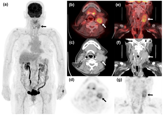
A pelvic MRI showed a 6.5 cm irregular mass involving the uterine cervix with bilateral parametrial involvement, without definitive pelvic LN enlargement, consistent with FIGO stage IIB disease. To assess for distant metastasis, an F-18 FDG PET/CT scan was performed using a PET/CT unit (Discovery PET/CT 710, GE Healthcare, Milwaukee, WI, USA). The primary cervical tumor exhibited intense F-18 FDG uptake, with a maximum standardized uptake value (SUVmax) of 16.3, and showed hypermetabolic infiltration into both parametria. Additionally, a hypermetabolic mass was identified in the left neck (levels II and III) with a SUVmax of 6.1, raising the possibility of metastatic LN, which could indicate distant nodal metastasis and potential upstaging (Figure 1).
Figure 1.
F-18 fluorodeoxyglucose (FDG) positron emission tomography/computed tomography (PET/CT) images of advanced uterine cervical cancer. Maximum intensity projection image (a) shows intense F-18 FDG uptake in the primary cervical tumor, as well as an additional hypermetabolic lesion in the left neck (black arrow). Axial (b–d) and sagittal (e–g) PET/CT images reveal a 4.2-cm hypermetabolic ovoid mass (SUVmax 6.1, white and black arrows) in the left level II/III region, raising suspicion of nodal metastasis.
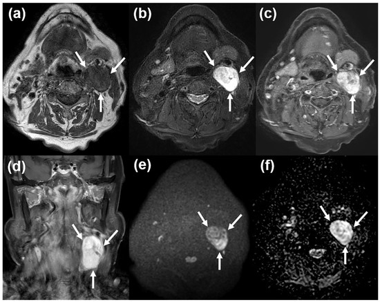
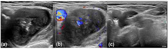
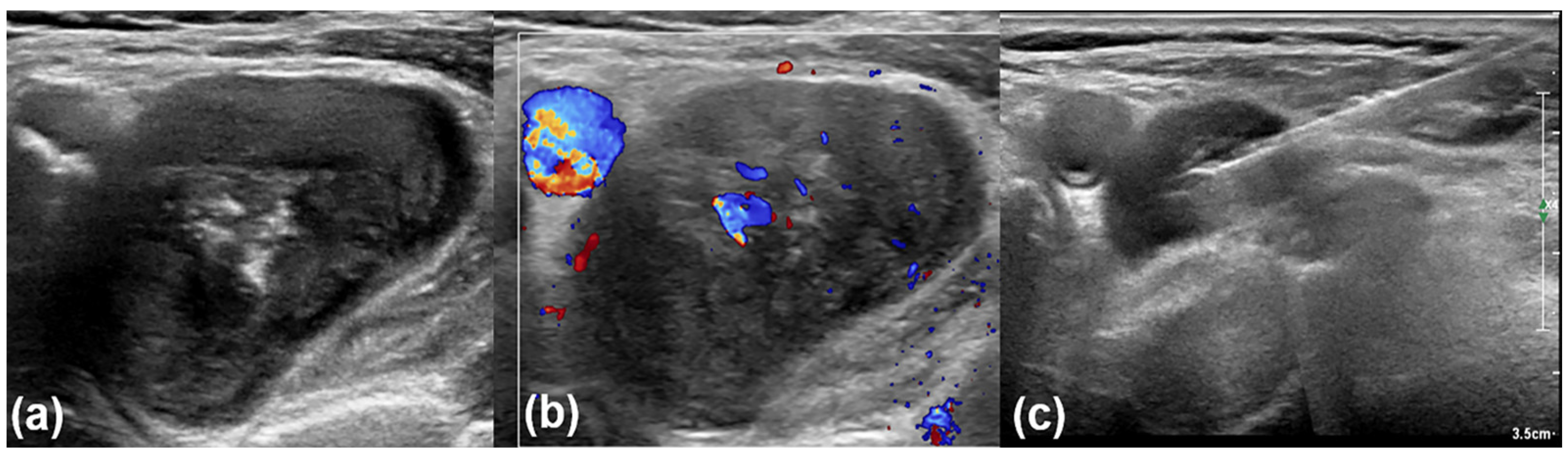
Due to concerns about neck nodal metastasis, a contrast-enhanced neck MRI was performed using a 3T system with a 20-channel head and neck coil (Ingenia 3.0 CX; Philips Healthcare, Best, The Netherlands). The protocol included Dixon-based T1- and T2-weighted sequences to obtain fat-suppressed images, as well as diffusion-weighted and contrast-enhanced fat-suppressed T1-weighted sequences. The MRI revealed a well-defined, ovoid mass approximately 3.8 cm in maximum diameter located in the left level II and III regions (Figure 2). The lesion exhibited isointense signal relative to muscle on T1-weighted images and heterogeneous high signal intensity on T2-weighted images. Diffusion-weighted imaging showed no definite diffusion restriction. Contrast-enhanced T1-weighted images indicated inhomogeneous but relatively strong enhancement, with no signs of adjacent tissue infiltration or extranodal extension. The mass had smooth margins and appeared distinct from nearby lymph nodes, suggesting a potential non-nodal origin, such as a peripheral nerve sheath tumor, like a schwannoma. The neck lesion was not assessed by dedicated physical examination before the imaging workup and was first detected on staging F-18 FDG PET/CT. During targeted ultrasonography (US) evaluation, manual palpation of the sonographically localized lesion revealed a firm, mobile mass in the left neck. High-resolution US was then performed using an EPIQ 7 scanner (Philips Medical Systems, Bothell, WA, USA) equipped with a 5–12 MHz linear-array transducer. Transverse ultrasound imaging showed a well-defined, oval hypoechoic mass with internal echogenic foci in the left level II and III regions, measuring approximately 4 cm in maximum dimension (Figure 3). Color Doppler imaging indicated multifocal internal vascularity. Based on the imaging findings and anatomical location, a schwannoma was included in the differential diagnosis. During the same session, a US-guided core needle biopsy was conducted using a disposable 18-gauge double-action spring-activated needle (1.6 cm excursion; TSK Acecut; TSK Laboratory, Tochigi-Ken, Japan) after local anesthesia with 1% lidocaine. Two tissue cores were obtained, and specimen adequacy was assessed by visual inspection. There was no immediate procedure-related complication during the biopsy.
Figure 2.
Contrast-enhanced neck magnetic resonance imaging (MRI). There is a well-circumscribed, ovoid mass in the left level II and III regions. The lesion exhibits an isointense signal relative to muscle on T1-weighted imaging (a) and shows heterogeneous high signal intensity on T2-weighted imaging (b). Post-contrast axial (c) and coronal (d) T1-weighted images demonstrate strong but inhomogeneous enhancement, with no evidence of extranodal extension or adjacent tissue infiltration. Axial diffusion-weighted imaging (e) and apparent diffusion coefficient map (f) show no definitive diffusion restriction. Arrows indicate the lesion.
Figure 3.
Transverse ultrasonography (US) with a 5–12 MHz linear-array transducer. There is a well-defined, oval hypoechoic mass containing central echogenic foci in the left level II and III regions (a). Transverse color Doppler imaging (b) shows multifocal internal vascularity within the lesion. Following the imaging evaluation, a US-guided core needle biopsy was performed during the same session (c).
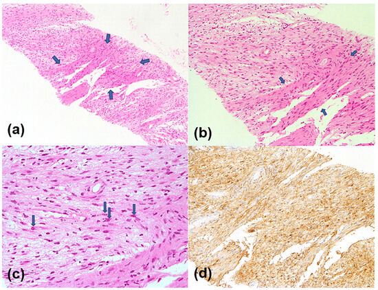
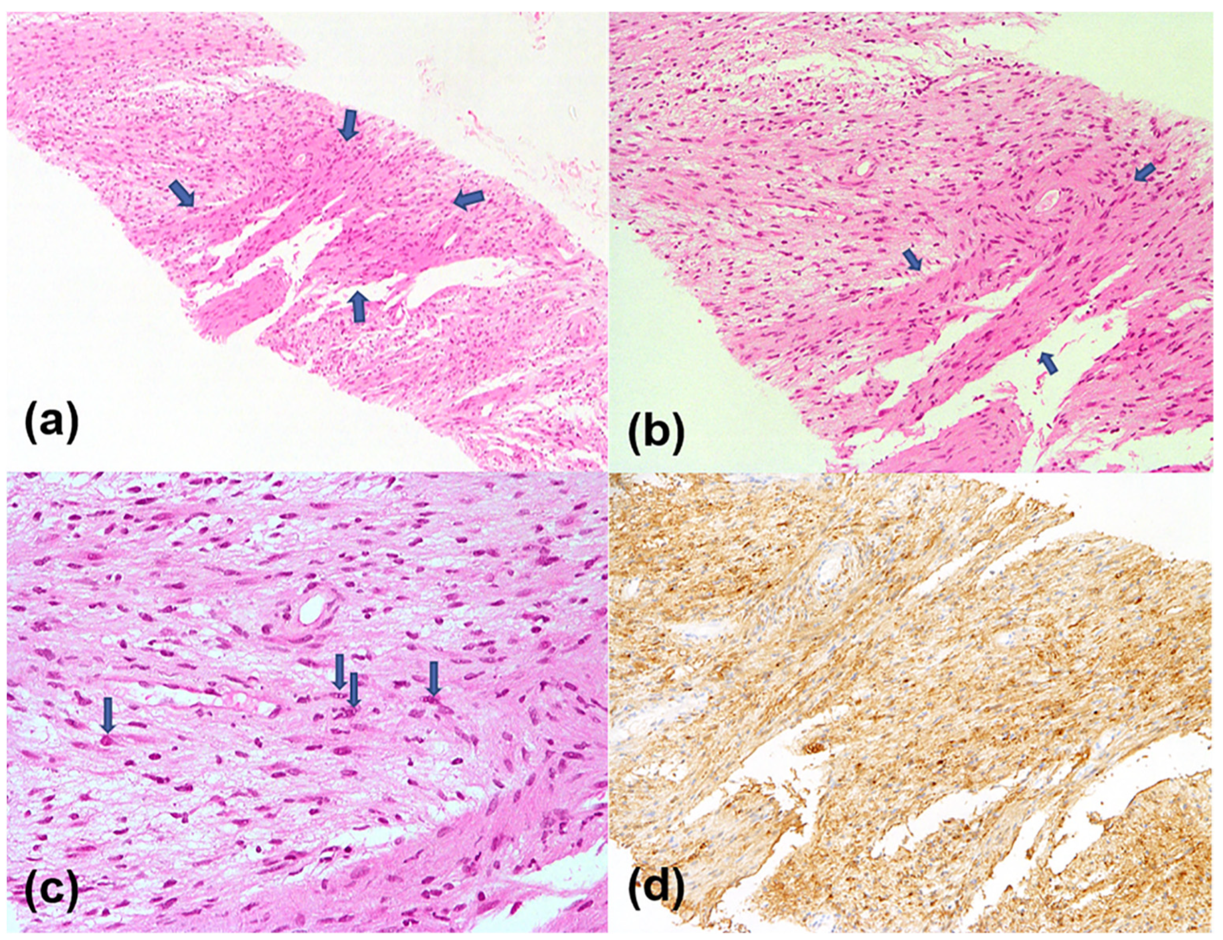
Histopathologic examination of the core biopsy specimen revealed a spindle cell neoplasm characterized by alternating hypercellular (Antoni A) and hypocellular (Antoni B) areas (Figure 4). The hypercellular regions contained spindle-shaped cells arranged in short fascicles with eosinophilic cytoplasm, while the hypocellular areas displayed loosely arranged spindle cells embedded in a myxoid stroma. Immunohistochemical staining showed diffuse positivity for S-100 protein, indicating a neurogenic tumor. Based on these findings, the final diagnosis was established as a schwannoma, likely arising from the vagus nerve.
Figure 4.
Histopathologic findings from the ultrasonography-guided core needle biopsy specimen. (a) Low-power view (×40, hematoxylin and eosin [H&E] stain) shows a spindle cell tumor featuring hypercellular Antoni A areas (arrows) and hypocellular Antoni B areas. (b) Higher magnification (×100, H&E) of the Antoni A area reveals spindle-shaped cells with fibrillary eosinophilic cytoplasm arranged in short fascicles. (c) High-power view (×400, H&E) of the Antoni B area demonstrates loosely arranged spindle and plump cells within an edematous myxoid stroma (arrows). (d) Immunohistochemical staining shows diffuse S-100 protein positivity (×100), indicating a neurogenic origin.
Given the patient’s advanced age, comorbidities, and repeated refusal of aggressive or definitive treatment, the treatment strategy was intentionally limited to symptom-directed palliative radiotherapy rather than systemic chemotherapy or definitive concurrent chemoradiotherapy. External beam radiotherapy was administered in 28 fractions of 180 cGy, culminating in a total dose of 5040 cGy. Four months after completing EBRT, a follow-up pelvic MRI indicated tumor progression with rectal invasion, and serum SCC-Ag levels rose to 18.9 ng/mL. High-dose-rate intracavitary brachytherapy was subsequently delivered in five fractions of 500 cGy, totaling 2500 cGy. Despite these treatments, the disease continued to progress, with serum SCC-Ag levels increasing to 31.0 ng/mL and rectal invasion confirmed by biopsy. Ultimately, the patient received conservative supportive care and was lost to follow-up.
3. Discussion
Schwannoma is a benign neurogenic tumor that originates from Schwann cells in the peripheral nerve sheath [8]. Extracranial head and neck schwannomas commonly present as well-circumscribed lateral neck masses, and accurate preoperative diagnosis is important because of their proximity to major neurovascular structures [7,8]. Histologically, schwannomas exhibit a distinctive biphasic architecture composed of hypercellular Antoni A and hypocellular Antoni B areas, and may undergo secondary degenerative changes such as cystic degeneration, hemorrhage, and calcification [7,19,20,21]. Immunohistochemically, diffuse positivity for S-100 protein supports the diagnosis of a neurogenic tumor [7,19]. In this case, US-guided core needle biopsy revealed the characteristic Antoni A and B areas with strong S-100 positivity, confirming the diagnosis.
From a metabolic imaging perspective, schwannomas pose a significant diagnostic challenge due to their highly variable F-18 FDG uptake, with reported SUVmax values extending into a range that overlaps with malignant lesions [9,10,11,12]. The exact mechanism behind this variability remains unclear. However, previous studies suggest that intense accumulation may have been attributed to a predominance of hypercellular Antoni A tissue, potentially linked to increased glucose transporter expression in Schwann cells [9,11,12]. Other proposed contributors include tumor microenvironmental factors and peritumoral inflammatory components [9,10,11,12]. As a result, F-18 FDG PET/CT alone has limited specificity in differentiating schwannomas from malignant lesions [10,11,12,22], and SUVmax should be interpreted as a marker of metabolic activity rather than as a stand-alone discriminator between benign and metastatic diseases. This limitation is particularly relevant in the neck, where a single presumed metastatic LN may upstage disease and meaningfully alter management.
In this regard, complementary anatomic imaging can provide clinically actionable information that metabolic intensity alone cannot. Metastatic lymphadenopathy typically presents with rounded morphology, loss of normal nodal architecture, necrosis or heterogeneous enhancement, and extranodal extension with infiltration of adjacent tissue planes in advanced cases. In contrast, schwannomas more often appear as solitary, well-defined, encapsulated masses with heterogeneous T2 hyperintensity reflecting mixed Antoni A/B components, a target-like internal architecture, or preserved surrounding fat planes [20,21]. Although these distinctions are not absolute, an anatomic pattern favoring a discrete non-nodal mass can help refine the differential diagnosis and support a targeted diagnostic workflow.
To date, only five cases of schwannoma mimicking metastatic cervical or supraclavicular lymph nodes in oncologic patients have been reported (Table 1) [13,14,15,16,17]: three in the supraclavicular fossa [13,14,15] and two in the lower neck [16,17]. In the four cases evaluated with F-18 FDG PET/CT, the hypermetabolic lesion was initially interpreted as metastatic nodal disease, but schwannoma was ultimately confirmed by biopsy or excision [13,14,15,17]. These reports also demonstrate variability in the diagnostic workflow. In one case, MRI and US findings suggested schwannoma and led to US-guided needle biopsy without altering the subsequent treatment course [17]. In contrast, limited anatomic correlation and persistent diagnostic uncertainty contributed to surgical removal in another PET/CT-based case [15] and in one case without PET/CT [16]. Hara et al. [14] similarly described a gastric cancer patient with a left supraclavicular schwannoma initially suspected as Virchow node metastasis on F-18 FDG PET/CT, and tissue diagnosis was essential to avoid misclassification. Collectively, these reports indicate that F-18 FDG avidity alone cannot reliably distinguish schwannoma from nodal metastasis when uptake overlaps, and that detailed anatomic imaging correlation may help direct patients toward minimally invasive biopsy rather than surgical excision when imaging findings suggest a non-nodal origin.
In the present case, multimodal imaging was crucial in reassessing the initial suspicion of nodal metastasis based on F-18 FDG PET/CT. Contrast-enhanced MRI revealed a well-circumscribed mass with heterogeneous T2 hyperintensity, no diffusion restriction, and no evidence of extranodal extension or adjacent tissue infiltration. High-resolution US further showed a solitary, well-defined oval hypoechoic mass with internal vascularity rather than a cluster of pathological LNs. Together, these morphological features supported a non-nodal origin and prompted targeted tissue sampling. In the neck, this biopsy-oriented workflow is particularly useful because US can provide real-time lesion characterization, guide a safe needle trajectory around adjacent vessels and neurovascular structures, and enable tissue confirmation without proceeding directly to surgical excision.
In addition, the SUVmax of 6.1 in our case was relatively higher than the values reported in several previously published oncologic cases of cervical or supraclavicular schwannoma mimicking nodal metastasis [13,14,15,17]. Importantly, the SUVmax values summarized in Table 1 refer to lesions that were ultimately confirmed as schwannomas, although they were initially interpreted or suspected as metastatic lymph nodes. Therefore, these values should not be regarded as metastatic nodal SUVmax values. Rather, they illustrate that schwannomas may show F-18 FDG uptake within a range that can overlap with malignant nodal disease. Although published oncologic data have proposed SUVmax-based thresholds for predicting cervical nodal metastasis in specific clinical settings [23,24,25], such thresholds cannot be directly extrapolated to a solitary hypermetabolic neck mass in a patient with a different primary malignancy. Thus, the key implication of the PET/CT finding in our case is not that the SUVmax favored metastasis by a particular threshold, but that F-18 FDG uptake alone had limited specificity and required complementary anatomic imaging and tissue confirmation.
This distinction is particularly relevant in advanced uterine cervical cancer, in which involvement of the left lower cervical or supraclavicular LNs may suggest distant metastatic spread and substantially influence treatment planning. Although such nodal involvement is uncommon overall, it has been reported more frequently in patients with para-aortic nodal disease or advanced-stage tumors [1,2,5,6]. In one PET study, occult hypermetabolic supraclavicular LNs were identified in 8% of patients with cervical cancer and were pathologically confirmed as metastatic [6]. Therefore, in a patient with advanced cervical cancer, a newly detected hypermetabolic lesion in the left lower neck may reasonably be suspected to represent metastatic nodal disease. However, as shown in the present case, benign hypermetabolic tumors such as schwannoma can closely mimic nodal metastasis on F-18 FDG PET/CT, creating a risk of overstaging if careful anatomical correlation is not performed. From a clinical perspective, a false-positive nodal interpretation may influence systemic therapy decisions, radiotherapy field design, or consideration of additional neck-directed interventions in other clinical settings. In such situations, multimodal imaging does not replace biopsy but can narrow the differential diagnosis, support a non-nodal hypothesis, and justify a minimally invasive sampling strategy.
In summary, this case demonstrates that integrating F-18 FDG PET/CT with MRI and high-resolution US can clarify the origin of a hypermetabolic neck lesion and guide an effective biopsy strategy during oncologic staging. In this patient, the final treatment course was determined mainly by the patient’s advanced age, comorbidities, disease extent, and preference for conservative management. Nevertheless, the multimodal imaging and biopsy-oriented workflow provided clinically meaningful diagnostic clarification by confirming that the PET/CT-positive neck lesion represented a benign schwannoma rather than nodal metastasis from cervical cancer. Thus, the principal message of this case is not that treatment escalation was definitively prevented, but that unsupported nodal upstaging can be avoided through careful anatomic correlation and minimally invasive tissue confirmation.
4. Conclusions
In oncologic patients, hypermetabolic neck lesions on F-18 FDG PET/CT are often presumed to represent metastatic nodal disease, particularly when located in a clinically relevant nodal station. However, benign tumors such as schwannomas may closely mimic this appearance and create a risk of false-positive nodal staging. This case highlights the value of multimodal imaging correlation, as complementary MRI and US can reveal morphologic features suggestive of a non-nodal origin that may not be apparent on metabolic imaging alone. In the present patient, tissue confirmation primarily provided diagnostic clarification rather than directly altering the delivered treatment strategy. The broader clinical implication is that a PET/CT-positive neck lesion with atypical morphology or non-nodal anatomic features should prompt targeted anatomic reassessment before being incorporated into oncologic staging. In such circumstances, MRI and high-resolution US can refine the differential diagnosis and guide minimally invasive US-guided tissue sampling, helping avoid unsupported nodal upstaging and supporting individualized treatment planning without overinterpreting PET/CT findings.
Author Contributions
Conceptualization, S.Y. and H.J.B.; methodology, S.Y. and B.C.; validation, H.J.B.; formal analysis, S.Y. and H.J.A.; writing—original draft preparation, S.Y. and H.J.B.; writing—review and editing, all authors. All authors have read and agreed to the published version of the manuscript.
Funding
This study received no external funding.
Institutional Review Board Statement
This study was conducted in accordance with the Declaration of Helsinki. The Institutional Review Board of Gyeongsang National University Changwon Hospital granted exemption from ethical review for this retrospective observational case report (Protocol Code: GNUCH 2026-02-014; Date of exemption determination: 5 March 2026), because it was solely observational in nature, the data were collected retrospectively, and neither the patient’s management nor outcome was affected by this case report.
Informed Consent Statement
Informed consent for publication was obtained from all identifiable human participants. Written informed consent was obtained from the patient for the publication of this case report and any accompanying images.
Data Availability Statement
No new data were created or analyzed in this study. Data sharing is not applicable to this article.
Conflicts of Interest
The authors declare no conflicts of interest.
References
- Fischerova, D.; Frühauf, F.; Burgetova, A.; Haldorsen, I.S.; Gatti, E.; Cibula, D. The Role of Imaging in Cervical Cancer Staging: ESGO/ESTRO/ESP Guidelines (Update 2023). Cancers 2024, 16, 775. [Google Scholar] [CrossRef]
- Dheur, A.; Kakkos, A.; Danthine, D.; Delbecque, K.; Goffin, F.; Gonne, E.; Lovinfosse, P.; Pleyers, C.; Thille, A.; Kridelka, F.; et al. Lymph node assessment in cervical cancer: Current approaches. Front. Oncol. 2024, 14, 1435532. [Google Scholar] [CrossRef]
- Olthof, E.P.; Bergink-Voorthuis, B.J.; Wenzel, H.H.B.; Mongula, J.; van der Velden, J.; Spijkerboer, A.M.; Adam, J.A.; Bekkers, R.L.M.; Beltman, J.J.; Slangen, B.F.M.; et al. Diagnostic accuracy of MRI, CT, and [18F]FDG-PET-CT in detecting lymph node metastases in clinically early-stage cervical cancer: A nationwide Dutch cohort study. Insights Imaging 2024, 15, 36. [Google Scholar] [CrossRef]
- Lopez, F.; Rodrigo, J.P.; Silver, C.E.; Haigentz, M., Jr.; Bishop, J.A.; Strojan, P.; Hartl, D.M.; Bradley, P.J.; Mendenhall, W.M.; Suarez, C.; et al. Cervical lymph node metastases from remote primary tumor sites. Head Neck 2016, 38, E2374–E2385. [Google Scholar] [CrossRef]
- Stehman, F.B.; Bundy, B.N.; Hanjani, P.; Fowler, W.C.; Abdulhay, G.; Whitney, C.W. Biopsy of the scalene fat pad in carcinoma of the cervix uteri metastatic to the periaortic lymph nodes. Surg. Gynecol. Obs. 1987, 165, 503–506. [Google Scholar]
- Tran, B.N.; Grigsby, P.W.; Dehdashti, F.; Herzog, T.J.; Siegel, B.A. Occult supraclavicular lymph node metastasis identified by FDG-PET in patients with carcinoma of the uterine cervix. Gynecol. Oncol. 2003, 90, 572–576. [Google Scholar] [CrossRef]
- Dosemane, D.; Kabekkodu, S.; Jaipuria, B.; Sreedharan, S.; Shenoy, V. Extracranial non-vestibular head and neck schwannomas: A case series with the review of literature. Braz. J. Otorhinolaryngol. 2022, 88, S9–S17. [Google Scholar] [CrossRef] [PubMed]
- Uerschels, A.K.; Dengler, N.F.; Chihi, M.; Lenkeit, A.; Dinger, T.F.; Jabbarli, R.; Sure, U.; Hagenacker, T.; Wrede, K.H. Benign peripheral nerve sheath tumors: An interdisciplinary diagnostic and therapeutic challenge. Neurosurg. Rev. 2023, 46, 205. [Google Scholar] [CrossRef]
- Miyashi, Y.; Oshiro, H.; Yoshikawa, Y.; Katsuki, R.; Tome, Y.; Nishida, K. Heterogeneity of Tumor Glucose Metabolism in Schwannomas Between Trunk and Extremities: An Imaging Study. In Vivo 2025, 39, 3428–3436. [Google Scholar] [CrossRef] [PubMed]
- Ugajin, M.; Yamashita, M.; Kani, H. PET Scanning May Not Distinguish Benign Schwannoma from Metastasis in a Patient with Lung Adenocarcinoma. Eur. J. Case Rep. Intern. Med. 2023, 10, 004048. [Google Scholar] [CrossRef]
- Beaulieu, S.; Rubin, B.; Djang, D.; Conrad, E.; Turcotte, E.; Eary, J.F. Positron emission tomography of schwannomas: Emphasizing its potential in preoperative planning. AJR Am. J. Roentgenol. 2004, 182, 971–974. [Google Scholar] [CrossRef]
- Miyake, K.K.; Nakamoto, Y.; Kataoka, T.R.; Ueshima, C.; Higashi, T.; Terashima, T.; Nakatani, K.; Saga, T.; Minami, S.; Togashi, K. Clinical, Morphologic, and Pathologic Features Associated With Increased FDG Uptake in Schwannoma. AJR Am. J. Roentgenol. 2016, 207, 1288–1296. [Google Scholar] [CrossRef] [PubMed]
- Gorospe, L.; Rioja-Martin, M.E.; Santos-Carreno, A.L.; Pacios-Blanco, R.E.; Munoz-Molina, G.M. Supraclavicular schwannoma mimicking a lymphadenopathy in a lung cancer patient: PET/CT findings. Lung Cancer 2017, 110, 74–76. [Google Scholar] [CrossRef]
- Hara, Y.; Nakamura, K.; Iwagami, S.; Ogawa, K.; Sawayama, H.; Iwatsuki, M.; Baba, Y.; Miyamoto, Y.; Yoshida, N.; Baba, H. A case of clinical stage I gastric cancer with a schwannoma on the left supraclavicular fossa suspected as Virchow’s node metastasis. Surg. Case Rep. 2022, 8, 95. [Google Scholar] [CrossRef]
- Perniola, G.; Tomao, F.; Fischetti, M.; Lio, S.; Pecorella, I.; Benedetti Panici, P. Benign schwannoma in supraclavicular region: A false-positive lymph node recurrence of breast cancer suspected by PET scan. Arch. Gynecol. Obs. 2014, 290, 583–586. [Google Scholar] [CrossRef] [PubMed]
- da Costa Vieira, R.A.; de Araujo Silva, I.; de Souza Coelho, R.D.; de Almeida Junior, C.R.; de Almeida Santos Yamashita, M.E. Brachial plexus schwannoma mimicking advanced breast carcinoma. Breast Dis. 2020, 39, 109–113. [Google Scholar] [CrossRef]
- Hsieh, T.C.; Wu, Y.C.; Hsu, C.N.; Yang, C.F.; Yen, K.Y.; Kao, C.H.; Sun, S.S. Lower neck neurilemmoma can masquerade as lymph node metastasis on FDG PET/CT in patient with nasopharyngeal carcinoma. Clin. Nucl. Med. 2011, 36, e217–219. [Google Scholar] [CrossRef]
- Bore, P.; Descourt, R.; Ollivier, L.; Le Roux, P.Y.; Abgral, R. False Positive 18F-FDG Positron Emission Tomography Findings in Schwannoma-A Caution for Reporting Physicians. Front. Med. 2018, 5, 275. [Google Scholar] [CrossRef]
- Colreavy, M.P.; Lacy, P.D.; Hughes, J.; Bouchier-Hayes, D.; Brennan, P.; O’Dwyer, A.J.; Donnelly, M.J.; Gaffney, R.; Maguire, A.; O’Dwyer, T.P.; et al. Head and neck schwannomas—A 10 year review. J. Laryngol. Otol. 2000, 114, 119–124. [Google Scholar] [CrossRef]
- Shirodkar, K.; Hussein, M.; Reddy, P.S.; Shah, A.B.; Raniga, S.; Pal, D.; Iyengar, K.P.; Botchu, R. Imaging of Peripheral Intraneural Tumors: A Comprehensive Review for Radiologists. Cancers 2025, 17, 246. [Google Scholar] [CrossRef]
- Li, S.; Wu, Z.; Lin, X.; Ye, L.; Zeng, X. Retroperitoneal schwannoma: A clinicopathological analysis of 14 cases. Ann. Diagn. Pathol. 2025, 78, 152497. [Google Scholar] [CrossRef]
- Kang, S. Benign Schwannoma Mimicking Metastatic Lesion on F-18 FDG PET/CT in Differentiated Thyroid Cancer. Nucl. Med. Mol. Imaging 2013, 47, 138–140. [Google Scholar] [CrossRef] [PubMed]
- Duvernay, J.; Schlund, M.; Majoufre, C. Contribution of FDG-PET in the diagnostic assessment of cervical lymph node metastasis in oral cavity squamous cell carcinoma (OCSCC). J. Stomatol. Oral Maxillofac. Surg. 2023, 124, 101659. [Google Scholar] [CrossRef] [PubMed]
- Banjare, A.K.; Arora, R.D.; Ravina, M.; Prajwal, S.D.; Rao, K.N.; Nagarkar, N.M. Role of the FDG PET CT Scan in Pretreatment Evaluation of Oral Carcinomas. Indian J. Otolaryngol. Head Neck Surg. 2024, 76, 5346–5352. [Google Scholar] [CrossRef]
- Yoon, S.; Ryu, K.H.; Baek, H.J.; Kim, T.H.; Moon, J.I.; Choi, B.H.; Park, S.E.; Ha, J.Y.; Song, D.H.; An, H.J.; et al. Cervical Lymph Nodes Detected by F-18 FDG PET/CT in Oncology Patients: Added Value of Subsequent Ultrasonography for Determining Nodal Metastasis. Medicina 2019, 56, 16. [Google Scholar] [CrossRef] [PubMed]

Disclaimer/Publisher’s Note: The statements, opinions and data contained in all publications are solely those of the individual author(s) and contributor(s) and not of MDPI and/or the editor(s). MDPI and/or the editor(s) disclaim responsibility for any injury to people or property resulting from any ideas, methods, instructions or products referred to in the content. |
© 2026 by the authors. Licensee MDPI, Basel, Switzerland. This article is an open access article distributed under the terms and conditions of the Creative Commons Attribution (CC BY) license.