1. Introduction
Polymyositis and dermatomyositis are idiopathic inflammatory myopathies (IIMs) characterised by immune-mediated muscle damage, presenting with progressive symmetric proximal weakness, elevated muscle enzymes, and systemic features, including skin involvement in dermatomyositis, dysphagia, interstitial lung disease, and cardiac complications [
1,
2,
3,
4,
5]. Diagnosis integrates clinical findings, biochemical markers, electromyography, imaging, serology (myositis-specific autoantibodies), and muscle biopsy [
1,
4,
6,
7].
Magnetic resonance imaging (MRI) is the preferred modality for detecting muscle oedema and guiding biopsy due to its excellent soft-tissue contrast. Although whole-body MRI is increasingly available and provides excellent soft-tissue characterisation, access, cost, scan duration, and protocol variability may still limit its routine use in some centres [
3,
8].
99mTc-MDP bone scintigraphy, although primarily used for evaluating skeletal pathology, can detect extraosseous uptake in inflamed or necrotic soft tissues through mechanisms involving calcium deposition, hyperaemia, and binding to denatured proteins [
3,
8]. In idiopathic inflammatory myopathies (IIMs), it characteristically demonstrates diffuse muscular accumulation that correlates with disease severity [
9,
10]. Previous studies have documented its usefulness in assessing disease extent and monitoring treatment response [
9,
10,
11]; however, its application for intraoperative guidance has not been previously described.
This case presents a patient with severe dermatomyositis exhibiting widespread extraosseous 99mTc-MDP uptake and introduces the novel use of gamma-probe-guided biopsy, thereby exploring its feasibility in challenging presentations of idiopathic inflammatory myopathies.
2. Case Presentation
A 49-year-old male presented with progressive proximal muscle weakness since June 2025, with muscle power graded 2/5 proximally and 5/5 distally. Symptoms began 2 months after influenza infection. He had a prior similar admission. He subsequently reported dysphagia for solids due to pharyngeal muscle weakness and developed characteristic dermatomyositis-associated rashes. He had difficulty standing and severe back pain. No reported history of trauma. No pyrexia, recent travel history, or significant comorbidities. Previous medication history included two doses of the Johnson & Johnson COVID-19 vaccine.
Biochemistry revealed elevated ESR (55 mm/hr), CRP (20 mg/L), and leucocytosis (16.58 × 10⁹/L). A quadriceps muscle biopsy (29 July 2025) showed no inflammatory myopathy. Laboratory investigations revealed markedly elevated creatine kinase (19,937 IU/L; reference range 38–174 IU/L) and troponin T levels (1421 ng/L; reference < 15 ng/L). A repeat biopsy was suggested due to sampling limitations.
Bone scintigraphy was performed after intravenous administration of ~1110 MBq
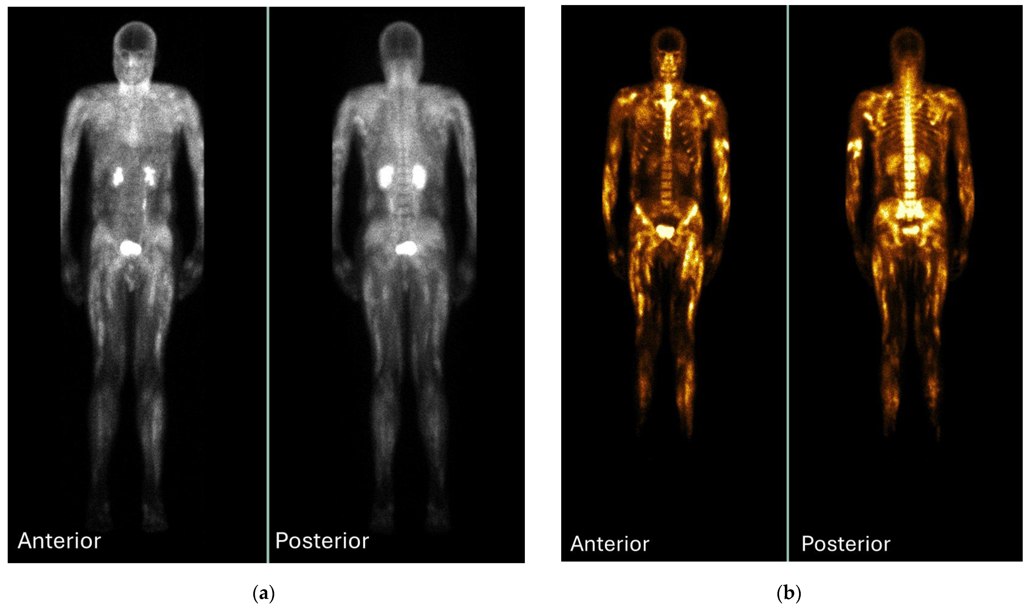
99mTc-MDP. A three-phase whole-body bone scan was acquired using a Discovery 630 Gamma Camera with LEHR collimator and Bone Evolution protocol. Blood-pool imaging (
Figure 1a) showed intense vascularity and variable tracer accumulation in multiple muscle groups. Delayed whole-body imaging (
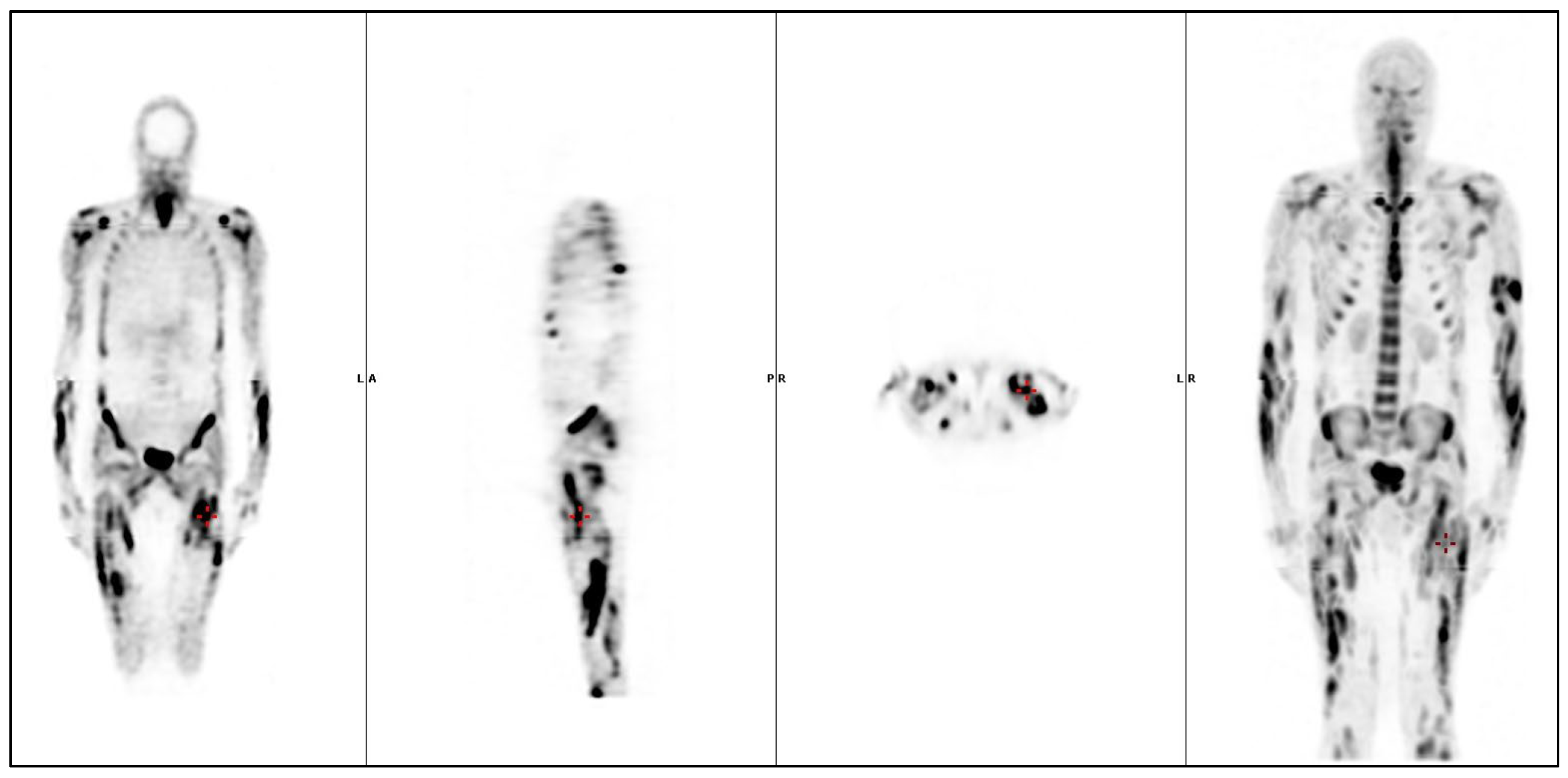
Figure 1b) and whole-body SPECT with triangulated view (
Figure 2) demonstrated extraosseous uptake in the bilateral deltoids (symmetric), biceps and triceps (patchy), paraspinal muscles (longitudinal along the spine), gluteal muscles, bilateral thigh muscles (quadriceps and hamstrings), and calf muscles (gastrocnemius). Delayed images showed preserved activity in the axial skeleton (pelvis, sternum, skull), with poor visualisation of the appendicular skeleton due to intense soft-tissue uptake and symmetrical renal excretion. Suppression of appendicular skeletal visualisation was noted. The findings were consistent with severe inflammatory myopathy (polymyositis/dermatomyositis spectrum).
A repeat muscle biopsy was performed under intraoperative gamma-probe guidance using 99mTc-MDP as a marker of active inflammatory muscle. Administration of approximately 1100 MBq 99mTc-MDP was performed prior to the intraoperative procedure to facilitate real-time gamma-probe localisation. The cumulative administered activity was approximately 2200 MBq, corresponding to an estimated effective dose of 8–10 mSv. This represents an important limitation of the technique. Formal organ-specific dosimetry was not performed, and additional radiation exposure to radiosensitive organs, including the kidneys, bladder, and bone marrow, should be considered. The procedure was undertaken following informed patient consent and in accordance with institutional radiation safety practices. Future studies would require formal dosimetric analysis and optimisation of tracer dosing protocols.
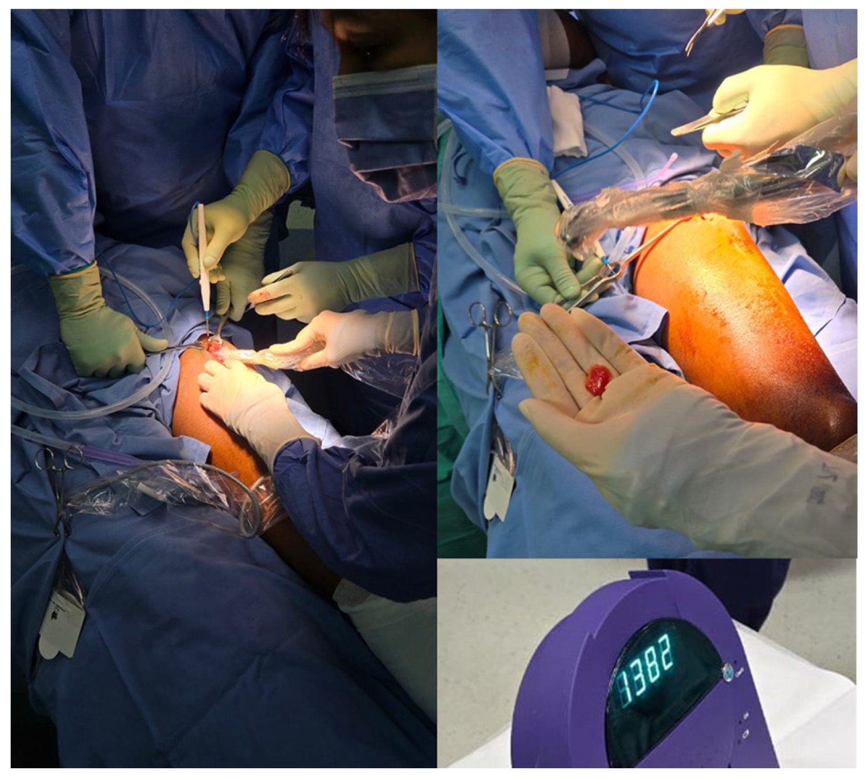
Real-time intraoperative localisation was performed with a Navigator 2.0 gamma probe. Two biopsy samples were obtained from regions demonstrating the highest tracer uptake, specifically the left quadriceps femoris and the distal triceps brachii. Intraoperative count measurements (1300–1400 counts compared with a background of approximately 500) facilitated precise localisation and ensured accurate sampling of metabolically active muscle tissue (
Figure 3).
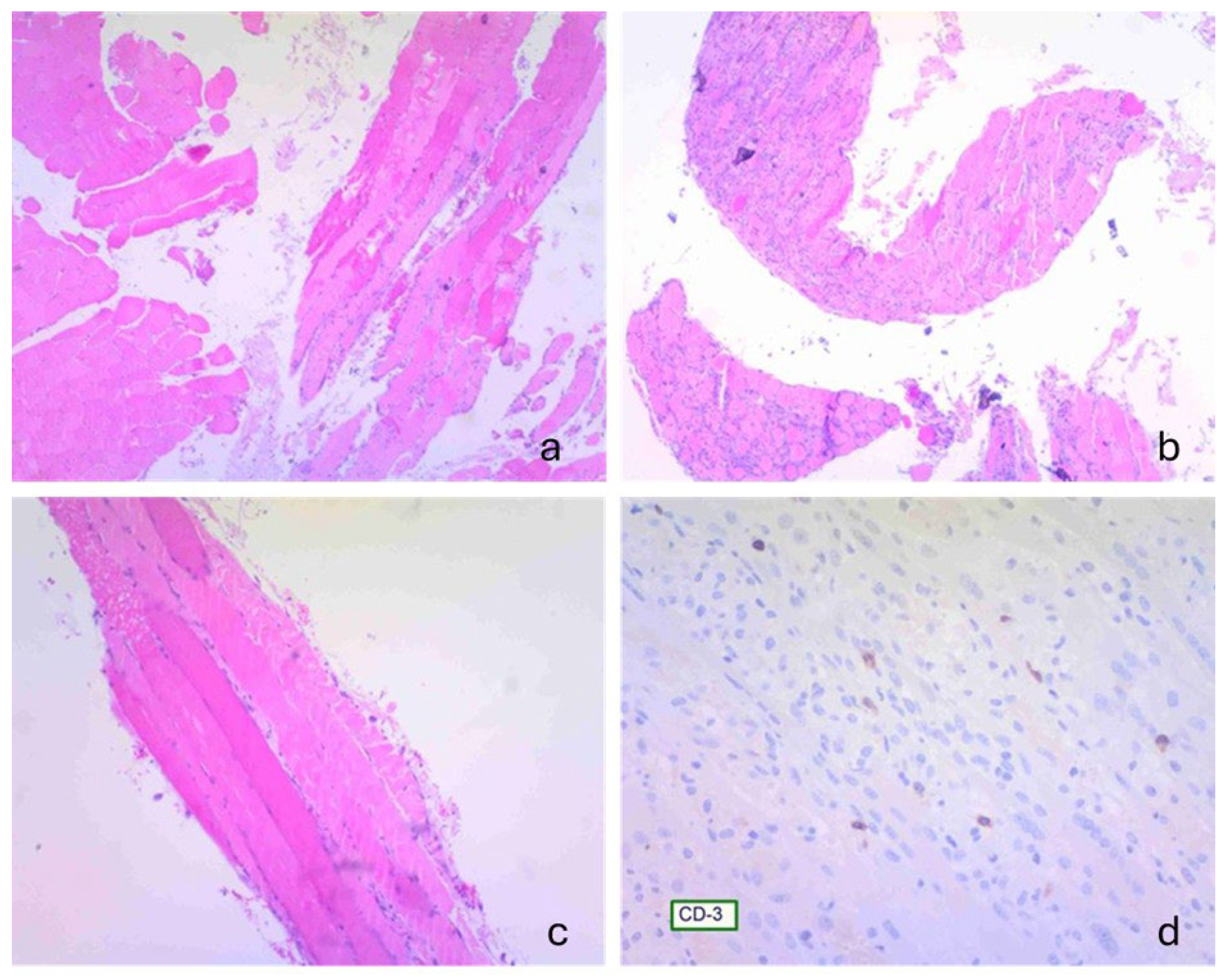
The biopsy findings were consistent with an inflammatory myopathy, characterised by a histiocyte-predominant inflammatory infiltrate with muscle fibre necrosis, histiocytic aggregates, a sparse lymphocytic component, and occasional CD4-positive cells. There was no evidence of chronic changes such as fibrosis or fatty infiltration. The pattern raised the possibility of an immune-mediated necrotising myopathy (which may be seropositive or seronegative, including anti-SRP or anti-HMGCR-associated forms). However, definitive classification requires correlation with clinical, serological, and other investigative findings. Toxic or metabolic necrotising myopathies remained important differential diagnoses (
Figure 4a–d). H&E sections of formalin-fixed muscle demonstrated varying degrees of inflammation, necrosis, regeneration, and loss of individual fibres; CD3 immunostaining highlighted T-cell lymphocytic inflammation.
Serological testing was positive for antinuclear antibodies (ANAs) and strongly positive for anti-Mi-2 antibodies, supporting and confirming the diagnosis of dermatomyositis. Initial treatment consisted of intravenous methylprednisolone 1 g daily for 3 days, followed by oral prednisone 60 mg daily with subsequent tapering. Additional immunosuppressive and supportive therapy included methotrexate 25 mg weekly (with folic acid 5 mg weekly), azathioprine 100 mg daily, celecoxib 200 mg twice daily, esomeprazole 40 mg daily, and intravenous immunoglobulin 34 g daily for 5 days. Rituximab (Ristova) was commenced in November 2025.
The patient responded very well to treatment. Methotrexate was reduced to 15 mg weekly, and prednisone was weaned to 5 mg daily. At follow-up on 30 March 2026, muscle power had returned to normal (5/5), and creatine kinase had normalised to 122 IU/L (reference range 38–174 IU/L). The patient remains in clinical remission, with no current indication for repeat imaging or biopsy. Should relapse occur, repeat imaging and a muscle biopsy may be considered. The diagnosis was established by integrating clinical, serological, imaging, and histopathological findings, allowing for the initiation of immunosuppressive therapy.
3. Discussion
Polymyositis (PM) and dermatomyositis (DM) typically present with gradually progressive, symmetric proximal muscle weakness, occasionally involving neck, respiratory, or pharyngeal muscles, leading to dysphagia or respiratory compromise in advanced disease. Late-stage distal weakness may occur, whereas early distal involvement favours alternative diagnoses such as sporadic inclusion body myositis (sIBM).
These conditions show a clear female predominance (female-to-male ratio approximately 2:1), with peak incidence in adults aged 40–60 years and, less commonly, in children (juvenile dermatomyositis) [
1,
4]. Predisposing factors include genetic susceptibility (certain HLA alleles), environmental triggers such as viral infections (including influenza), vaccinations, ultraviolet radiation exposure, smoking, and certain medications. There is also a well-recognised association with underlying malignancy (particularly in dermatomyositis) and overlap with other autoimmune diseases, including systemic lupus erythematosus, scleroderma, rheumatoid arthritis, and antisynthetase syndrome. Diagnostic overlap with other inflammatory, metabolic, toxic, and genetic myopathies—particularly sIBM, immune-mediated necrotising myopathy, and antisynthetase syndromes—poses significant challenges and contributes to frequent misclassification. A combination of clinical assessment, targeted laboratory and serological testing, neurological evaluation, and—most importantly—muscle biopsy is essential for distinguishing polymyositis and dermatomyositis from other autoimmune and non-inflammatory myopathies [
4].
The Bohan and Peter criteria diagnose polymyositis and dermatomyositis based on symmetric proximal muscle weakness, characteristic electromyographic abnormalities, and elevated muscle enzymes. Definitive diagnosis requires supportive muscle biopsy findings, while dermatomyositis is distinguished by the presence of characteristic cutaneous features such as heliotrope rash and Gottron’s papules. Disease certainty (definite, probable, or possible) depends on the number and combination of these clinical, biochemical, electrophysiological, and histological criteria met [
6,
7].
Supportive muscle biopsy findings in inflammatory myopathies include endomysial CD8
+ T-cell infiltration with or without invasion of non-necrotic fibres, diffuse MHC-I upregulation, perifascicular atrophy, perivascular or perimysial inflammation, muscle fibre necrosis and regeneration, and complement (MAC) deposition on capillaries or the sarcolemma. Features such as rimmed vacuoles and mitochondrial abnormalities suggest alternative diagnoses such as inclusion body myositis [
12,
13,
14].
Dalakas and Hohlfeld expanded diagnostic criteria in 2003 [
5] by emphasising muscle biopsy pathology and defining distinct immunohistochemical and histological features for definite and probable polymyositis, dermatomyositis, and amyopathic dermatomyositis. In the same year, the Muscle Study Group (MSG) and 119th ENMC workshop [
15] proposed comprehensive classification criteria for idiopathic inflammatory myopathies, integrating clinical features, EMG, laboratory markers, MRI, and muscle biopsy findings. These criteria also highlighted ongoing challenges in diagnosis, study design, and outcome assessment, underscoring the need for standardised and sensitive measures in myositis research.
The clinical and diagnostic challenges highlight the complexity of inflammatory myopathies and the importance of a multimodal approach. Advances in muscle immunopathology and myositis-specific autoantibody (MSA) testing have significantly facilitated the diagnostic accuracy and classification of idiopathic inflammatory myopathies (IIMs). Modern immunopathology allows precise characterisation of inflammatory patterns, including endomysial CD8+ T-cell infiltration in polymyositis, perifascicular atrophy and complement deposition in dermatomyositis, and prominent myofibre necrosis with minimal lymphocytic inflammation in immune-mediated necrotising myopathy.
The discovery and routine use of myositis-specific autoantibodies have further refined diagnosis. These highly specific antibodies, such as anti-Mi-2 (strongly associated with classic dermatomyositis), enable clinicians to correlate clinical phenotypes, predict prognosis, and guide treatment with greater precision. Together with modern imaging techniques, particularly MRI and contrast-enhanced ultrasound, these advances have transformed the diagnostic approach to IIMs from broad clinical categories to more accurate, serologically and histologically defined subtypes [
1,
4,
5].
Magnetic resonance imaging (MRI) is increasingly used to evaluate polymyositis (PM) and dermatomyositis (DM). It effectively detects muscle oedema, necrosis, and inflammation, typically appearing as increased signal intensity on short tau inversion recovery (STIR) sequences [
3,
8]. Although whole-body MRI is increasingly available, differences in access, protocol availability, scan duration, and cost may still limit routine whole-body implementation in some centres. In contrast, whole-body
99mTc-MDP bone scintigraphy provides a comprehensive metabolic overview of disease activity, aiding biopsy site selection and potentially therapeutic monitoring [
16,
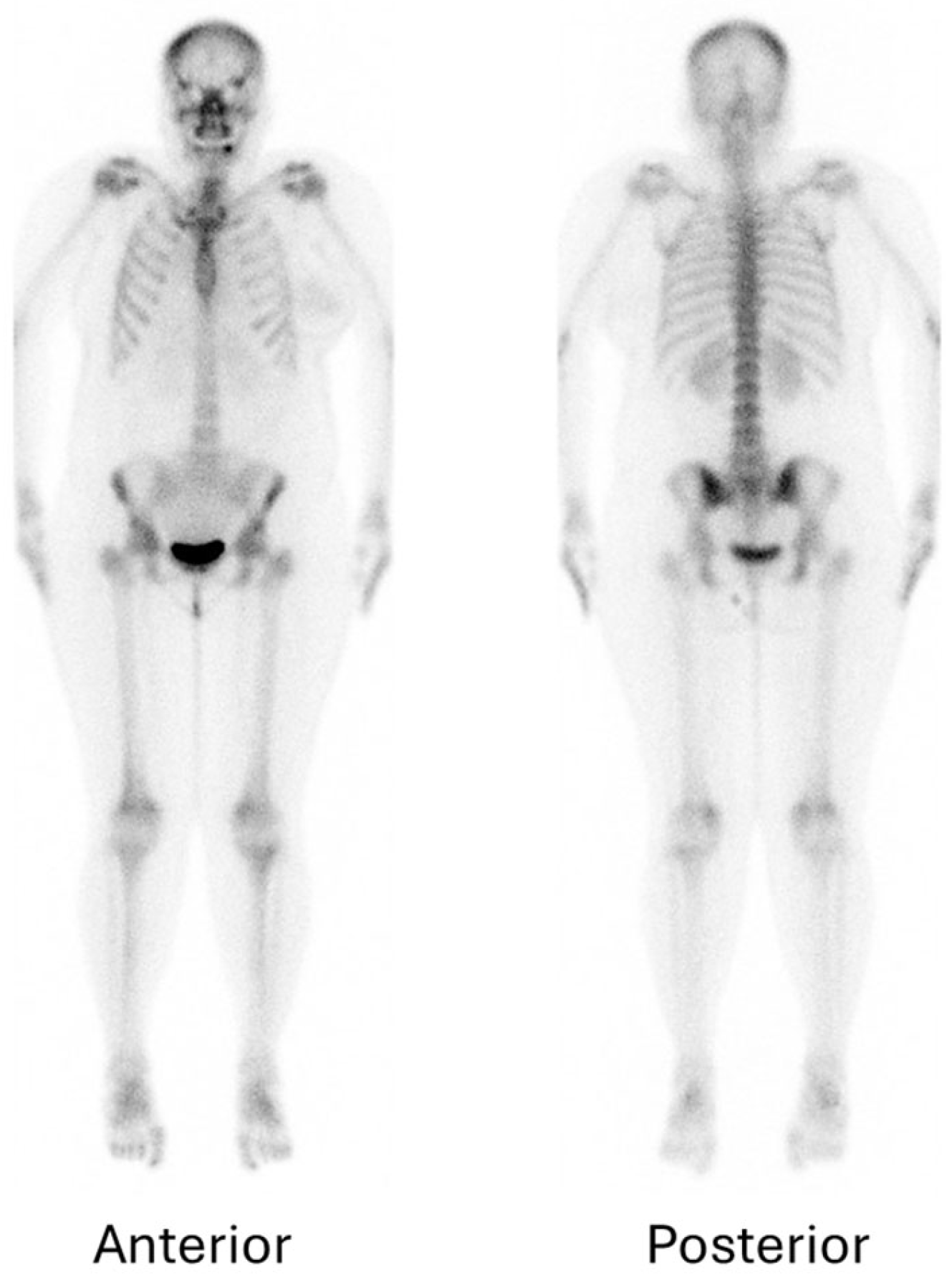
17]. A representative normal whole-body
99mTc-MDP scan is shown in
Figure 5 to highlight the contrast between physiological tracer distribution and the abnormal muscular uptake observed in our patient. Extraosseous uptake of
99mTc-MDP is a well-documented phenomenon resulting from increased capillary permeability, calcium deposition, and binding to denatured proteins in inflamed or necrotic tissue [
16,
17]. In PM and DM, repeated cycles of muscle fibre necrosis and regeneration create conditions favourable for this uptake, producing characteristic diffuse or patchy muscular patterns that correlate with disease severity and extent [
9,
10]. Recognition of these scintigraphic findings can offer critical diagnostic information beyond conventional imaging or clinical examination. Muscle biopsy in PM/DM remains prone to sampling error due to the focal and heterogeneous nature of inflammation [
4,
18]. Whole-body
99mTc-MDP scintigraphy, therefore, serves as a valuable adjunct by enabling global assessment of disease activity and facilitating targeted biopsy, particularly after inconclusive histological sampling.
3.1. Pathophysiology of Extraosseous 99mTc-MDP Uptake in Inflammatory Myopathies
Extraosseous
99mTc-MDP uptake is a well-recognised phenomenon observed in a variety of non-skeletal conditions, including soft-tissue inflammation, trauma, heterotopic ossification, dystrophic calcification, tumours, and myocardial infarction [
16,
17]. In idiopathic inflammatory myopathies, the loss of muscle fibre integrity leads to repeated cycles of necrosis and regeneration, accompanied by intense inflammatory changes that create a permissive microenvironment for tracer accumulation. Under normal conditions,
99mTc-MDP primarily binds to hydroxyapatite crystals in bone. In inflamed or necrotic skeletal muscle, uptake is mediated by several pathophysiological mechanisms: increased vascular permeability and hyperaemia, expansion of extracellular fluid volume, calcium influx into damaged myocytes, and direct binding of the radiotracer to denatured proteins and calcium deposits within necrotic tissue [
16,
17]. In the context of myositis, these mechanisms are particularly relevant, as immune-mediated muscle injury triggers ongoing myofibre necrosis, regeneration, and perivascular/perimysial inflammation.
In the presented case, the intense diffuse muscular uptake—symmetric in the deltoids, patchy in the biceps and triceps, longitudinal along the paraspinal muscles, and involving the gluteal, thigh (quadriceps and hamstrings), and calf (gastrocnemius) muscles—accompanied by marked suppression of appendicular skeletal visualisation, directly reflects the profound soft-tissue metabolic activity that overwhelms skeletal binding. This pattern is characteristic of severe, widespread myositis and correlates with the extensive inflammation and necrosis seen histologically in dermatomyositis.
Although
99mTc-MDP scintigraphy provides valuable whole-body functional information and complements MRI in distinguishing active inflammation from chronic damage, emerging data also compare it to FDG-PET. FDG uptake in inflamed muscle occurs due to increased glucose metabolism in activated inflammatory cells (particularly macrophages and lymphocytes) via overexpression of glucose transporters (GLUTs) and hexokinase activity. FDG-PET demonstrates similar patterns of muscular hypermetabolism but at a higher cost and greater radiation exposure [
19].
Compared with MRI, which primarily detects structural changes, such as muscle oedema (hyperintensity on T2-weighted or STIR sequences), fatty infiltration, or atrophy [
3,
8],
99mTc-MDP bone scintigraphy offers complementary functional information. MRI excels at high-resolution regional assessment of individual muscle groups and is excellent at distinguishing oedema from chronic damage. However, it is limited by its restricted field of view, higher cost, and its inability to provide a whole-body metabolic overview. In contrast, bone scintigraphy provides a comprehensive, whole-body functional map of active inflammation and necrosis, readily identifying subclinical or multifocal involvement and correlating well with disease severity and serum markers, such as CK [
9,
10]. Although MRI remains the preferred modality for detailed anatomic characterisation,
99mTc-MDP scintigraphy provides complementary functional information regarding metabolic activity and whole-body disease distribution.
3.2. Comparison with Previously Reported Cases
Severe extraosseous
99mTc-MDP uptake in polymyositis and dermatomyositis is uncommon but well documented in case reports and small series [
9,
10,
20].
Table 1 summarises relevant cases, including those with widespread muscular accumulation, often involving proximal and lower limb groups, similar to the present patient. These cases share common features, including symmetric or diffuse uptake correlating with disease severity, elevated creatine kinase levels, and inflammatory markers, with suppression of skeletal visualisation in extensive disease [
9,
10,
11,
21].
Notably, several reported cases involve extensive soft-tissue uptake and highlight diagnostic challenges when biopsy is inconclusive. Standard features include proximal-predominant patterns, correlation with clinical activity, and utility in disease extent assessment [
9]. Treatment in all cases centred on immunosuppression (corticosteroids, methotrexate, azathioprine, intravenous immunoglobulin), with scintigraphy aiding monitoring through reduced uptake post-therapy in some [
11]. Outcomes were generally favourable with prompt recognition. All patients exhibited active myositis with metabolic tracer uptake secondary to inflammation or necrosis. Each required an integrated multimodal approach due to biopsy limitations. Cases with profound uptake and suppressed skeletal visualisation particularly emphasised disease severity. Consistent findings included muscle weakness, elevated muscle enzymes, and heterogeneous histology. Management uniformly involved immunosuppression and strategies to overcome biopsy sampling error. In some cases, adjunctive imaging-guided therapy and definitive confirmation often required repeat or targeted biopsy, as performed in the present case.
3.3. Diagnostic Challenges and Novel Guidance in the Current Case
The diagnosis of idiopathic inflammatory myopathies (IIMs), including polymyositis (PM) and dermatomyositis (DM), is inherently challenging due to the patchy and heterogeneous nature of muscle involvement. Muscle biopsy, although the histopathological gold standard, is susceptible to significant sampling error. The focal distribution of inflammation means that even clinically affected muscles may yield non-diagnostic or non-specific results if the sampled area is spared or shows only end-stage changes [
4,
18]. Reported false-negative rates for muscle biopsy in suspected IIMs range from 10% to 45%, even when imaging-guided, and can be higher (up to 40–50% indeterminate or non-inflammatory) depending on pre-test probability and biopsy site selection [
4].
From a surgical perspective, intraoperative visualisation adds another layer of difficulty. Macroscopically, inflamed or damaged muscle often appears indistinguishable from normal tissue during open biopsy. Surgeons typically rely on clinical palpation, preoperative imaging (e.g., MRI for oedema), or electromyography-guided site selection, as affected muscle may lack gross discolouration, oedema, or atrophy visible to the naked eye, particularly in early or patchy disease. This lack of reliable macroscopic distinction exacerbates sampling error, as biopsied fascicles may inadvertently come from relatively spared regions, resulting in normal or minimally abnormal histology despite widespread active myositis elsewhere [
4,
18]. Larger open biopsies or multiple samples are sometimes used to mitigate this, but they do not fully resolve the issue in heterogeneous disease.
Compared with MRI-guided biopsy, which relies on structural changes such as oedema on STIR sequences, the gamma-probe method provides functional, count-based confirmation of metabolically active inflammation. The literature indicates that blind or clinically guided muscle biopsy has a false-negative rate of approximately 10–45% (commonly 10–23%) in suspected IIM [
22]. In a prospective study, MRI-guided biopsies used as a triage tool reduced the false-negative rate to around 19%, while MRI as an add-on test after a non-diagnostic biopsy further lowered it to ~6% [
23]. In our patient, gamma-probe-guided biopsy yielded diagnostic tissue after an initial non-diagnostic biopsy; however, whether this reflected improved targeting, sampling variability, operator-related factors, or disease heterogeneity cannot be determined from a single case. Although no conclusions regarding diagnostic superiority can be drawn, this observation suggests that functional metabolic targeting using
99mTc-MDP scintigraphy with intraoperative gamma-probe guidance may be technically feasible in selected cases of diagnostically challenging or heterogeneous inflammatory myopathy and warrants further prospective evaluation.
Gamma probes, such as the Navigator 2.0 system, are widely used in radio-guided surgery for sentinel lymph node detection, parathyroid localisation, and occult tumour resection, owing to their high sensitivity, portability, and ability to deliver real-time quantitative counts. Their flexibility—supported by various probe designs (straight, angled, laparoscopic, and wireless)—allows adaptation to diverse surgical fields. In the context of muscle biopsy, gamma probes provide significant clinical benefit by enabling precise localisation of metabolically active tissue. This reduces operative exploration time, minimises unnecessary tissue dissection, limits damage to surrounding healthy muscle, and may decrease the need for repeat biopsies and associated histopathology costs. Although the application of gamma probes in inflammatory myopathies remains largely unexplored, this case illustrates their potential to enhance surgical efficiency and diagnostic accuracy in challenging scenarios where conventional visual or palpation-guided approaches are often inadequate [
24].
Procedurally, the technique was straightforward and used standard nuclear-medicine equipment already available in most centres (Discovery 630 Gamma Camera with LEHR collimator and Bone Evolution protocol plus Navigator 2.0 gamma probe). The procedure required a single additional radiotracer reinjection and additional radiation exposure, while enabling real-time quantitative localisation during open biopsy. In this case, 99mTc-MDP scintigraphy mapped widespread extraosseous uptake, and reinjection of ~1100 MBq 99mTc-MDP (90–120 min prior to surgery) enabled gamma-probe guidance. This targeted high-uptake sites (left quadriceps femoris and distal triceps brachii), with intraoperative counts of 1300–1400 versus background ~500, yielding supportive histopathological findings showing histiocyte-predominant inflammation, muscle fibre necrosis, regeneration, sparse CD4+ T-cells, and absence of chronic changes.
Anti-Mi-2 is a myositis-specific autoantibody (MSA) highly specific for classic dermatomyositis; while typically associated with perifascicular pathology, anti-Mi-2-positive DM can show prominent necrotising features and severe muscle involvement [
1,
4,
5]. The biopsy findings were consistent with an inflammatory myopathy showing histiocyte-predominant inflammation, muscle fibre necrosis, regeneration, sparse CD4
+ T-cells, and absence of chronic changes. The final diagnosis of dermatomyositis was based on the combination of characteristic cutaneous features, strong anti-Mi-2 positivity, and clinical phenotype, with the biopsy providing supportive (but not pathognomonic) evidence. It is also possible that gamma-probe guidance preferentially sampled a region of active necrosis rather than a classic perifascicular area, representing a limitation of targeted biopsy in heterogeneous disease [
1,
4,
5]. Reproducibility appears feasible in centres with nuclear medicine and surgical collaboration. However, larger multi-centre studies are needed to confirm generalisability, assess long-term diagnostic yield relative to MRI- or ultrasound-guided biopsy, and evaluate cost-effectiveness. To the best of our knowledge, no prior reports have described the use of gamma-probe-guided muscle biopsy in idiopathic inflammatory myopathies, making this a novel interventional application.
3.4. Clinical Implications and Management Considerations
Extraosseous uptake on 99mTc-MDP scintigraphy provided diagnostic clarity in this severe case. Gamma-probe-guided biopsy facilitated targeted tissue sampling in this case and contributed supportive histopathological information alongside the clinical and serological findings. Conservative escalation over days enabled therapy initiation. Without targeted sampling, diagnostic delay could have persisted, risking clinical decompensation. The novel interventional use suggests potential value in selected refractory cases, enhancing yield in heterogeneous IIMs.
In selected challenging cases, 99mTc-MDP bone scintigraphy may serve as a useful adjunct to MRI. Potential indications include guiding repeat biopsy after a non-diagnostic result, assessing whole-body disease extent in severe or multifocal myositis, evaluating disease activity when serum markers are discordant, monitoring treatment response in refractory cases, and providing an alternative imaging option in patients with MRI contraindications or limited access. In the present case, the scenario of suspected IIM with an inconclusive initial muscle biopsy was directly applicable. 99mTc-MDP scintigraphy enabled functional targeting of metabolically active muscle, resulting in successful histopathological confirmation after the first biopsy had failed.
The mainstay of therapy for DM and PM consists of immunosuppression, physical therapy, monitoring for adverse events from medications, and prevention of complications. Therapy for polymyositis and dermatomyositis is centred on high-dose corticosteroids, with intravenous methylprednisolone reserved for severe or rapidly progressive disease. Steroid-refractory or steroid-dependent cases require adjunctive immunomodulatory agents, including intravenous immunoglobulin and steroid-sparing immunosuppressants such as methotrexate, azathioprine, mycophenolate mofetil, and calcineurin inhibitors. In refractory disease, biologic therapies such as rituximab or enrolment in clinical trials targeting specific immune pathways may be considered [
1,
4,
25,
26].
A U.S. cohort study reported a 10-year survival rate of 62% in patients with polymyositis and dermatomyositis, with mortality primarily due to cardiac and pulmonary complications, infections, and malignancy. Prognosis is influenced by factors such as age at diagnosis, gender, Raynaud phenomenon, interstitial lung disease, dysphagia, and cardiac or respiratory muscle involvement. Long-term outcome data in juvenile dermatomyositis remain limited, though affected adults demonstrate reduced quality of life and impaired muscle fitness compared with age-matched controls [
19,
26,
27].
4. Conclusions
This case illustrates the technical feasibility of integrating 99mTc-MDP bone scintigraphy with intraoperative gamma-probe guidance in a patient with severe dermatomyositis and a prior non-diagnostic biopsy. Whole-body scintigraphy provided comprehensive metabolic mapping of disease extent, while real-time gamma-probe localisation facilitated targeted sampling of metabolically active muscle.
At follow-up on 30 March 2026 (approximately 8 months after presentation), the patient achieved excellent clinical recovery with normal muscle power (5/5) and normalisation of creatine kinase to 122 IU/L (reference range 38–174 IU/L) and remains in clinical remission.
By extending scintigraphy beyond its traditional diagnostic and monitoring roles, this report highlights the interventional potential of gamma-probe guidance in selected diagnostically challenging cases. However, as this is a single-case observation, larger prospective studies are needed to evaluate reproducibility, diagnostic yield, radiation risk–benefit profile, and comparative performance with established MRI- or ultrasound-guided biopsy techniques.