


Abstract
A 75-year-old man, with a history of descending thoracic aortic rupture and dissection treated with aortic stenting at 73 years old, was admitted for rehabilitation following recurrent cerebral ischemic attacks. Upon admission, blood tests revealed elevated inflammatory markers, including a C-reactive protein (CRP) level of 10.75 mg/dL and a D-dimer level of 4.2 µg/mL, alongside microcytic anemia. Despite thorough evaluations using computed tomography (CT) and ultrasound, the origin of these abnormalities remained unidentified. Two months later, MRI using diffusion-weighted whole-body imaging with background body signal suppression (DWIBS) revealed hyperintensities in the thoracic aorta. He remained asymptomatic and progressed well during rehabilitation, prompting continued observation. However, three months after admission, he developed hemoptysis. Contrast-enhanced CT showed pneumonia, as well as enhanced lesions in the aortic wall, confirming aortic inflammation. Due to concerns about aortic stent ulceration, an emergency stent graft insertion extending to the superior mesenteric artery was performed. He recovered uneventfully and was discharged. DWIBS is an MRI-based tool that avoids exposure to radiation or contrast agents and is cost-effective. MRI using DWIBS demonstrated high signal accumulations in the aortic wall, indicative of inflammation. These findings suggest that DWIBS holds significant potential as a powerful imaging tool for detecting and assessing inflammation, particularly in the aorta.
Figure 1.
The results of magnetic resonance imaging using diffusion-weighted whole-body imaging with background body signal suppression (DWIBS). (A) Three-dimensional rendering of the DWIBS data demonstrates high signal intensity surrounding the aorta (red arrows). (B,C) Axial slices from the DWIBS dataset (original images) show hyperintensity within the aortic wall (white arrows). DWIBS, pioneered by Takahara et al. in 2004, is an advanced MRI-based technique for comprehensive whole-body cancer screening [1]. This technique employs a short τ inversion recovery echo-planar imaging sequence with free breathing for the highly sensitive detection of diffusion-restricted lesions, such as cancers and abscesses, by leveraging the movement of water molecules to create sharp contrasts between abnormal and normal structures across the entire body simultaneously [1,2,3,4]. This provides a level of convenience comparable to positron emission tomography–computed tomography (PET-CT) [3]. DWIBS offers both cost-effectiveness and the advantage of being non-invasive, as it eliminates the need for expensive radionuclides that may otherwise pose a risk of radiation exposure [3]. Additionally, DWIBS has been reported to detect inflammatory pathology, with its most common application being Takayasu aortitis [5,6]. In addition, there is a case of aortic inflammation related to giant cell arteritis detected by DWIBS [7]. In this case, high DWI signals were observed along the aortic wall, indicating such inflammation. Cardiovascular surgeons suspected that the inflammation was associated with an aortic stent ulcer, but conclusive evidence to support this was unavailable. Nevertheless, based on the previous reports and our case, we believe that DWIBS holds promise as a useful modality for detecting inflammatory lesions, particularly in the aorta.
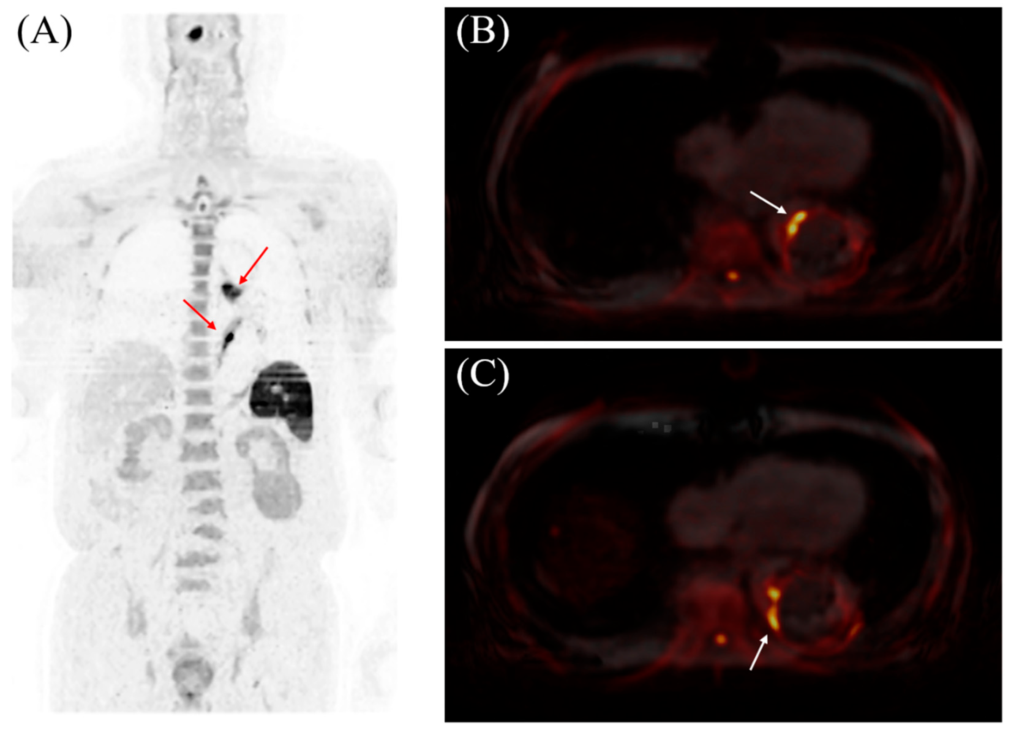
Figure 2.
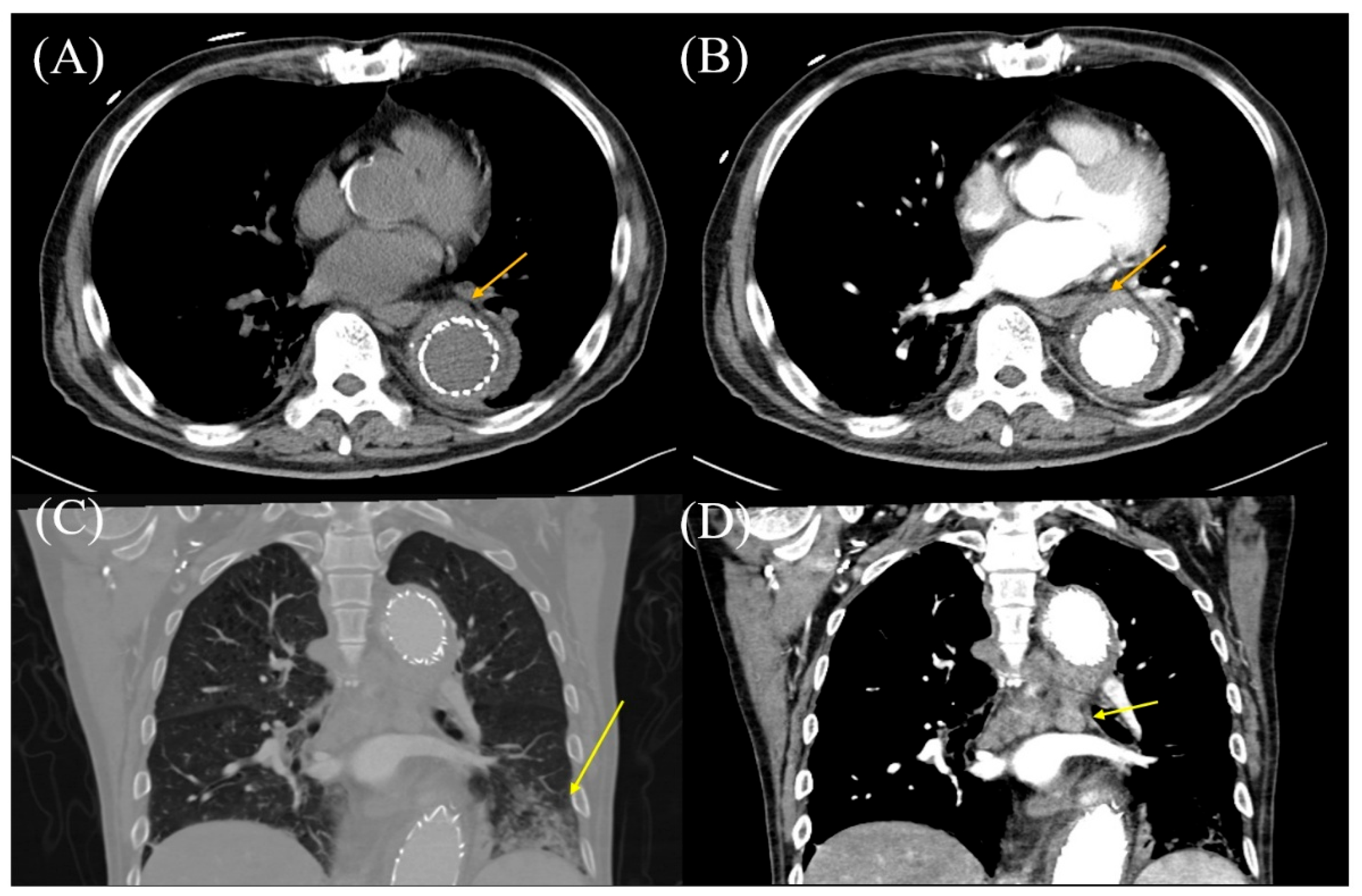
The results of computed tomography (CT) with or without contrast agent. (A) Thoracic CT without a contrast agent reveals that the aortic wall has Hounsfield units (HU) of 21 (arrow). (B) Thoracic CT with a contrast agent reveals areas of enhancement in the aortic wall, located outside the aortic stent, with an HU of 82 (arrow). (C) Thoracic CT in the coronal section reveals pneumonia (arrow). (D) Contrast-enhanced thoracic CT reveals mediastinal lymph node enlargement (arrow).
Author Contributions
Conceptualization, K.H.; methodology, K.H.; software, not applicable; validation, A.S., K.H. and M.S.; formal analysis, not applicable; investigation, A.S., K.H., M.S., T.M., Y.N. and N.T.; resources, M.S. and Y.K.; data curation, not applicable; writing—original draft preparation, A.S. and K.H.; writing—review and editing, M.S., Y.N., N.T., T.M. and Y.K.; visualization, T.I.; supervision, M.S. and Y.K.; project administration, not applicable; funding acquisition, M.S. and Y.K. All authors have read and agreed to the published version of the manuscript.
Funding
This research received no external funding.
Institutional Review Board Statement
According to the guidelines issued by the Japanese Ministry of Health, Labour and Welfare, case reports generally do not require approval from an Ethics Committee. Therefore, the ethical review and approval were waived for this study.
Informed Consent Statement
Informed consent was obtained from all subjects involved in the study.
Data Availability Statement
The data presented in this study are available on request from the corresponding author. Due to patient privacy and ethical considerations, the data are not publicly accessible.
Acknowledgments
The authors thank the patient for granting permission to publish this information.
Conflicts of Interest
The authors declare no conflicts of interest.
References
- Takahara, T.; Imai, Y.; Yamashita, T.; Yasuda, S.; Nasu, S.; Van Cauteren, M. Diffusion weighted whole body imaging with background body signal suppression (DWIBS): Technical improvement using free breathing, STIR and high resolution 3D display. Radiat. Med. 2004, 22, 275–282. [Google Scholar] [PubMed]
- Kwee, T.C.; Takahara, T.; Ochiai, R.; Nievelstein, R.A.J.; Luijten, P.R. Diffusion-weighted whole-body imaging with background body signal suppression (DWIBS): Features and potential applications in oncology. Eur. Radiol. 2008, 18, 1937–1952. [Google Scholar] [PubMed]
- Schicho, A.; Habicher, W.; Wendl, C.; Stroszczynski, C.; Strotzer, Q.; Dollinger, M.; Schreyer, A.G.; Schleder, S. Clinical Value of Diffusion-Weighted Whole-Body Imaging with Background Body Signal Suppression (DWIBS) for Staging of Patients with Suspected Head and Neck Cancer. Tomography 2022, 8, 2522–2532. [Google Scholar] [CrossRef] [Scilit] [PubMed]
- Nakata, M.; Yokota, N.; Kenzaka, T. Diffusion-weighted whole-body magnetic resonance imaging with background body signal suppression was useful in a patient with isolated myocardial abscess confined to the right atrial wall: A case report. BMC Cardiovasc. Disord. 2023, 23, 341. [Google Scholar] [CrossRef] [Scilit]
- Davidovic, L.; Tsay, V. Potential Application of Diffusion-Weighted Whole-Body Imaging with Background Body Signal Suppression for Disease Activity Assessment in Takayasu Arteritis-In Search of the “Golden Mean”: Case Report. Ann. Vasc. Surg. 2019, 61, 468.e9–468.e12. [Google Scholar] [CrossRef] [Scilit] [PubMed]
- Oguro, E.; Ohshima, S.; Kikuchi-Taura, A.; Murata, A.; Kuzuya, K.; Okita, Y.; Matsuoka, H.; Teshigawara, S.; Yoshimura, M.; Yoshida, Y.; et al. Diffusion-weighted Whole-body Imaging with Background Body Signal Suppression (DWIBS) as a Novel Imaging Modality for Disease Activity Assessment in Takayasu’s Arteritis. Intern. Med. 2019, 58, 1355–1360. [Google Scholar] [CrossRef] [Scilit] [PubMed]
- Matsuoka, H.; Yoshida, Y.; Oguro, E.; Murata, A.; Kuzuya, K.; Okita, Y.; Teshigawara, S.; Yoshimura, M.; Isoda, K.; Harada, Y.; et al. Diffusion Weighted Whole Body Imaging with Background Body Signal Suppression (DWIBS) was useful for the diagnosis and follow-up of giant cell arteritis. Intern. Med. 2019, 58, 2095–2099. [Google Scholar] [CrossRef] [Scilit] [PubMed]
Disclaimer/Publisher’s Note: The statements, opinions and data contained in all publications are solely those of the individual author(s) and contributor(s) and not of MDPI and/or the editor(s). MDPI and/or the editor(s) disclaim responsibility for any injury to people or property resulting from any ideas, methods, instructions or products referred to in the content. |
© 2025 by the authors. Licensee MDPI, Basel, Switzerland. This article is an open access article distributed under the terms and conditions of the Creative Commons Attribution (CC BY) license (https://creativecommons.org/licenses/by/4.0/).