Article

Clinical Application of High-Frame-Rate Vector Flow Imaging in Evaluation of Carotid Atherosclerotic Stenosis

1
Department of Ultrasound, Zhongshan Hospital, Fudan University, Shanghai 200032, China
2
Department of Ultrasound, Xinhua Hospital Affiliated to Shanghai Jiaotong University School of Medicine, Shanghai 200092, China
3
Department of Interventional Radiology, Zhongshan Hospital, Fudan University, Shanghai 200032, China
*
Authors to whom correspondence should be addressed.
These authors contributed equally to this work.
Diagnostics 2023, 13(3), 519; https://doi.org/10.3390/diagnostics13030519

Submission received: 4 December 2022 / Revised: 16 January 2023 / Accepted: 27 January 2023 / Published: 31 January 2023
(This article belongs to the Section Medical Imaging and Theranostics)

Abstract

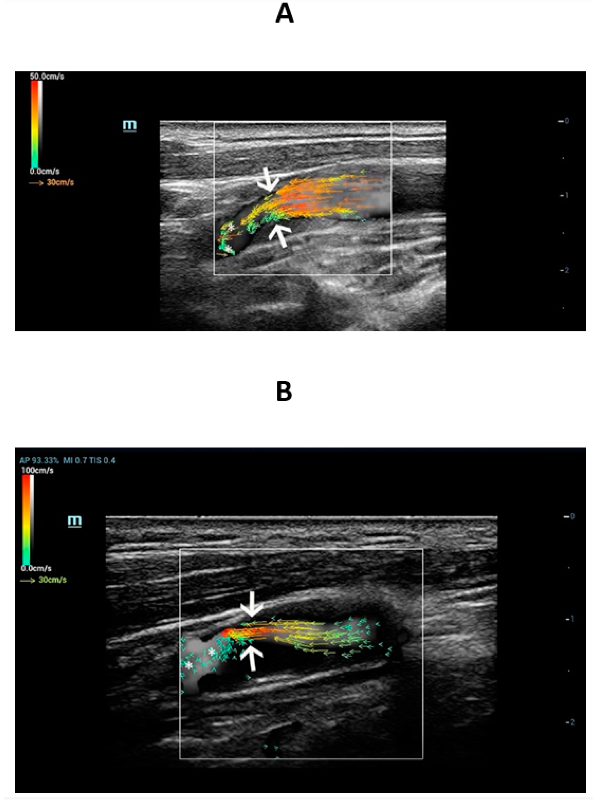
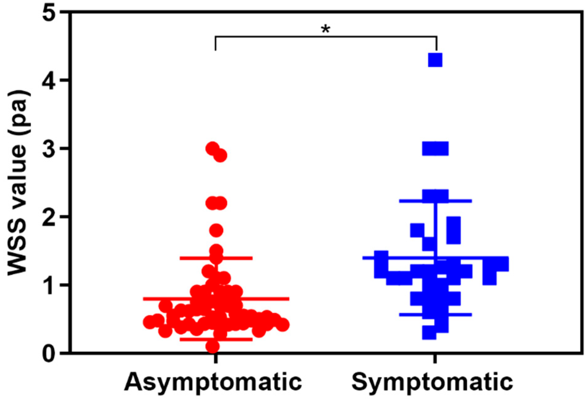
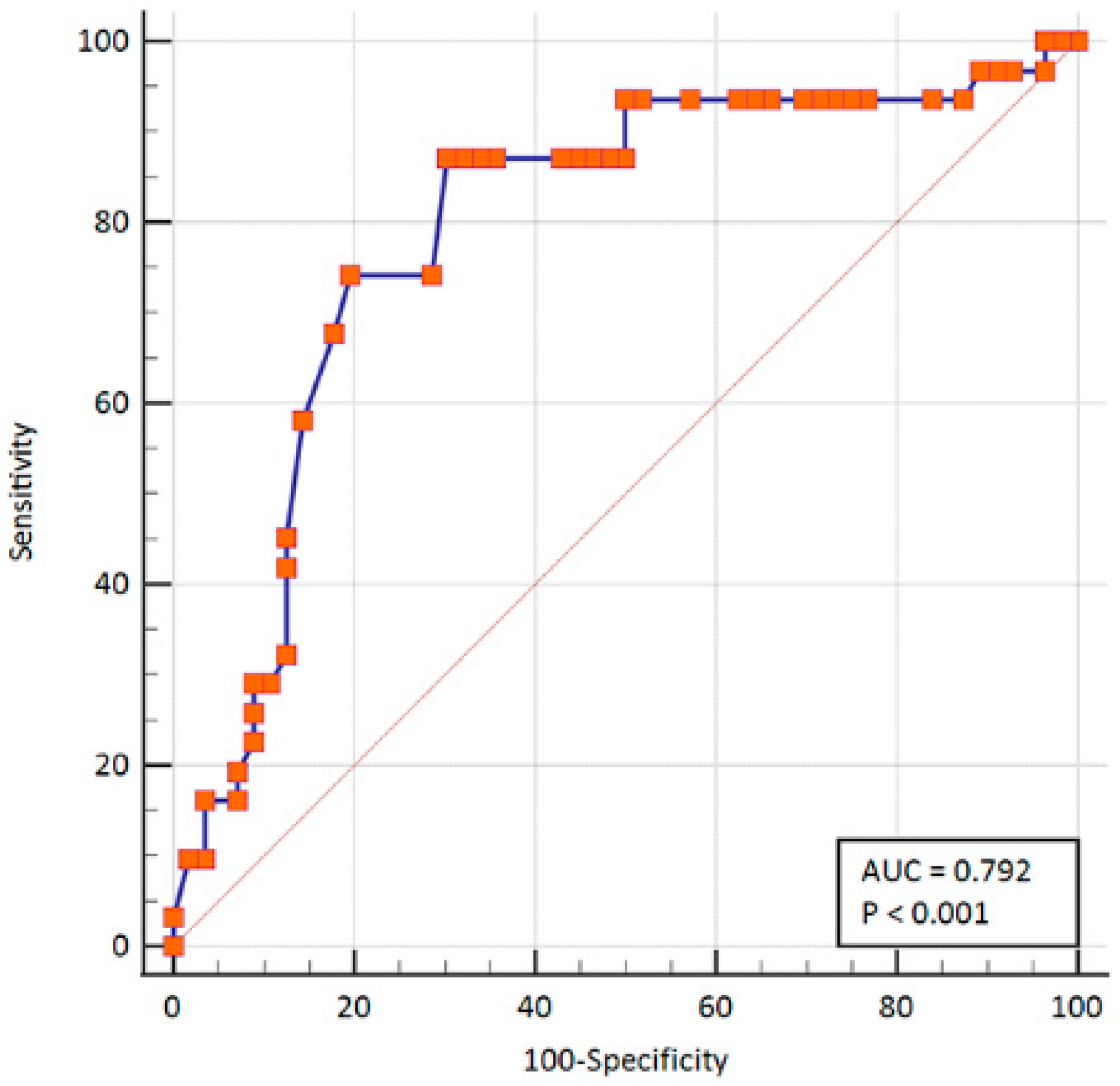
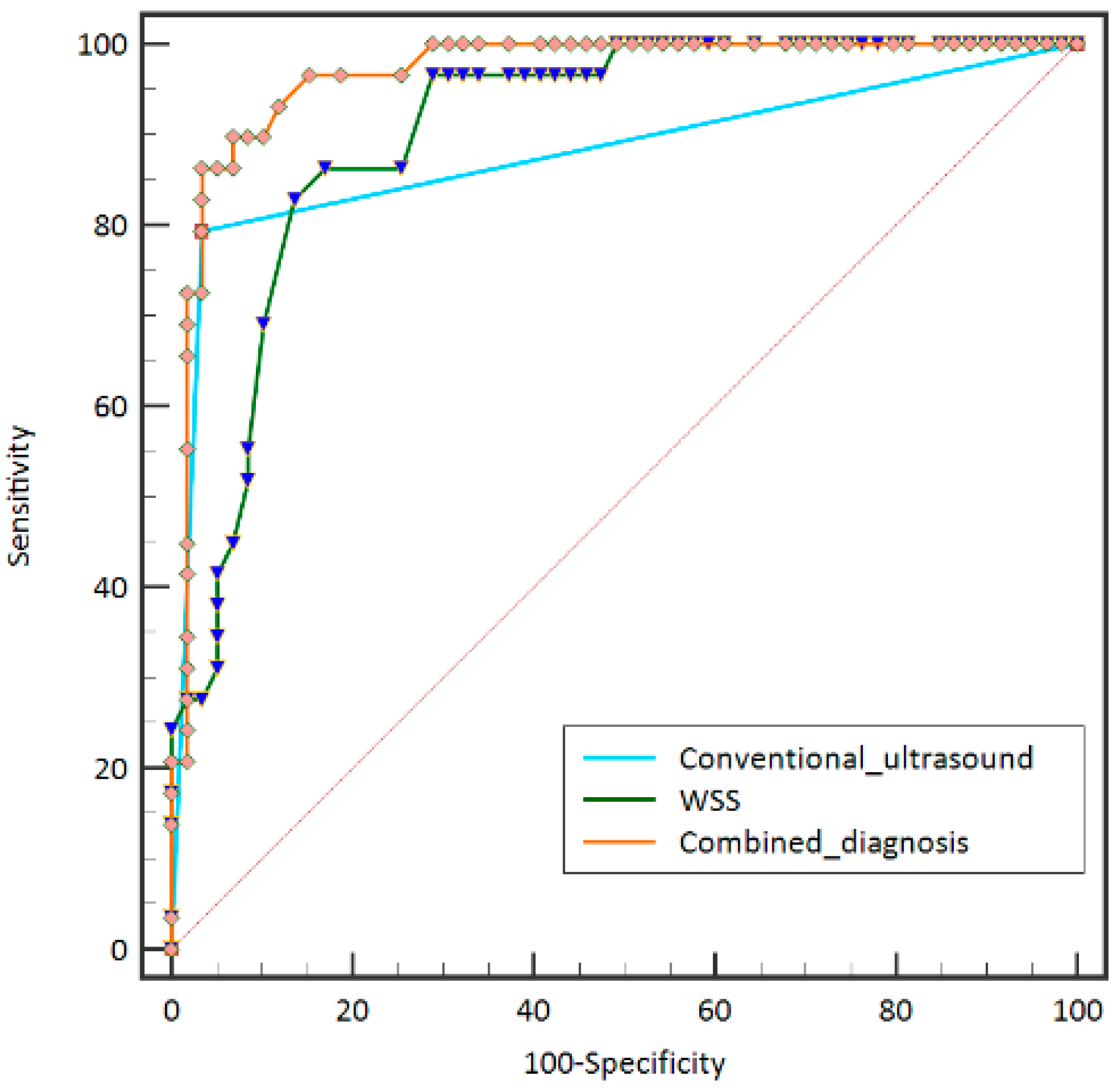
Objective: This study seeks to evaluate the value of the high-frame-rate vector flow imaging technique in assessing the hemodynamic changes of carotid atherosclerotic stenosis in aging people (>60 years old). Methods: Aging patients diagnosed with carotid atherosclerotic stenosis who underwent carotid high-frame-rate vector flow imaging examination were prospectively enrolled. A Mindray Resona7s ultrasound machine equipped with high-frame-rate vector flow function was used for ultrasound evaluation. First, B mode ultrasound and color Doppler flow imaging were used to evaluate carotid stenosis. Then, the vector arrows and flow streamline detected by V Flow were analyzed and the wall shear stress values (Pa) at the carotid stenosis site were measured. All patients were divided into symptomatic and asymptomatic groups according to whether they had acute/subacute stroke or other clinical symptoms within 2 weeks before ultrasound examination. The results of digital subtraction angiography or computed tomography angiography were used as the gold standard. The stenosis rate was calcified, according to North American Symptomatic Carotid Endarterectomy Trial criteria. The diagnostic values of wall shear stress, conventional ultrasound, and the combined diagnosis in carotid atherosclerotic stenosis were compared. Results: Finally, 88 patients with carotid atherosclerotic plaque were enrolled (71 males (80.7%), mean age 67.6 ± 5.4 years). The success rate of high-frame-rate vector flow imaging was 96.7% (88/91). The WSS value of symptomatic carotid stenosis (1.4 ± 0.15 Pa) was significantly higher than that of asymptomatic carotid stenosis (0.80 ± 0.08 Pa) (p < 0.05). Taking the wall shear stress value > 0.78 Pa as the diagnostic criteria for symptomatic carotid atherosclerotic plaque, the area under receiver operating characteristic curves was 0.79 with 87.1% sensitivity and 69.6% specificity. The area under receiver operating characteristic curves of the combined diagnosis (0.966) for differentiating severe carotid atherosclerotic stenosis was significantly higher than that of conventional ultrasound and WSS value, with 89.7% sensitivity and 93.2% specificity (p < 0.05). Conclusion: As a non-invasive imaging method, the high-frame-rate vector flow imaging technique showed potential value in the preoperative assessment of the symptomatic carotid atherosclerotic stenosis and diagnosing carotid atherosclerotic stenosis in aging patients.
Keywords: vector flow (V flow); high frame rate; carotid atherosclerotic stenosis; wall shear stress (WSS); ultrasound vector flow (V flow); high frame rate; carotid atherosclerotic stenosis; wall shear stress (WSS); ultrasound