


Abstract
We used nasopharyngeal swab samples of patients with a symptomatic (n = 82) or asymptomatic (n = 20) coronavirus disease 2019 (COVID-19) diagnosis to assess the ability of antigen detection tests to infer active (potentially transmissible) or inactive (potentially non-transmissible) infection by severe acute respiratory syndrome coronavirus 2 (SARS-CoV-2). Using the subgenomic RNA (sgRNA) as an active replication marker of SARS-CoV-2, 48 (76.2%), 56 (88.9%), and 63 (100%) of 63 samples with sgRNA positive results tested positive with the SD BIOSENSOR STANDARD Q COVID-19 Ag (Standard Q), the SD BIOSENSOR STANDARD F COVID-19 Ag FIA (Standard F), or the Fujirebio LUMIPULSE G SARS-CoV-2 Ag (Lumipulse) assay, respectively. Conversely, 37 (94.9%), 29 (74.4%), and 7 (17.9%) of 39 samples with sgRNA negative results tested negative with Standard Q, Standard F, or Lumipulse, respectively. Stratifying results by the number of days of symptoms before testing revealed that most antigen positive/sgRNA positive results were among samples tested at 2–7 days regardless of the assay used. Conversely, most antigen negative/sgRNA negative results were among samples tested at 16–30 days only when Standard Q or Standard F were used. In conclusion, based on our findings, a negative antigen test, especially with the Lumipulse assay, or a positive antigen test, especially with the Standard F assay, may suggest, respectively, the absence or presence of replication-competent SARS-CoV-2.
1. Introduction
Reverse transcription-PCR (RT-PCR)-based molecular assays offer a reliable means of detecting the severe acute respiratory syndrome coronavirus 2 (SARS-CoV-2) genomic RNA (gRNA) in respiratory tract samples from patients who develop coronavirus disease 2019 (COVID-19) within 1 to 2 weeks of SARS-CoV-2 exposure [1]. Whereas there is no established RT-PCR cycle threshold (Ct) value at which individuals are likely to no longer be infectious [2], growing evidence shows that RT-PCR detection of SARS-CoV-2 subgenomic RNA (sgRNA), as with viral culture (i.e., the currently accepted reference standard for virus replication) [3], may be reliably used to measure infectivity [4,5,6]. Unlike diagnostic (gRNA-detecting) RT-PCR, detection of the SARS-CoV-2 antigen, which is per se an indicator of active infection, offers an accurate but less sensitive means of identifying acutely infected individuals [7] and, therefore, the possibility to identify individuals who are shedding the infectious virus and are likely to transmit SARS-CoV-2 [2]. Compared to RT-PCR assays, which have widely replaced viral culture to diagnose respiratory infections [2], antigen assays have 100% specificity, whereas their sensitivity may be moderately high (84%) or low (53%) for symptomatic (i.e., tested within the first 7 days of illness) or asymptomatic patients, respectively [8].
Only one study so far [9], to the best of our knowledge, has compared SARS-CoV-2 sgRNA with SARS-CoV-2 antigen detection in symptomatic COVID-19 patients, showing that antigen was concordant with sgRNA for samples from a midturbinate nasal swab but not from a nasopharyngeal swab (NPS). To clarify this matter, we used NPS samples of asymptomatic or symptomatic COVID-19 patients to assess the antigen detection tests’ ability for differentiating between active (potentially transmissible) and inactive (potentially non-transmissible) SARS-CoV-2 infection. We assessed the SD BIOSENSOR STANDARD Q COVID-19 Ag, the SD BIOSENSOR STANDARD F COVID-19 Ag FIA, and the Fujirebio LUMIPULSE G SARS-CoV-2 Ag in comparison with SARS-CoV-2 sgRNA, here used as a marker of active virus replication.
2. Materials and Methods
Study design and clinical samples. This retrospective study was carried out at the Fondazione Policlinico Universitario A. Gemelli IRCCS hospital of Rome, Italy, and approved by the Institutional Ethics Committee (reference no. 0040205/21), during a 2021 one-month (24 November through 24 December) period. We randomly selected NPS samples from patients who had a laboratory-confirmed COVID-19 diagnosis, which relied on a positive RT-PCR result through a Panther Fusion SARS-CoV-2 assay (Hologic S.r.l., Roma, Italy). Residual 2-mL aliquots from originally collected samples in 3 mL of universal transport medium (UTM, Copan, Brescia, Italy) were put into anonymized blank vials and immediately processed for or kept at −80 °C until testing (see below). One hundred and two NPS samples from patients diagnosed with asymptomatic (n = 20), mild (n = 14), moderate (n = 17), severe (n = 32), or critical (n = 19) COVID-19, according to a median (interquartile range) RT-PCR Ct value of 24.6 (18.6–30.2) for SARS-CoV-2 gRNA detection, were included (Table 1). Only for symptomatic patients (n = 82), disease severity was established according to the criteria from US National Institutes of Health (https://www.covid19treatmentguidelines.nih.gov accessed on 10 May 2022).
SARS-CoV-2 RNA and antigen testing. Following diagnostic RT-PCR (i.e., using a molecular assay not allowing Ct value determination as above specified), samples (i.e., residual NPS samples) were used to detect SARS-CoV-2 gRNA (i.e., using a molecular assay allowing Ct value determination), sgRNA, and antigen. For gRNA, Ct values for three SARS-CoV-2 targets, such as envelope (E), RNA-dependent RNA polymerase (RdRP)/spike (S), and nucleoprotein (N) encoding genes, were determined using an updated version of the Allplex™ 2019-nCoV assay [10], now designated as Allplex™ SARS-CoV-2 assay. For sgRNA (i.e., E-gene sgRNA) detection, we used an in-house RT-PCR assay [11], which was essentially in accordance with a method described elsewhere [4]. For antigen detection, we used STANDARD Q COVID-19 Ag (SD BIOSENSOR, https://www.sdbiosensor.com accessed on 10 May 2022), STANDARD F COVID-19 Ag FIA (SD BIOSENSOR, https://www.sdbiosensor.com accessed on 10 May 2022), and LUMIPULSE G SARS-CoV-2 Ag (Fujirebio, https://www.fujirebio.com accessed on 10 May 2022) immunoassays (hereafter referred to as Standard Q, Standard F, and Lumipulse, respectively) according to the respective manufacturer’s instructions. Briefly, the Standard Q is a lateral flow assay that detects SARS-CoV-2 N antigen in a cassette-based format with a visual read-out, allowing interpretation of results as positive (i.e., test band detected) or negative (i.e., test band not detected). The Standard F is a fluorescence immunoassay that detects SARS-CoV-2 N antigen with STANDARD F Analyzer, allowing interpretation of results as positive (i.e., cutoff index (COI) value of ≥1.0) or negative (i.e., COI of <1.0), according to the manufacturer’s instructions (https://www.sdbiosensor.com accessed on 10 May 2022). The Lumipulse uses a CLEIA (chemiluminescent enzyme immunoassay) technology to measure SARS-CoV-2 S antigen by a LUMIPULSE® G System automated instrument, allowing interpretation of results as positive (>10–>5000 pg/mL), gray-zone positive (1.34–10 pg/mL), or negative (<1.34 pg/mL) [12].
Statistical analysis. For SARS-CoV-2 antigen or sgRNA assays’ results, differences between a priori established groups were assessed using the chi-square test or the Wilcoxon test, as appropriate. Percent agreement values, with their respective confidence intervals (CIs), were calculated comparing results from antigen assays with those from the sgRNA assay, which was used as the reference method. Statistical analysis was conducted using Stata 15 (StataCorp, College Station, TX, USA) or GraphPad Prism 7 (GraphPad Software, San Diego, CA, USA) software. p < 0.05 was considered statistically significant.
3. Results
For the 102 patients studied, ages ranged from 45.7 to 78.8 years (median 62.8), and 53 (52.0%) out of 102 were male (Table 1). Excluding asymptomatic patients (n = 20), time from the onset of symptoms to sample collection ranged from 1 day to >30 days, with most patients (32/82; 39.0%) being sampled 2–7 days after symptoms became apparent (Table 1). Accordingly, in this subset of patients, Standard Q, Standard F, or Lumipulse antigen positivity rates were found to be the highest (28/32 [87.5%], 29/32 [90.6%], and 32/32 [100%], respectively). Lower rates were, indeed, noticed in subsets of patients sampled at 8–15 days (7/28 (25.0%), 18/28 (64.3%), and 28/28 (100%), respectively) or 16–30 days (0/13 (0.0%), 2/13 (15.4%), and 9/13 (69.2%), respectively) after the onset of symptoms, which accounted for 28 (34.1%) and 13 (15.8%) of the 82 samples collected in total.
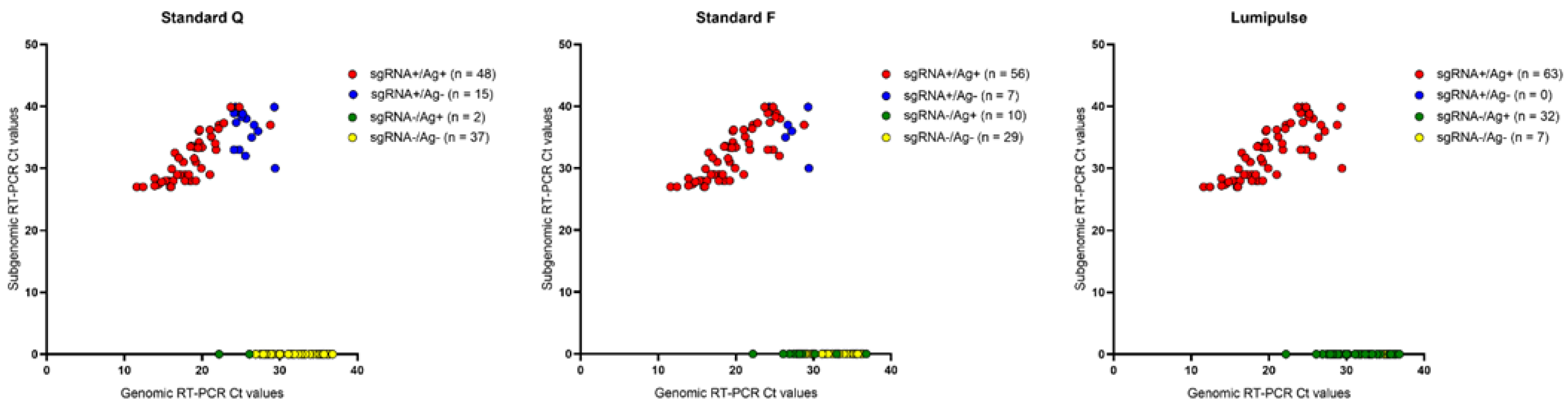
Of the 102 samples studied (Table 1), 95 (93.1%) tested positive with Lumipulse, with 50 (49.0%) and 66 (64.7%) found to be concomitantly positive with the Standard Q or the Standard F, respectively. In parallel, 63 (61.8%) samples tested positive with the sgRNA detection assay, and their median (interquartile range) RT-PCR Ct value was 33.0 (28.4–36.4). As with diagnostic RT-PCR Ct values (p value < 0.001 for all comparisons), sgRNA RT-PCR Ct values differed significantly between antigen positive and antigen negative samples’ groups (p values < 0.001 and 0.008 for Standard Q’s or Standard F’s comparisons, respectively). Seven (6.9%) samples with Lumipulse antigen (and sgRNA) negative results had a diagnostic RT-PCR Ct median value of 35.1, which was higher than the median value of 32.4 noticed in 39 (38.2%) samples with sgRNA negative results. Except for seven samples, detectable sgRNA levels were noticed in the group of 52 (51.0%) samples with Standard Q antigen negative results (RT-PCR Ct median value, 37.4) or the group of 36 (35.3%) samples with Standard F antigen negative results (RT-PCR Ct median value, 37.0).
Compared to sgRNA results (Table 2 and Figure 1), 48 (76.2%), 56 (88.9%), and 63 (100%) of 63 samples with sgRNA positive results tested positive with Standard Q, Standard F, or Lumipulse, respectively. Conversely, 37 (94.9%), 29 (74.4%), and 7 (17.9%) out of 39 samples with sgRNA negative results tested negative with Standard Q, Standard F, or Lumipulse, respectively
Table 1.
Demographic and clinical characteristics of COVID-19 patients who had NPS samples analyzed for SARS-CoV-2 antigen and sgRNA a.
Table 2.
Comparison of SARS-CoV-2 antigen and sgRNA results for NPS samples tested at indicated day(s) from symptom onset a.
Figure 1.
Distribution of gRNA or sgRNA reverse transcription-PCR Ct values for NPS samples from 102 COVID-19 patients tested for antigen detection with Standard Q, Standard F, or Lumipulse. Colors indicate groups of results stratified as sgRNA positive/Ag positive (red), sgRNA positive/Ag negative (blue), sgRNA negative/Ag positive (green), or sgRNA negative/Ag negative (yellow). Ag, antigen; Ct, cycle threshold; COVID-19, coronavirus disease 2019; gRNA, genomic RNA; NPS, nasopharyngeal swab; sgRNA, subgenomic RNA.
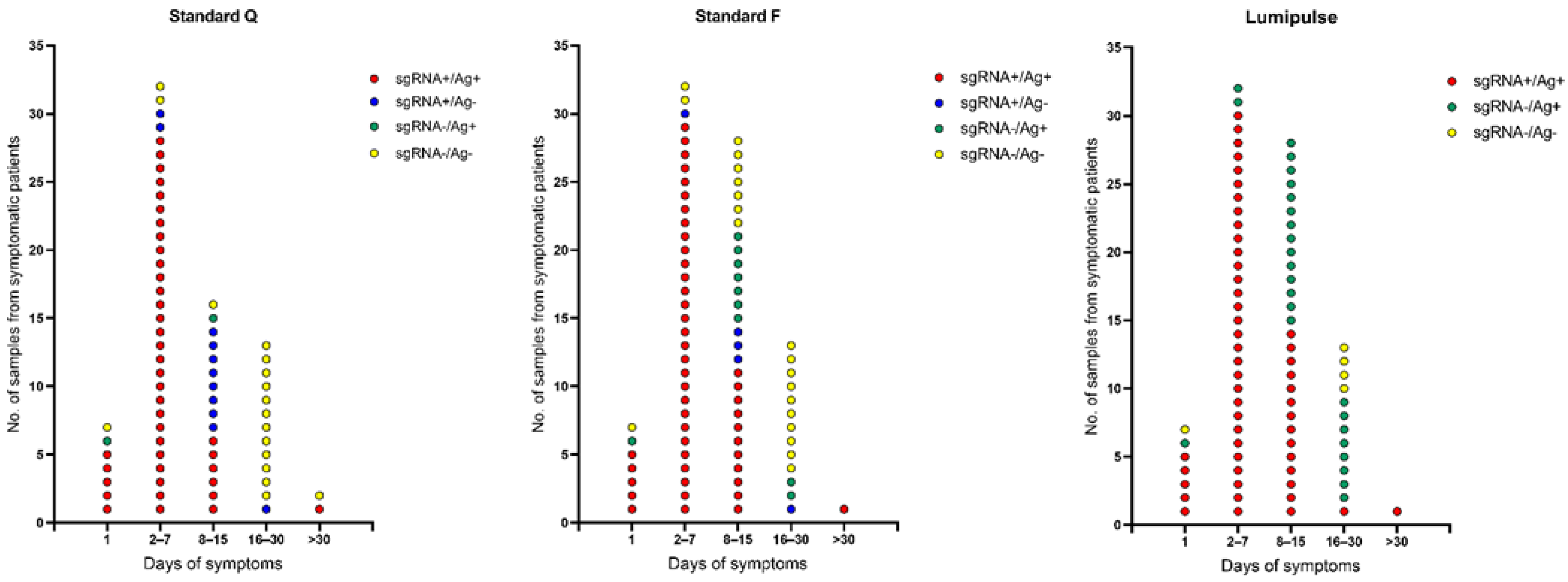
Stratifying antigen results by number of days of symptoms before sample collection (Table 2 and Figure 2) showed that Standard F for samples tested at 2–7 days had positive and negative percent agreement values of 96.7% and 100%, which in combination were found to be better than those of Standard Q (93.3% and 100%) or Lumipulse (100% and 0.0%). As illustrated in Figure 2, antigen positive/sgRNA positive results were most frequent among the samples tested at 2–7 days regardless of the (Standard Q, Standard F, or Lumipulse) test used, and antigen negative/sgRNA negative results were most frequent among the samples tested at 16–30 days only when Standard Q or Standard F were used.
Figure 2.
Distribution of NPS samples from 82 symptomatic COVID-19 patients tested for (reverse transcription-PCR) sgRNA and (Standard Q, Standard F, or Lumipulse) antigen detection according to number of days of symptoms before sample collection. Colors indicate groups of results stratified as sgRNA positive/Ag positive (red), sgRNA positive/Ag negative (blue), sgRNA negative/Ag positive (green), or sgRNA negative/Ag negative (yellow). Ag, antigen; COVID-19, coronavirus disease 2019; Ct, cycle threshold; NPS, nasopharyngeal swab; sgRNA, subgenomic RNA.
4. Discussion
We comparatively assessed SARS-CoV-2 antigen detection tests, i.e., three among a myriad of commercially available tests [13], whose analytical sensitivity (Lumipulse > Standard F > Standard Q) depends on how advanced the immunoassay’s technology is (lateral flow assay < immunofluorescence < chemiluminescence). Two are rapid antigen tests [13] and the third (Lumipulse) is a laboratory-based antigen test [12]. Our findings show that a negative antigen test, especially with the Lumipulse assay, or a positive antigen test, especially with the Standard F assay, may be highly suggestive of the absence or presence of replication-competent SARS-CoV-2, respectively. Differentiation between the persistent shedding of viral RNA (i.e., non-infectious status), which is usually associated with a positive SARS-CoV-2 molecular assay [7], and active virus replication (i.e., infectious status) is essential for the clinical management, discontinuation of isolation, and/or work reintegration of SARS-CoV-2-infected individuals. A negative antigen result may be particularly important in the context of a previously positive SARS-CoV-2 molecular assay, i.e., within 10–14 days following symptom onset when both the detection of the infectious virus and contagiousness (or transmissibility) are almost unlikely [14].
Previous studies’ findings supported us to adopt SARS-CoV-2 sgRNA RT-PCR as the reference method for our antigen tests’ assessment [6,15,16,17]. One study [15] using respiratory tract samples obtained from COVID-19 patients within 7 days of symptom onset showed the correlation between the SARS-CoV-2 antigen (using the Becton Dickinson BD Veritor System for Rapid Detection of SARS-CoV-2 test) and SARS-CoV-2 culture (using a VeroE6TMPRSS2 cell line-based high-sensitive culture test) positivity. Another study [16] showed that three lateral flow assays and a (microfluidic) immunofluorescence assay correlated with cell-culture infectivity more significantly than SARS-CoV-2 gRNA RT-PCR. Finally, in Santos Bravo et al.’s study [6], sgRNA RT-PCR proved to detect replication-competent SARS-CoV-2 in COVID-19 patients’ NPS or other respiratory samples with a sensitivity of 97% and a positive predictive value of 94% compared to SARS-CoV-2 culture. However, to comply with the assumption that culture may be suboptimal to detect the presence of the infectious virus due to poor sensitivity [2], we used a highly sensitive RT-PCR method (i.e., the sgRNA detection assay) to assess antigen assays in our study. Similar to us, Santos Bravo et al. [6] chose to measure E-gene sgRNA, i.e., the sgRNA species known as the best option to detect infectivity so far [2].
Our study has some intrinsic limitations. First, the sample size is relatively small, particularly for patients without COVID-19 compatible symptoms who represent only 19.6% of the patients included in the study. This precludes our results from being informative for study populations that primarily consist of asymptomatic patients. Second, antigen assays studied by us are very different platforms, implying their analytical sensitivities are bound to be different. This was especially noted for asymptomatic patients, but it could also explain the high rates of Standard Q or Standard F disagreement with the sgRNA RT-PCR assay that occurred for samples tested after 7 days or more of symptoms. Third, our results suggest that antigen assays can be very helpful at the onset of symptoms, which is pretty much expected.
In conclusion, our findings reinforce the potential of SARS-CoV-2 antigen tests to serve as a marker for active SARS-CoV-2 replication. However, further studies are required before using such tests to predict SARS-CoV-2 infectivity and, thus, inform the discontinuation of COVID-19 transmission-based precautions.
Author Contributions
Conceptualization, G.D.A., G.M., F.M.L., B.P. and M.S.; methodology, S.M., A.S., A.V. and R.S.; formal analysis, B.P. and R.S.; resources, M.S.; data curation, S.M., R.S. and M.S.; writing—original draft preparation, B.P.; writing—review and editing, all authors. All authors have read and agreed to the published version of the manuscript.
Funding
This research received no external funding.
Institutional Review Board Statement
The study was conducted in accordance with the Declaration of Helsinki and approved by the Ethics Committee of the Fondazione Policlinico Universitario A. Gemelli IRCCS, Rome, Italy (protocol code 0040205/21, date of approval 16 November 2021).
Informed Consent Statement
Patient consent was waived due to the nature of the study according to which only residual samples were tested for each patient.
Data Availability Statement
The data presented in this study are available on request from the corresponding author.
Conflicts of Interest
The authors declare no conflict of interest.
References
- van Kasteren, P.B.; van Der Veer, B.; van den Brink, S.; Wijsman, L.; de Jonge, J.; van den Brandt, A.; Molenkamp, R.; Reusken, C.B.; Meijer, A. Comparison of seven commercial RT-PCR diagnostic kits for COVID-19. J. Clin. Virol. 2020, 128, 104412. [Google Scholar] [CrossRef] [Scilit] [PubMed]
- Binnicker, M.J. Can testing predict SARS-CoV-2 infectivity? The potential for certain methods to be surrogates for replication-competent virus. J. Clin. Microbiol. 2021, 59, e0046921. [Google Scholar] [CrossRef] [Scilit] [PubMed]
- Stelzer-Braid, S.; Walker, G.J.; Aggarwal, A.; Isaacs, S.R.; Yeang, M.; Naing, Z.; Stella, A.O.; Turville, S.G.; Rawlinson, W.D. Virus isolation of severe acute respiratory syndrome coronavirus 2 (SARS-CoV-2) for diagnostic and research purposes. Pathology 2020, 52, 760–763. [Google Scholar] [CrossRef] [Scilit] [PubMed]
- Wölfel, R.; Corman, V.M.; Guggemos, W.; Seilmaier, M.; Zange, S.; Müller, M.A.; Niemeyer, D.; Jones, T.C.; Vollmar, P.; Rothe, C.; et al. Virological assessment of hospitalized patients with COVID-2019. Nature 2020, 581, 465–469. [Google Scholar] [CrossRef] [Scilit] [PubMed]
- Perera, R.A.; Tso, E.; Tsang, O.T.; Tsang, D.N.; Fung, K.; Leung, Y.W.; Chin, A.W.; Chu, D.K.; Cheng, S.M.; Poon, L.L.; et al. SARS-CoV-2 virus culture and subgenomic RNA for respiratory specimens from patients with mild coronavirus disease. Emerg. Infect. Dis. 2020, 26, 2701–2704. [Google Scholar] [CrossRef] [Scilit] [PubMed]
- Santos Bravo, M.; Berengua, C.; Marín, P.; Esteban, M.; Rodriguez, C.; Del Cuerpo, M.; Miró, E.; Cuesta, G.; Mosquera, M.; Sánchez-Palomino, S.; et al. Viral culture confirmed SARS-CoV-2 subgenomic RNA value as a good surrogate marker of infectivity. J. Clin. Microbiol. 2022, 60, e0160921. [Google Scholar] [CrossRef] [Scilit] [PubMed]
- Hanson, K.E.; Altayar, O.; Caliendo, A.M.; Arias, C.A.; Englund, J.A.; Hayden, M.K.; Lee, M.J.; Loeb, M.; Patel, R.; El Alayli, A.; et al. The Infectious Diseases Society of America guidelines on the diagnosis of COVID-19: Antigen testing. Clin. Infect. Dis. 2021, ciab557. [Google Scholar] [CrossRef] [Scilit] [PubMed]
- Fragkou, P.C.; De Angelis, G.; Menchinelli, G.; Can, F.; Garcia, F.; Morfin-Sherpa, F.; Dimopoulou, D.; Mack, E.; de Salazar, A.; Grossi, A.; et al. ESCMID COVID-19 guidelines: Diagnostic testing for SARS-CoV-2. Clin. Microbiol. Infect. 2022, 28, 812–822. [Google Scholar] [CrossRef] [Scilit] [PubMed]
- Immergluck, K.; Gonzalez, M.D.; Frediani, J.K.; Levy, J.M.; Figueroa, J.; Wood, A.; Rogers, B.B.; O’Neal, J.; Elias-Marcellin, R.; Suessmith, A.; et al. Correlation of SARS-CoV-2 subgenomic RNA with antigen detection in nasal midturbinate swab specimens. Emerg. Infect. Dis. 2021, 27, 2887–2891. [Google Scholar] [CrossRef] [Scilit] [PubMed]
- Liotti, F.M.; Menchinelli, G.; Marchetti, S.; Morandotti, G.A.; Sanguinetti, M.; Posteraro, B.; Cattani, P. Evaluation of three commercial assays for SARS-CoV-2 molecular detection in upper respiratory tract samples. Eur. J. Clin. Microbiol. Infect. Dis. 2021, 40, 269–277. [Google Scholar] [CrossRef] [Scilit] [PubMed]
- Liotti, F.M.; Menchinelli, G.; Marchetti, S.; Posteraro, B.; Landi, F.; Sanguinetti, M.; Cattani, P. Assessment of SARS-CoV-2 RNA test results among patients who recovered from COVID-19 with prior negative results. JAMA Intern. Med. 2021, 181, 702–704. [Google Scholar] [CrossRef] [Scilit] [PubMed]
- Menchinelli, G.; Bordi, L.; Liotti, F.M.; Palucci, I.; Capobianchi, M.R.; Sberna, G.; Lalle, E.; Romano, L.; De Angelis, G.; Marchetti, S.; et al. Lumipulse G SARS-CoV-2 Ag assay evaluation using clinical samples from different testing groups. Clin. Chem. Lab. Med. 2021, 59, 1468–1476. [Google Scholar] [CrossRef] [Scilit] [PubMed]
- Scheiblauer, H.; Filomena, A.; Nitsche, A.; Puyskens, A.; Corman, V.M.; Drosten, C.; Zwirglmaier, K.; Lange, C.; Emmerich, P.; Müller, M.; et al. Comparative sensitivity evaluation for 122 CE-marked rapid diagnostic tests for SARS-CoV-2 antigen, Germany, September 2020 to April 2021. Eurosurveillance 2021, 26, 2100441. [Google Scholar] [CrossRef] [Scilit] [PubMed]
- He, X.; Lau, E.H.; Wu, P.; Deng, X.; Wang, J.; Hao, X.; Lau, Y.C.; Wong, J.Y.; Guan, Y.; Tan, X.; et al. Temporal dynamics in viral shedding and transmissibility of COVID-19. Nat. Med. 2020, 26, 672–675. [Google Scholar] [CrossRef] [Scilit] [PubMed]
- Pekosz, A.; Parvu, V.; Li, M.; Andrews, J.C.; Manabe, Y.C.; Kodsi, S.; Gary, D.S.; Roger-Dalbert, C.; Leitch, J.; Cooper, C.K. Antigen-based testing but not real-time polymerase chain reaction correlates with severe acute respiratory syndrome coronavirus 2 viral culture. Clin. Infect. Dis. 2021, 73, e2861–e2866. [Google Scholar] [CrossRef] [Scilit] [PubMed]
- Kohmer, N.; Toptan, T.; Pallas, C.; Karaca, O.; Pfeiffer, A.; Westhaus, S.; Widera, M.; Berger, A.; Hoehl, S.; Kammel, M.; et al. The comparative clinical performance of four SARS-CoV-2 rapid antigen tests and their correlation to infectivity in vitro. J. Clin. Med. 2021, 10, 328. [Google Scholar] [CrossRef] [Scilit] [PubMed]
- Mencacci, A.; Gili, A.; Gidari, A.; Schiaroli, E.; Russo, C.; Cenci, E.; Camilloni, B.; Graziani, A.; Melelli-Roia, A.; Francisci, D.; et al. Role of nucleocapsid protein antigen detection for safe end of isolation of SARS-CoV-2 infected patients with long persistence of viral RNA in respiratory samples. J. Clin. Med. 2021, 10, 4037. [Google Scholar] [CrossRef] [Scilit] [PubMed]
Publisher’s Note: MDPI stays neutral with regard to jurisdictional claims in published maps and institutional affiliations. |
© 2022 by the authors. Licensee MDPI, Basel, Switzerland. This article is an open access article distributed under the terms and conditions of the Creative Commons Attribution (CC BY) license (https://creativecommons.org/licenses/by/4.0/).