


Highlights
What are the main findings?
- An established method for trace, simple, rapid, cheap, sensitive, accurate, robust, and high-throughput for methylmalonic acid quantification.
- Good chromatographic separation of MMA and its intrinsic isomer and good signals of MMA were achieved using a simple isocratic elution strategy.
- Materials and reagents that are complex or not always accessible and procedures in previous methods such as derivatization, multistep SPE, incubations, evaporations, drying, or reconstitutions are not required in this MMA quantification method.
What is the implication of the main finding?
- This method is suitable for large-scale MMA testing.
Abstract
Background: Methylmalonic acid (MMA) is an essential indicator of vitamin B12 (VB12) deficiency and inherited metabolic disorders (IMDs). The increasing number of requests for MMA testing call for higher requirements for convenient MMA testing methods. This study aims to develop a convenient quantification method for serum MMA. Methods: The method was established based on the stable isotope-dilution liquid chromatography–tandem mass spectroscopy (ID-LC-MS/MS) technique. The LC-MS/MS parameters and sample preparation were optimized. Specificity, sensitivity, robustness, accuracy, and clinical applicability were validated according to CLSI C62-A guidelines. MMA levels in VB12-sufficient subjects and VB12-deficient subjects were measured. Results: MMA and its intrinsic isomer, i.e., succinic acid (SA), were completely separated. The average slope, intercept, and correlation relationship (R) with 95% confidence intervals, during the two months, were 0.992 (0.926–1.059), −0.004 (−0.012–0.004), and 0.997 (0.995–0.999), respectively. The limit of detection and quantification were <0.058 μmol/L and 0.085 μmol/L, respectively. Intra-run, inter-run, and total imprecisions were 1.42–2.69%, 3.09–5.27%, and 3.22–5.47%, respectively. The mean spiked recoveries at the three levels were 101.51%, 92.40%, and 105.95%, respectively. The IS-corrected matrix effects were small. The VB12-deficient subjects showed higher MMA levels than VB12-sufficient subjects. Conclusions: A convenient LC-MS/MS method for serum MMA measurement was developed and validated, which could be suitable for large-scale MMA testing and evaluating MMA levels in VB12-deficient patients.
1. Introduction
Methylmalonic acid (MMA), an abnormal metabolic product of defective cobalamin metabolism and methylmalonate, is considered to be a specific diagnostic biomarker of vitamin B12 (VB12) deficiency [1,2,3,4] and methylmalonic acidemia [5]. VB12 (i.e., cobalamin) is a key cofactor of the enzymatic conversion of methylmalonyl-CoA to succinyl-CoA and the conversion of homocysteine to methionine [6]. An insufficiency of VB12 can lead to elevated methylmalonyl-CoA and homocysteine, causing high levels of MMA and total homocysteine (tHcy) in the blood [3,4]. VB12 deficiency is a common and serious condition [3,4,7] with a high prevalence across many populations, for example, 70–80% in African and Asian children, 40% in Latin American children and adults, and close to 20% in the elderly [3,7]. The measurement of MMA level is also helpful in the screening, confirmation diagnosis, and therapy monitoring of inherited metabolic disorder (IMD) [8].
Early identification of VB12 deficiency is vital for early determination of causes and early prevention and remission of serious presentations; VB12, tHcy, and MMA are all helpful diagnostic indicators for these disorders. The VB12 assay is the most commonly used test. However, previous studies have suggested that MMA is a more sensitive and representative biomarker for VB12 deficiency than VB12 and tHcy [3,4], because: (1) Testing for tHcy is less specific since its concentrations can be easily influenced by many environmental factors [3,9]; (2) VB12 is a less stable indicator and is easily affected by environmental factors [10], while MMA is very stable [11]; (3) MMA is more sensitive than VB12 because MMA can increase before VB12 falls [12,13]. (4) Elevated MMA can persist for several days even after replacement is started [14]. (5) VB12 measurements cannot reflect the true status of VB12. VB12 is measured by automated immunoassays based on competitive-binding immune chemiluminescence [3,15,16]. However, immunoassays lack specificity since they simultaneously measure VB12, as well as holohaptocorrin and holotranscobalamin [3,10]. Under these circumstances, increasingly more requests for MMA testing, and therefore, effective and convenient MMA testing is in demand.
MMA is a “mass spectrometry (MS)-based disease biomarker” [17]. Currently, there are no routine economic immunoassays that can quantify MMA. Gas chromatography–mass spectroscopy (GC-MS) [18] was the gold standard for MMA measurements. However, GC-MS-based MMA testing has several drawbacks (including high costs, cumbersome and laborious procedures, large sample volume, and long analytical time) [19,20]. Liquid chromatography–tandem mass spectroscopy (LC-MS/MS) methods are currently preferred for MMA measurements [14,21,22,23,24,25,26,27,28,29,30,31,32,33,34,35,36,37,38].
There are many published papers on MMA measurements by LC-MS/MS, using derivatized, non-derivatized, more or less complicated sample extraction, etc. Table 1 summarizes the reported analytical procedures of MMA measurement based on LC-MS/MS (from 2000 to 2022 [14,21,22,23,24,25,26,27,28,29,30,31,32,33,34,35,36,37,38]). Given a comprehensive consideration of all the method procedures as well as the aspects of cost-effectiveness, testing speeding, convenience, method performance, purpose, degree of difficulty for introducing the testing, robustness, accuracy, safety, and environmental protection, it is evident that all of these methods have their advantages and disadvantages. The improvement history of MMA testing shows that the method procedures basically consist of one or more of the following steps: multistep solid-phase extraction (SPE), derivations, protein precipitation, and ultrafiltration, while to complete the above step, at least one of the processes of evaporations, incubations, heating, dryings, reconsititutions, or centrifugations is required [14,21,22,23,24,25,26,27,28,29,30,31,32,33,34,35,36,37,38]. Improvements in procedures have usually focused on reducing these steps by changing different reagents or materials or by applying new materials [14,21,22,23,24,25,26,27,28,29,30,31,32,33,34,35,36,37,38]. These improvements have reduced time and cost. However, with the deepening understanding of the clinical significance of MMA and the widespread popularization of mass spectrometry technology in clinical laboratories, requests for MMA testing are evidently increasing, which calls for more convenient testing methods.
Table 1.
A literature review of previous methylmalonic acid quantification based on the liquid chromatography–tandem mass spectrometry method (from 2000 to 2022). Abbreviations of the names of equipment/columns are not listed and abbreviations of reagents are list in the table for reading convenience. Note: LC-MS/MS, liquid chromatography–tandem mass spectrometry; LC, liquid chromatography; MS, mass spectrometry; MMA, methylmalonic acid; LOD, limit of detection; LOQ, limit of quantification; HCl, hydrochloride; SPE, solid phase extraction; SRM, selected reaction monitoring; CVs, coefficient of variations; NA, not available; MRM, multiple reaction monitoring; DBS, dried blood spot; SIM, single-ion monitoring; HPLC, high-performance liquid chromatography; tHcy, total homocysteine.
Different from the previous paths of improvement, we took advantage of a simple mobile phase strategy to improve simple MMA detection, which required fewer reagents and a smaller sample. There was no need for complex and dangerous derivation reagents, costly/not always accessible ultrafiltration materials, or processes that are time-consuming and laborious, such as evaporations, incubations, dryings, and reconstitutions. The established method in this study demonstrated several advantages for MMA detection, for example, convenient, environmentally friendly, economical, and more cost-effective for the assessment of VB12 deficiency; only several simple reagents in small volume were needed and sample preparation could be completed in 20 min. Since LC-MS/MS testing is currently mainly manual, such improvements are significant, especially, when there are numerous requests for MMA testing.
2. Materials and Methods
2.1. Chemicals
Methylmalonic acid (1.0 mg/mL in acetonitrile, 1 mL ampule, certified reference material, Cerilliant®, purity 99%) was purchased from Sigma-Aldrich (Burlington, MA, USA). Isotope-labeled internal standard (IS) methylmalonic acid-13C4 in acetonitrile solution (certified reference material, CAS: 1173019-21-0, product no. M-173-1ML, purity 99%) was purchased from Sigma-Aldrich (TX, USA). HPLC-grade methanol, acetonitrile, and isopropanol were purchased from Fisher Scientific (Waltham, MA, USA). Formic acid was purchased from Honeywell (San Francisco, CA, USA). Deionized water (18 Ω) was produced from a Milli-Q Advantage system (Millipore Corp., Bedford, MA, USA).
2.2. Samples
Pooled and individual serum samples for method establishment and individual specimens for clinical application were obtained from fresh leftover specimens in the Department of Laboratory Medicine, Beijing Hospital, Beijing, China. The collection of leftover sera was approved by the Ethics Committee of the Beijing Hospital.
2.3. Calibrators, Internal Standard (IS), and Quality Control (QC) Materials Preparation
Working standard solutions of MMA (218.69 ng/g) used for calibration and working standard solutions of MMA-13C4 (148.95 ng/g) were prepared gravimetrically in water. All solutions were aliquoted into 2 mL brown ampoules and stored at −80 °C. For each sample batch, the calibrators were freshly prepared. Eight working calibrators were prepared using 100 μL working standard solutions and subsequently diluted with 800, 700, 600, 500, 400, 300, 200, and 100 μL water, respectively.
Standard solutions of MMA (2003.60 ng/g) used as QC additive standard solutions were prepared gravimetrically in water. To make low-, medium-, and high-levels of QC materials, 3 μL, 5 μL, and 8 μL of QC additive solutions were gravimetrically added to three gravimetrically prepared 400 μL serum pools, respectively.
2.4. Sample Preparation
Fifty microliters of IS working solution, 50 μL of samples/calibrators, and 300 μL methanol were added to a 2.0 mL Eppendorf tube. The mixture was vortexed for 20 s and centrifuged at 148,000 rpm, at 4 °C, for 15 min. The upper phase of the mixture was poured into a new 2.0 mL Eppendorf tube and centrifuged at 148,000 rpm, at 4 °C, for 5 min. Fifty microliters of the upper phase was used for the LC-MS/MS analysis. The injection volume was 1 μL.
2.5. LC-MS/MS Conditions
The LC-MS/MS analysis was performed on a 6500 plus triple quadrupole mass spectrometer (AB Sciex, USA) coupled with an ExionLC™ AD ultra-high-performance liquid chromatography system (Applied Biosystems, CA, USA). The Analyst 1.7.2 software (Applied Biosystems, CA, USA) was used for data processing.
A Shim-pack GIST-HP C18-AQ column (3 μm, 2.1 × 100 mm, SHIMADZU, Japan) with a guard column (Shim-pack GIST-HP (G) C18, 3 μm, 2.1 × 10 with Cartridge (2pcs) and Holder) was used for separation. An isocratic method (100% A phase) was developed for separation. The mobile phase was water containing 0.1% formic acid and 5% isopropanol. The flow rate of the mobile phase was 0.45 mL/min. The column temperature was 35 °C, and the autosampler temperature was set at 8 °C. Acetonitrile was the wash solution of the LC system. The injection volume was 1 μL. A diverter valve was used from 1.9 min to 2.8 min.
The mass spectrometer was operated in the negative electrospray ionization (ESI) mode with multiple reaction monitoring (MRM). The transitions at m/z 116.9→72.8 for MMA and m/z 120.9→75.8 for MMA-13C4 were monitored for quantification. A source temperature of 450 °C and an ion spray voltage of −4500 V were used. Nitrogen gas was used as the curtain gas (CUR), nebulizer gas (GS1), auxiliary gas (GS2), and collision gas (CAD), and the pressures of the gases were set at 35, 35, 45, and low mode, respectively. The declustering potential (DP), entrance potential (EP), and collision exit potential (CXP) were set at −26 V, −12.6 V, and −11.6 V, respectively. The collision energy (CE) was set at −10.9 eV.
2.6. Method Validation
The method performance was validated according to the Clinical and Laboratory Standards Institute (CLSI) C62-A guideline [39].
2.6.1. Limits of Detection (LOD) and Limits of Quantification (LOQ)
Fifty microliters of standard solution of MMA (218.69 ng/g) was diluted with water to generate a series of concentrations. The diluted solutions were treated according to the sample preparation described previously. A signal-to-noise ratio (S/N) ≥ 3 with a coefficient of variation (CV) ≤ 20% for 20 injections was defined as the LOD, while an S/N ≥ 10 and a CV ≤ 20% for 20 injections was defined as the LOQ [39,40].
2.6.2. Analytical Precision and Recovery
Pooled serum was filtered using a disposable Corning bottle-top vacuum filter with a 0.22 μm membrane and aliquoted. Four pooled samples with different concentrations of MMA were prepared for the precision and recovery evaluation, that is, sample pools (Level 1), low- (Level 2), medium- (Level 3), and high-levels (Level 4) of QC materials described before. All samples were aliquoted into 2.0 mL Corning vials (1 mL/vial) and stored at −40 °C before analysis. Each sample was measured five times per day for five days.
2.6.3. Matrix Effect
The matrix addition mixing experiment was designed according to CLSI C62A [39] to assess the sample matrix effect before and after IS correction. Two different matrices were prepared: (X) the mixed standard solution of the neat analyte and IS and (Z) the extracted matrix, i.e., the matrix of serum pools (Level 1, Level 2, and Level 3) which were processed based on the established sample preparation procedure. Then, the defined amount of the mixed standard solution (i.e., X) was added to the same amount of extracted matrix (i.e., Z) to prepare the solution (Y), and the same amount of IS solution was added to the extracted matrix (i.e., Z) to prepare the solution (W). The IS in acetonitrile was previously dried under nitrogen gas and reconstituted with the primary mobile phase. The absolute matrix effects and IS-corrected matrix effects were calculated by the following equations:
where A is the peak area of MMA;
where R is the peak area ratio of MMA to MMA-13C4.
2.7. Method Applications
Fresh individual serum samples from healthy controls (n = 18, physical examination subjects with normal biochemical indicators, VB12-sufficient subjects (n = 24, VB12 > 240 pg/mL, measured by a routine immunoassay), VB12-deficient subjects (n = 13, VB12 < 240 pg/mL), patients diagnosed with anemia (n = 25), patients diagnosed with vitamin deficiency (n = 13), and patients diagnosed with colon cancer (n = 11) were randomly collected and measured in a random order. MMA levels in these populations were investigated using the established LC-MS/MS method. One positive urine sample from methylmalonic academia was measured alongside five urine samples from non-IMDs subjects.
2.8. Statistical Analysis
Statistical analysis was completed using Microsoft Excel 2016 (Microsoft Corporation, Redmond, WA, USA), SPSS 25.0 (IBM Inc., Armonk, NY, USA), and GraphPad Prism (version 8.0.0 for Windows, GraphPad Software, San Diego, CA, USA). The intra-assay, interassays, and total imprecision were calculated using one-way analysis of variance (ANOVA). Analytical performance specifications were established based on the within-subject (CVI) and between-subject (CVG) biological variations of serum MMA, i.e., 7.2% and 21.1%, respectively [41]. An allowable imprecision (CVA) = 0.75 ∗ CVI was employed to evaluate the precision criterion (i.e., 5.5%). An allowable bias of 10.61%, derived from allowable bias = 0.375 ∗ (CVI + CVG)1/2, was used as the bias criterion. A Mann–Whitney U analysis was used to examine the differences in MMA levels of two subjects.
3. Results
3.1. Optimization of the Mobile Phase Strategy
MMA is a small polar compound with high retention properties, making the acquisition of good chromatographic behavior of MMA difficult. Searching for a suitable mobile phase component and an isocratic method cost most of the time in the method development. The first mobile phase strategy was 0.1% formic acid water solution (A phase) and methanol (B phase). An isocratic method of 50% A phase was initially chosen and the optimization determined 95% of A phase. This strategy (95% A phase) was friendly to both SA and MMA, but SA had a higher signal than MMA (see Supplementary Figure S1). Due to the strong retention properties of MMA, isopropanol was added to the A phase and its percentage was optimized from 50% to 2% (5% was adopted). The addition of isopropanol increased the MMA signal. Then, the percentage of A phase was optimized from 50% to 100% (100% was adopted).
The drying step before the LC-MS/MS analysis was optimized. The dried residue was initially reconstituted by a 5% methanol water solution containing 0.1% formic acid. Interestingly, MMA is a pH-sensitive compound because we found that when the residue was reconstituted by 5% methanol water solution containing 2% formic acid, there was no peak of MMA. Since MMA was monitored in a negative model, a low pH environment could enhance the ion suppression effects leading to a low signal. Reconstitution solutions were optimized from 5% methanol to 100% methanol without formic acid. Better chromatographic behavior, with a smaller peak width (<0.2 min), symmetrical peak, and higher signal of MMA, was obtained when the dried residue was reconstituted with 100% methanol. Thus, the drying step is no longer necessary.
Notably, since MMA is a high retention compound, the rinse solutions for the LC system were carefully optimized. Acetonitrile, methanol, and isopropanol were chosen for initial optimization. When methanol was employed for the rinse solution, obvious carryover existed after an injection of serum samples. When isopropanol was employed for rinsing, the carryover disappeared but the MMA peak in the next injection also disappeared. When acetonitrile was employed for rinsing, robust and good chromatographic behavior of MMA was obtained and no obvious carryover existed.
The injection volume was determined to be 1 μL, as the obvious peak tailing problems started to be presented when the injection volume was set to more than 1 μL.
3.2. Optimization of Sample Preparation
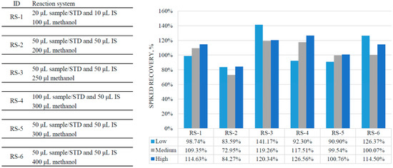
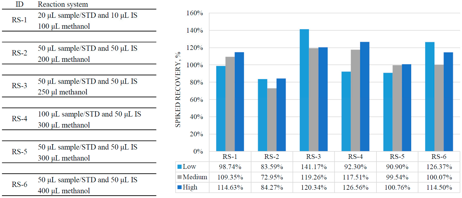
Protein precipitation was the main preparation step. The type of precipitation reagent and volume of the reaction system were optimized. Acetonitrile, methanol, and isopropanol were chosen for initial optimization while acetonitrile and isopropanol were excluded because their relative spiked recovery rates were less than 80%. Five reaction systems (RS) were initially explored for a good recovery rate and high signals (the large precipitation reagent volume can reduce the signals because of the dilution effects). A relatively good recovery was obtained in RS-5 (see Figure 1).
Figure 1.
Optimization results of reaction systems in sample preparation.
3.3. Method Validation
3.3.1. Chromatographic Separation
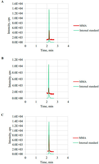
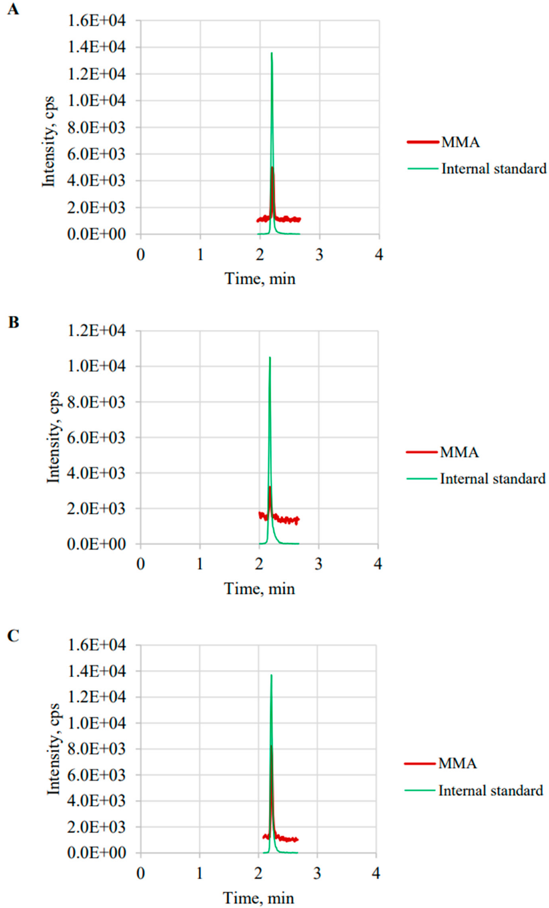
The total run time of the LC-MS/MS analysis was 4.0 min per sample. The intrinsic isomer, i.e., succinic acid (SA), can be completely separated from MMA by chromatography. SA had a relatively higher signal than MMA. MMA can be significantly distinguished from SA. The retention time of MMA and SA were 2.21 min and 1.80 min, respectively. Figure 2 presents representative chromatographs of MMA and MMA-13C4 in standard solutions (A), serum from a healthy control serum (B), and serum from a patient with VB12 deficiency (C).
Figure 2.
Representative chromatographs of: (A) calibrators; (B) healthy controls; (C) patients.
3.3.2. Linearity, LOD, and LOQ
The average slope, intercept, and correlation relationship (R) with their 95% confidence interval (CI) obtained from 12 inconsecutive calibration curves used for analysis during two months were 0.992 (0.926 to 1.059), −0.004 (−0.012 to 0.004), and 0.997 (0.995 to 0.999), respectively. The LOD was estimated as <7.05 ng/g (0.058 μmol/L), and the CV for 20 consecutive injections (S/N > 3) was 5.24%. The LOQ was estimated as <10.41 ng/g (0.085 μmol/L), and the CV for 20 consecutive injections (S/N > 10) was 4.14%.
3.3.3. Precision and Recovery
Table 2 summarizes the precision and spiked recovery of the LC-MS/MS method at four levels of MMA on five consecutive days. The interrun CV at Level 1 (native pool), Level 2, Level 3, and Level 4 ranged from 3.09% to 5.27%; intrarun CV ranged from 1.42% to 2.69%; and the total CV ranged from 3.22% to 5.47%. All imprecision performace met the allowable precision criterion. The recoveries of MMA at the three levels were 101.51%, 92.40%, and 105.95%, respectively.
Table 2.
Spiked recovery and precision performance of the LC-MS/MS method for methylmalonic acid quantification.
3.3.4. Matrix Effect
The absolute matrix effects for Level 1 serum (native, ~0.3 μmol/L), Level 2 serum (spiked, ~0.5 μmol/L), and Level 3 serum (spiked, ~1.0 μmol/L) were −4.74%, −32.95%, and −84.59%, respectively, indicating that ion suppression effects existed in spiked serum without IS correction. IS-corrected matrix effects for Levels 1, 2 and to 3 serum were −0.19%, 6.84%, and 4.06%, respectively, suggesting IS correction could adequately compensate for the observed matrix effect.
3.4. Clinical Application
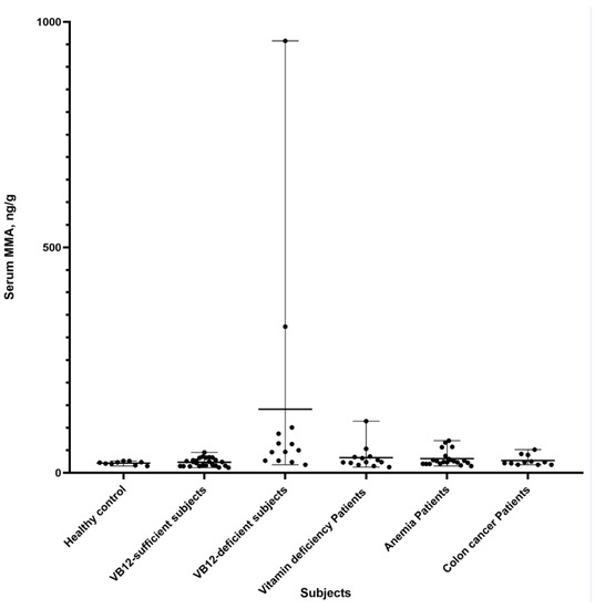
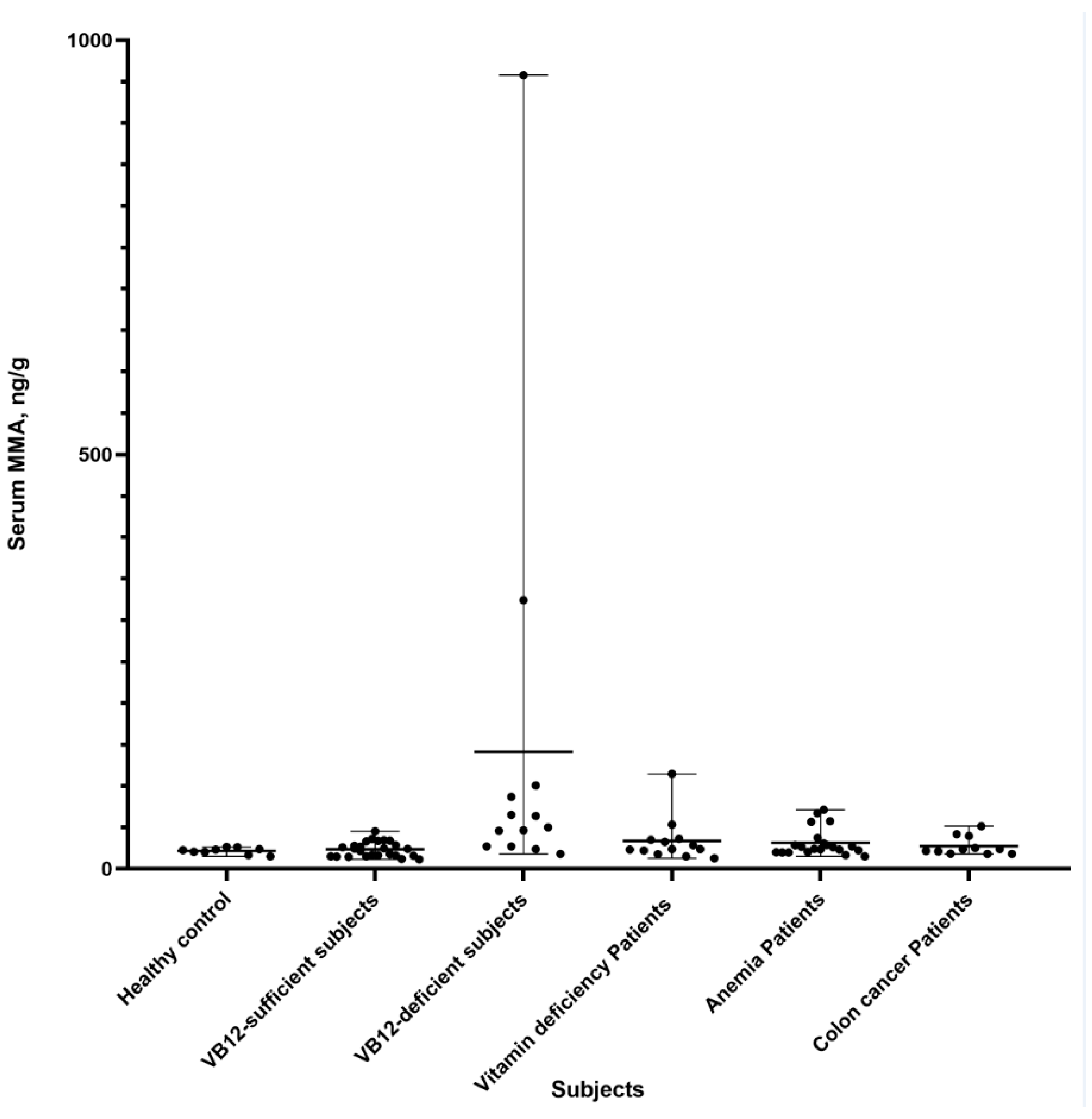
Figure 3 shows the MMA levels in different subjects. VB12-deficient subjects (VB12 < 240 pg/mL) had significantly higher MMA levels than VB12-sufficient subjects (VB12 > 240 pg/mL) (p < 0.05). Additionally, elevated MMA levels were observed in some patients diagnosed with anemia (4/25) and vitamin deficiency (2/13). The MMA levels in these populations were investigated using the established LC-MS/MS method.
Figure 3.
Methylmalonic acid levels in clinical subjects.
4. Discussion
In this study, we established an LC-MS/MS method for MMA quantification that had clear advantages over the previously established LC-MS/MS methods. It is trace, simple, fast, cheap, sensitive, accurate, robust, economic, environmentally friendly, and cost-effective for MMA measurement and is more suitable for laboratories that have large request volumes of MMA testing. These improvements were achieved using protein precipitation combined with a simple mobile phase strategy. Good chromatographic separation of MMA and its isomer was achieved by using an isocratic elution strategy, i.e., 100% A phase (deionzed water containing 0.1% formic acid and 8% isopropanol). Materials and reagents, which are complex or not always accessible, and procedures in previous methods, such as derivatization, multistep SPE, incubation, evaporation, drying, or reconstitution, were not required in this MMA quantification method. We believe this LC-MS/MS method for serum MMA is suitable and convenient for the evaluation of MMA status for VB12 deficiency and can be a reference for laboratories intending to improve their established methods as well as laboratories that plan to introduce MMA testing programs.
The analytical performances of the previous LC-MS/MS methods have been studied [14,21,22,23,24,25,26,27,28,29,30,31,32,33,34,35,36,37,38]. The LOD and LOQ of previous methods have ranged from 0.03–1.95 μmol/L and 0.03–4.20 μmol/L, respectively. The LOD and LOQ of our simplified method were 0.058 μmol/L and 0.085 μmol/L, respectively. The mean recoveries of previous methods varied from 90% to 111%, whereas the mean recoveries of our method ranged from 92.40% to 105.95%. The intra-assay, inter-assay, and total imprecisions of previous methods were 1.3–8.0%, 3.8–8.5%, and 4.6–10.7%, respectively, while the imprecision indicators of our method were 1.42% to 2.69%, 3.09% to 5.27%, and 3.22% to 5.47%, respectively. The LC-MS/MS run time of previous methods varied from 1.0 min to 10 min per sample and the time for sample preparation for a sample ranged from 4 h to 7.5 h, whereas the throughput of our method was 4.0 min per sample with around 20 min for sample preparation.
MMA can be a specific diagnostic biomarker of VB12 deficiency and some inborn errors of metabolism (IEM) [1,2]. It has been reported that moderately elevated MMA (over 0.4 μmol/L in serum, ~46 ng/g) was an early indicator of acquired vitamin B12 insufficiency, and a massive elevation of MMA (over 40 μmol/L in serum, ~4613 ng/g) could strongly indicate IMDs, e.g., methylmalonic acidemia (an IMD with a relatively high prevalence) [22]. We compared MMA levels in VB12-sufficient patients and VB12-deficient patients and observed higher MMA levels in VB12-deficient patients. Additionally, we observed that some patients diagnosed with colon cancer, anemia, diabetes, and coronary disease had elevated MMA.
One of the limitations of this study and previous studies was the lack of measurements of reference materials which have ceritificed values and uncertainty for truness. Current MMA measurements lack qualified certified reference measurement procedures and reference materials. The standard materials for calibrators and IS that we employed are certified reference materials with certified purities and uncertainties, thus, obtaining metrological traceability to the SI (System International) unit. [34,35]
5. Conclusions
In this study, we established a trace, simple, fast, cheap, sensitive, accurate, robust, economic, environmentally friendly, and cost-effective LC-MS/MS method for serum MMA quantification. This LC-MS/MS method can act as an easy assay and provide fast report results to evaluate the MMA status for vitamin B12 deficiency patients, and is especially applicable for large-scale MMA testing.
Supplementary Materials
The following supporting information can be downloaded at: https://www.mdpi.com/article/10.3390/diagnostics12102273/s1. Figure S1: Chromatographic separation by isocratic elution with 95% A phase (0.1% formic acid water solution) and 5% B phase (methanol). This procedure needs drying and residue reconstitution by 5% methanol.
Author Contributions
Conceptualization, L.J.; Data curation, L.J., W.Z., J.Z., M.W., Y.Z., T.Z., F.H. and C.Z.; Formal analysis, L.J. and Z.L.; Funding acquisition, F.H. and C.Z.; Methodology, Z.L.; Project administration, F.H. and C.Z.; Validation, L.J. and Z.L.; Visualization, L.J. and Z.L.; Resources, M.W., Y.Z., F.H. and C.Z. Writing—original draft, L.J.; Writing—review & editing, L.J. and Z.L.; All authors have accepted responsibility for the entire content of this manuscript and approved its submission. All authors have read and agreed to the published version of the manuscript.
Funding
The National Key Research and Development Program of China (2021YFC1005304 and 2018YFC1002204).
Institutional Review Board Statement
The study was conducted in accordance with the Declaration of Helsinki, and approved by the Ethics Committee of Beijing Hospital (protocol code: 2018YFC1002204, date of approval: 18 July 2022).
Informed Consent Statement
This study was approved for the exemption from informed consent.
Acknowledgments
We are grateful for valuable comments from the reviewers and the kind help from Xueyan Han (Department of Medical Statistics, Peking University First Hospital, Beijing, China) in language editing.
Conflicts of Interest
The authors state no conflict of interest.
References
- Cox, E.V.; White, A.M. Methylmalonic acid excretion: An index of vitamin-B12 deficiency. Lancet 1962, 2, 853–856. [Google Scholar] [CrossRef] [Scilit]
- Rosenberg, L.E.; Lilljeqvist, A.; Hsia, Y.E. Methylmalonic aciduria: Metabolic block localization and vitamin B 12 dependency. Science 1968, 162, 805–807. [Google Scholar] [CrossRef] [Scilit] [PubMed]
- Hunt, A.; Harrington, D.; Robinson, S. Vitamin B12 deficiency. BMJ 2014, 349, g5226. [Google Scholar] [CrossRef] [Scilit] [PubMed]
- Stabler, S. Vitamin B12 deficiency. N. Engl. J. Med. 2013, 368, 149–160. [Google Scholar] [CrossRef] [Scilit] [PubMed]
- Haijes, H.A.; Jans, J.J.M.; Tas, S.Y.; Verhoeven-Duif, N.M.; van Hasselt, P.M. Pathophysiology of propionic and methylmalonic acidemias. Part 1: Complications. J. Inherit. Metab. Dis. 2019, 42, 730–744. [Google Scholar] [CrossRef] [Scilit] [PubMed]
- Elin, R.J.; Winter, W.E. Methylmalonic acid: A test whose time has come? Arch. Pathol. Lab. Med. 2001, 125, 824–827. [Google Scholar] [CrossRef] [Scilit]
- Allen, L. How common is vitamin B-12 deficiency? Am. J. Clin. Nutr. 2009, 89, S693–S696. [Google Scholar] [CrossRef] [Scilit]
- Longo, N.; Sass, J.O.; Jurecka, A.; Vockley, J. Biomarkers for drug development in propionic and methylmalonic acidemias. J. Inherit. Metab. Dis. 2022, 45, 132–143. [Google Scholar] [CrossRef] [Scilit]
- Devalia, V.; Hamilton, M.; Molloy, A. Guidelines for the diagnosis and treatment of cobalaminand folate disorders. Br. J. Haematol. 2014, 166, 496–513. [Google Scholar] [CrossRef] [Scilit]
- Mastropaolo, W.; Wilson, M.A. Effect of light on serum B12 and folate stability. Clin. Chem. 1993, 39, 913. [Google Scholar] [CrossRef] [Scilit]
- Reineks, E.Z.; Hughes, A.; Wang, S. Effect of storage temperature or collection tube type on blood methylmalonic acid concentrations measured by gas chromatography-mass spectrometry. Clin. Chem. Lab. Med. 2010, 48, 1351–1353. [Google Scholar] [CrossRef] [Scilit] [PubMed]
- Monsen, A.L.; Refsum, H.; Markestad, T.; Ueland, P.M. Cobalamin status and its biochemical markers methylmalonic acid and homocysteine in different age groups from 4 days to 19 years. Clin. Chem. 2003, 49, 2067–2075. [Google Scholar] [CrossRef] [Scilit]
- Holleland, G.; Schneede, J.; Ueland, P.M.; Lund, P.K.; Refsum, H.; Sandberg, S. Cobalamin deficiency in general practice. Assessment of the diagnostic utility and cost-benefit analysis of methylmalonic acid determination in relation to current diagnostic strategies. Clin. Chem. 1999, 45, 189–198. [Google Scholar] [CrossRef] [Scilit] [PubMed]
- Blom, H.J.; van Rooij, A.; Hogeveen, M. A simple high-throughput method for the determination of plasma methylmalonic acid by liquid chromatography-tandem mass spectrometry. Clin. Chem. Lab. Med. 2007, 45, 645–650. [Google Scholar] [CrossRef] [Scilit] [PubMed]
- Valente, E.; Scott, J.; Ueland, P.; Cunningham, C.; Casey, M.; Molloy, A. Diagnostic accuracy of holotranscobalamin, methylmalonic acid, serum cobalamin, and other indicators of tissue vitamin B12 status in the elderly. Clin. Chem. 2011, 57, 856–863. [Google Scholar] [CrossRef] [Scilit]
- Sobczynńska-Malefora, A.; Gorska, R.; Pelisser, M.; Ruwona, P.; Witchlow, B.; Harrington, D. An audit of holotranscobalamin (“Active” B12) and methylmalonic acid assays for the assessment of vitamin B12 status: Application in a mixed patient population. Clin. Biochem. 2014, 47, 82–86. [Google Scholar] [CrossRef] [Scilit]
- Banerjee, S. Empowering Clinical Diagnostics with Mass Spectrometry. ACS Omega 2020, 5, 2041–2048. [Google Scholar] [CrossRef] [Scilit]
- McCann, M.T.; Thompson, M.M.; Gueron, I.C.; Lemieux, B.; Giguère, R.; Tuchman, M. Methylmalonic acid quantification by stable isotope dilution gas chromatography-mass spectrometry from filter paper urine samples. Clin. Chem. 1996, 42 Pt 1, 910–914. [Google Scholar] [CrossRef] [Scilit]
- Inoue, Y.; Ohse, M. Prenatal diagnosis of methylmalonic aciduria by measuring methylmalonic acid in dried amniotic fluid on filter paper using gas chromatography-mass spectrometry. Anal. Bioanal. Chem. 2011, 400, 1953–1958. [Google Scholar] [CrossRef] [Scilit]
- Stabler, S.P.; Marcell, P.D.; Podell, E.R.; Allen, R.H.; Lindenbaum, J. Assay of methylmalonic acid in the serum of patients with cobalamin deficiency using capillary gas chromatography-mass spectrometry. J. Clin. Investig. 1986, 77, 1606–1612. [Google Scholar] [CrossRef] [Scilit]
- Magera, M.J.; Helgeson, J.K.; Matern, D.; Rinaldo, P. Methylmalonic acid measured in plasma and urine by stable-isotope dilution and electrospray tandem mass spectrometry. Clin. Chem. 2000, 46, 1804–1810. [Google Scholar] [CrossRef] [Scilit] [PubMed]
- Kushnir, M.M.; Komaromy-Hiller, G.; Shushan, B.; Urry, F.M.; Roberts, W.L. Analysis of dicarboxylic acids by tandem mass spectrometry. High-throughput quantitative measurement of methylmalonic acid in serum, plasma, and urine. Clin. Chem. 2001, 47, 1993–2002. [Google Scholar] [CrossRef] [Scilit]
- Schmedes, A.; Brandslund, I. Analysis of methylmalonic acid in plasma by liquid chromatography-tandem mass spectrometry. Clin. Chem. 2006, 52, 754–757. [Google Scholar] [CrossRef] [Scilit] [PubMed]
- la Marca, G.; Malvagia, S.; Pasquini, E.; Innocenti, M.; Donati, M.A.; Zammarchi, E. Rapid 2nd-tier test for measurement of 3-OH-propionic and methylmalonic acids on dried blood spots: Reducing the false-positive rate for propionylcarnitine during expanded newborn screening by liquid chromatography-tandem mass spectrometry. Clin. Chem. 2007, 53, 1364–1369. [Google Scholar] [CrossRef] [Scilit] [PubMed]
- Lakso, H.A.; Appelblad, P.; Schneede, J. Quantification of methylmalonic acid in human plasma with hydrophilic interaction liquid chromatography separation and mass spectrometric detection. Clin. Chem. 2008, 54, 2028–2035. [Google Scholar] [CrossRef] [Scilit] [PubMed]
- Carvalho, V.M.; Kok, F. Determination of serum methylmalonic acid by alkylative extraction and liquid chromatography coupled to tandem mass spectrometry. Anal. Biochem. 2008, 381, 67–73. [Google Scholar] [CrossRef] [Scilit]
- Fasching, C.; Singh, J. Quantitation of methylmalonic acid in plasma using liquid chromatography-tandem mass spectrometry. Methods Mol. Biol. 2010, 603, 371–378. [Google Scholar]
- Pedersen, T.L.; Keyes, W.R.; Shahab-Ferdows, S.; Allen, L.H.; Newman, J.W. Methylmalonic acid quantification in low serum volumes by UPLC-MS/MS. J. Chromatogr. B Analyt. Technol. Biomed. Life Sci. 2011, 879, 1502–1506. [Google Scholar] [CrossRef] [Scilit]
- Yuan, C.; Gabler, J.; El-Khoury, J.M.; Spatholt, R.; Wang, S. Highly sensitive and selective measurement of underivatized methylmalonic acid in serum and plasma by liquid chromatography-tandem mass spectrometry. Anal. Bioanal. Chem. 2012, 404, 133–140. [Google Scholar] [CrossRef] [Scilit]
- Fu, X.; Xu, Y.K.; Chan, P.; Pattengale, P.K. Simple, Fast, and Simultaneous Detection of Plasma Total Homocysteine, Methylmalonic Acid, Methionine, and 2-Methylcitric Acid Using Liquid Chromatography and Mass Spectrometry (LC/MS/MS). JIMD Rep. 2013, 10, 69–78. [Google Scholar]
- Hempen, C.; Wanschers, H.; van der Sluijs Veer, G. A fast liquid chromatographic tandem mass spectrometric method for the simultaneous determination of total homocysteine and methylmalonic acid. Anal. Bioanal. Chem. 2008, 391, 263–270. [Google Scholar] [CrossRef] [Scilit] [PubMed]
- Kushnir, M.M.; Nelson, G.J.; Frank, E.L.; Rockwood, A.L. High-Throughput Analysis of Methylmalonic Acid in Serum, Plasma, and Urine by LC-MS/MS. Method for Analyzing Isomers without Chromatographic Separation. Methods Mol. Biol. 2016, 1378, 159–173. [Google Scholar]
- Ambati, C.S.; Yuan, F.; Abu-Elheiga, L.A.; Zhang, Y.; Shetty, V. Identification and Quantitation of Malonic Acid Biomarkers of In-Born Error Metabolism by Targeted Metabolomics. J. Am. Soc. Mass Spectrom 2017, 28, 929–938. [Google Scholar] [CrossRef] [Scilit] [PubMed]
- Ma, X.; Zou, Y.; Tang, Y.; Wang, D.; Zhou, W.; Yu, S.; Qiu, L. High-throughput analysis of total homocysteine and methylmalonic acid with the efficiency to separate succinic acid in serum and urine via liquid chromatography tandem mass spectrometry. J. Chromatogr. B Analyt. Technol. Biomed. Life Sci. 2022, 1193, 123135. [Google Scholar] [CrossRef] [Scilit] [PubMed]
- Ueyanagi, Y.; Setoyama, D.; Kawakami, D.; Mushimoto, Y.; Matsumoto, S.; Hotta, T.; Kang, D. Fully Automated Quantitative Measurement of Serum Organic Acids via LC-MS/MS for the Diagnosis of Organic Acidemias: Establishment of an Automation System and a Proof-of-Concept Validation. Diagnostics 2021, 11, 2195. [Google Scholar] [CrossRef] [Scilit]
- Boutin, M.; Presse, N.; Martineau, T.; Perreault, A.; Gaudreau, P.; Auray-Blais, C. Mass spectrometry analysis of urinary methylmalonic acid to screen for metabolic vitamin B12 deficiency in older adults. Bioanalysis 2020, 12, 693–705. [Google Scholar]
- Boutin, M.; Presse, N.; Allard, D.; Martineau, T.; Gaudreau, P.; Auray-Blais, C. Methylmalonic acid analysis using urine filter paper samples to screen for metabolic vitamin B12 deficiency in older adults. Bioanalysis 2022, 14, 615–626. [Google Scholar] [CrossRef] [Scilit] [PubMed]
- Zheng, Y.; Bergström, M. Validation of an automated UPLC-MS/MS method for methylmalonic acid in serum/plasma and its application on clinical samples. Scand. J. Clin. Lab Investig. 2022, 82, 342–347. [Google Scholar] [CrossRef] [Scilit]
- CLSI. C-62A: Liquid Chromatography-Mass Spectrometry Methods, Approved Guideline; Linical and Laboratory Standards Institute: Wayne, PA, USA, 2014. [Google Scholar]
- Yu, S.; Wang, D.; Ma, X.; Zhang, Y.; Sun, D.; Zou, Y.; Yu, J.; Zhang, F.; Yang, G.; Xie, S.; et al. Analytical and Clinical Performance of a Liquid Chromatography-Tandem Mass Spectrometry Method for Measuring Gastrin Subtypes G34 and G17 in Serum. Clin. Chem. 2021, 67, 1220–1229. [Google Scholar] [CrossRef] [Scilit]
- Lindberg, M.; Borgstrøm Hager, H.; Brokner, M. Week-to-week biological variation of methylmalonic acid and homocysteine in healthy women. Scand. J. Clin. Lab Investig. 2019, 79, 247–250. [Google Scholar] [CrossRef] [Scilit]

Publisher’s Note: MDPI stays neutral with regard to jurisdictional claims in published maps and institutional affiliations. |
© 2022 by the authors. Licensee MDPI, Basel, Switzerland. This article is an open access article distributed under the terms and conditions of the Creative Commons Attribution (CC BY) license (https://creativecommons.org/licenses/by/4.0/).