


Abstract
Background: Biallelic loss-of-function variants in LAMC3, encoding laminin gamma-3, cause occipital cortical malformation (OMIM#614115). White matter disease and intracranial neoplasia have not been reported in this spectrum. We report a novel homozygous LAMC3 frameshift variant, expanding its phenotypic and molecular spectrum. Methods: Two adolescent siblings from a consanguineous Turkish family underwent whole-exome sequencing, with segregation confirmed by NGS/IGV and classification per ACMG/AMP criteria. In silico analyses included multiple sequence alignment, AlphaFold modeling of wild-type and mutant proteins, and docking against nidogen-1 (NID1). Results: Both siblings had a novel homozygous LAMC3 variant frameshift variant (NM_006059.4: c.1852_1882del; p.(Pro618Serfs*5)), classified as pathogenic (PVS1, PM2, PP3, PP1) with full cosegregation. Proband II.III, a 17-year-old female, developed postoperative epilepsy after resection of a tectal low-grade glioneuronal tumor harboring a somatic KRAS (NM_004985.3) p.(Gln61Lys) variant (VAF 42.9%), with periventricular white matter gliosis. Proband II.IV, a 15-year-old male, presented with confluent leukoencephalopathy, occipital pachygyria, parietal polymicrogyria, and subcortical band heterotopia, illustrating striking intrafamilial discordance. Conclusions: Docking analysis revealed that the cleavage removes the C-terminal nidogen-binding region, eliminates the predicted wild-type interface (residues 906–1029), and shifts the binding to an unnatural N-terminal surface. This finding is a hypothesis-generating result consistent with loss of function. This study expands the LAMC3 phenotype to include leukoencephalopathy and reports a co-occurring low-grade tectal glioneuronal tumor as a novel, single-case observation, supporting inclusion of LAMC3 in the differential diagnosis of pediatric leukoencephalopathies, particularly with consanguinity.
1. Introduction
Malformations of cortical development (MCDs) are a heterogeneous group of disorders characterized by abnormal cerebral cortex formation, most commonly presenting with epilepsy and neurodevelopmental delay [1,2]. Recessive LAMC3 gene variants cause occipital cortical malformations (OMIM #614115), a rare autosomal recessive condition first described by [3] in consanguineous Turkish families presenting with complex bilateral occipital gyration abnormalities. LAMC3 encodes the laminin gamma-3 chain, a critical extracellular matrix protein involved in cell differentiation, migration, and adhesion, playing an essential role in cortical organization and gyration [1,3].
LAMC3-related disease is exceptionally rare, with fewer than fifteen unrelated families reported in the literature to date. The emerging literature has progressively expanded the phenotypic spectrum beyond its originally defined occipital boundaries, encompassing frontal polymicrogyria, periventricular nodular heterotopia, temporal cobblestone malformation, and seizures with entirely normal MRI. However, white matter disease as a primary neuroradiological feature and the co-occurrence of intracranial neoplasia have not been previously described in association with LAMC3 variants [4,5,6].
In the present study, we report two adolescent siblings from a consanguineous Turkish family harboring a novel homozygous LAMC3 frameshift variant (c.1852_1882del; p.(Pro618Serfs*5)) identified by whole-exome sequencing. Both siblings exhibited features not previously associated with LAMC3-related disease, namely confluent leukoencephalopathy as the primary neuroradiological finding in one sibling and a low-grade tectal glioneuronal tumor in the other. We aimed to define their clinical, neuroradiological, and molecular characteristics, to establish the pathogenicity of the variant through familial segregation and ACMG/AMP classification, and to characterize its structural consequences in silico by conservation analysis, AlphaFold modeling of the wild-type and mutant proteins, and docking against nidogen-1 (NID1). In doing so, we expand the phenotypic and molecular spectrum of LAMC3-related disease and highlight LAMC3 in the differential diagnosis of pediatric leukoencephalopathies of undetermined etiology.
2. Methods
2.1. Genetic Analysis
Written informed consent was obtained from the patients, their parents, and all available family members for clinical evaluation, genetic testing, and publication. The study was approved by the Başakşehir Çam and Sakura City Hospital Non-Interventional Clinical Research Ethics Committee (Approval No: KAEK/11.03.2026.89; Date: 11 March 2026) and conducted in accordance with the principles of the Declaration of Helsinki. Detailed clinical histories and neurological assessments were completed for all available family members.
Genomic DNA was extracted from peripheral blood leukocytes of the affected siblings and available family members using the QIAGEN DNeasy Blood DNA Extraction Kit (Qiagen, Hilden, Germany). Whole-exome sequencing was performed on the index patients; exonic and flanking splice-site regions were enriched with the QIAseq xHYB Actionable Exome (Qiagen, Hilden, Germany) and sequenced on the Element AVITI System (Element Biosciences, San Diego, CA, USA) with 100 bp paired-end reads at a mean on-target depth of approximately 100×. Reads were quality-trimmed, aligned to the human reference genome (GRCh37/hg19) using BWA-MEM, and processed for duplicate marking and variant calling with a GATK-based pipeline. Variants were annotated with ANNOVAR/VEP (v116) and filtered against population databases (gnomAD, 1000 Genomes), excluding those with a minor allele frequency above 0.5%. Given the parental consanguinity and the autosomal recessive inheritance of LAMC3-related disease, rare homozygous protein-altering variants were prioritized. The candidate variant in LAMC3 (NM_006059.4) was evaluated using in silico predictors within dbNSFP and classified according to the ACMG/AMP guidelines [7]. Segregation of the c.1852_1882del variant was assessed in all available family members by next-generation sequencing, and read alignments were reviewed in the Integrative Genomics Viewer (IGV) to confirm zygosity in each individual. A structured literature search was performed in PubMed and Google Scholar to identify all previously reported LAMC3-related cases, using the search terms “LAMC3” combined with “cortical malformation,” “polymicrogyria,” “pachygyria,” or “epilepsy,” restricted to publications in English through June 2026. Case reports, case series, and original research articles reporting individual-level phenotypic and genotypic data were included; review articles without new patient-level data, conference abstracts without full-text availability, and studies lacking molecular confirmation of LAMC3 variants were excluded. When multiple publications described overlapping cohorts or the same family, the most complete and recent report was used as the primary source, cross-referenced against earlier reports to avoid case duplication in Table 1.
Table 1.
Summary of all reported LAMC3-associated cases including present cases.
2.2. Bioinformatic Analysis
The canonical protein sequences of human laminin subunit gamma-3 (LAMC3; UniProt Q9Y6N6; 1575 aa) and nidogen-1 (NID1; UniProt P14543; 1247 aa) were retrieved from the UniProt database. The three-dimensional structures of NID1, the wild-type LAMC3 protein, and the mutant protein resulting from the c.1852_1882del p.(Pro618Serfs*5) variant were predicted using the AlphaFold Server (https://alphafoldserver.com, accessed on 19 June 2026). The mutant sequence was generated in silico by introducing the deletion at the coding level and translating the resulting open reading frame, yielding a truncated protein of 621 residues in which the native sequence diverges at codon 618 and terminates after four aberrant residues. Models were ranked by their predicted local distance difference test (pLDDT) and predicted TM-score (pTM) confidence metrics, and the top-ranked model was retained for downstream analysis.
To evaluate the effect of the truncation on partner binding, protein-to-protein docking was performed on the HDOCK server [13]. NID1 was selected as the binding partner based on the well-characterized laminin-to-nidogen interaction that anchors laminin networks within the basement membrane. The wild-type and mutant LAMC3 models were each docked as the receptor against the NID1 structure as the ligand. For each run, the resulting models were ranked by the HDOCK docking score and confidence score, and the top-ranked complex was retained. Interface residues of the receptor and ligand, together with their inter-residue contacts, were extracted from the HDOCK output using a 5 Å distance cutoff.
All predicted structures and docking complexes were visualized in PyMOL (v3.1.1). The wild-type and mutant models were superposed to illustrate the structural consequences of the truncation, and the interface residues at the LAMC3 to NID1 binding surface were mapped and rendered for both complexes.
2.3. Brain Tumor Molecular Analysis
Tumor molecular profiling was performed on formalin-fixed paraffin-embedded tissue from the surgically resected tectal lesion of Proband II.III. RNA and DNA were isolated using the Maxwell RSC RNA FFPE Kit and Maxwell RSC FFPE Plus DNA Kit (Promega, Madison, WI, USA), respectively. Targeted next-generation sequencing was performed using the Acibadem University Brain Tumor Research Group FusionPlex Brain Panel V1 (RNA) and VariantPlex Brain Panel V1 (DNA) on the NextSeq 500 Sequencing System (Illumina, San Diego, CA, USA). MGMT promoter methylation status was assessed by real-time PCR analysis.
2.4. AI Assistance Disclosure
During preparation of this manuscript, the authors used Claude Opus 4.8 (Anthropic) for language editing and grammar checking of the draft text; the tool was not used for data generation, analysis, or interpretation. All AI-assisted output was reviewed and verified by the authors, who take full responsibility for the content.
3. Results
A comprehensive review of the literature identified fewer than fifteen unrelated families with LAMC3-related disease reported to date, summarized together with the present cases in Table 1. Reported variants are predominantly protein-truncating (nonsense, frameshift, and canonical splice-site), consistent with a uniform loss-of-function mechanism, with rare missense changes occurring only in the compound heterozygous state. The associated phenotype is dominated by posterior cortical malformation, most commonly occipital pachygyria and polymicrogyria, accompanied by epilepsy of variable severity and largely preserved to mildly impaired cognition. White matter signal abnormalities have been described in only a minority of previously reported individuals, and consistently as a secondary or incidental feature, whereas intracranial neoplasia has never been reported. Against this background, the two present siblings, both homozygous for the novel c.1852_1882del p.(Pro618Serfs*5) variant, are distinguished as the first cases in which white matter disease constitutes the primary neuroimaging phenotype and the first to harbor an associated intracranial tumor (Table 1). For both siblings, whole-exome sequencing data were additionally reviewed for pathogenic or likely pathogenic variants in genes associated with established leukodystrophies and metabolic white matter disorders (including PLP1, GFAP, EIF2B1-5, ASPA, GALC, ARSA, and mitochondrial and peroxisomal disease genes), none of which were identified, further supporting LAMC3 as the causative gene.
3.1. Clinical Findings
3.1.1. Proband II.III
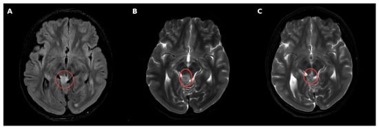
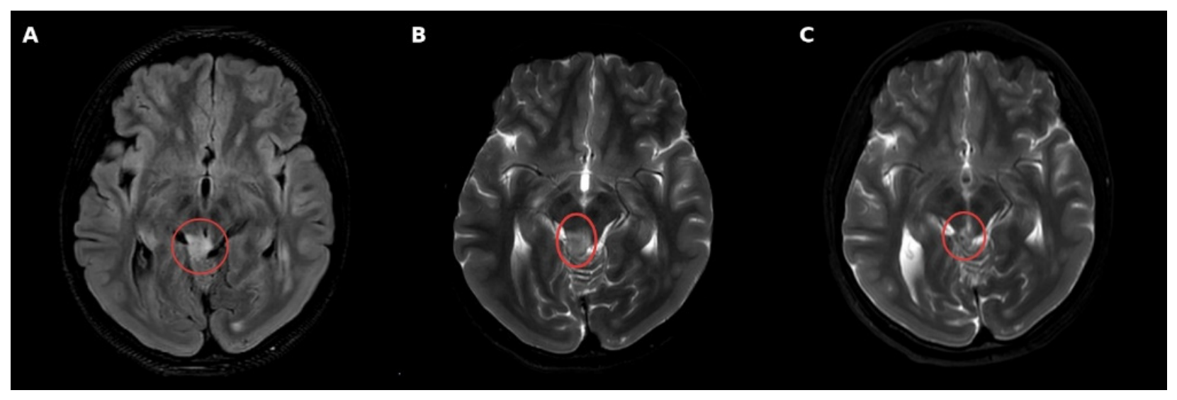
A 17-year-old and 6-month-old female patient, born to second-degree consanguineous Turkish parents, presented with a history of epilepsy. Antenatal and perinatal histories were unremarkable, with no familial history of genetic, metabolic, or neurological diseases. Academic performance was comparable to peers. Following a syncopal episode, investigations revealed a pineal region mass lesion, for which the patient underwent two surgical interventions. Postoperative epileptic seizures were controlled with levetiracetam. Neurological examination revealed no intellectual disability. Visual field examination demonstrated a shift and visual blurring in the right eye, anatomically consistent with the lesion. Electroencephalography revealed 2–2.5 Hz sharp-slow wave paroxysms originating from the left central and bilateral temporal regions, showing secondary generalization lasting 20–30 s, accompanied by generalized fast wave discharges during non-REM sleep. Brain MRI demonstrated a nodular lesion in the right lateral half of the inferior mesencephalic tectum consistent with a presumed tectal glioma, with periventricular white matter gliotic T2/FLAIR hyperintense signal changes (Figure 1). Histopathological examination of the surgical excision material identified a low-grade glioneuronal tumor. Molecular analysis revealed a somatic KRAS (NM_004985.3) c.180_181delinsAA p.(Gln61Lys) pathogenic variant at VAF 42.9%, without pathogenic fusions, copy number variants, or MGMT promoter hypermethylation.
Figure 1.
Brain Magnetic Resonance Imaging (MRI) features of the low-grade tectal glioneuronal tumor. (A) Axial Fluid-Attenuated Inversion Recovery (FLAIR) image demonstrates a well-circumscribed, hyperintense expansile lesion located in the tectal plate, with adjacent periventricular white matter signal change (red circle) (Pre-op). (B) Axial T2-weighted image shows an intrinsically hyperintense tectal mass (red circle) protruding into the quadrigeminal cistern, with narrowing of the cerebral aqueduct and no significant surrounding perilesional vasogenic edema (Pre-op). (C) Postoperative axial T2-weighted follow-up image depicts significant surgical debulking with a small, stable focus of residual tumor/postoperative changes along the tectal margin (red circle), and decompression of adjacent CSF pathways without significant surrounding edema.
3.1.2. Proband II.IV
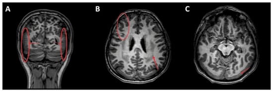
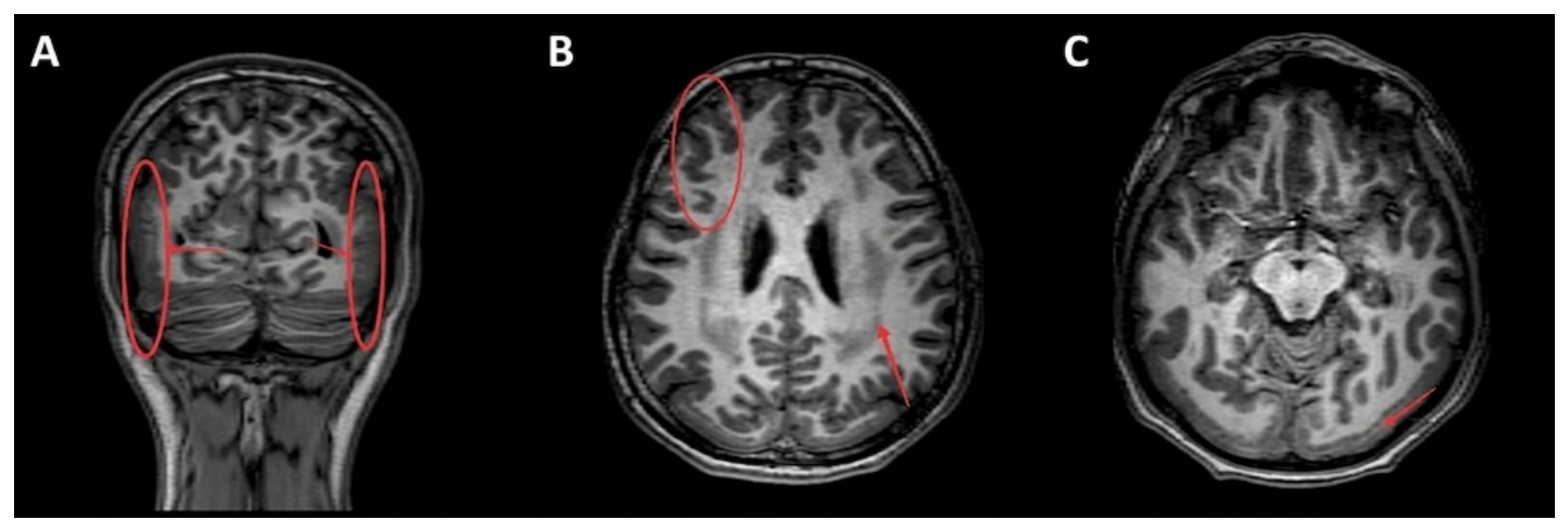
A 15-year-old and 4-month-old male patient, the sibling of Proband II.III, born of the same consanguineous union, presented with a history of epilepsy with onset at age 9 years. Antenatal and perinatal histories were unremarkable. Academic achievement was described as moderate. Seizures were controlled with carbamazepine. Neurological examination revealed mild cognitive deficit; cranial nerve examination, deep tendon reflexes, cerebellar tests, and visual field examination were normal. Electroencephalography demonstrated bilateral synchronous centrotemporal discharges, more prominent on the right side, absent during wakefulness and more pronounced during sleep. Brain MRI demonstrated bilateral periventricular deep white matter symmetric confluent T2/FLAIR hyperintense lesions at the level of the ventricular atria, without diffusion restriction, with relative sparing of U-fibers, basal ganglia, corticospinal tracts, and thalami, consistent with leukoencephalopathy (Figure 2). Additional findings included bilateral occipital pachygyria, parietal polymicrogyria, and subcortical band heterotopia. MR spectroscopy demonstrated mildly elevated Cho/Cr and Cho/NAA ratios consistent with ongoing demyelination and axonal injury. Cerebrospinal fluid analysis revealed an elevated protein level of 174 mg/dL with acellular cytology and normal glucose, chloride, and LDH values; CSF pressure was 18 cmH2O. Lysosomal enzyme panel was within normal limits, reducing the likelihood of a lysosomal storage disorder as the underlying etiology.
Figure 2.
Cranial Magnetic Resonance Imaging (MRI) demonstrating complex malformations of cortical development, subcortical leukodystrophy-like changes, and subcortical U-fiber preservation. (A) Coronal T1-weighted image demonstrates bilateral symmetrical subcortical band heterotopia (red ovals) beneath the thickened cortex, accompanied by confluent subcortical white matter signal alterations mimicking a leukodystrophic pattern. (B) Axial T1-weighted image at the level of the lateral ventricles demonstrates an area of parietal polymicrogyria with irregular, closely packed gyri and shallow sulci overlying subcortical U-fibers that remain well-preserved (red circle), despite extensive adjacent leukodystrophy-like white matter abnormalities and posterior band heterotopia (red arrow). (C) Axial T1-weighted image at the posterior occipitotemporal level highlights marked posterior occipital pachygyria and a contiguous thick layer of heterotopic gray matter beneath the cortex (red arrow), confirming bilateral posterior-predominant band heterotopia (double cortex syndrome).
3.1.3. Comparative Clinical Summary
Given the marked intrafamilial phenotypic discordance despite an identical homozygous genotype, the demographic, clinical, radiological, and molecular findings of the two siblings are summarized comparatively in Table 2. This side-by-side comparison highlights that, beyond their shared LAMC3 genotype, the probands diverged across nearly every clinical domain, including the presence versus absence of intracranial neoplasia, the pattern and severity of white matter and cortical involvement, seizure semiology and pharmacological response, and cognitive outcome.
Table 2.
Comparative clinical summary of the two LAMC3 probands.
3.2. Genetic Findings
Whole-exome sequencing of the affected siblings identified a novel homozygous frameshift variant in LAMC3 (NM_006059.4), c.1852_1882del; p.(Pro618Serfs*5), located in exon 13. This 31-base-pair deletion shifts the reading frame at codon 618, replacing proline with serine and introducing a premature termination codon after four aberrant residues. The variant is predicted to trigger nonsense-mediated mRNA decay or, alternatively, to yield a severely truncated protein of 621 residues that lacks approximately 61% of the 1575-residue wild-type chain, including the entire C-terminal region required for laminin network assembly. The variant was absent from population databases (gnomAD, 1000 Genomes) and had not been previously reported in ClinVar or HGMD.
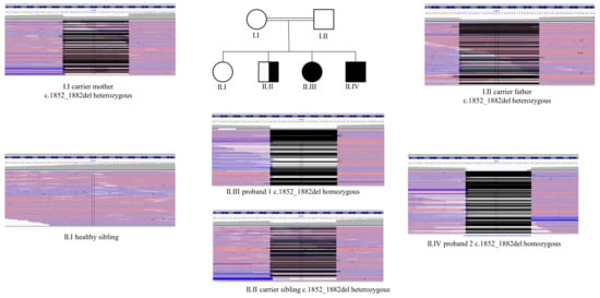
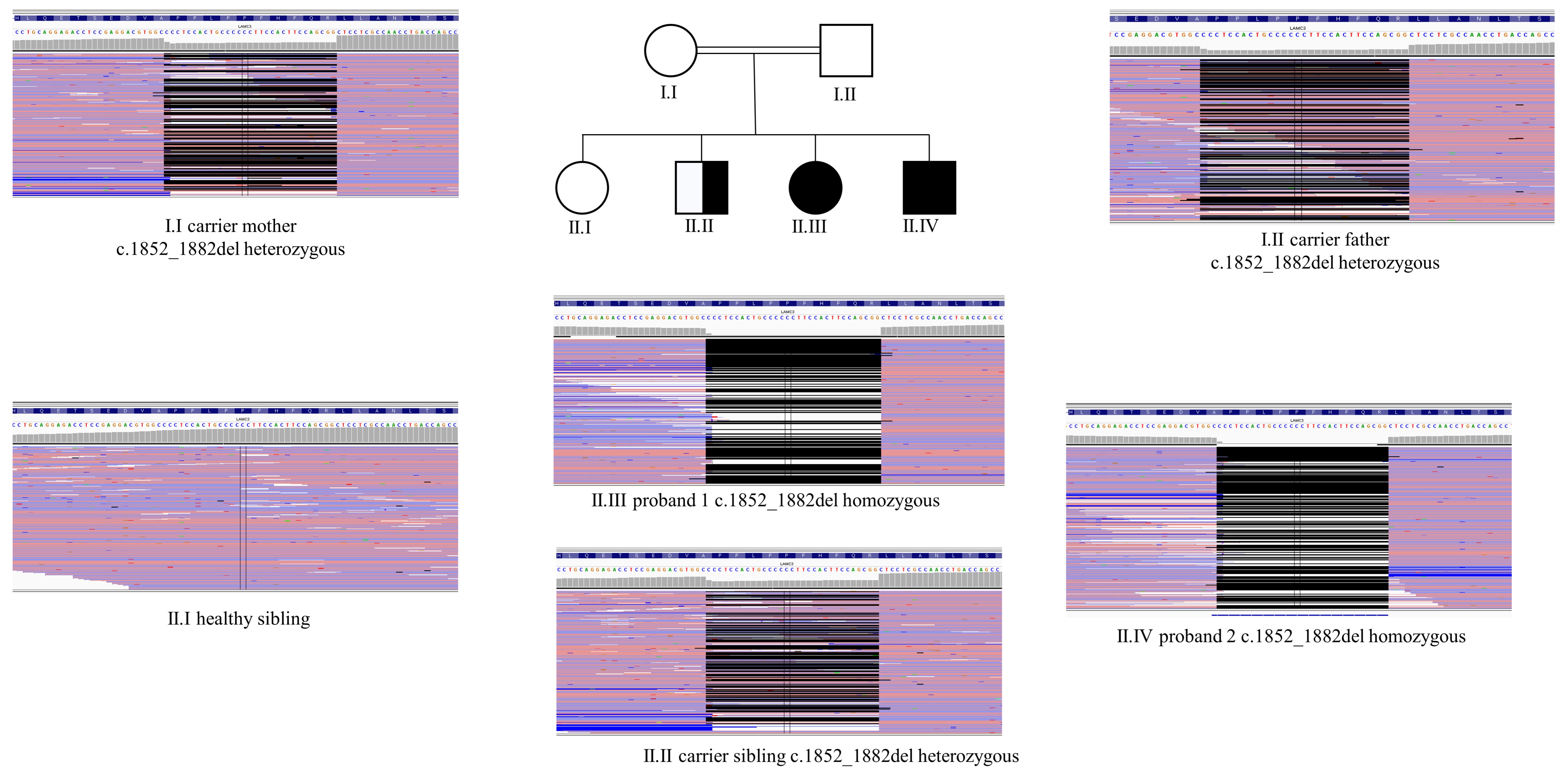
Segregation analysis was performed in all available family members by next-generation sequencing, with read alignments reviewed in the Integrative Genomics Viewer (IGV) to confirm zygosity in each individual Figure 3. The two affected siblings (Proband II.III and II.IV) were homozygous for the deletion, whereas both consanguineous parents were heterozygous carriers. Of the two unaffected siblings, one was a heterozygous carrier, and the other did not carry the variant. The variant therefore cosegregated fully with the disease phenotype in a manner consistent with autosomal recessive inheritance.
Figure 3.
Pedigree and segregation of the LAMC3 c.1852_1882del variant. The central pedigree depicts the consanguineous family; filled symbols denote the two affected siblings and the half-filled symbol denotes an unaffected heterozygous carrier. Surrounding panels show Integrative Genomics Viewer (IGV) read alignments across the variant region for each genotyped individual. Both parents (I.I, carrier mother; I.II, carrier father) and one unaffected sibling (II.II) are heterozygous for the 31 bp deletion; the two affected siblings (Proband II.III, Proband II.IV) are homozygous; and one unaffected sibling (II.I) does not carry the variant. The variant thus fully cosegregates with the disease phenotype in a manner consistent with autosomal recessive inheritance.
On the basis of these findings, the variant was classified as pathogenic according to the ACMG/AMP guidelines, meeting the criteria PVS1 (a null variant in a gene in which loss of function is an established disease mechanism, with the premature termination codon predicted to undergo nonsense-mediated decay), PM2 (absence from population databases), PP3 (computational prediction tools unanimously support a deleterious effect on the gene) and PP1 (cosegregation with disease in multiple affected family members).
3.3. Bioinformatic Findings
Multiple sequence alignment has revealed that the residues in and around Pro618 are highly conserved among vertebrate orthologs; this indicates that the affected region lies within a functionally restricted segment of the protein. The variant is predicted to affect the expression of the laminin/attractin EGF and laminin EGF domain regions located between residues 707–748 and 673–1010.
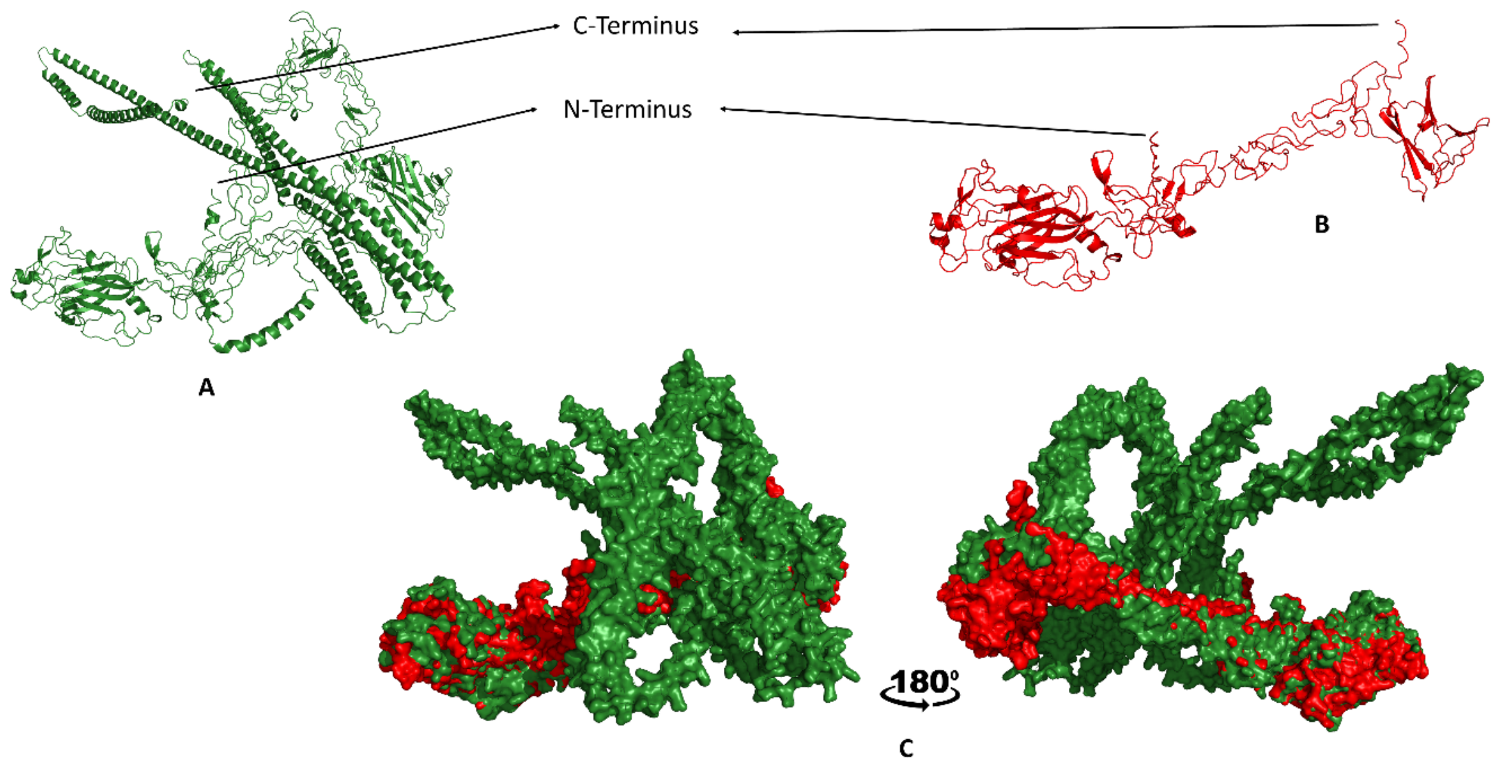
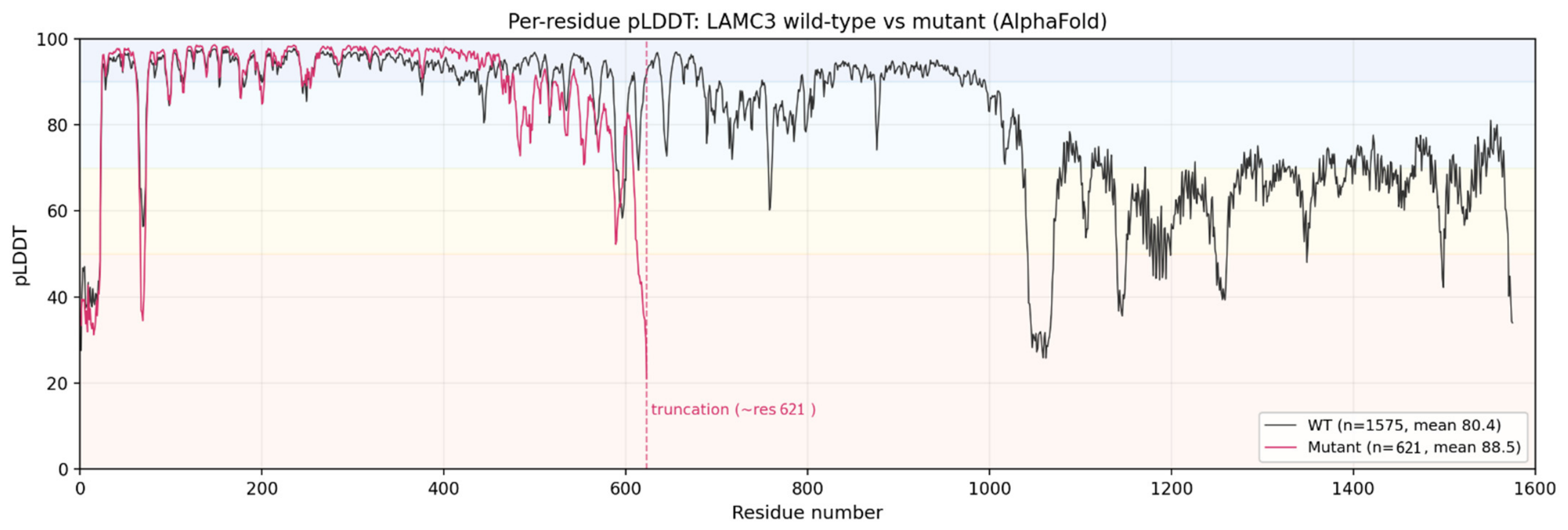
AlphaFold modeling of the wild-type LAMC3 (pTM = 0.38) chain reproduced the expected elongated, multidomain laminin architecture, comprising a compact N-terminal globular region followed by an extended C-terminal arm (Figure 4A). The relatively low pTM score of the wild-type model (0.38) reflects reduced confidence in the relative inter-domain arrangement of this large, multidomain protein, and constitutes an additional limitation when interpreting the docking results derived from this model. In contrast, the model of the p.(Pro618Serfs*5) mutant (pTM = 0.73) was reduced to a compact N-terminal fragment of 621 residues that entirely lacked the C-terminal half of the chain, including the extended arm and the domains required for heterotrimer assembly and laminin network formation (Figure 4B). Superposition of the two models confirmed that the truncation removes approximately 61% of the native chain while leaving the retained N-terminal region essentially unchanged (Figure 4C). AlphaFold modeling of the wild-type LAMC3 protein yielded a full-length structure of 1575 residues with a mean per-residue pLDDT of 80.4, indicating overall reliable folding. The p.(Pro618Serfs*5) variant produced a severely truncated model of only 621 residues, consistent with premature termination shortly downstream of codon 618 and the loss of 954 C-terminal residues, corresponding to roughly 61% of the protein.
Figure 4.
AlphaFold structural models of wild-type and mutant LAMC3. (A) Cartoon representation of the wild-type LAMC3 chain (green), showing the elongated multidomain laminin architecture, with the N-terminal and C-terminal regions indicated. (B) Cartoon representation of the mutant protein resulting from the c.1852_1882del (p.(Pro618Serfs*5)) variant (red), truncated to a compact 621-residue N-terminal fragment that lacks the entire C-terminal half of the chain. (C) Surface representation of the mutant model (red) superposed on the wild-type model (green), shown in two orientations related by a 180-degree rotation, illustrating that the retained region corresponds to only the N-terminal portion of the native chain.
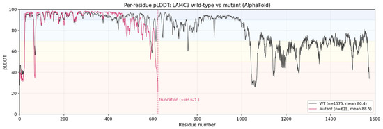
This truncation eliminated the C-terminal region encompassing the laminin coiled-coil domain that mediates assembly of the γ3 chain into the laminin heterotrimer. Across the shared N-terminal segment (residues 1 to 621), both models folded with comparably high confidence (wild-type 90.4, mutant 88.6), confirming that the retained region is modeled reliably and that the structural divergence between the two is driven by truncation rather than local misfolding. The higher global mean pLDDT of the mutant model relative to the full-length wild-type (88.6 versus 80.4) reflects the removal of intrinsically low-confidence C-terminal domains and should not be interpreted as improved structural integrity (Figure 5).
Figure 5.
Per-residue pLDDT profiles of wild-type and mutant LAMC3 from AlphaFold modeling. The wild-type protein (1575 residues) is shown in black and the p.(Pro618Serfs*5) mutant (621 residues) in red; the dashed line marks the truncation point (The black lines show the pLDDT values for the wild-type protein and the red lines show those for the mutant protein).
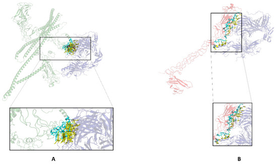
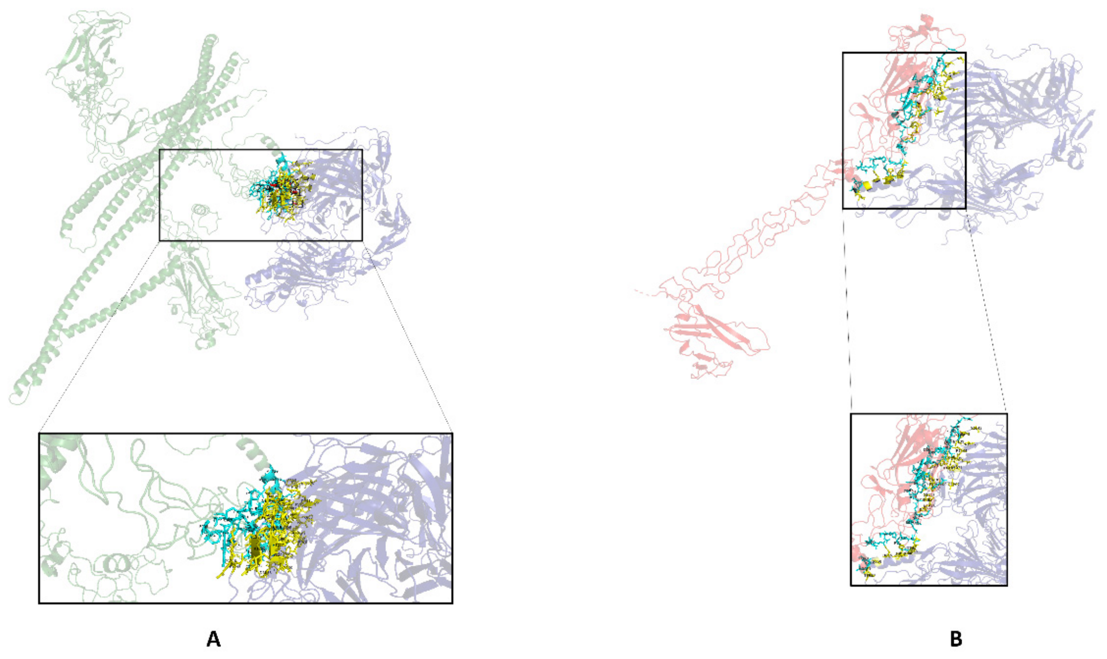
To assess the impact of the truncation on the interaction with nidogen-1, the wild-type and mutant LAMC3 models were each docked against NID1 using HDOCK (Figure 6, Table 3). Both complexes yielded high-confidence top-ranked models. In the wild-type complex, the LAMC3 interface localized to the C-terminal region of the chain (residues 906 to 1029), engaging NID1 through 114 inter-residue contacts. This region falls within the laminin EGF-like (LE) domain segment that, in laminin gamma chains, harbors the canonical nidogen-binding module, consistent with a physiologically relevant interface.
Figure 6.
HDOCK docking of wild-type and mutant LAMC3 with nidogen-1 (NID1). (A) Top-ranked docking model of wild-type LAMC3 (green) with NID1 (blue); the boxed interface is enlarged below, with interface residues shown as sticks (LAMC3 in cyan, NID1 in yellow) and labeled. The interaction localizes to the C-terminal region of LAMC3 (residues 906 to 1029), corresponding to the physiological nidogen-binding region. (B) Top-ranked docking model of the mutant LAMC3 (red) with NID1 (blue); the boxed interface is enlarged. The truncation abolishes the native interface, and the residual interaction relocates to a non-native N-terminal surface of the truncated chain (residues 16 to 348) (The dashed lines indicate the enlarged areas of interaction regions in protein docking. Frames are a zoomed-in view of the interaction area.).
Table 3.
HDOCK docking of wild-type and mutant LAMC3 with nidogen-1 (NID1), top-ranked model.
In the mutant complex, this native interface was necessarily absent, as the truncation terminates the protein at residue 621, well upstream of residues 906 to 1029. Consequently, docking engaged an entirely different, N-terminal surface of the truncated chain (residues 16 to 348), corresponding to the laminin N-terminal (LN) domain region, through 105 contacts. Although the mutant complex returned a nominally more favorable docking score (−338.96 versus −308.37) and confidence score (0.98 versus 0.96), these values reflect the shape and energetic complementarity of the top-ranked pose rather than physiological binding affinity or biological validity. The relocation of the interface from the native C-terminal nidogen-binding region to a non-native N-terminal surface indicates that the truncated protein cannot reconstitute the physiological laminin-to-nidogen interface, and that any residual interaction is aberrant and unlikely to support functional basement membrane assembly. These docking-based inferences are computational and hypothesis-generating; direct experimental confirmation (e.g., co-immunoprecipitation, surface plasmon resonance, or isothermal titration calorimetry) will be required to establish definitive loss of physiological binding.
4. Discussion
The loss-of-function consequence predicted for the c.1852_1882del p.(Pro618Serfs*5) variant is supported by convergent genetic, clinical, and computational evidence, and is further consistent with previously published experimental studies on the laminin-nidogen interaction and LAMC3 ablation in mice. Frameshift variants that introduce a premature termination codon upstream of the final exon–exon junction are efficiently recognized by the nonsense-mediated mRNA decay (NMD) pathway, which degrades the aberrant transcript and precludes synthesis of the truncated protein [14]. Genome-wide bioinformatic modeling of these NMD rules has confirmed that such protein-truncating variants predominantly act through loss of function across human disease genes [15]. The present variant lies in exon 13, well upstream of the final exon junction, and is therefore predicted to elicit NMD. Even in the alternative scenario of NMD escape, the resulting protein would lack the entire C-terminal half of the chain, including the laminin EGF-like (LE) modules that harbor the nidogen-binding site experimentally defined by crystallography of the laminin-to-nidogen complex [16] consistent with this, our wild-type docking localized LAMC3 to the NID1 interface to residues 906 to 1029, a region entirely absent from the truncated mutant. Both outcomes therefore converge on abolition of laminin gamma-3 function, in agreement with experimental ablation of LAMC3 in mice, which disrupts cortical basement membrane integrity and lamination [17], and with the exclusively loss-of-function mutational spectrum reported for LAMC3-related disease.
Premature truncation is a recurrent mechanism in LAMC3-related disease. Barak et al. first established recessive loss-of-function LAMC3 variants as a cause of occipital cortical malformation in consanguineous families ascertained through first-cousin marriage, subsequently identified across three unrelated Turkish families with occipital polymicrogyria and epilepsy [3]. Later reports repeatedly implicated nonsense and frameshift variants that cause premature truncation of the laminin γ3 chain, and the associated phenotype has progressively broadened beyond the occipital lobe, including a novel stop-gain variant (p.Arg1291*) with polymicrogyria extending into temporal regions and biallelic variants with predominantly temporal rather than occipital involvement. The variant reported here terminates the protein considerably earlier than most previously described alleles, removing the entire C-terminal half, including the assembly-critical coiled-coil domain, which provides a plausible structural basis for complete loss of function [4,10,11]. Our analysis also underscores an interpretive caveat for structural studies of truncating variants: AlphaFold confidence scores such as pLDDT and pTM are length-dependent, and a truncated model may return a higher global mean simply because the disordered, low-confidence C-terminal regions of the full-length protein are no longer present. Confidence metrics should therefore be compared over the shared sequence rather than globally, as done here, to avoid mistaking the loss of protein for an improvement in model quality.
LAMC3-associated cortical malformations represent an exceptionally rare autosomal recessive condition caused by biallelic loss-of-function variants in the gene encoding the laminin γ3 chain, a critical component of the extracellular matrix. Since their initial description by [3] in consanguineous Turkish families presenting with complex bilateral occipital gyration abnormalities, fewer than fifteen unrelated families have been reported in the global literature. LAMC3 is located on chromosome 9q34.12 and encodes a 1575-amino acid protein that assembles into heterotrimeric laminin complexes through non-covalent interactions with laminin α and β chains [5,10]. These laminin isoforms are essential components of basement membranes and play indispensable roles in cell adhesion, differentiation, migration, and extracellular matrix organization throughout development [18]. In the developing human fetal brain, LAMC3 expression peaks between late gestation and late infancy, localizing to the cell bodies and apical dendrites of pyramidal neurons in the cortical plate [3].
The phenotypic spectrum of LAMC3-related disease has expanded considerably since its initial description. Barak et al. established bilateral occipital pachygyria and polymicrogyria as the defining phenotype [3]. Zambonin et al. reported generalized polymicrogyria extending to the frontal, parietal, and temporal cortices with posterior deep white matter signal changes [5]. Kasper et al. described frontal-only polymicrogyria with adult-onset epilepsy, demonstrating that occipital involvement is not obligatory [9]. De Angelis et al. reported posterior periventricular nodular heterotopia [6]; Abe et al. described temporal cobblestone malformation with ventral pontine involvement [11]; and Falcicchio et al. reported drug-resistant epilepsy with pontocerebellar atrophy [4]. Most recently, Saarela et al. described a broad phenotypic spectrum ranging from Lennox–Gastaut syndrome to focal epilepsy with preserved cognition [12]. Against this background of progressive phenotypic expansion, the probands add two entirely novel clinical dimensions: intracranial neoplasia and primary leukoencephalopathy.
The first major novel finding of the present study is the low-grade glioneuronal tumor identified in the tectal region of Proband II.III. Histopathological examination identified spindle cell morphology consistent with a low-grade glioneuronal tumor; molecular analysis revealed a somatic KRAS c.180_181delinsAA p.(Gln61Lys) pathogenic variant at VAF 42.9%. Tectal gliomas are known to harbor high rates of activating KRAS mutations [19]; rare KRAS missense variants at codon 61 have been previously described [20,21]. The co-occurrence of LAMC3-associated cortical malformation with intracranial neoplasia has not been previously reported in the literature. It is conceivable that germline loss-of-function of LAMC3 may disrupt the integrity of the extracellular matrix microenvironment surrounding glial progenitors, potentially predisposing to somatic oncogenic events such as activating KRAS [10,22]. This hypothesis is speculative and based on a single observation; a coincidental co-occurrence of a sporadic KRAS driver mutation, unrelated to the germline LAMC3 variant, cannot be excluded. Systematic evaluation of tumor occurrence in additional LAMC3-related disease cohorts will be necessary to determine whether this represents a genuine biological association. Furthermore, the temporal relationship between the patient’s multiple surgical interventions and the white matter signal abnormalities documented on follow-up imaging raises the possibility that a proportion of the white matter signal abnormalities may have been treatment-related. Notably, preoperative FLAIR imaging (Figure 1A) already demonstrated periventricular white matter signal change prior to surgical intervention, indicating that this finding, at least in part, reflects an underlying process related to the LAMC3 variant rather than being purely a postoperative artifact, although a treatment-related contribution to signal evolution on subsequent follow-up imaging cannot be entirely excluded.
The dominant neuroimaging finding in Proband II.IV was confluent leukoencephalopathy with symmetric, merging T2/FLAIR hyperintense lesions bilaterally involving the corona radiata, centrum semiovale, and peritrigonal white matter, with relative sparing of U-fibers, basal ganglia, corticospinal tracts, and thalami, and without diffusion restriction. This pattern initially raised the consideration of leukodystrophic conditions—most notably metachromatic leukodystrophy, given the symmetric posterior predominance, the patient’s age, and the absence of diffusion restriction—underscoring the diagnostic challenge posed by white matter-predominant presentations of LAMC3-related disease. Comprehensive metabolic workup, including lysosomal enzyme panel analysis, returned within normal limits; whole-exome sequencing data were further reviewed for pathogenic variants in nuclear genes associated with mitochondrial and peroxisomal disorders, none of which were identified, reducing the likelihood of these alternative metabolic etiologies. However, functional biochemical testing and evaluation of mitochondrial DNA-encoded genes were not performed in this study and cannot be formally excluded by sequencing alone. Cerebrospinal fluid analysis demonstrated an elevated protein level of 174 mg/dL with acellular cytology and normal glucose, chloride, and LDH values; CSF pressure was 18 cmH2O. While CSF protein elevation is a non-specific finding, it is consistent with an active white matter process and has been described in other genetic leukoencephalopathies. MR spectroscopy demonstrated mildly elevated Cho/Cr and Cho/NAA ratios without tumor-specific alterations, interpreted as consistent with ongoing demyelination and axonal injury, further supporting an active rather than purely static white matter pathology. Taken together, these findings suggest that the leukoencephalopathy in this patient may harbor a progressive or dynamic component, meriting longitudinal neuroimaging and biochemical surveillance. The genetic etiology was ultimately confirmed by identification of the homozygous LAMC3 frameshift variant, highlighting the importance of whole-exome sequencing in the diagnostic workup of pediatric leukoencephalopathies of undetermined etiology, particularly in the setting of consanguinity. Proband II.III demonstrated periventricular white matter gliosis without overt leukoencephalopathy, representing a milder end of the same white matter pathology spectrum. Although white matter signal abnormalities have been reported as incidental findings in prior LAMC3 reports [5,12], the probands are the first in which they constitute the primary neuroimaging phenotype. Demirayak et al. documented marked microstructural disruption across multiple major white matter tracts in a patient with homozygous LAMC3 [23], while Urgen et al. and Roberto et al. demonstrated that structural abnormalities in LAMC3 carriers extend well beyond the occipital cortex [20,24].
From a genetic standpoint, the c.1852_1882del p(.Pro618Serfs*5) variant represents a novel 31-base-pair deletion in exon 13 of LAMC3, predicted to result in loss of function through protein truncation or nonsense-mediated mRNA decay. The variant is absent from population databases (gnomAD, ClinVar, HGMD) and was classified as pathogenic (PVS1, PM2, PP3, PP1) under the ACMG/AMP framework [7]. The marked phenotypic discordance between the two homozygous siblings—Proband II.III presenting with tectal glioma and periventricular white matter gliosis, Proband II.IV with confluent leukoencephalopathy, occipital pachygyria, parietal polymicrogyria, and subcortical band heterotopia—represents a striking example of intrafamilial phenotypic variability in LAMC3-related disease. Similar variability has been previously described by Afawi et al. and Saarela et al. [8,12]. The mechanisms underlying this intrafamilial variability remain incompletely understood but likely involve modifier gene effects, stochastic variation in developmental processes, epigenetic regulation of LAMC3 expression, and the influence of somatic mutational events on the ultimate clinical phenotype.
5. Conclusions
In conclusion, the present study expands the phenotypic spectrum of LAMC3-related disease to include primary leukoencephalopathy, and reports the co-occurrence of a low-grade tectal glioneuronal tumor as a novel, single-case observation whose causal relationship to the LAMC3 variant remains unproven and warrants further study, in two siblings harboring a novel homozygous frameshift variant. LAMC3 should be considered in the differential diagnosis of pediatric leukoencephalopathies of uncertain etiology, particularly in the presence of consanguinity. Accumulation of additional cases through international collaborative registries will be essential to elucidate the relationship between germline LAMC3 loss-of-function and intracranial neoplasia, and to fully delineate the phenotypic spectrum of this rare disorder.
Author Contributions
Conceptualization, S.B. (Serdar Bozlak) and C.Y.; methodology, S.B. (Serdar Bozlak), C.Y., H.I.Y., O.S., P.P., and T.E.E.; software, C.Y., H.I.Y., and O.S.; validation, S.B. (Serdar Bozlak), C.Y., H.I.Y., O.S., P.P., and T.E.E.; formal analysis, S.B. (Serdar Bozlak), C.Y., H.I.Y., O.S., P.P., T.E.E., D.C., and T.D.; investigation, S.B. (Serdar Bozlak), C.Y., H.I.Y., O.S., P.P., T.E.E., and S.B. (Sirin Bozlak); resources, S.B. (Serdar Bozlak), P.P., and S.B. (Sirin Bozlak); data curation, S.B. (Serdar Bozlak), C.Y., H.I.Y., and O.S.; writing—original draft prep-aration, S.B. (Serdar Bozlak), and C.Y.; writing—review and editing, S.B. (Serdar Bozlak), C.Y., H.I.Y., O.S., P.P., D.C., T.D., T.E.E., and S.B. (Sirin Bozlak); visualization, C.Y., O.S., D.C., and T.D.; supervision, S.B. (Serdar Bozlak), and C.Y.; project administration, S.B. (Serdar Bozlak) and C.Y. All authors have read and agreed to the published version of the manuscript.
Funding
This research received no external funding.
Institutional Review Board Statement
The study protocol received ethical approval from the Başakşehir Çam and Sakura City Hospital Non-Interventional Clinical Research Ethics Committee (Reference No: KAEK/11.03.2026.89; Date: 11 March 2026). The research was conducted in strict accordance with the ethical principles set forth in the Declaration of Helsinki by the World Medical Association.
Informed Consent Statement
Informed consent was obtained from all subjects involved in the study.
Data Availability Statement
The original contributions presented in this study are included in the article material. Further inquiries can be directed to the corresponding author.
Acknowledgments
During the preparation of this article/paper, the authors used Claude Opus 4.8 for text editing and grammar checking. The authors have reviewed and edited the output and assume full responsibility for the content of this publication.
Conflicts of Interest
The authors declare no conflicts of interest.
References
- Barkovich, A.J.; Guerrini, R.; Kuzniecky, R.I.; Jackson, G.D.; Dobyns, W.B. A developmental and genetic classification for malformations of cortical development: Update 2012. Brain 2012, 135, 1348–1369. [Google Scholar] [CrossRef] [Scilit]
- Severino, M.; Geraldo, A.F.; Utz, N.; Tortora, D.; Pogledic, I.; Klonowski, W.; Triulzi, F.; Arrigoni, F.; Mankad, K.; Leventer, R.J.; et al. Definitions and classification of malformations of cortical development: Practical guidelines. Brain 2020, 143, 2874–2894. [Google Scholar] [CrossRef] [Scilit]
- Barak, T.; Kwan, K.Y.; Louvi, A.; Demirbilek, V.; Saygı, S.; Tüysüz, B.; Choi, M.; Boyacı, H.; Doerschner, K.; Zhu, Y. Recessive LAMC3 mutations cause malformations of occipital cortical development. Nat. Genet. 2011, 43, 590–594. [Google Scholar] [CrossRef] [Scilit]
- Falcicchio, G.; Riva, A.; La Neve, A.; Iacomino, M.; Lastella, P.; Suppressa, P.; Sciruicchio, V.; Trojano, M.; Striano, P. Case report: LAMC3-associated cortical malformations: Case report of a novel stop-gain variant and literature review. Front. Genet. 2022, 13, 990350. [Google Scholar] [CrossRef] [Scilit]
- Zambonin, J.L.; Dyment, D.A.; Xi, Y.; Lamont, R.E.; Hartley, T.; Miller, E.; Kerr, M.; Care4Rare Canada Consortium; Boycott, K.M.; Parboosingh, J.S.; et al. A novel mutation in LAMC3 associated with generalized polymicrogyria of the cortex and epilepsy. Neurogenetics 2018, 19, 61–65. [Google Scholar] [CrossRef] [Scilit]
- De Angelis, C.; Byrne, A.B.; Morrow, R.; Feng, J.; Ha, T.; Wang, P.; Schreiber, A.W.; Babic, M.; Taranath, A.; Manton, N.; et al. Compound heterozygous variants in LAMC3 in association with posterior periventricular nodular heterotopia. BMC Med. Genom. 2021, 14, 64. [Google Scholar] [CrossRef] [Scilit]
- Richards, S.; Aziz, N.; Bale, S.; Bick, D.; Das, S.; Gastier-Foster, J.; Grody, W.W.; Hegde, M.; Lyon, E.; Spector, E.; et al. Standards and guidelines for the interpretation of sequence variants: A joint consensus recommendation of the American College of Medical Genetics and Genomics and the Association for Molecular Pathology. Genet. Med. 2015, 17, 405–424. [Google Scholar] [CrossRef] [Scilit]
- Afawi, Z.; Oliver, K.L.; Kivity, S.; Mazarib, A.; Blatt, I.; Neufeld, M.Y.; Helbig, K.L.; Goldberg-Stern, H.; Misk, A.J.; Straussberg, R.; et al. Multiplex families with epilepsy: Success of clinical and molecular genetic characterization. Neurology 2016, 86, 713–722. [Google Scholar] [CrossRef] [Scilit]
- Kasper, B.S.; Kraus, C.; Schwarz, M.; Rosch, J.; Thiel, C.T.; Reis, A.; Zweier, C. A novel splice variant expands the LAMC3-associated cortical phenotype to frontal only polymicrogyria and adult-onset epilepsy. Am. J. Med. Genet. A 2020, 182, 2761–2764. [Google Scholar] [CrossRef] [Scilit]
- Qian, X.; Liu, X.; Zhu, Z.; Wang, S.; Song, X.; Chen, G.; Wu, J.; Cao, Y.; Luan, X.; Tang, H.; et al. Variants in LAMC3 Causes Occipital Cortical Malformation. Front. Genet. 2021, 12, 616761. [Google Scholar] [CrossRef] [Scilit]
- Abe, K.; Ando, K.; Kato, M.; Saitsu, H.; Nakashima, M.; Aoki, S.; Kimura, T. A New Case With Cortical Malformation Caused by Biallelic Variants in LAMC3. Neurol. Genet. 2022, 8, e680. [Google Scholar] [CrossRef] [Scilit]
- Saarela, A.; Timonen, O.; Kirjavainen, J.; Liu, Y.; Silvennoinen, K.; Mervaala, E.; Kalviainen, R. Novel LAMC3 pathogenic variant enriched in Finnish population causes malformations of cortical development and severe epilepsy. Epileptic Disord. 2024, 26, 498–509. [Google Scholar] [CrossRef] [Scilit]
- Yan, Y.; Tao, H.; He, J.; Huang, S.Y. The HDOCK server for integrated protein-protein docking. Nat. Protoc. 2020, 15, 1829–1852. [Google Scholar] [CrossRef] [Scilit]
- Kurosaki, T.; Popp, M.W.; Maquat, L.E. Publisher Correction: Quality and quantity control of gene expression by nonsense-mediated mRNA decay. Nat. Rev. Mol. Cell Biol. 2019, 20, 384. [Google Scholar] [CrossRef] [Scilit]
- Lindeboom, R.G.H.; Vermeulen, M.; Lehner, B.; Supek, F. The impact of nonsense-mediated mRNA decay on genetic disease, gene editing and cancer immunotherapy. Nat. Genet. 2019, 51, 1645–1651. [Google Scholar] [CrossRef] [Scilit]
- Takagi, J.; Yang, Y.; Liu, J.H.; Wang, J.H.; Springer, T.A. Complex between nidogen and laminin fragments reveals a paradigmatic beta-propeller interface. Nature 2003, 424, 969–974. [Google Scholar] [CrossRef] [Scilit]
- Radner, S.; Banos, C.; Bachay, G.; Li, Y.N.; Hunter, D.D.; Brunken, W.J.; Yee, K.T. beta2 and gamma3 laminins are critical cortical basement membrane components: Ablation of Lamb2 and Lamc3 genes disrupts cortical lamination and produces dysplasia. Dev. Neurobiol. 2013, 73, 209–229. [Google Scholar] [CrossRef] [Scilit]
- Durbeej, M. Laminins. Cell Tissue Res. 2010, 339, 259–268. [Google Scholar] [CrossRef] [Scilit]
- Chiang, J.; Li, X.; Liu, A.P.Y.; Qaddoumi, I.; Acharya, S.; Ellison, D.W. Tectal glioma harbors high rates of KRAS G12R and concomitant KRAS and BRAF alterations. Acta Neuropathol. 2020, 139, 601–602. [Google Scholar] [CrossRef] [Scilit]
- Roberto, K.; Keith, J.; Levine, A.; Pirouzmand, F.; Soliman, H.; Lim-Fat, M.J. MEK Inhibition in a Pilocytic Astrocytoma with a Rare KRAS Q61R Mutation in a Young Adult Patient: A Case Report. JCO Precis. Oncol. 2024, 8, e2400174. [Google Scholar] [CrossRef] [Scilit]
- Chau, L.Q.; Levy, M.L.; Crawford, J.R. Unusual KRAS missense mutation (p.E63K) in patient with juvenile pilocytic astrocytoma of the tectum. BMJ Case Rep. 2019, 12, e228128. [Google Scholar] [CrossRef] [Scilit]
- Dogan, C.; Akosman, C.; Sonmez, M.; Yilmaz, B.; Demirag, G.; Abur, U. Somatic mutation profiling by NGS-based liquid biopsy in advanced non-small cell lung cancer: Frequency, clinical correlates, and prognostic significance. Mol. Biol. Rep. 2026, 53, 818. [Google Scholar] [CrossRef] [Scilit]
- Demirayak, P.; Karli Oguz, K.; Ustun, F.S.; Urgen, B.M.; Topac, Y.; Gilani, I.; Kansu, T.; Saygi, S.; Ozcelik, T.; Boyaci, H.; et al. Cortical connectivity in the face of congenital structural changes-A case of homozygous LAMC3 mutation. Brain Behav. 2021, 11, e2241. [Google Scholar] [CrossRef] [Scilit]
- Urgen, B.M.; Topac, Y.; Ustun, F.S.; Demirayak, P.; Oguz, K.K.; Kansu, T.; Saygi, S.; Ozcelik, T.; Boyaci, H.; Doerschner, K. Homozygous LAMC3 mutation links to structural and functional changes in visual attention networks. Neuroimage 2019, 190, 242–253. [Google Scholar] [CrossRef] [Scilit]

Disclaimer/Publisher’s Note: The statements, opinions and data contained in all publications are solely those of the individual author(s) and contributor(s) and not of MDPI and/or the editor(s). MDPI and/or the editor(s) disclaim responsibility for any injury to people or property resulting from any ideas, methods, instructions or products referred to in the content. |
© 2026 by the authors. Licensee MDPI, Basel, Switzerland. This article is an open access article distributed under the terms and conditions of the Creative Commons Attribution (CC BY) license.