


Abstract
The proven involvement of the sinuvertebral nerve (SVN) in discogenic low-back pain and the demonstration that its blockade has been effective in reducing the intensity and frequency of diffuse low back pain have led to an increase in publications related to the characterization of this nerve. However, there is a huge disparity in the observations resulting from the studies carried out, probably due to the technical difficulty of accessing this structure. In recent years, the number of studies in large samples has increased but some important data in relation to the nature of the sinuvertebral nerve remain unpublished. We studied 100 vertebral column segments between L1 and L5, corresponding to both sides of 10 adult cadavers donated to the Body Donation Centre and Dissection Rooms of the Complutense University of Madrid. All levels were carefully dissected to study sinuvertebral nerve origins and some samples of SVN were selected to be routinely paraffin-embedded and serially sectioned with a Minot-type microtome at a 7 µm thickness. Immediately after dewaxing following the standard histology lab protocols, sections from the selected SVN (well-preserved morphology and histologic condition) were subjected to an immunohistochemical protocol to detect CGRP-IH and VIP-IH. Data analysis was performed using IBM SPSS Statistics version 27 and RStudio. The SVN was observed with a single branch (pattern I) in 82 cases (85.4%) and with two branches (pattern II) at the same level in 14 cases (14.6%). Statistical differences were not found in relation to vertebral levels, side or sex. All sinuvertebral nerve samples that underwent immunohistochemical study were positive for CGRP and VIP, suggesting a mixed autonomic (VIP+) and sensory (CGRP+) fibre composition along the nerve trunk. This study confirms the neurochemistry profile of the SVN due to the realization of the immunochemistry characterization directly in the SVN, not in its innervated structures. This information supports the usage of SVN blocking from a pathophysiological point of view for diagnostic and treatment techniques (e.g., Percutaneous Transforaminal Endoscopic Radiofrequency Ablation of the SVN) in discogenic lumbar pain.
1. Introduction
The proven involvement of the sinuvertebral nerve (SVN) in discogenic low-back pain [1,2,3,4,5,6,7,8] and the demonstration that its blockade has been effective in reducing the intensity and frequency of diffuse low back pain [8,9,10] have led to an increase in publications related to the characterization of this nerve [11,12].
The first thing that stands out after analyzing the publications related to the sinuvertebral nerve is the disparity in the observations resulting from the studies carried out, probably due to the technical difficulty of accessing this structure. Studies in human cadaveric material began with small samples [1,2,4,5,6,13,14,15,16,17,18] and continued with larger sample sizes [11,19] which, however, have not yielded more homogeneous results.
Despite efforts to further its study, considerable controversy remains regarding the origin, course, and distribution of the nerve. Most authors define the origin as a neural branch emerging from the spinal nerve and a postganglionic sympathetic branch developing from the communicating branch [1,4,5,6,7,15,20]. Other authors define a single origin as a spinal branch [1,5] or an exclusively sympathetic branch [15,21,22]. Its course is described as ascending branches [1,4,5,18], descending branches [1,4,5,7], oblique horizontal branches [6,7], and mixed branches dividing into a descending and an ascending branch [4,5,20,21,22,23,24], although plexiform patterns have also been described [12,21,22,23,24].
The SVN has been described as forming ipsilateral [4,5,21] or contralateral [4,5,21,22,23,24] connections, or showing no connections [1,11,15,18,20].
Regarding its immunohistochemistry, even today there are profound discrepancies in the precise somatic, sympathetic, or mixed composition of the sinuvertebral nerve [18,21,22,23,25]. Some authors have performed immunohistochemical studies with the sole purpose of determining the nervous nature of the branches they observed and thus being able to consider them as deputy branches of the sinuvertebral nerve [12]. The histological characterization of the sinuvertebral nerve has also been proposed in various studies to discern its sensory, motor, and/or autonomic nature, and even as a confirmatory method for identifying structures that could initially be described as sinuvertebral nerves [19,26,27].
While previous studies have successfully mapped a variety of neuropeptides at the nerve endings within its innervated target structures—such as the intervertebral disc and the posterior longitudinal ligament—the intrinsic neurochemical profile directly on the isolated nerve trunk itself remains completely unverified [2,4,5,6,13,14,15,18]. Performing immunohistochemistry directly on the main nerve trunk represents a highly novel approach, as it definitively isolates its true functional nature from surrounding tissue contamination and avoids the confounding signal noise inherent to peripheral targets. This study aims to systematically characterize the neurochemical composition of the lumbar SVN. We hypothesize that the human lumbar SVN trunk contains a mixed population of nerve fibres exhibiting both VIP and CGRP immunoreactivity, which would be consistent with a complex autonomic and sensory structural composition.
2. Material and Methods
We studied 100 vertebral column segments between L1 and L5, corresponding to both sides of 10 adult cadavers donated to the Body Donation Centre and Dissection Rooms of the Complutense University of Madrid. Five were male and five were female, aged between 53 and 94 years, all preserved in a solution containing ethanol, formaldehyde, phenol, and glycerin for a minimum fixation period of 6 months. Subsequently, those branches that appeared thicker at different levels were cut and fixed in 4% paraformaldehyde for 24 h at 4 °C for immunohistochemical testing.
All specimens were obtained through a voluntary body donation programme. We strictly adhere to Spanish legislation regarding anatomical donation, medical research, and data protection. This program operates in rigorous compliance with the regulatory framework governing the use of anatomical material and clinical data, including Decree 2263/1974 (Reglamento de Policía Sanitaria Mortuoria), the foundational Law of 18 December 1950 (Obtención de piezas anatómicas para injertos), Law 41/2002 (Autonomía del Paciente) for informed consent and clinical records, and Organic Law 3/2018 (LOPDGDD) for data privacy. Explicit, written informed consent was obtained from every donor prior to death. All handled donor data have been strictly pseudonymized to maintain absolute confidentiality.
After removing the dorsal musculature, neural arch, dura mater, and spinal cord, we exposed the anterior vertebral plexus, spinal artery, and sinuvertebral nerve. The dissection was performed using a Zeiss surgical microscope (Carl Zeiss AG, Oberkochen, Germany) (0.6× to 2.5×).
The lumbar sympathetic trunk was identified and dissected along its length, including (as far as magnification allowed) all its branches. Visceral and aortic branches were not studied. All branches passing around the vertebral column were dissected as far as possible along their course. Communicating rami were marked from their junction with the ventral primary rami of the spinal nerves.
For the identification and anatomical dissection of the ventral spinal nerves, the sympathetic chains and spinal nerves of the lumbar spine were first identified.
Next, microdissection of the selected segments was performed to locate the ventral spinal nerve roots of origin and their initial course. Once the sympathetic chains and lumbar roots were identified, the lumbar spine was cut, and the posterior arches were removed at the level of the pedicles. Using microdissection, the recurrent branches on each side were identified. Subsequently, those branches that appeared thicker at different levels were cut and fixed in 4% paraformaldehyde for immunohistochemical testing.
The SVNs were routinely paraffin-embedded and serially sectioned with a Minot-type microtome at a 7 µm thickness. Immediately after dewaxing following the standard histology lab protocols, sections from selected SVN (well-preserved morphology and histologic condition) were subjected to an immunohistochemical protocol to detect CGRP-IH, and VIP-IH. The staining of the antibodies CGRP (mouse monoclonal, Abcam 4901) (Abcam, Cambridge, UK) and VIP (rabbit polyclonal, Boster RP1108) (Boster Biological Technology, Pleasanton, CA, USA) was done onto a Ventana Discovery Ultra® (Roche Diagnostics, Mannheim, Germany) as following: sections were pre-treated for 32 min (VIP), or 48 min (CGRP), at 97 °C with a pH 8.5 Tris-Borat-EDTA buffer (Ventana, 950-500) (Ventana Medical Systems, Inc., Tucson, AZ, USA). Prior to primary antibody incubation, the endogen peroxidase was blocked for 12 min in Dic Inhibitor (Ventana® 760-4840). Sections where incubated either in VIP serum (final dilution 1 µg/mL; 1 h at 37 °C) or CGRP serum (final dilution 2 µg/mL; 2 h at RT). For the signal detection the Ultravision LP Detection Kit (Labvision, TL-060-HL) (Lab Vision Corp., Fremont, CA, USA) and the ImmPACT™ DAB EqV Peroxidase Substrate (VectorLabs, 4103-100) (Vector Laboratories, Newark, CA, USA) were used.
Positive controls where developed, with the same protocol, in human colon (CGRP) and pancreas (VIP).
Inmunohistochemical controls:
Immunohistochemical validation was performed using external human positive controls, specifically colon tissue for CGRP and pancreas tissue for VIP. Separate negative reagent controls (omission of the primary antibody) were not conducted. Instead, tissue specificity was continuously monitored via internal negative controls within each analyzed spinal nerve section; non-target structures, including the connective tissue of the epineurium and adjacent vascular walls, consistently demonstrated a complete absence of immunoreactivity, confirming the high specificity of the primary antibodies and the automated detection system (Ventana Discovery Ultra®) (Roche Tissue Diagnostics, Ventana Medical Systems, Inc., Tucson, AZ, USA).
Sections were selected for immunohistochemical processing based on strict semi-quantitative criteria for well-preserved morphology. Specifically, tissue integrity was defined by the complete retention of nerve trunk epineurium, absence of extensive tissue fragmentation or detachment from the slide, and minimal post-mortem autolysis artefact under light microscopy examination. Any sample failing to meet these baseline microstructural criteria was discarded prior to staining.
Due to the inherent nature of gross spinal microdissection, investigators could not be blinded to the vertebral level or anatomical side during the initial tissue-harvesting phase. However, to prevent bias during histological evaluation, complete blinding was strictly implemented for the subsequent microscopic analysis. All immunohistochemical slides were assigned alphanumeric codes by an independent laboratory member. The two investigators evaluating fibre immunoreactivity and composition remained completely blinded to the vertebral level, side, sex, and age of the source specimens until all data collection was finalized.
A sample size justification was performed based on the expected prevalence of the SVN from historical anatomical data (>90%). Investigating a total of 100 segments guarantees a margin of error below 5% at a 95% confidence level, ensuring the generalizability of the findings. Dissection integrity was verified for each segment; any level exhibiting irreversible mechanical or structural damage during tissue preparation was strictly excluded a priori from the final morphometric and pattern analysis to prevent exclusion bias.
Statistical analysis:
Data analysis was performed using IBM SPSS Statistics version 27 and RStudio (RStudio software version 2026.06.0; Posit Software, PBC, Boston, MA, USA). For the comparison analysis by side, type and lumbar region, Chi-squared and Anova tables were considered. Statistical significance was defined using a stringent alpha level of 0.005 to reduce type I errors of false positives. The strength and direction of the relationship among variables was evaluated by means of the non-parametric Spearman Correlation statistic. To evaluate the strength of the association between multiple independent variables, multiple correspondence analysis was considered.
3. Results
Ten vertebral columns were dissected between segments L1 and L5, constituting an initial sample of 50 levels on both sides (left and right). Out of these, four levels were excluded prior to analysis due to procedural microdissection issues (uncontrollable tearing during the removal of high-calibre vertebral veins), yielding a final sample size of N = 96 levels for evaluation. The presence of the sinuvertebral nerve was successfully confirmed in 100% of these analyzed levels (n = 96).
In all cases, the SVN originated from the union of a somatic root from the corresponding spinal nerve and a sympathetic branch from the communicating ramus. After its origin, the nerve followed a recurrent course to enter the intervertebral canal.
The SVN was observed with a single branch (pattern I) in 82 cases (85.4%) and with two branches (pattern II) at the same level in 14 cases (14.6%).
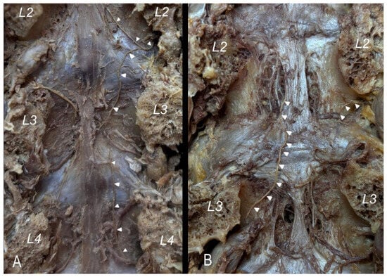
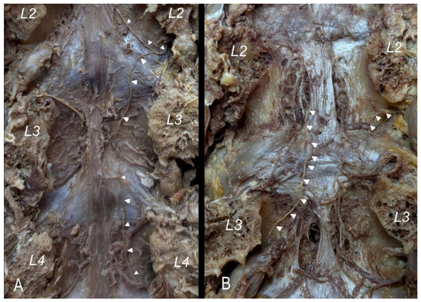
The pattern I cases presented variable arrangements, with four distinct situations classified into four types (Figure 1):
Figure 1.
Vertebral canal posterior views showing pattern 1. Sinuvertebral branches labelled with white arrow heads. Vertebral pedicles are numbered. (A) Type 1a. One right single sinuvertebral nerve ascends from L4 to L3 and type 1b right L2 sinuvertebral nerve divided into two branches, one ascending and one descending terminating at the midline. (B) Type 1c a left single branch arises from L3 level and ascends two levels above its entry into the intervertebral canal (L2 vertebral body) and type 1d a single right descending L2 sinuvertebral nerve.
Type 1a: The sinuvertebral nerve ascends to approximately the middle third of the vertebral body of the vertebra immediately above, covered by the posterior longitudinal ligament. This situation was observed in 64.6% of cases (n = 62).
Type 1b: The sinuvertebral nerve divided into two branches, one ascending and one descending, terminating at the midline. This situation was observed in 17 cases (17.7%).
Type 1c: In two cases (2.1%), a single branch ascended, terminating two levels above its entry into the intervertebral canal.
Type 1d: In one case (1%), the sinuvertebral nerve showed a descending course.
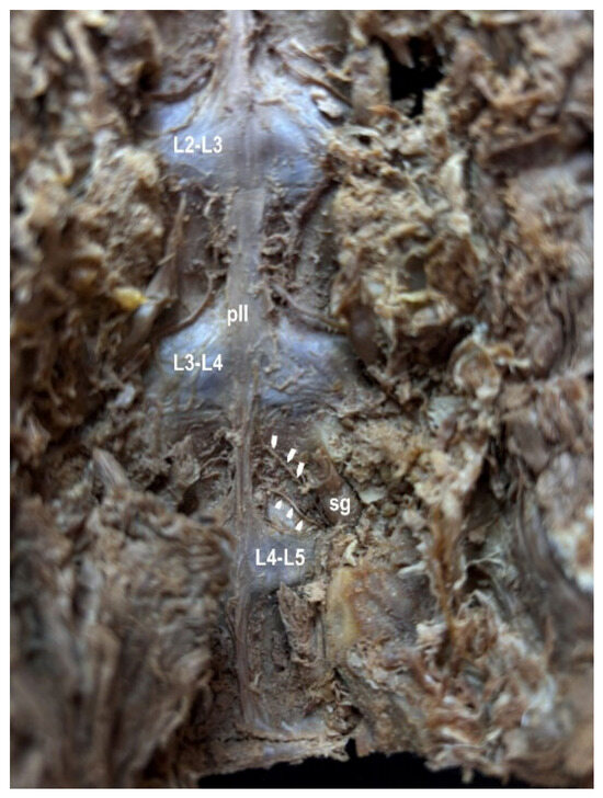
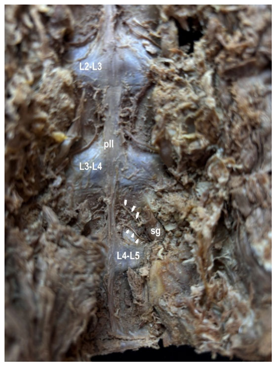
Only one disposition was observed in cases of patten 2 (Figure 2).
Figure 2.
Vertebral canal posterior view showing a double sinuvertebral nerve in the right side. (pattern 2). Both branches are labelled with white arrow heads. Intervertebral discs are numbered. (pll) Posterior longitudinal ligament, (sg) spinal ganglion.
The different patterns and types were assessed according to the vertebral level, observing a homogeneous distribution at all levels. Statistical differences were not found in relation to vertebral levels (Table 1).
Table 1.
Frequency of each of the patterns and types at each of the vertebral levels studied.
Table 2 shows the degree of association between the different types according to Spearman’s correlation. A negative association was only found between the L3 and L4 vertebrae (rho = −0.623; p-value = 0.003). No significant correlation was found between the side and the types considered without taking the lumbar vertebra into account (Spearman’s rho = −0.068; p-value = 0.503).
Table 2.
Spearman’s correlation between vertebrae according to the types considered.
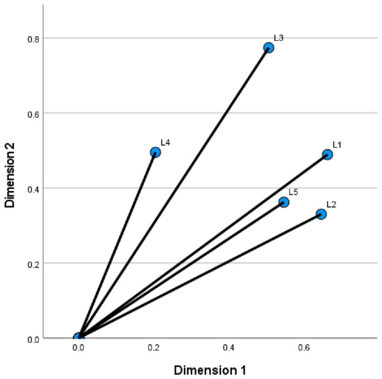
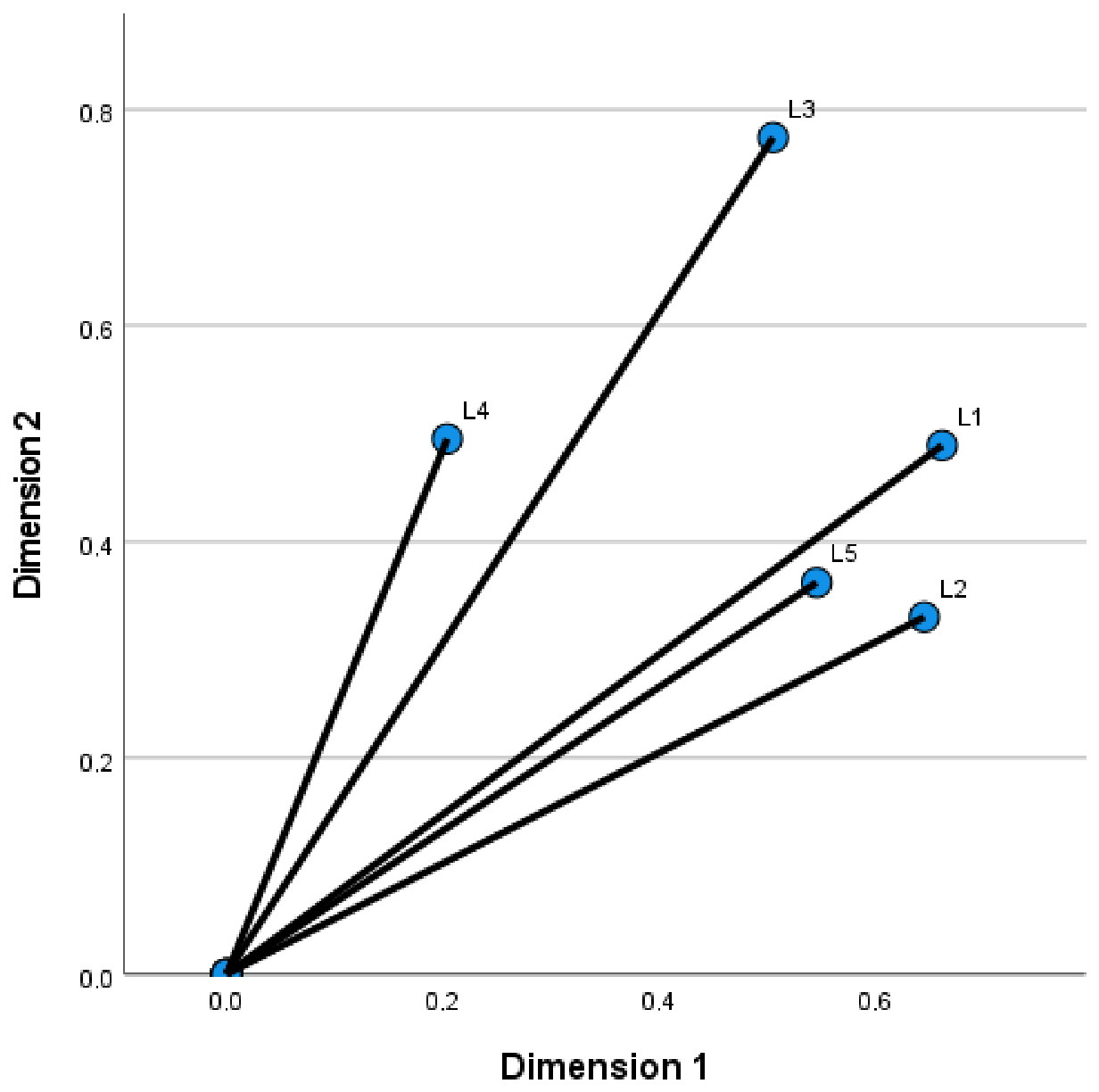
A multiple correspondence study was conducted to examine the association of the different types considered according to the lumbar vertebrae. The results show a Cronbach’s alpha of 0.762 for the first dimension and 0.740 for the second dimensions (resulting in a high degree of association). The first component grouped lumbar vertebrae L1, L2, and L5. The second component grouped lumbar vertebrae L3 and L4 (Figure 3).
Figure 3.
Bispatial diagram of multiple correspondence analysis according to the type considered and the lumbar vertebrae.
Ipsilateral nerve connections were observed in 31 cases at different levels. No statistical differences were observed by level, side or sex.
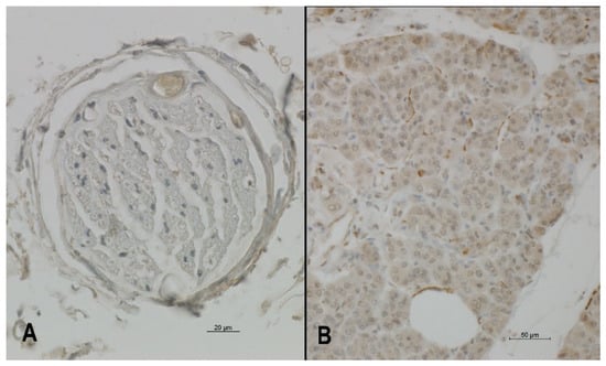
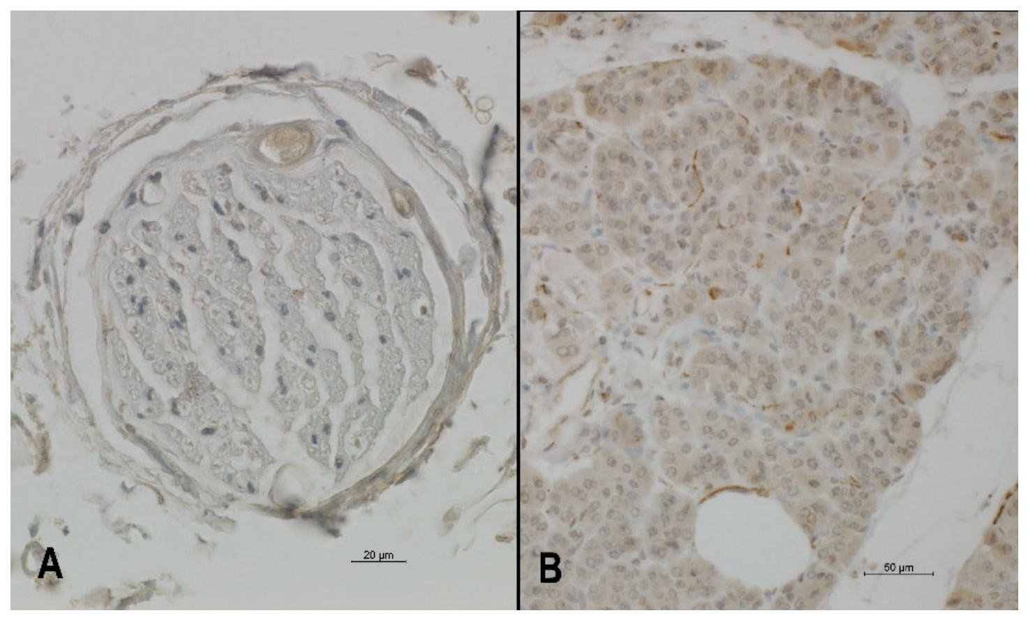
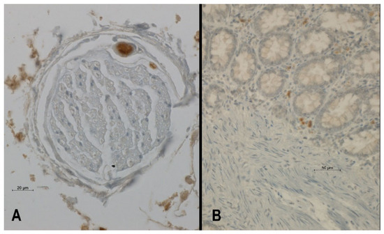
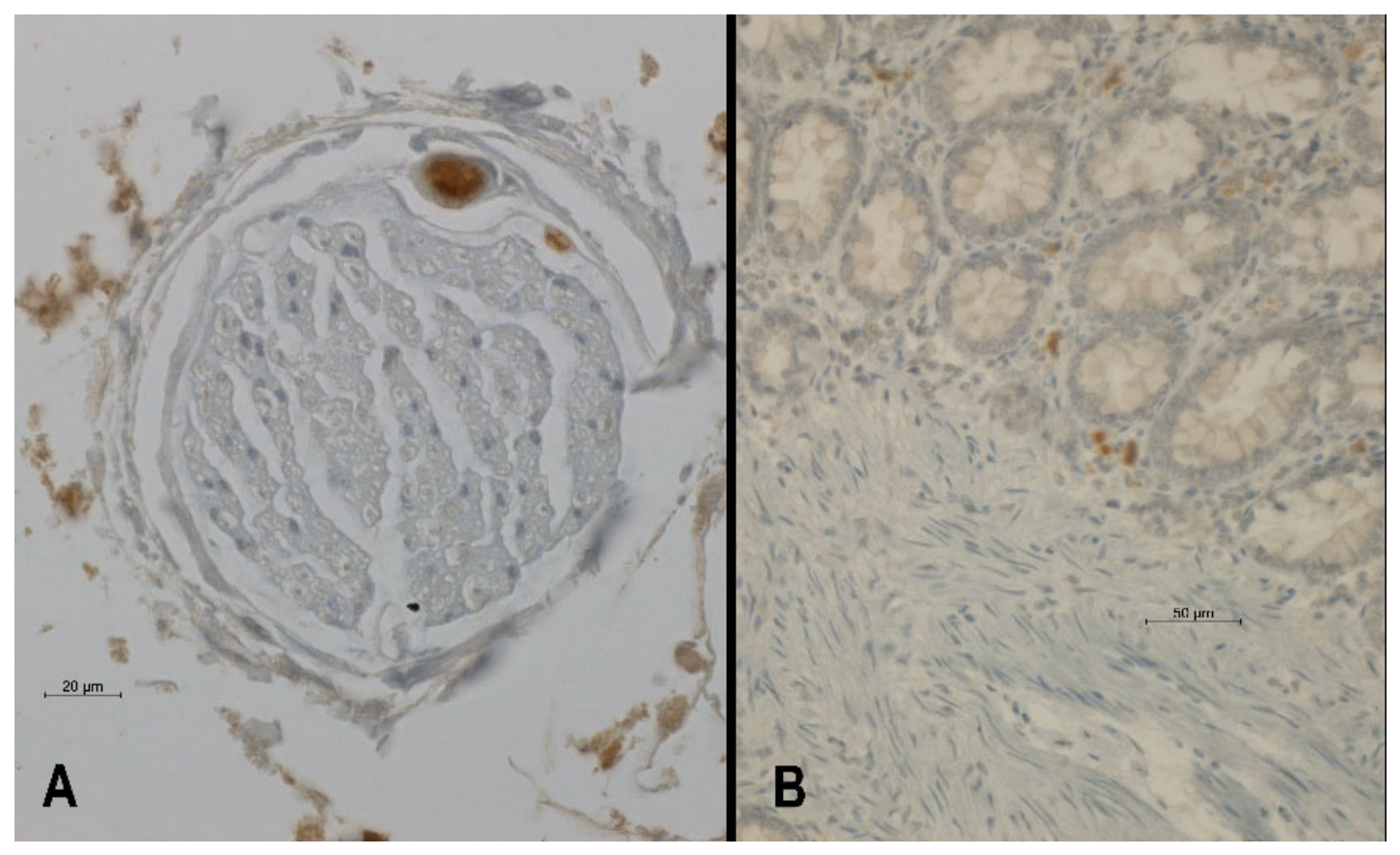
All sinuvertebral nerve samples that underwent immunohistochemical study were positive for CGRP and VIP, suggesting a mixed autonomic (VIP+) and sensory (CGRP+) fibre composition along the nerve trunk (Figure 4 and Figure 5).
Figure 4.
(A) Immunohistochemical localization of VIP within the human lumbar sinuvertebral nerve trunk. Dense VIP-positive chromogen precipitation is confined to the cytoplasmic matrix and elongating processes of Schwann cells (arrowheads) enveloping unmyelinated axons. Intranuclear spaces remain strictly negative, matching the secretory profile of the neuropeptide. (B) Positive control of VIP made in pancreas (20×).
Figure 5.
(A) Immunohistochemical localization of CGRP within the human lumbar sinuvertebral nerve trunk. Micrographs show intense CGRP-immunoreactivity localized within the axoplasm of fine nerve fibres and the perinuclear cytoplasm of associated Schwann cells. Note the complete absence of staining within the connective tissue epineurium. No true nuclear accumulation is observed. (B) Positive control of CGRP made in colon (20×).
4. Discussion
Regarding its origin, several theories have been described that consider an exclusively sympathetic origin [18,21] or a mixed origin in some cases [5,15] or in all cases [4,6,7,11,19,20]. In 100% of cases, we observed a mixed origin (sympathetic and spinal) for the sinuvertebral nerve, as reported by other authors and as observed in previous studies made by our laboratory [19].
The difficulty of the dissection technique, as well as the fact that the nature of the observed nerves has not been histologically verified, may explain these differences [19,26].
Our immunohistochemical analysis reveals that the human lumbar sinuvertebral nerve trunk exhibits consistent VIP-immunoreactivity (VIP-ir) and CGRP-immunoreactivity (CGRP-ir). While VIP is not exclusively sympathetic and can be found in other autonomic or sensory pathways, and CGRP mediates broader sensory and vasodilatory functions rather than purely nociceptive transmission, their combined presence is consistent with a mixed autonomic and sensory fibre composition. These findings suggest a complex, dual structural architecture within the nerve trunk, though they do not provide definitive functional classification of individual fibre subtypes.
The distribution of the different patterns and types is homogeneous, with no significant differences by vertebral level, side, or sex. These results are consistent with those observed by most authors but differ from those published by Raoul et al. [18], who describe a variable distribution depending on the vertebral level and a greater number of sinuvertebral nerve branches at L2. Regarding this level (L2), we found type 1a distributions more frequently, but this difference was not statistically significant as has been shown in Table 1.
Connections between sinuvertebral nerves at adjacent levels have also been classically described. In our study, we observed this in 32% of cases, always ipsilaterally. We did not observe any branches crossing the midline, although it is worth reiterating the difficulty of the dissection technique in these very small structures, which could explain the differences.
Many authors have studied the immunochemical characterization based on the structures innervated by the SVN, such as the posterior surface of intervertebral disc (IVD), posterior longitudinal ligament and anterior aspect of meninges [2,4,5,6,13,14,15,18,21,28,29,30,31,32,33].
In the case of the human IVD, the immunohistochemical characterization of these fibres’ endings have yielded the presence of a variety of neuropeptides involved in nociceptive neurotransmission, PGP 9.5, SP, CGR, NPY, and VIP [16,17,18,34,35,36,37,38].
In addition, in degenerated human IVD, from patients with clinical symptoms, there is an extensive spread of vessels and nerve fibres even reaching the nucleus pulposus [39,40], that correlates with the expression of nerve growth factor [41] and inflammatory markers such as tumour necrosis factor-alpha [42].
In the case of annulus fibrosus, some results have shown immunoreactivity to general nerve markers (synaptophysin gene product and protein 9.5) and to neuropeptides (substance P and C terminal flanking peptide of neuropeptide Y) [17].
Previous observations have found in the posterior longitudinal ligament the presence of different nervous endings containing substance P, and occasionally finding the presence of enkephalin+, indicating modulation of pain signals [43].
Other authors have based their research in fetuses, for example Groen et al. (1990) with a choline acetyl esterase staining, having as a main limitation the inability to determine if these patterns are maintained within the adult population [33].
From a clinical and neurosurgical perspective, establishing a robust neurochemical and morphological profile of the human lumbar SVN trunk is vital for resolving the widespread diagnostic mismatch frequently observed between lumbar spine neuroimaging and a patient’s subjective complaints. Incidental radiological findings, such as lumbar facet joint abnormalities, ligamentous cysts, or even rare developmental remnants like cystic dilation of the ventriculus terminalis (CDVT), are frequently uncovered on routine MRI exams. As highlighted by Ganau et al. (2012) [44] in their updated classification of CDVT, patients categorized under Type 1a exhibit nonspecific symptoms that do not mechanically correlate with the central lesion itself. In these complex clinical scenarios, localized entrapment, microvascular engorgement, or peptidergic inflammation of the mixed autonomic-sensory SVN trunk may serve as the true underlying source of axial discomfort. Recognizing the consistent distribution of nociceptive and autonomic pathways along the SVN trunk can help operating spine surgeons avoid aggressive, unnecessary central spinal procedures, steering the management plan toward targeted diagnostic nerve blocks or conservative therapies instead.
Some studies have also suggested that the terminal branches of the SVN could act as mechanoreceptors, pressure receptors, and nociceptors or thermal terminals [14]. Therefore, even today, in the case of the SVN and its neurochemical profile there are still profound discrepancies in the somatic, sympathetic, or mixed composition. For this reason, the present study confirms the neurochemistry profile of the SVN thanks due to the realization of the immunochemistry characterization directly in the SVN, not in its innervated structures. This information supports the usage of the SVN blocking from a pathophysiological point of view for diagnostic and treatment techniques (e.g., Percutaneous Transforaminal Endoscopic Radiofrequency Ablation of the SVN) in discogenic lumbar pain [8,10].
Finally, a minor limitation of this study includes the lack of dedicated slide-omission negative controls during the immunohistochemical runs; however, the absolute lack of staining in the internal connective and vascular tissues within the nerve trunk strongly validates the specificity of our neurochemical findings.
Author Contributions
L.A., S.Q., P.A., R.E.-M.: conceptualization, formal analysis, resources, investigation, methodology, visualization and supervision. F.V., J.F.R.-V.: conceptualization, formal analysis, visualization, supervision and reviewing of manuscript. M.K., M.L.-C.: Immunohistochemical study design and performance, visualization, supervision and reviewing of manuscript. C.S.d.B.: Study design for statistical analysis, supervision of original draft and review. T.V.-O.: conceptualization, project administration, formal analysis, resources, investigation, methodology, visualization, supervision and writing original draft and writing—review and editing. All authors have read and agreed to the published version of the manuscript.
Funding
This research has been supported by grant of Spanish Spine Society (GEER).
Institutional Review Board Statement
All specimens managed by the authors in the Bodies Donation Centre at the Complutense University of Madrid are obtained through a voluntary body donation program. We strictly adhere to Spanish legislation regarding anatomical donation, medical research, and data protection. This program operates in rigorous compliance with the regulatory framework governing the use of anatomical material and clinical data, including Decree 2263/1974 (Reglamento de Policía Sanitaria Mortuoria), the foundational Law of 18 December 1950 (Obtención de piezas anatómicas para injertos), Law 41/2002 (Autonomía del Paciente) for informed consent and clinical records, and Organic Law 3/2018 (LOPDGDD) for data privacy. This procedure complies with current Spanish law and does not require approval from the ethics committee.
Informed Consent Statement
Informed consent is obtained from every donor prior to death. All handled donor data will be strictly pseudonymized to maintain absolute confidentiality.
Data Availability Statement
The original contributions presented in this study are included in the article. Further inquiries can be directed to the corresponding author.
Acknowledgments
The authors wish to express their sincere gratitude to the anatomical body-donors, who bequeathed their bodies for medical education and basic science research.
Conflicts of Interest
The authors declare no conflicts of interest.
References
- Hovelacque, A. Le Nerf Sinu-Vertebral; Masson: Paris, France, 1925. [Google Scholar]
- Roofe, P.G. Innervation of annulus fibrosus and posterior longitudinal ligament. Fourth and fifth lumbar level. Arch. Neur. Psych. 1940, 44, 100–103. [Google Scholar]
- Van Buskirk, C. Nerves in the vertebral canal: Their relation to the sympathetic innervation of the upper extremities. Arch. Surg. 1941, 43, 427–432. [Google Scholar] [CrossRef] [Scilit]
- Wiberg, G. Back pain in relation to the nerve supply of the intervertebral disc. Acta Orthop. Scand. 1949, 19, 211–221. [Google Scholar] [CrossRef] [Scilit] [PubMed]
- Pedersen, H.E.; Blunck, C.F.; Gardner, E. The Anatomy of Lumbosacral Posterior Rami and Meningeal Branches of Spinal Nerve (Sinu-Vertebral Nerves); With an Experimental Study of Their Functions. J. Bone Jt. Surg. Am. 1956, 38, 377–391. [Google Scholar]
- Bogduk, N.; Tynan, W.; Wilson, A.S. The nerve supply to the human lumbar intervertebral discs. J. Anat. 1981, 132, 39–56. [Google Scholar] [PubMed]
- Bogduk, N. The innervation of the lumbar spine. Spine 1983, 8, 286–293. [Google Scholar] [CrossRef] [Scilit] [PubMed]
- Shayota, B.; Wong, T.L.; Fru, D.; David, G.; Iwanaga, J.; Loukas, M.; Tubbs, R.S. A Comprehensive Review of the Sinuvertebral Nerve with Clinical Applications. Anat. Cell Biol. 2019, 52, 128–133. [Google Scholar] [CrossRef] [Scilit] [PubMed]
- Kim, H.S.; Kashlan, O.N.; Singh, R.; Adsul, N.M.; Yong, Z.; Oh, S.W.; Noh, J.H.; Jang, I.T.; Oh, S.H. Percutaneous transforaminal endoscopic radiofrequency ablation of the sinuvertebral nerve in an olympian with a Left L5 pedicle/pars interarticularis fracture-associated left L5–S1 disk desiccation. World Neurosurg. 2019, 3, 100032. [Google Scholar] [CrossRef] [Scilit] [PubMed]
- Kim, H.S.; Wu, P.H.; Jang, I.-T. Lumbar degenerative disease part 1: Anatomy and pathophysiology of intervertebral discogenic pain and radiofrequency ablation of basivertebral and sinuvertebral nerve treatment for chronic discogenic back pain: A prospective case series and review of literature. Int. J. Mol. Sci. 2020, 21, 1483. [Google Scholar] [CrossRef] [Scilit] [PubMed]
- Zhao, Q.; Cheng, L.; Yan, H.; Deng, S.; Zhao, J.; Liu, Z.; Tan, X.; Zhong, E.; Zhuo, W.; Jin, D. The anatomical study and clinical significance of the sinuvertebral nerves at the lumbar levels. Spine 2020, 45, E61–E66. [Google Scholar] [CrossRef] [Scilit] [PubMed]
- Chen, J.; Liu, Z.; Xu, Y.; Liu, Z.; Zheng, Z.; Zhang, Z.; Fan, C.; Li, Q.; Zhao, Q. Anatomic zone division and clinical significance of the lumbar sinuvertebral nerves. Spine J. 2023, 23, 1223–1233. [Google Scholar] [CrossRef] [Scilit] [PubMed]
- Malinsky, J. The Ontogenetic Development of Nerve Terminations in the Intervertebral Discs of Man. (Histology of Intervertebral Discs, 11th Communication). Acta Anat. 1959, 38, 96–113. [Google Scholar] [CrossRef] [Scilit] [PubMed]
- Korkala, O.; Grönblad, M.; Liesi, P.; Karaharju, E. Immunohistochemical Demonstration of Nociceptors in the Ligamentous Structures of the Lumbar Spine. Spine 1985, 10, 156–157. [Google Scholar] [CrossRef] [Scilit] [PubMed]
- Chen, X.Q.; Bo, S.; Zhong, S.Z. Nerves accompanying the vertebral artery and their clinical relevance. Spine 1988, 13, 1360–1364. [Google Scholar] [CrossRef] [Scilit] [PubMed]
- Coppes, M.; Marani, E.; Thomeer, R.; Oudega, M.; Groen, G. Innervation of annulus fibrosis in low back pain. Lancet 1990, 336, 189–190. [Google Scholar] [CrossRef] [Scilit] [PubMed]
- Palmgren, T.; Grönblad, M.; Virri, J.; Kääpä, E.; Karaharju, E. An immunohistochemical study of nerve structures in the anulus fibrosus of human normal lumbar intervertebral discs. Spine 1999, 24, 2075–2079. [Google Scholar] [CrossRef] [Scilit] [PubMed]
- Raoul, S.; Faure, A.; Robert, R.; Rogez, J.-M.; Hamel, O.; Cuillère, P.; Le Borgne, J. Role of the Sinu-Vertebral Nerve in Low Back Pain and Anatomical Basis of Therapeutic Implications. Surg. Radiol. Anat. 2003, 24, 366–371. [Google Scholar] [PubMed]
- Quinones, S.; Konschake, M.; Aguilar, L.L.; Simon, C.; Aragones, P.; Hernández, L.M.; Abramovic, A.; Tubbs, R.S.; Bouzada, J.; Valderrama-Canales, F.J.; et al. Clinical anatomy of the lumbar sinuvertebral nerve with regard to discogenic low back pain and review of literature. Eur. Spine J. 2021, 30, 2999–3008, Erratum in Eur. Spine J. 2021, 30, 2409. https://doi.org/10.1007/s00586-021-06894-1. [Google Scholar] [CrossRef] [Scilit] [PubMed]
- Kimmel, D.L. Innervation of spinal dura mater and dura mater of the posterior cranial fossa. Neurology 1961, 11, 800–809. [Google Scholar] [CrossRef] [Scilit] [PubMed]
- Groen, G.J.; Baljet, B.; Drukker, J. The innervation of the spinal dura mater: Anatomy and clinical implications. Acta Neurochir. 1988, 92, 39–46. [Google Scholar] [CrossRef] [Scilit] [PubMed]
- Suseki, K.; Takahashi, Y.; Takahashi, K.; Chiba, T.; Yamagata, M.; Moriya, H. Sensory nerve fibres from lumbar intervertebral discs pass through rami communicantes: A possible pathway for discogenic low back pain. J. Bone Jt. Surg. Br. Vol. 1998, 80, 737–742. [Google Scholar] [CrossRef] [Scilit]
- Kojima, Y.; Maeda, T.; Arai, R.; Shichikawa, K. Nerve supply to the posterior longitudinal ligament and the intervertebral disc of the rat vertebral column as studied by acetylcholinesterase histochemistry. I. Distribution in the lumbar region. J. Anat. 1990, 169, 237–246. [Google Scholar] [PubMed]
- Nakamura, S.-I.; Takahashi, K.; Takahashi, Y.; Morinaga, T.; Shimada, Y.; Moriya, H. Origin of nerves supplying the posterior portion of lumbar intervertebral discs in rats. Spine 1996, 21, 917–924. [Google Scholar] [CrossRef] [Scilit] [PubMed]
- Sekiguchi, Y.; Konnai, Y.; Kikuchi, S.; Sugiura, Y. An anatomic study of neuropeptide immunoreactivities in the lumbar dura mater after lumbar sympathectomy. Spine 1996, 21, 925–930. [Google Scholar] [CrossRef] [Scilit] [PubMed]
- Breemer, M.; Notenboom, R.; Malessy, M. Letter to the Editor concerning “Clinical anatomy of the lumbar sinuvertebral nerve with regard to discogenic low back pain and review of literature” by Sara Quinones et al. (Eur Spine J [2021]; 30(10): 2999–3008). Eur. Spine J. 2022, 31, 1931–1932. [Google Scholar] [CrossRef] [Scilit] [PubMed]
- Konschake, M.; Abramovic, A.; Quinones, S.; Sanudo, J.R. Reply to the Letter to the Editor of Breemer MC; et al. concerning “Clinical anatomy of the lumbar sinuvertebral nerve with regard to discogenic low back pain and review of literature” by Sara Quinones et al. (Eur Spine J [2021]; 30(10); 2999-3008). Eur. Spine J. 2022, 31, 1933–1935. [Google Scholar] [CrossRef] [Scilit] [PubMed]
- Edgar, M.A.; Nundy, S. Innervation of the spinal dura mater. Neurol. Neurosurg. Psychiatry 1966, 29, 530–534. [Google Scholar] [CrossRef] [Scilit]
- Bogduk, N. The anatomy of the lumbar intervertebral disc syndrome. Med. J. Aust. 1976, 1, 878–881. [Google Scholar] [PubMed]
- Bogduk, N. The innervation of the intervertebral discs. In The Biology of the Intervertebral Disc, Gosh, P., Ed.; 1st ed.; CRC Press: Boca Raton, FL, USA, 1988; Volume I, pp. 22–40. [Google Scholar]
- Bogduk, N. Clinical Anatomy of the Lumbar Spine and Sacrum, 3rd ed.; Churchill Livingstone: Philadelphia, PA, USA, 1997. [Google Scholar]
- Bogduk, N. Functional anatomy of the spine. Handb. Clin. Neurol. 2016, 136, 675–688. [Google Scholar] [CrossRef] [Scilit] [PubMed]
- Groen, G.J.; Baljet, B.; Drukker, J. Nerves and nerve plexuses of the human vertebral column. Am. J. Anat. 1990, 188, 282–296. [Google Scholar] [CrossRef] [Scilit] [PubMed]
- Youssef, P.; Loukas, M.; Chapman, J.R.; Oskouian, R.J.; Tubbs, R.S. Comprehensive anatomical and immunohistochemical review of the innervation of the human spine and joints with application to an improved understanding of back pain. Childs Nerv. Syst. 2016, 32, 243–251. [Google Scholar] [PubMed]
- Konttinen, Y.T.; Grönblad, M.; Antti-Poika, I.; Seitsalo, S.; Santavirta, S.; Hukkanen, M.; Polak, J.M.M. Neuroimmunohistochemical analysis of peridiscal nociceptive neural elements. Spine 1990, 15, 383–386. [Google Scholar] [CrossRef] [Scilit] [PubMed]
- Ashton, I.K.; Roberts, S.; Jaffray, D.C.; Polak, J.M.; Eisenstein, S.M. Neuropeptides in the human intervertebral disc. J. Orthop. Res. 1994, 12, 186–192. [Google Scholar] [CrossRef] [Scilit] [PubMed]
- Roberts, S.; Eisenstein, S.M.; Menage, J.; Evans, E.H.; Ashton, I.K. Mechanoreceptors in intervertebral discs. Spine 1995, 20, 2645–2651. [Google Scholar] [CrossRef] [Scilit] [PubMed]
- Brown, M.F.; Hukkanen, M.V.J.; McCarthy, I.D.; Redfern, D.R.M.; Batten, J.J.; Crock, H.V.; Hughes, S.P.F.; Polak, J.M. Sensory and sympathetic innervation of the vertebral endplate in patients with degenerative disc disease. J. Bone Jt. Surg. Br. 1997, 79, 147–153. [Google Scholar] [CrossRef] [Scilit]
- Peng, B.; Wu, W.; Hou, S.; Li, P.; Zhang, C.; Yang, Y. The pathogenesis of discogenic low back pain. J. Bone Jt. Surg. Br. 2005, 87, 62–67. [Google Scholar] [CrossRef] [Scilit]
- García-Cosamalón, J.; E Del Valle, M.; Calavia, M.G.; García-Suárez, O.; López-Muñiz, A.; Otero, J.; A Vega, J. Intervertebral Disc, Sensory Nerves and Neurotrophins: Who Is Who in Discogenic Pain? J. Anat. 2010, 217, 1–15. [Google Scholar] [CrossRef] [Scilit] [PubMed]
- Freemont, A.J.; Watkins, A.; Le Maitre, C.; Baird, P.; Jeziorska, M.; Knight, M.T.N.; Ross, E.R.S.; O’Brien, J.P.; Hoyland, J.A. Nerve growth factor expression and innervation of the painful intervertebral disc. J. Pathol. 2002, 197, 286–292. [Google Scholar] [CrossRef] [Scilit] [PubMed]
- Weiler, C.; Nerlich, A.G.; Bachmeier, B.E.; Boos, N. Expression and Distribution of Tumor Necrosis Factor Alpha in Human Lumbar Intervertebral Discs: A Study in Surgical Specimen and Autopsy Controls. Spine 2005, 30, 44–53. [Google Scholar] [CrossRef] [Scilit] [PubMed]
- Liesi, P.; Grönblad, M.; Korkala, O.; Karahaju, E.; Rusanen, M. Substance P: A neuropeptide involved in low-pain? Lancet 1983, 1, 1328–1329. [Google Scholar] [CrossRef] [Scilit] [PubMed]
- Ganau, M.; Talacchi, A.; Cecchi, P.C.; Ghimenton, C.; Gerosa, M.; Faccioli, F. Cystic dilation of the ventriculus terminalis. J. Neurosurg. Spine 2012, 17, 86–92. [Google Scholar] [CrossRef] [Scilit] [PubMed]

Disclaimer/Publisher’s Note: The statements, opinions and data contained in all publications are solely those of the individual author(s) and contributor(s) and not of MDPI and/or the editor(s). MDPI and/or the editor(s) disclaim responsibility for any injury to people or property resulting from any ideas, methods, instructions or products referred to in the content. |
© 2026 by the authors. Licensee MDPI, Basel, Switzerland. This article is an open access article distributed under the terms and conditions of the Creative Commons Attribution (CC BY) license.