


Abstract
Artificial intelligence (AI) is increasingly transforming gastrointestinal endoscopy by supporting lesion detection, lesion characterization, quality assessment, and clinical risk prediction. Hemostatic decision-making represents a particularly complex field for AI integration, as therapeutic decisions are often made rapidly in the presence of active bleeding, impaired visualization, unstable patients, and variable lesion accessibility. This review critically examines the current evidence for AI-assisted decision-making in gastrointestinal endoscopy and endoscopic hemostasis, with emphasis on gastrointestinal bleeding, prediction of hemostatic therapy requirements, bleeding-risk stratification, rebleeding prediction, transfusion support, and post-procedural monitoring. Available studies suggest that machine learning and deep learning models may outperform conventional scoring systems in selected retrospective or validation cohorts, improve recognition of high-risk lesions, support less experienced endoscopists, and contribute to more individualized management of non-variceal bleeding, variceal bleeding, and capsule endoscopy findings. However, prospective interventional evidence remains sparse, and most available models are limited by retrospective design, single-center datasets, incomplete external validation, black-box decision-making, heterogeneous reporting, workflow barriers, and uncertain cost-effectiveness. AI should therefore be regarded as an adjunctive decision-support tool rather than an autonomous replacement for clinical judgment. Its future value will depend on prospective multicenter validation, explainability, real-time usability, regulatory clarity, post-deployment surveillance, and evidence of improved patient-centered outcomes before widespread implementation in emergency endoscopy practice.
1. Introduction
In recent years, medical interest in artificial intelligence (AI) has expanded considerably, particularly in specialties that depend on image interpretation and pattern recognition. The most visible progress has been reported in radiology, gastroenterology (especially endoscopy), surgery, and dermatology, where AI systems are increasingly explored as tools for detection, classification, and clinical decision support [1,2]. In gastrointestinal (GI) endoscopy, this development is particularly relevant because diagnostic and therapeutic decisions frequently depend on real-time visual interpretation, procedural quality, and rapid clinical judgment.
In GI endoscopy, AI has moved from experimental image analysis toward clinically oriented applications, including lesion detection, lesion characterization, quality assessment, and prediction of relevant outcomes [3,4]. Upper GI endoscopy is particularly suitable for its integration because diagnostic accuracy often depends on visual recognition, completeness of mucosal inspection, operator experience, and real-time interpretation of subtle endoscopic findings [5]. These features are especially important in bleeding-related scenarios, where incomplete visualization, active blood, clots, patient instability, and time pressure may affect both lesion recognition and therapeutic decision-making.
Endoscopic hemostasis represents a particularly demanding clinical context because decisions must often be made rapidly in the presence of active bleeding, impaired visualization, patient instability, and variable lesion accessibility [6,7]. In non-variceal upper GI bleeding, early risk stratification, accurate recognition of high-risk bleeding stigmata, and timely selection of endoscopic therapy are essential for reducing persistent bleeding, rebleeding, and adverse outcomes [8].
In this setting, AI may support several stages of the hemostatic pathway, including pre-endoscopic triage, prediction of the need for urgent endoscopy, recognition of high-risk lesions, estimation of hemostatic therapy requirement, transfusion support, and post-procedural monitoring [9,10,11]. Recent evidence suggests that machine learning models may improve the assessment and management of acute GI bleeding by supporting risk prediction, intervention planning, and individualized decision-making, but most applications remain at an early stage of clinical validation and implementation [12].
Although not all AI applications in GI endoscopy are hemostasis-specific, several broader endoscopic functions remain relevant to bleeding-related care. AI-assisted lesion detection, lesion characterization, and quality assessment may help reduce operator-dependent variability, improve recognition of subtle or high-risk findings, and support more consistent documentation during both routine and emergency endoscopic assessment [13,14,15]. However, these indirect applications should be distinguished from AI tools designed specifically for bleeding-risk stratification, prediction of hemostatic therapy, transfusion support, or rebleeding assessment.
Accordingly, AI should be interpreted as an adjunctive decision-support tool rather than an autonomous substitute for expert judgment. Its current value lies primarily in standardizing assessment, supporting risk prediction, and assisting clinical decision-making, while its future role will depend on prospective validation, explainability, integration into emergency workflows, and evidence of improved patient-centered outcomes [16,17].
This review critically evaluates the current evidence supporting AI-assisted decision-making in gastrointestinal endoscopy and hemostatic care, with emphasis on lesion detection, bleeding-risk stratification, prediction of hemostatic therapy requirement, rebleeding assessment, transfusion support, and barriers to clinical implementation.
2. Search Strategy
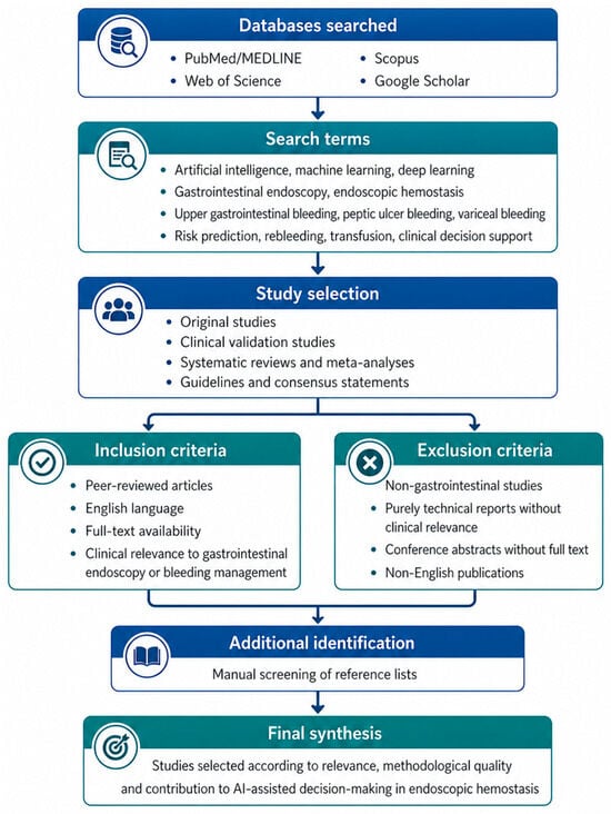
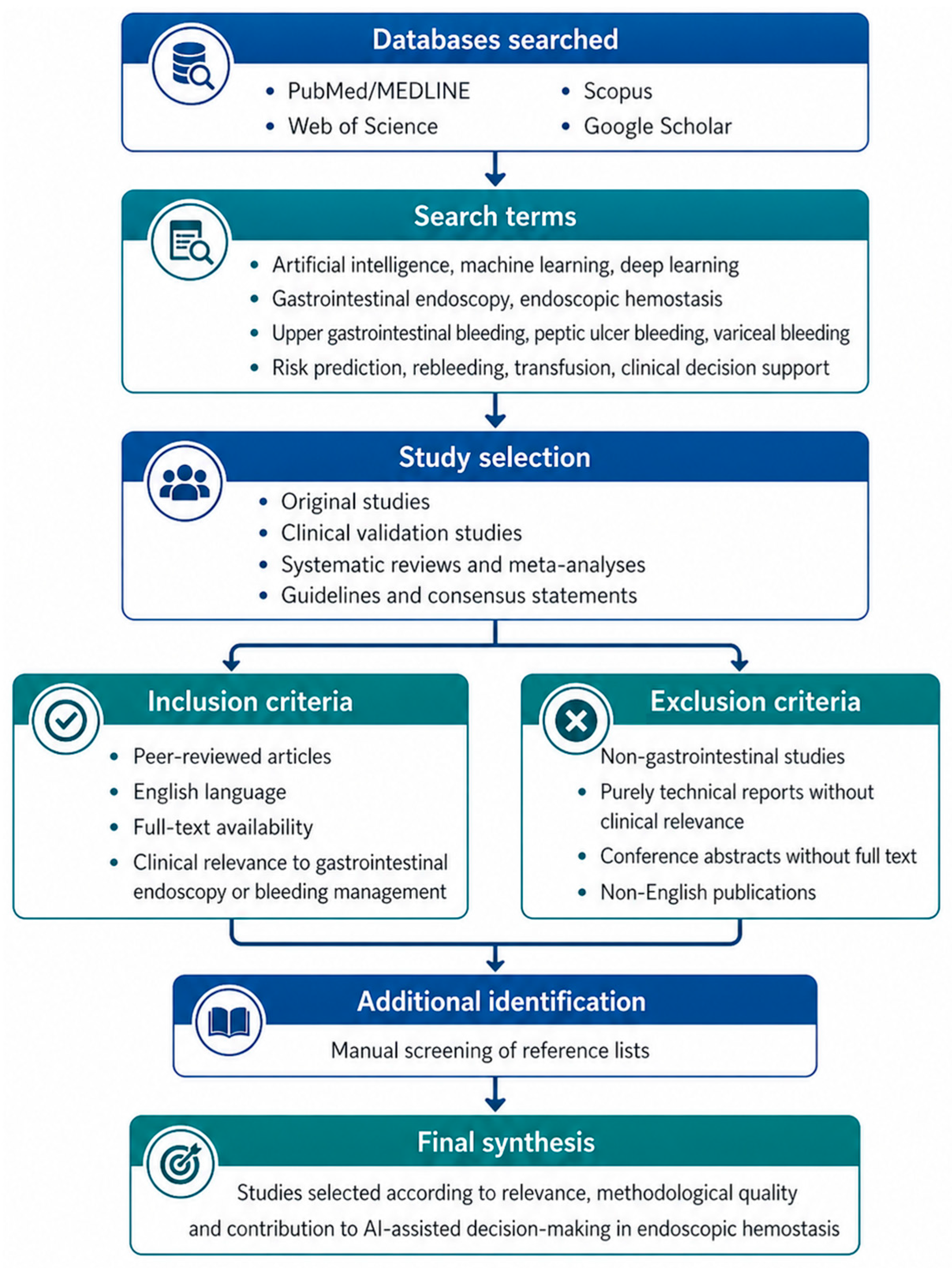
This structured narrative review evaluated current evidence on artificial intelligence applications in gastrointestinal endoscopy and hemostatic decision-making. The literature search was performed in PubMed/MEDLINE, Web of Science, Scopus, and Google Scholar for publications available up to April 2026. Search terms included combinations of “artificial intelligence”, “machine learning”, “deep learning”, “computer-aided detection”, “computer-aided diagnosis”, “gastrointestinal endoscopy”, “upper gastrointestinal bleeding”, “peptic ulcer bleeding”, “variceal bleeding”, “endoscopic hemostasis”, “hemostatic therapy”, “rebleeding”, “transfusion”, “clinical decision support” and “explainable artificial intelligence.”
Eligible publications included original studies, validation studies, systematic reviews, meta-analyses, guidelines, and consensus statements relevant to gastrointestinal bleeding, endoscopic hemostasis, bleeding-risk stratification, prediction of hemostatic therapy, rebleeding, transfusion support, or implementation barriers. Non-GI studies, purely technical reports without clinical relevance, non-peer-reviewed articles, conference abstracts with insufficient clinical information, and non-English publications were excluded. Additional articles were identified through reference-list screening.
After eligibility screening, 137 references were considered eligible and included in the final narrative synthesis. Eligibility was determined primarily from a clinical and hemostatic decision-making perspective. Publications were retained when they addressed gastrointestinal bleeding, endoscopic hemostasis, bleeding-risk stratification, prediction of hemostatic or endoscopic intervention, rebleeding, transfusion support, mortality prediction, or implementation barriers relevant to clinical decision support.
Studies were prioritized according to their clinical relevance to hemostatic decision-making. Evidence directly related to gastrointestinal bleeding and endoscopic hemostasis was summarized in Table 1, while broader AI applications in gastrointestinal endoscopy were included only when relevant to lesion recognition, procedural quality, operator variability, or therapeutic decision-making. Methodological limitations were assessed descriptively, including study design, external validation, explainability, and implementation readiness. The search and selection process is summarized in Figure 1.
Table 1.
AI-based decision support in gastrointestinal bleeding and hemostatic decision-making.
Figure 1.
Structured literature search strategy.
3. Conceptual Foundations of Artificial Intelligence in Endoscopic Decision Support
Artificial intelligence (AI) refers to a computer’s capacity to perform cognitive tasks typically associated with humans, particularly learning, interpretation, and decision-making [29,30]. In medical applications, AI systems may be rule-based, relying on predefined algorithms, or data-driven, relying mainly on machine learning models that identify patterns from clinical or image-based datasets [31]. Machine learning models are designed to learn from available data and generate outputs that can be applied to new, previously unseen cases [31,32,33].
In gastrointestinal endoscopy, the most relevant AI approaches include machine learning and deep learning. Conventional machine learning generally depends on predefined clinical or image-derived features, whereas deep learning can automatically extract relevant visual features from endoscopic images, most often through convolutional neural networks [34,35,36]. This is particularly relevant in endoscopic decision support, where lesion recognition, bleeding-risk estimation, and prediction of therapeutic need may depend on complex combinations of visual findings, clinical variables, and laboratory parameters.
For hemostatic decision-making, AI tools can be broadly divided into image-based models and clinical prediction models. Image-based models may support recognition of high-risk bleeding stigmata, variceal features, ulcers, vascular lesions, or potentially hemorrhagic lesions on capsule endoscopy. Clinical prediction models may integrate vital signs, comorbidities, medication exposure, laboratory parameters, risk scores, and endoscopic findings to estimate outcomes, such as need for hemostatic intervention, rebleeding, transfusion, or mortality [37,38].
However, deep learning systems also have important limitations. Many models function as “black boxes,” meaning that their internal reasoning is difficult to interpret despite known inputs and outputs [39,40]. This lack of transparency raises scientific, ethical, and legal concerns, because clinical decisions require not only accurate predictions but also understandable explanations [41,42]. Explainability methods, including heat maps, activation maps, and input-modification techniques, may improve transparency by showing which image regions or variables influenced model output [43]. Nevertheless, in emergency bleeding scenarios, AI outputs should be interpreted as decision-support signals rather than definitive therapeutic instructions.
4. Artificial Intelligence in Endoscopy
In endoscopy, AI applications are commonly organized into three clinically relevant domains: computer-aided detection (CADe), computer-aided diagnosis (CADx), and computer-aided quality assessment (CADq) [44,45]. CADe supports the identification of abnormal findings, CADx assists lesion characterization, and CADq evaluates procedural quality, completeness of examination, and standardization of documentation. In upper GI endoscopy, these domains follow the clinical workflow from mucosal visualization to lesion recognition, interpretation, and therapeutic decision-making [46].
Although many early AI systems were developed for colorectal polyp detection and other diagnostic indications, the same technical principles are relevant to bleeding-related care when they support rapid recognition of abnormal mucosal findings, vascular lesions, ulcers, blood clots, or small-bowel bleeding sources [29,47]. In hemostatic decision-making, the value of AI therefore extends beyond diagnosis, because lesion recognition, risk stratification, and therapeutic prioritization may directly influence the timing and type of intervention.
CADx systems may further assist endoscopists by classifying detected lesions into clinically meaningful categories, including features relevant to malignancy, invasion depth, lesion extent, or technical treatment difficulty [48,49]. Although these applications are not always hemostasis-specific, they may inform therapeutic planning when bleeding occurs in complex lesions or when the distinction between benign, malignant, and high-risk lesions influences subsequent management.
CADq tools are also indirectly relevant to emergency endoscopy because missed lesions, incomplete mucosal visualization, and inconsistent documentation may affect downstream management. AI-based quality tools have been used to support mucosal exposure, landmark recognition, blind-spot detection, bowel preparation assessment, and standardized photo-documentation [50,51,52,53,54,55,56,57]. In bleeding scenarios, these functions may help reduce operator-dependent variability, especially when visualization is impaired by blood clots, bubbles, or rapid scope movement.
Broader AI applications in upper GI endoscopy, including neoplasia detection, targeted biopsy support, H. pylori assessment, and anatomical landmark recognition, remain clinically relevant but should be distinguished from tools designed specifically for gastrointestinal bleeding or hemostatic decision-making [58,59,60,61]. In this review, these applications are considered only insofar as they illustrate how AI may improve lesion recognition, procedural quality, and consistency of endoscopic interpretation.
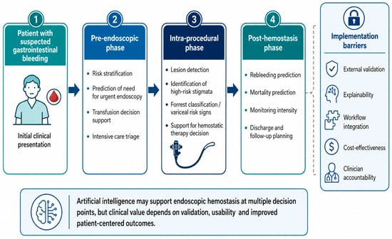
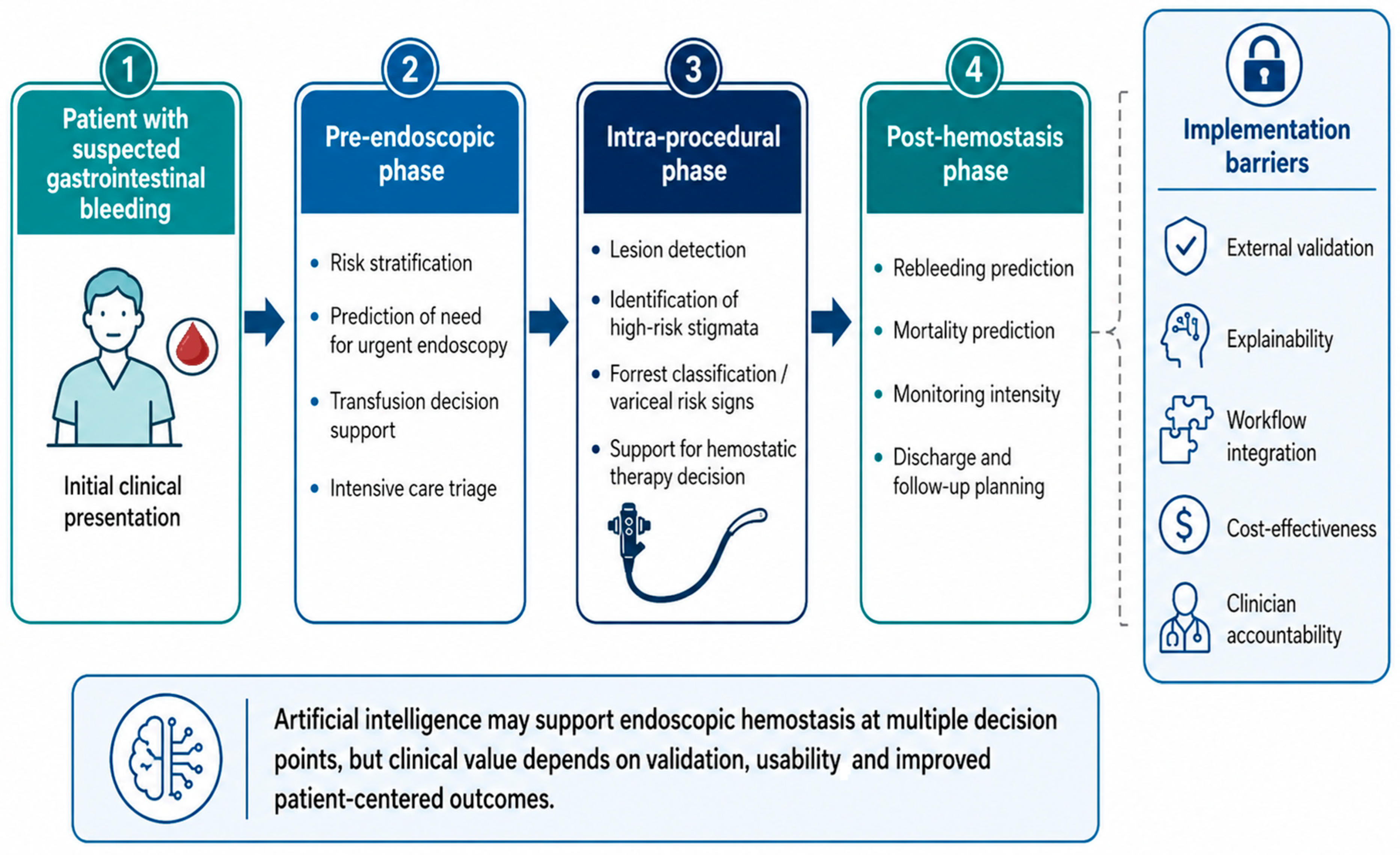
Taken together, these applications can be integrated into a broader clinical pathway in which AI supports decision-making before, during, and after endoscopic hemostasis, from initial risk stratification to post-procedural monitoring, as illustrated in Figure 2.
Figure 2.
Proposed role of artificial intelligence across the endoscopic hemostasis pathway.
The main studies evaluating AI applications relevant to GI bleeding, hemostatic decision-making, bleeding-risk stratification, and post-procedural outcomes are summarized in Table 1.
5. Disease-Specific Applications of Artificial Intelligence
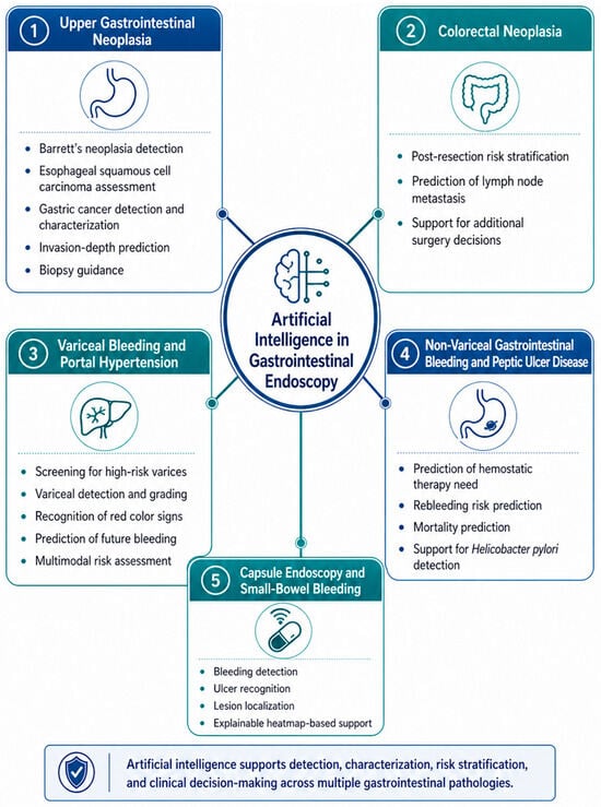
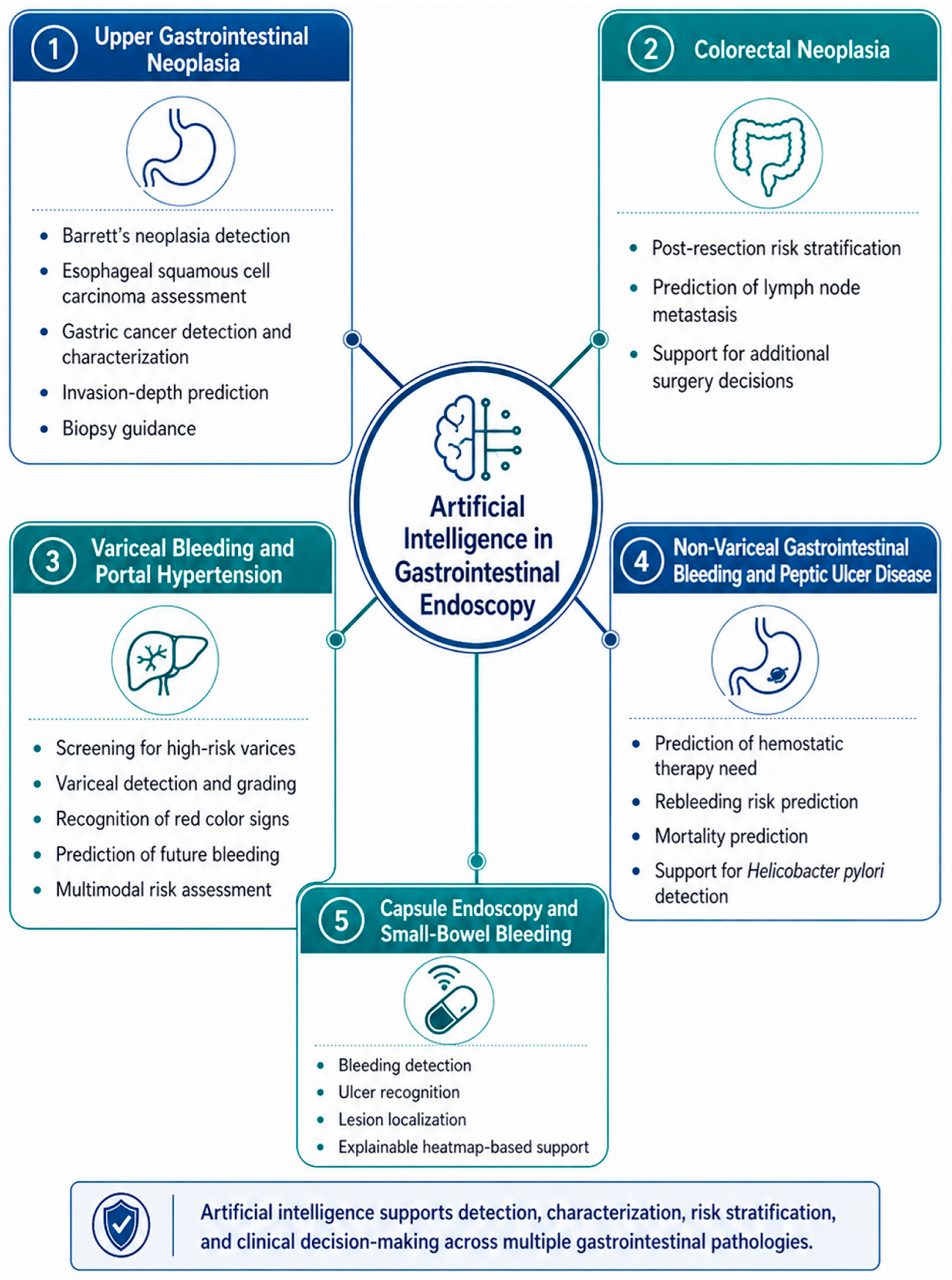
The main disease-specific applications of AI relevant to GI endoscopy, bleeding-related disorders, and therapeutic decision-making are summarized in Figure 3. Applications directly related to GI bleeding and hemostasis are discussed separately from broader endoscopic applications that are only indirectly relevant to hemostatic care.
Figure 3.
Disease-specific applications of artificial intelligence in gastrointestinal endoscopy and bleeding-related decision-making.
5.1. Upper Gastrointestinal Neoplasia
These applications are included only as indirectly relevant to hemostatic decision-making, particularly when lesion characterization, invasion-depth assessment, or therapeutic planning may influence the management of bleeding, potentially hemorrhagic or technically complex upper gastrointestinal lesions [62,63].
AI-assisted CADx systems may help distinguish neoplastic Barrett’s lesions from non-dysplastic mucosa and guide targeted biopsies, but in this review, their relevance is limited to situations in which lesion characterization may influence bleeding-related therapeutic planning [64,65].
For esophageal squamous cell carcinoma, AI systems have been used to estimate invasion depth and differentiate superficial from deeply invasive disease, with accuracy comparable to expert endoscopists in some studies [66,67]. Similar models applied to gastric cancer have also shown good performance in assessing invasion depth and distinguishing malignant from benign mucosal lesions [68,69,70].
Esophageal squamous cell carcinoma remains highly lethal, particularly when diagnosed at an advanced stage, making early recognition a priority [71]. AI-based systems have demonstrated high sensitivity for detecting early lesions, often outperforming non-expert endoscopists and reaching performance levels close to experienced specialists [72].
Additional AI-supported methods, including high-resolution microendoscopy, narrow-band imaging analysis, and endocytoscopy-based models, have shown strong diagnostic potential [73,74,75,76]. However, performance may decrease when models trained on static images are applied to real-time video, mainly because of false-positive findings, indicating the need for further training and validation [77].
Gastric cancer remains a leading cause of cancer-related mortality, making early recognition of premalignant and malignant gastric lesions essential for improving clinical outcomes [78]. AI-based systems have demonstrated high sensitivity in gastric cancer detection, although false-positive results may occur in the presence of chronic atrophic gastritis or intestinal metaplasia [79]. More advanced approaches using magnifying endoscopy with narrow-band imaging have shown particular promise for early gastric cancer detection, achieving performance comparable to that of expert endoscopists [80,81]. In addition, these models can localize suspicious mucosal areas, thereby improving interpretability and increasing their potential usefulness as decision-support tools in clinical practice. AI assistance also improved diagnostic performance among both junior and senior physicians, suggesting its value as a decision-support tool rather than a replacement for endoscopists [81]. Similar models have also been used to assess the depth of gastric cancer invasion, which may help guide treatment selection between endoscopic and surgical approaches [82].
Other AI models have improved the recognition of chronic atrophic gastritis, differentiation between cancer and gastritis, and classification of early gastric mucosal lesions during magnifying endoscopy [83,84,85,86,87]. When combined with magnifying endoscopy and narrow-band imaging, AI can reach senior endoscopist-level performance and may improve the diagnostic accuracy of less experienced clinicians [81].
In gastric disease assessment, AI models have been developed to distinguish gastric neoplasms, gastric cancer, and gastric ulcers on endoscopic images [88,89]. Expanding training datasets with ulcer images markedly improved diagnostic accuracy, while broader systems have shown strong potential in detecting early gastric cancer, estimating invasion depth, and assessing differentiation status, supporting follow-up and decision-making after bleeding episodes [90].
5.2. Colorectal Neoplasia
Colorectal neoplasia is discussed only as an adjacent therapeutic endoscopy application rather than a direct hemostasis-specific indication. Its relevance to this review is limited to post-endoscopic therapeutic decision-making, particularly when AI may help identify patients who can safely avoid additional surgery after endoscopic treatment [91]. In T1 colorectal cancer, en bloc endoscopic resection may be sufficient when there are no clear signs of deep invasion, but lymph node metastasis is still present in about 10% of cases, creating uncertainty about the need for subsequent surgical resection with lymph node dissection [92,93,94].
To address this issue, an AI model was developed using data from 690 patients with T1 colorectal cancer, including 45 clinicopathological variables such as demographic features, comorbidities, endoscopic appearance, and histological findings [95]. Compared with American, European, and Japanese guideline-based criteria, the AI system reduced unnecessary additional surgery caused by falsely classifying lymph node-negative patients as high-risk, without missing cases with lymph node metastasis [95]. These findings suggest that AI could assist interventional endoscopists in making faster and more individualized therapeutic decisions after endoscopic resection.
5.3. Variceal Bleeding and Portal Hypertension
AI-based methods, including ML, neural networks, radiomics, and multimodal models, have increasingly been applied to the assessment of esophageal varices and bleeding risk in patients with liver cirrhosis. Models based on clinical, laboratory, elastography, ultrasound, computed tomography, and endoscopic data have shown better performance than several conventional scores and criteria, including Baveno VI and traditional endoscopic classifications, while helping identify patients who may require closer monitoring or treatment [96,97,98,99,100,101,102].
Newly developed tools for detecting esophageal varices have shown performance comparable to endoscopy, although larger validation studies remain necessary [81]. ENDOANGEL-GEV, for example, outperformed many endoscopists in variceal detection and improved the recognition of high-risk features [97,103]. The EVendo score has also shown potential for safely reducing unnecessary endoscopies compared with the Baveno VI criteria by estimating the likelihood of varices and varices requiring treatment using clinical and laboratory variables [104,105].
Deep learning systems using endoscopic images have also demonstrated strong potential for detecting esophageal and gastric varices, grading their severity, and recognizing red color signs. Advanced systems such as AI-assisted endoscopic platforms achieved performance comparable to endoscopists for variceal detection and were superior in identifying certain high-risk features, especially red color signs [12,106].
AI has been further explored for predicting variceal bleeding within one year. Image-based models were able to estimate bleeding risk from endoscopic findings and, when used as assistance tools, improved the diagnostic accuracy of endoscopists [22]. Multimodal models combining endoscopic images with clinical and laboratory data achieved even stronger predictive performance, suggesting that integrated AI systems may offer more individualized risk stratification [107].
Computed tomography-based AI has also shown value in assessing portal hypertension and high-risk esophageal varices. Automated spleen volume analysis, combined with platelet count, may help identify patients at increased risk and support non-invasive evaluation before or alongside endoscopic assessment [108].
5.4. Non-Variceal and Small-Bowel Gastrointestinal Bleeding
In non-variceal upper GI bleeding, particularly peptic ulcer bleeding, clinical decision-making depends on the accurate recognition of high-risk stigmata, estimation of rebleeding risk, and timely selection of endoscopic therapy [8]. AI-based systems may support this process by standardizing lesion assessment, identifying patients likely to require hemostatic intervention, and assisting post-procedural risk stratification [109].
ML models have been used to improve mortality prediction in patients with GI bleeding, showing better performance than conventional intensive care risk scores and helping identify low-risk patients who may not require prolonged intensive care monitoring [110]. Other AI models combining baseline clinical data with serial laboratory parameters have also predicted long-term mortality after peptic ulcer bleeding and helped identify relevant risk factors for personalized management [111,112].
AI has also been applied to recurrence prevention after peptic ulcer bleeding by supporting the detection of Helicobacter pylori, one of the main preventable risk factors alongside nonsteroidal anti-inflammatory drugs and low-dose aspirin [113]. In endoscopic diagnosis, deep learning systems have shown performance comparable to human endoscopists for Helicobacter pylori detection.
AI has shown strong performance in detecting GI bleeding and ulcers on capsule endoscopy images [114,115]. These systems can also visually indicate the suspected bleeding or ulcerated areas, making the results easier to interpret [116].
Explainable AI methods, such as heatmaps and activation maps, have also been used to identify the image regions that influenced the model’s decision [117,118,119]. This improves clinical transparency and may support lesion localization, although some systems still detect abnormalities without clearly differentiating between lesion types [118].
6. The Hype: Limitations, Validation Gaps, and Implementation Barriers
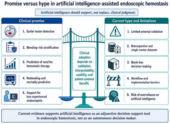
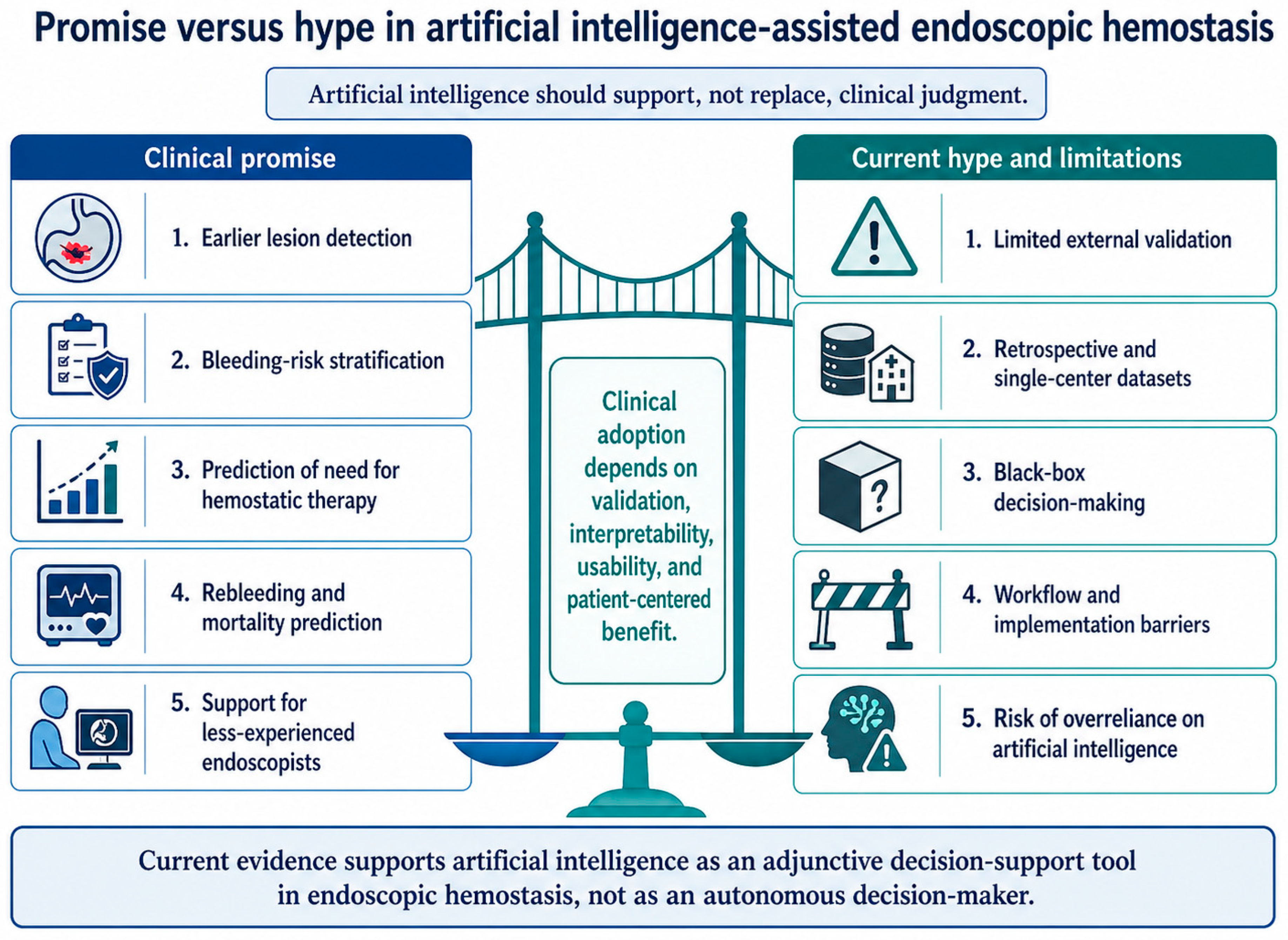
Despite the rapid expansion of AI in GI endoscopy, its translation into endoscopic hemostasis remains more limited than its technical performance might suggest [120]. High sensitivity, specificity, or area-under-the-curve values obtained in development datasets should not be interpreted as proof of clinical benefit [121]. In acute GI bleeding, the key question is not only whether an algorithm can recognize a lesion or predict risk, but whether its use changes patient management, reduces delayed or unnecessary procedures, prevents rebleeding, improves survival, or optimizes resource allocation [122]. This distinction is essential because many AI studies in medicine still evaluate diagnostic or predictive performance rather than clinically meaningful outcomes [123,124]. The balance between the potential benefits of AI and the main barriers to its clinical implementation in endoscopic hemostasis is illustrated in Figure 4.
Figure 4.
Potential benefits of artificial intelligence in endoscopic hemostasis and the main limitations, validation gaps, and implementation barriers affecting clinical adoption.
A major limitation is the persistent gap between model development and external validation. Many ML models for GI bleeding have been derived from retrospective cohorts, administrative databases, single-center registries, or selected image datasets [125,126]. Such models may perform well in the environment in which they were trained, but their reliability may decrease when applied to different hospitals, endoscopy platforms, patient populations, bleeding etiologies, or clinical workflows. This is particularly relevant in endoscopic hemostasis, where real-world decisions are influenced by hemodynamic instability, anticoagulant exposure, comorbidities, lesion accessibility, operator experience, and device availability—variables that are not always fully captured in model development [127,128].
Generalizability is further threatened by dataset shift and data drift. Endoscopic images obtained in emergency bleeding conditions differ substantially from curated training datasets: blood, clots, bubbles, poor insufflation, unstable visualization, active peristalsis, suboptimal lighting, and rapid scope movement can all modify image quality and model input [129]. Similarly, clinical prediction models may degrade when laboratory practices, transfusion policies, hemostatic devices, admission thresholds, or patient case-mix change over time [130]. Without prospective monitoring after deployment, an AI system that was accurate at validation may become unreliable during routine clinical use.
Another source of hype is incomplete and heterogeneous reporting. AI studies may omit essential details such as algorithm version, preprocessing methods, exclusion criteria for poor-quality inputs, threshold selection, code accessibility, calibration, handling of missing data, or subgroup performance. This weakens reproducibility and makes it difficult to compare models across studies. The emergence of reporting frameworks such as CONSORT-AI, TRIPOD + AI, DECIDE-AI, and STARD-AI reflects the need for higher methodological standards before AI tools are considered clinically mature [124,126,131,132].
The “black box” nature of many deep learning systems remains a major barrier in hemostatic decision-making [133]. In GI bleeding, an incorrect recommendation may delay urgent therapy, lead to unnecessary intervention, or falsely reassure clinicians after apparently successful hemostasis [7,132]. Explainability methods such as heatmaps, activation maps, local interpretable model-agnostic explanations, and SHapley additive explanations may improve transparency, but they do not necessarily prove that the model is using clinically valid reasoning [40,131]. Visual attention on a suspicious region does not equal causal interpretation, and feature attribution does not eliminate the need for clinical judgment.
Medicolegal responsibility is another unresolved issue in AI-assisted hemostatic decision-making. In emergency bleeding scenarios, delayed therapy, unnecessary intervention, or inappropriate reassurance after apparent hemostasis may have immediate consequences for patient safety. Therefore, the final responsibility for clinical decisions should remain with the treating physician, while institutions and manufacturers must define clear rules for validation, monitoring, documentation, software updates, and accountability when AI output contributes to patient management [17,134]. AI recommendations should be documented as supportive information rather than autonomous therapeutic decisions.
Implementation is also constrained by human factors. In emergency endoscopy, AI must operate in a high-pressure environment where the endoscopist is simultaneously responsible for diagnosis, suction, irrigation, device selection, hemostatic technique, patient instability, and communication with the anesthesia or intensive care team [17,132,135]. Poorly integrated alerts, excessive false positives, delayed outputs, or unclear recommendations may increase cognitive load rather than reduce it. For this reason, early-stage evaluation of AI systems should include usability, workflow integration, safety, and clinician interaction, not only model accuracy.
A further implementation risk is overreliance on AI. The assumption that it will automatically improve clinician performance ignores the possibility of automation bias, deskilling, or reduced vigilance. Recent evidence from colonoscopy suggests that continuous exposure to AI assistance may reduce unaided endoscopist performance, raising concerns that similar effects could occur in other visually dependent endoscopic tasks [136,137]. In hemostasis, where rapid independent judgment is essential when AI fails, is unavailable, or produces uncertain outputs, preserving endoscopist expertise remains critical.
Economic and regulatory barriers are equally important, because AI systems require acquisition costs, hardware integration, maintenance, software updates, cybersecurity safeguards, data governance, training, and post-market surveillance [134]. Their value, therefore, depends not only on diagnostic performance but also on whether they reduce unnecessary endoscopies, shorten time to hemostasis, decrease rebleeding, rationalize transfusion, reduce intensive care admissions, or improve survival [17]. Without cost-effectiveness analyses and clear reimbursement pathways, even technically successful systems may remain difficult to implement on a large scale.
Therefore, current evidence supports AI as an adjunctive tool rather than an autonomous decision-maker in endoscopic hemostasis. Its clinical value will depend on prospective validation, real-time usability, transparent reporting, integration into emergency workflows, and demonstrable improvement in patient-centered outcomes.
Limitations
Several limitations should be considered when interpreting the findings of this review. Although the literature search followed a structured approach, the evidence was synthesized narratively, with a descriptive appraisal of methodological quality rather than a formal risk-of-bias assessment. This approach was appropriate for the broad and heterogeneous nature of the topic, but it limits the ability to compare studies systematically or to quantify the overall strength of evidence. The available literature varies considerably in terms of clinical setting, patient population, AI architecture, input data, validation strategy, outcome definition, and reporting standards. As a result, models developed for lesion detection, bleeding-risk stratification, transfusion prediction, rebleeding assessment, or mortality estimation cannot be interpreted as equivalent in clinical maturity or implementation readiness.
A further limitation is that much of the current evidence remains derived from retrospective cohorts, selected image datasets, or internally validated prediction models. Prospective multicenter validation and real-time interventional studies remain limited, particularly in emergency endoscopy and hemostatic decision-making. Moreover, many studies primarily report diagnostic or predictive performance metrics, such as accuracy, sensitivity, specificity, or area under the curve, whereas fewer studies demonstrate that AI-assisted workflows improve clinically meaningful outcomes, including time to hemostasis, rebleeding prevention, transfusion optimization, resource allocation, intensive care use, or survival. Therefore, the conclusions of this review should be interpreted as a critical synthesis of an evolving evidence base rather than as definitive confirmation of clinical effectiveness. Further prospective validation, standardized reporting, and outcome-oriented trials are needed before AI-assisted hemostatic decision-making can be widely adopted in routine practice.
7. Conclusions
Artificial intelligence shows clear potential to support endoscopic hemostasis by improving lesion recognition, risk stratification, prediction of hemostatic therapy needs, and post-procedural monitoring. Current evidence suggests that these tools may reduce operator-dependent variability and assist clinical decision-making, particularly in complex or high-risk bleeding scenarios.
However, AI is not yet ready to replace expert judgment. Most available models still require stronger external validation, prospective testing, workflow integration, and proof of benefit on clinically relevant outcomes such as rebleeding, intervention timing, resource use, and survival. Its use in endoscopic hemostasis represents a promising adjunct rather than an autonomous solution. Its future value will depend on whether technical performance can be translated into safer, faster, and more individualized patient care.
Author Contributions
Conceptualization and design: O.B., P.A. and C.I.; methodology: A.B., O.B. and D.O.; formal analysis: P.A.; investigation: D.O.; resources: A.B. and C.I.; software and data analysis: O.B.; supervision: D.O.; validation: D.O. and O.B.; writing—original draft: P.A., C.I. and O.B. All authors have read and agreed to the published version of the manuscript.
Funding
This research received no external funding.
Institutional Review Board Statement
Not applicable.
Informed Consent Statement
Not applicable.
Data Availability Statement
Not applicable.
Conflicts of Interest
The authors have no conflicts of interest to declare.
References
- Li, Z.; Koban, K.C.; Schenck, T.L.; Giunta, R.E.; Li, Q.; Sun, Y. Artificial Intelligence in Dermatology Image Analysis: Current Developments and Future Trends. J. Clin. Med. 2022, 11, 6826. [Google Scholar] [CrossRef] [Scilit] [PubMed] [PubMed Central]
- Chadebecq, F.; Lovat, L.B.; Stoyanov, D. Artificial intelligence and automation in endoscopy and surgery. Nat. Rev. Gastroenterol. Hepatol. 2023, 20, 171–182. [Google Scholar] [CrossRef] [Scilit]
- Parasa, S.; Berzin, T.; Leggett, C.; Gross, S.; Repici, A.; Ahmad, O.F.; Chiang, A.; Coelho-Prabhu, N.; Cohen, J.; Dekker, E.; et al. Consensus statements on the current landscape of artificial intelligence applications in endoscopy, addressing roadblocks, and advancing artificial intelligence in gastroenterology. Gastrointest. Endosc. 2025, 101, 2–9.e1. [Google Scholar] [CrossRef] [Scilit]
- Parasa, S.; Repici, A.; Berzin, T.; Leggett, C.; Gross, S.A.; Sharma, P. Framework and metrics for the clinical use and implementation of artificial intelligence algorithms into endoscopy practice: Recommendations from the American Society for Gastrointestinal Endoscopy Artificial Intelligence Task Force. Gastrointest. Endosc. 2023, 97, 815–824.e1. [Google Scholar] [CrossRef] [Scilit]
- Januszewicz, W.; Witczak, K.; Wieszczy, P.; Socha, M.; Turkot, M.H.; Wojciechowska, U.; Didkowska, J.; Kaminski, M.F.; Regula, J. Prevalence and risk factors of upper gastrointestinal cancers missed during endoscopy: A nationwide registry-based study. Endoscopy 2022, 54, 653–660. [Google Scholar] [CrossRef] [Scilit]
- Holster, I.L.; Kuipers, E.J. Update on the endoscopic management of peptic ulcer bleeding. Curr. Gastroenterol. Rep. 2011, 13, 525–531. [Google Scholar] [CrossRef] [Scilit] [PubMed] [PubMed Central]
- Barkun, A.N.; Almadi, M.; Kuipers, E.J.; Laine, L.; Sung, J.; Tse, F.; Leontiadis, G.I.; Abraham, N.S.; Calvet, X.; Chan, F.K.L.; et al. Management of Nonvariceal Upper Gastrointestinal Bleeding: Guideline Recommendations from the International Consensus Group. Ann. Intern. Med. 2019, 171, 805–822. [Google Scholar] [CrossRef] [Scilit]
- Gralnek, I.M.; Stanley, A.J.; Morris, A.J.; Camus, M.; Lau, J.; Lanas, A.; Laursen, S.B.; Radaelli, F.; Papanikolaou, I.S.; Cúrdia Gonçalves, T.; et al. Endoscopic diagnosis and management of nonvariceal upper gastrointestinal hemorrhage (NVUGIH): European Society of Gastrointestinal Endoscopy (ESGE) Guideline—Update 2021. Endoscopy 2021, 53, 300–332. [Google Scholar] [CrossRef] [Scilit] [PubMed]
- Nazarian, S.; Lo, F.P.W.; Qiu, J.; Patel, N.; Lo, B.; Ayaru, L. Development and validation of machine learning models to predict the need for haemostatic therapy in acute upper gastrointestinal bleeding. Clin. Med. Insights Gastroenterol. 2024, 17, 26317745241246899. [Google Scholar] [CrossRef] [Scilit] [PubMed]
- Raghareutai, K.; Tanchotsrinon, W.; Sattayalertyanyong, O.; Kaosombatwattana, U. Development and validation of a machine learning model to predict hemostatic intervention in patients with acute upper gastrointestinal bleeding. BMC Med. Inform. Decis. Mak. 2025, 25, 145. [Google Scholar] [CrossRef] [Scilit]
- Levi, R.; Carli, F.; Arévalo, A.R.; Altinel, Y.; Stein, D.J.; Naldini, M.M.; Grassi, F.; Zanoni, A.; Finkelstein, S.; Vieira, S.M.; et al. Artificial intelligence-based prediction of transfusion in the intensive care unit in patients with gastrointestinal bleeding. BMJ Health Care Inform. 2021, 28, e100245. [Google Scholar] [CrossRef] [Scilit] [PubMed] [PubMed Central]
- Nigam, G.B.; Murphy, M.F.; Travis, S.P.L.; Stanley, A.J. Machine learning in the assessment and management of acute gastrointestinal bleeding. BMJ Med. 2024, 3, e000699. [Google Scholar] [CrossRef] [Scilit]
- Ebigbo, A.; Mendel, R.; Probst, A.; Manzeneder, J.; Prinz, F.; De Souza, L.A., Jr.; Papa, J.; Palm, C.; Messmann, H. Real-time use of artificial intelligence in the evaluation of cancer in Barrett’s oesophagus. Gut 2020, 69, 615–616. [Google Scholar] [CrossRef] [Scilit] [PubMed]
- De Groof, A.J.; Struyvenberg, M.R.; Fockens, K.N.; Van Der Putten, J.; Van Der Sommen, F.; Boers, T.G.; Zinger, S.; Bisschops, R.; De With, P.H.; Pouw, R.E.; et al. Deep learning algorithm detection of Barrett’s neoplasia with high accuracy during live endoscopic procedures: A pilot study (with video). Gastrointest. Endosc. 2020, 91, 1242–1250. [Google Scholar] [CrossRef] [Scilit]
- Popovic, D.; Glisic, T.; Milosavljevic, T.; Panic, N.; Marjanovic-Haljilji, M.; Mijac, D.; Stojkovic Lalosevic, M.; Nestorov, J.; Dragasevic, S.; Savic, P.; et al. The Importance of Artificial Intelligence in Upper Gastrointestinal Endoscopy. Diagnostics 2023, 13, 2862. [Google Scholar] [CrossRef] [Scilit]
- Renna, F.; Martins, M.; Neto, A.; Cunha, A.; Libânio, D.; Dinis-Ribeiro, M.; Coimbra, M. Artificial Intelligence for Upper Gastrointestinal Endoscopy: A Roadmap from Technology Development to Clinical Practice. Diagnostics 2022, 12, 1278. [Google Scholar] [CrossRef] [Scilit]
- El-Sayed, A.; Lovat, L.B.; Ahmad, O.F. Clinical Implementation of Artificial Intelligence in Gastroenterology: Current Landscape, Regulatory Challenges, and Ethical Issues. Gastroenterology 2025, 169, 518–530. [Google Scholar] [CrossRef] [Scilit] [PubMed]
- Yen, H.-H.; Wu, P.-Y.; Su, P.-Y.; Yang, C.-W.; Chen, Y.-Y.; Chen, M.-F.; Lin, W.-C.; Tsai, C.-L.; Lin, K.-P. Performance Comparison of the Deep Learning and the Human Endoscopist for Bleeding Peptic Ulcer Disease. J. Med. Biol. Eng. 2021, 41, 504–513. [Google Scholar] [CrossRef] [Scilit]
- Yen, H.-H.; Wu, P.-Y.; Wu, T.-L.; Huang, S.-P.; Chen, Y.-Y.; Chen, M.-F.; Lin, W.-C.; Tsai, C.-L.; Lin, K.-P. Forrest Classification for Bleeding Peptic Ulcer: A New Look at the Old Endoscopic Classification. Diagnostics 2022, 12, 1066. [Google Scholar] [CrossRef] [Scilit]
- Seo, D.-W.; Yi, H.; Park, B.; Kim, Y.-J.; Jung, D.H.; Woo, I.; Sohn, C.H.; Ko, B.S.; Kim, N.; Kim, W.Y. Prediction of Adverse Events in Stable Non-Variceal Gastrointestinal Bleeding Using Machine Learning. J. Clin. Med. 2020, 9, 2603. [Google Scholar] [CrossRef] [Scilit]
- Li, R.; Zhu, Y.; Xu, W. Development and validation of an interpretable predictive model for short-term rebleeding after endoscopic hemostasis in peptic ulcer: A retrospective single-center cohort study. Surg. Endosc. 2026, 40, 2398–2413. [Google Scholar] [CrossRef] [Scilit]
- Hong, Y.; Yu, Q.; Mo, F.; Yin, M.; Xu, C.; Zhu, S.; Lin, J.; Xu, G.; Gao, J.; Liu, L.; et al. Deep learning to predict esophageal variceal bleeding based on endoscopic images. J. Int. Med. Res. 2023, 51, 03000605231200371. [Google Scholar] [CrossRef] [Scilit]
- Zhao, X.; Wei, S.; Pan, Y.; Qu, K.; Yan, G.; Wang, X.; Song, Y. Early prognosis prediction for non-variceal upper gastrointestinal bleeding in the intensive care unit: Based on interpretable machine learning. Eur. J. Med. Res. 2024, 29, 442. [Google Scholar] [CrossRef] [Scilit]
- Liu, Z.; Zhang, L.; Jiang, G.J.; Chen, Q.Q.; Hou, Y.G.; Wu, W.; Malik, M.; Li, G.; Zhan, L. Machine Learning-Based Mortality Prediction for Acute Gastrointestinal Bleeding Patients Admitted to Intensive Care Unit. Curr. Med. Sci. 2025, 45, 70–81. [Google Scholar] [CrossRef] [Scilit]
- Boros, E.; Pintér, J.; Molontay, R.; Prószéky, K.G.; Vörhendi, N.; Simon, O.A.; Teutsch, B.; Pálinkás, D.; Frim, L.; Tari, E.; et al. New machine-learning models outperform conventional risk assessment tools in Gastrointestinal bleeding. Sci. Rep. 2025, 15, 6371. [Google Scholar] [CrossRef] [Scilit]
- Li, Q.; Chen, G.; Li, Q. Multi-task machine learning for transfusion decision support in acute upper gastrointestinal bleeding: A novel ensemble approach with clinical validation. J. Transl. Med. 2025, 23, 979. [Google Scholar] [CrossRef] [Scilit]
- Rosa, B.; Mascarenhas Saraiva, M.J.; Afonso, J.; Cúrdia Gonçalves, T.; Mendes, F.; Moreira, M.J.; Martins, M.; Dias De Castro, F.; Ribeiro, T.; Cardoso, P.; et al. Artificial intelligence-assisted versus conventional reading in pan-intestinal capsule endoscopy for suspected mid-lower gastrointestinal bleeding: A retrospective analysis of a prospective cohort. BMJ Open Gastroenterol. 2025, 12, e001906. [Google Scholar] [CrossRef] [Scilit] [PubMed]
- Aoyama, N.; Nakajo, K.; Sasabe, M.; Inaba, A.; Nakanishi, Y.; Seno, H.; Yano, T. Effects of artificial intelligence assistance on endoscopist performance: Comparison of diagnostic performance in superficial esophageal squamous cell carcinoma detection using video-based models. DEN Open 2026, 6, e70083. [Google Scholar] [CrossRef] [Scilit] [PubMed]
- Okagawa, Y.; Abe, S.; Yamada, M.; Oda, I.; Saito, Y. Artificial Intelligence in Endoscopy. Dig. Dis. Sci. 2022, 67, 1553–1572. [Google Scholar] [CrossRef] [Scilit] [PubMed]
- Tokat, M.; Van Tilburg, L.; Koch, A.D.; Spaander, M.C.W. Artificial Intelligence in Upper Gastrointestinal Endoscopy. Dig. Dis. 2022, 40, 395–408. [Google Scholar] [CrossRef] [Scilit]
- Visaggi, P.; De Bortoli, N.; Barberio, B.; Savarino, V.; Oleas, R.; Rosi, E.M.; Marchi, S.; Ribolsi, M.; Savarino, E. Artificial Intelligence in the Diagnosis of Upper Gastrointestinal Diseases. J. Clin. Gastroenterol. 2022, 56, 23–35. [Google Scholar] [CrossRef] [Scilit]
- Le Berre, C.; Sandborn, W.J.; Aridhi, S.; Devignes, M.-D.; Fournier, L.; Smaïl-Tabbone, M.; Danese, S.; Peyrin-Biroulet, L. Application of Artificial Intelligence to Gastroenterology and Hepatology. Gastroenterology 2020, 158, 76–94.e2. [Google Scholar] [CrossRef] [Scilit]
- Ebigbo, A.; Palm, C.; Probst, A.; Mendel, R.; Manzeneder, J.; Prinz, F.; De Souza, L.A.; Papa, J.P.; Siersema, P.; Messmann, H. A technical review of artificial intelligence as applied to gastrointestinal endoscopy: Clarifying the terminology. Endosc. Int. Open 2019, 07, E1616–E1623. [Google Scholar] [CrossRef] [Scilit] [PubMed]
- Huang, L.-M.; Yang, W.-J.; Huang, Z.-Y.; Tang, C.-W.; Li, J. Artificial intelligence technique in detection of early esophageal cancer. World J. Gastroenterol. 2020, 26, 5959–5969. [Google Scholar] [CrossRef] [Scilit] [PubMed] [PubMed Central]
- Hamade, N.; Sharma, P. Artificial intelligence in Barrett’s Esophagus. Clin. Med. Insights Gastroenterol. 2021, 14, 26317745211049964. [Google Scholar] [CrossRef] [Scilit]
- Mori, Y.; Kudo, S.; Mohmed, H.E.N.; Misawa, M.; Ogata, N.; Itoh, H.; Oda, M.; Mori, K. Artificial intelligence and upper gastrointestinal endoscopy: Current status and future perspective. Dig. Endosc. 2019, 31, 378–388. [Google Scholar] [CrossRef] [Scilit]
- Smith, E.R.; Shah, J. Applications of Artificial Intelligence to Eosinophilic Esophagitis. Gastroenterol. Insights 2022, 13, 218–227. [Google Scholar] [CrossRef] [Scilit]
- Li, X.-R.; Kong, M.-W.; Guan, X.-F.; Gao, Y. Revolutionizing upper gastrointestinal disease diagnosis: The transformative role of artificial intelligence in endoscopy. World J. Gastrointest. Endosc. 2025, 17, 108293. [Google Scholar] [CrossRef] [Scilit] [PubMed]
- Mascarenhas, M.; Mendes, F.; Martins, M.; Ribeiro, T.; Afonso, J.; Cardoso, P.; Ferreira, J.; Fonseca, J.; Macedo, G. Explainable AI in Digestive Healthcare and Gastrointestinal Endoscopy. J. Clin. Med. 2025, 14, 549. [Google Scholar] [CrossRef] [Scilit]
- Kim, C.; Gadgil, S.U.; Lee, S.-I. Transparency of medical artificial intelligence systems. Nat. Rev. Bioeng. 2025, 4, 11–29. [Google Scholar] [CrossRef] [Scilit]
- Chan, B. Black-box assisted medical decisions: AI power vs. ethical physician care. Med. Health Care Philos. 2023, 26, 285–292. [Google Scholar] [CrossRef] [Scilit] [PubMed] [PubMed Central]
- Raposo, V.L. The fifty shades of black: About black box AI and explainability in healthcare. Med. Law Rev. 2025, 33, fwaf005. [Google Scholar] [CrossRef] [Scilit]
- Glover, B.; Teare, J.; Patel, N. A systematic review of the role of non-magnified endoscopy for the assessment of H. pylori infection. Endosc. Int. Open 2020, 8, E105–E114. [Google Scholar] [CrossRef] [Scilit]
- Roshan, A.; Byrne, M.F. Artificial intelligence in colorectal cancer screening. CMAJ 2022, 194, E1481–E1484. [Google Scholar] [CrossRef] [Scilit] [PubMed]
- Byrne, M.F.; Shahidi, N.; Rex, D.K. Will Computer-Aided Detection and Diagnosis Revolutionize Colonoscopy? Gastroenterology 2017, 153, 1460–1464.e1. [Google Scholar] [CrossRef] [Scilit] [PubMed]
- Arif, A.A.; Jiang, S.X.; Byrne, M.F. Artificial intelligence in endoscopy: Overview, applications, and future directions. Saudi J. Gastroenterol. 2023, 29, 269–277. [Google Scholar] [CrossRef] [Scilit]
- Taghiakbari, M.; Mori, Y.; Von Renteln, D. Artificial intelligence-assisted colonoscopy: A review of current state of practice and research. World J. Gastroenterol. 2021, 27, 8103–8122. [Google Scholar] [CrossRef] [Scilit] [PubMed]
- Barua, I.; Wieszczy, P.; Kudo, S.; Misawa, M.; Holme, Ø.; Gulati, S.; Williams, S.; Mori, K.; Itoh, H.; Takishima, K.; et al. Real-Time Artificial Intelligence–Based Optical Diagnosis of Neoplastic Polyps during Colonoscopy. NEJM Evid. 2022, 1, EVIDoa2200003. [Google Scholar] [CrossRef] [Scilit]
- Von Renteln, D.; Djinbachian, R.; Zarandi-Nowroozi, M.; Taghiakbari, M. Measuring size of smaller colorectal polyps using a virtual scale function during endoscopies. Gut 2023, 72, 417–420. [Google Scholar] [CrossRef] [Scilit]
- Wu, L.; Zhang, J.; Zhou, W.; An, P.; Shen, L.; Liu, J.; Jiang, X.; Huang, X.; Mu, G.; Wan, X.; et al. Randomised controlled trial of WISENSE, a real-time quality improving system for monitoring blind spots during esophagogastroduodenoscopy. Gut 2019, 68, 2161–2169. [Google Scholar] [CrossRef] [Scilit]
- Yao, L.; Zhang, L.; Liu, J.; Zhou, W.; He, C.; Zhang, J.; Wu, L.; Wang, H.; Xu, Y.; Gong, D.; et al. Effect of an artificial intelligence-based quality improvement system on efficacy of a computer-aided detection system in colonoscopy: A four-group parallel study. Endoscopy 2022, 54, 757–768. [Google Scholar] [CrossRef] [Scilit] [PubMed]
- Samnani, S.; Khan, R.; Heitman, S.J.; Hilsden, R.J.; Byrne, M.F.; Grover, S.C.; Forbes, N. Optimizing adenoma detection in screening-related colonoscopy. Expert Rev. Gastroenterol. Hepatol. 2023, 17, 589–602. [Google Scholar] [CrossRef] [Scilit]
- Choi, S.J.; Khan, M.A.; Choi, H.S.; Choo, J.; Lee, J.M.; Kwon, S.; Keum, B.; Chun, H.J. Development of artificial intelligence system for quality control of photo documentation in esophagogastroduodenoscopy. Surg. Endosc. 2022, 36, 57–65. [Google Scholar] [CrossRef] [Scilit] [PubMed]
- Takiyama, H.; Ozawa, T.; Ishihara, S.; Fujishiro, M.; Shichijo, S.; Nomura, S.; Miura, M.; Tada, T. Automatic anatomical classification of esophagogastroduodenoscopy images using deep convolutional neural networks. Sci. Rep. 2018, 8, 7497. [Google Scholar] [CrossRef] [Scilit]
- Chen, D.; Wu, L.; Li, Y.; Zhang, J.; Liu, J.; Huang, L.; Jiang, X.; Huang, X.; Mu, G.; Hu, S.; et al. Comparing blind spots of unsedated ultrafine, sedated, and unsedated conventional gastroscopy with and without artificial intelligence: A prospective, single-blind, 3-parallel-group, randomized, single-center trial. Gastrointest. Endosc. 2020, 91, 332–339.e3. [Google Scholar] [CrossRef] [Scilit]
- Liu, W.; Wu, Y.; Yuan, X.; Zhang, J.; Zhou, Y.; Zhang, W.; Zhu, P.; Tao, Z.; He, L.; Hu, B.; et al. Artificial intelligence-based assessments of colonoscopic withdrawal technique: A new method for measuring and enhancing the quality of fold examination. Endoscopy 2022, 54, 972–979. [Google Scholar] [CrossRef] [Scilit]
- Zhou, W.; Yao, L.; Wu, H.; Zheng, B.; Hu, S.; Zhang, L.; Li, X.; He, C.; Wang, Z.; Li, Y.; et al. Multi-step validation of a deep learning-based system for the quantification of bowel preparation: A prospective, observational study. Lancet Digit. Health 2021, 3, e697–e706. [Google Scholar] [CrossRef] [Scilit]
- Yen, H.-H.; Wu, P.-Y.; Chen, M.-F.; Lin, W.-C.; Tsai, C.-L.; Lin, K.-P. Current Status and Future Perspective of Artificial Intelligence in the Management of Peptic Ulcer Bleeding: A Review of Recent Literature. J. Clin. Med. 2021, 10, 3527. [Google Scholar] [CrossRef] [Scilit]
- Alemam, A.; Tamanna, R.; Ali, M.; Ibraheem, N.; Swealem, A. Artificial Intelligence in Upper Gastrointestinal Endoscopy: Current Evidence, Practice, and Future Directions. Cureus 2025, 17, e96657. [Google Scholar] [CrossRef] [Scilit] [PubMed]
- De Groof, A.J.; Struyvenberg, M.R.; Van Der Putten, J.; Van Der Sommen, F.; Fockens, K.N.; Curvers, W.L.; Zinger, S.; Pouw, R.E.; Coron, E.; Baldaque-Silva, F.; et al. Deep-Learning System Detects Neoplasia in Patients with Barrett’s Esophagus with Higher Accuracy Than Endoscopists in a Multistep Training and Validation Study with Benchmarking. Gastroenterology 2020, 158, 915–929.e4. [Google Scholar] [CrossRef] [Scilit]
- Shichijo, S.; Nomura, S.; Aoyama, K.; Nishikawa, Y.; Miura, M.; Shinagawa, T.; Takiyama, H.; Tanimoto, T.; Ishihara, S.; Matsuo, K.; et al. Application of Convolutional Neural Networks in the Diagnosis of Helicobacter pylori Infection Based on Endoscopic Images. EBioMedicine 2017, 25, 106–111. [Google Scholar] [CrossRef] [Scilit]
- Ishihara, R.; Matsuura, N.; Hanaoka, N.; Yamamoto, S.; Akasaka, T.; Takeuchi, Y.; Higashino, K.; Uedo, N.; Iishi, H. Endoscopic imaging modalities for diagnosing invasion depth of superficial esophageal squamous cell carcinoma: A systematic review and meta-analysis. BMC Gastroenterol. 2017, 17, 24. [Google Scholar] [CrossRef] [Scilit] [PubMed] [PubMed Central]
- Vincze, Á. Endoscopic diagnosis and treatment in gastric cancer: Current evidence and new perspectives. Front. Surg. 2023, 10, 1122454. [Google Scholar] [CrossRef] [Scilit]
- Van Der Sommen, F.; Zinger, S.; Curvers, W.; Bisschops, R.; Pech, O.; Weusten, B.; Bergman, J.; De With, P.; Schoon, E. Computer-aided detection of early neoplastic lesions in Barrett’s esophagus. Endoscopy 2016, 48, 617–624. [Google Scholar] [CrossRef] [Scilit]
- Qi, X.; Sivak, M.V.; Isenberg, G.; Willis Joseph, E.; Rollins, A.M. Computer-aided diagnosis of dysplasia in Barrett’s esophagus using endoscopic optical coherence tomography. J. Biomed. Opt. 2006, 11, 044010. [Google Scholar] [CrossRef] [Scilit] [PubMed]
- Tokai, Y.; Yoshio, T.; Aoyama, K.; Horie, Y.; Yoshimizu, S.; Horiuchi, Y.; Ishiyama, A.; Tsuchida, T.; Hirasawa, T.; Sakakibara, Y.; et al. Application of artificial intelligence using convolutional neural networks in determining the invasion depth of esophageal squamous cell carcinoma. Esophagus 2020, 17, 250–256. [Google Scholar] [CrossRef] [Scilit] [PubMed]
- Nakagawa, K.; Ishihara, R.; Aoyama, K.; Ohmori, M.; Nakahira, H.; Matsuura, N.; Shichijo, S.; Nishida, T.; Yamada, T.; Yamaguchi, S.; et al. Classification for invasion depth of esophageal squamous cell carcinoma using a deep neural network compared with experienced endoscopists. Gastrointest. Endosc. 2019, 90, 407–414. [Google Scholar] [CrossRef] [Scilit] [PubMed]
- Miyaki, R.; Yoshida, S.; Tanaka, S.; Kominami, Y.; Sanomura, Y.; Matsuo, T.; Oka, S.; Raytchev, B.; Tamaki, T.; Koide, T.; et al. A Computer System to Be Used with Laser-based Endoscopy for Quantitative Diagnosis of Early Gastric Cancer. J. Clin. Gastroenterol. 2015, 49, 108–115. [Google Scholar] [CrossRef] [Scilit]
- Kanesaka, T.; Lee, T.-C.; Uedo, N.; Lin, K.-P.; Chen, H.-Z.; Lee, J.-Y.; Wang, H.-P.; Chang, H.-T. Computer-aided diagnosis for identifying and delineating early gastric cancers in magnifying narrow-band imaging. Gastrointest. Endosc. 2018, 87, 1339–1344. [Google Scholar] [CrossRef] [Scilit]
- Zhu, Y.; Wang, Q.-C.; Xu, M.-D.; Zhang, Z.; Cheng, J.; Zhong, Y.-S.; Zhang, Y.-Q.; Chen, W.-F.; Yao, L.-Q.; Zhou, P.-H.; et al. Application of convolutional neural network in the diagnosis of the invasion depth of gastric cancer based on conventional endoscopy. Gastrointest. Endosc. 2019, 89, 806–815.e1. [Google Scholar] [CrossRef] [Scilit]
- Jamel, S.; Tukanova, K.; Markar, S. Detection and management of oligometastatic disease in oesophageal cancer and identification of prognostic factors: A systematic review. World J. Gastrointest. Oncol. 2019, 11, 741–749. [Google Scholar] [CrossRef] [Scilit]
- Cai, S.-L.; Li, B.; Tan, W.-M.; Niu, X.-J.; Yu, H.-H.; Yao, L.-Q.; Zhou, P.-H.; Yan, B.; Zhong, Y.-S. Using a deep learning system in endoscopy for screening of early esophageal squamous cell carcinoma (with video). Gastrointest. Endosc. 2019, 90, 745–753.e2. [Google Scholar] [CrossRef] [Scilit]
- Horie, Y.; Yoshio, T.; Aoyama, K.; Yoshimizu, S.; Horiuchi, Y.; Ishiyama, A.; Hirasawa, T.; Tsuchida, T.; Ozawa, T.; Ishihara, S.; et al. Diagnostic outcomes of esophageal cancer by artificial intelligence using convolutional neural networks. Gastrointest. Endosc. 2019, 89, 25–32. [Google Scholar] [CrossRef] [Scilit]
- Tang, D.; Wang, X.; Wang, L.; He, G.; Fu, Y.; Li, X.; Zhang, Y.; Guo, H.; Zhu, H.; Xu, G.; et al. Tu2004 artificial intelligence network to aid the diagnosis of early esophageal squamous cell carcinoma and esophageal inflammations in white light endoscopic images. Gastrointest. Endosc. 2019, 89, AB654. [Google Scholar] [CrossRef] [Scilit]
- Quang, T.; Schwarz, R.A.; Dawsey, S.M.; Tan, M.C.; Patel, K.; Yu, X.; Wang, G.; Zhang, F.; Xu, H.; Anandasabapathy, S.; et al. A tablet-interfaced high-resolution microendoscope with automated image interpretation for real-time evaluation of esophageal squamous cell neoplasia. Gastrointest. Endosc. 2016, 84, 834–841. [Google Scholar] [CrossRef] [Scilit] [PubMed]
- Kumagai, Y.; Takubo, K.; Kawada, K.; Aoyama, K.; Endo, Y.; Ozawa, T.; Hirasawa, T.; Yoshio, T.; Ishihara, S.; Fujishiro, M.; et al. Diagnosis using deep-learning artificial intelligence based on the endocytoscopic observation of the esophagus. Esophagus 2019, 16, 180–187. [Google Scholar] [CrossRef] [Scilit]
- Shiroma, S.; Yoshio, T.; Aoyama, K.; Ishiyama, A.; Tsuchida, T.; Fujisaki, J.; Tada, T. Sa1252 the application of artificial intelligence to detect esophageal squamous cell carcinoma in movies using convoluntional neural networks. Gastrointest. Endosc. 2019, 89, AB189. [Google Scholar] [CrossRef] [Scilit]
- Jin, P.; Ji, X.; Kang, W.; Li, Y.; Liu, H.; Ma, F.; Ma, S.; Hu, H.; Li, W.; Tian, Y. Artificial intelligence in gastric cancer: A systematic review. J. Cancer Res. Clin. Oncol. 2020, 146, 2339–2350. [Google Scholar] [CrossRef] [Scilit]
- Hirasawa, T.; Aoyama, K.; Tanimoto, T.; Ishihara, S.; Shichijo, S.; Ozawa, T.; Ohnishi, T.; Fujishiro, M.; Matsuo, K.; Fujisaki, J.; et al. Application of artificial intelligence using a convolutional neural network for detecting gastric cancer in endoscopic images. Gastric Cancer 2018, 21, 653–660. [Google Scholar] [CrossRef] [Scilit]
- Ueyama, H.; Kato, Y.; Akazawa, Y.; Yatagai, N.; Komori, H.; Takeda, T.; Matsumoto, K.; Ueda, K.; Matsumoto, K.; Hojo, M.; et al. Application of artificial intelligence using a convolutional neural network for diagnosis of early gastric cancer based on magnifying endoscopy with narrow-band imaging. J. Gastro Hepatol. 2021, 36, 482–489. [Google Scholar] [CrossRef] [Scilit]
- Hu, H.; Gong, L.; Dong, D.; Zhu, L.; Wang, M.; He, J.; Shu, L.; Cai, Y.; Cai, S.; Su, W.; et al. Identifying early gastric cancer under magnifying narrow-band images with deep learning: A multicenter study. Gastrointest. Endosc. 2021, 93, 1333–1341.e3. [Google Scholar] [CrossRef] [Scilit]
- Cho, B.-J.; Bang, C.S.; Lee, J.J.; Seo, C.W.; Kim, J.H. Prediction of Submucosal Invasion for Gastric Neoplasms in Endoscopic Images Using Deep-Learning. J. Clin. Med. 2020, 9, 1858. [Google Scholar] [CrossRef] [Scilit]
- Zhang, Y.; Li, F.; Yuan, F.; Zhang, K.; Huo, L.; Dong, Z.; Lang, Y.; Zhang, Y.; Wang, M.; Gao, Z.; et al. Diagnosing chronic atrophic gastritis by gastroscopy using artificial intelligence. Dig. Liver Dis. 2020, 52, 566–572. [Google Scholar] [CrossRef] [Scilit]
- Horiuchi, Y.; Aoyama, K.; Tokai, Y.; Hirasawa, T.; Yoshimizu, S.; Ishiyama, A.; Yoshio, T.; Tsuchida, T.; Fujisaki, J.; Tada, T. Convolutional Neural Network for Differentiating Gastric Cancer from Gastritis Using Magnified Endoscopy with Narrow Band Imaging. Dig. Dis. Sci. 2020, 65, 1355–1363. [Google Scholar] [CrossRef] [Scilit]
- Li, L.; Chen, Y.; Shen, Z.; Zhang, X.; Sang, J.; Ding, Y.; Yang, X.; Li, J.; Chen, M.; Jin, C.; et al. Convolutional neural network for the diagnosis of early gastric cancer based on magnifying narrow band imaging. Gastric Cancer 2020, 23, 126–132. [Google Scholar] [CrossRef] [Scilit]
- Wu, L.; Zhou, W.; Wan, X.; Zhang, J.; Shen, L.; Hu, S.; Ding, Q.; Mu, G.; Yin, A.; Huang, X.; et al. A deep neural network improves endoscopic detection of early gastric cancer without blind spots. Endoscopy 2019, 51, 522–531. [Google Scholar] [CrossRef] [Scilit] [PubMed]
- Guimarães, P.; Keller, A.; Fehlmann, T.; Lammert, F.; Casper, M. Deep-learning based detection of gastric precancerous conditions. Gut 2020, 69, 4–6. [Google Scholar] [CrossRef] [Scilit] [PubMed]
- Namikawa, K.; Hirasawa, T.; Nakano, K.; Ikenoyama, Y.; Ishioka, M.; Shiroma, S.; Tokai, Y.; Yoshimizu, S.; Horiuchi, Y.; Ishiyama, A.; et al. Artificial intelligence-based diagnostic system classifying gastric cancers and ulcers: Comparison between the original and newly developed systems. Endoscopy 2020, 52, 1077–1083. [Google Scholar] [CrossRef] [Scilit] [PubMed]
- Cho, B.-J.; Bang, C.S.; Park, S.W.; Yang, Y.J.; Seo, S.I.; Lim, H.; Shin, W.G.; Hong, J.T.; Yoo, Y.T.; Hong, S.H.; et al. Automated classification of gastric neoplasms in endoscopic images using a convolutional neural network. Endoscopy 2019, 51, 1121–1129. [Google Scholar] [CrossRef] [Scilit]
- Wu, L.; Wang, J.; He, X.; Zhu, Y.; Jiang, X.; Chen, Y.; Wang, Y.; Huang, L.; Shang, R.; Dong, Z.; et al. Deep learning system compared with expert endoscopists in predicting early gastric cancer and its invasion depth and differentiation status (with videos). Gastrointest. Endosc. 2022, 95, 92–104.e3. [Google Scholar] [CrossRef] [Scilit]
- El Hajjar, A.; Rey, J.-F. Artificial intelligence in gastrointestinal endoscopy: General overview. Chin. Med. J. 2020, 133, 326–334. [Google Scholar] [CrossRef] [Scilit]
- Bosch, S.; Teerenstra, S.; De Wilt, J.; Cunningham, C.; Nagtegaal, I. Predicting lymph node metastasis in pT1 colorectal cancer: A systematic review of risk factors providing rationale for therapy decisions. Endoscopy 2013, 45, 827–841. [Google Scholar] [CrossRef] [Scilit]
- Overwater, A.; Kessels, K.; Elias, S.G.; Backes, Y.; Spanier, B.W.M.; Seerden, T.C.J.; Pullens, H.J.M.; De Vos Tot Nederveen Cappel, W.H.; Van Den Blink, A.; Offerhaus, G.J.A.; et al. Endoscopic resection of high-risk T1 colorectal carcinoma prior to surgical resection has no adverse effect on long-term outcomes. Gut 2018, 67, 284–290. [Google Scholar] [CrossRef] [Scilit]
- Miyachi, H.; Kudo, S.; Ichimasa, K.; Hisayuki, T.; Oikawa, H.; Matsudaira, S.; Kouyama, Y.; Kimura, Y.J.; Misawa, M.; Mori, Y.; et al. Management of T1 colorectal cancers after endoscopic treatment based on the risk stratification of lymph node metastasis. J. Gastro Hepatol. 2016, 31, 1126–1132. [Google Scholar] [CrossRef] [Scilit]
- Ichimasa, K.; Kudo, S.; Mori, Y.; Misawa, M.; Matsudaira, S.; Kouyama, Y.; Baba, T.; Hidaka, E.; Wakamura, K.; Hayashi, T.; et al. Artificial intelligence may help in predicting the need for additional surgery after endoscopic resection of T1 colorectal cancer. Endoscopy 2018, 50, 230–240. [Google Scholar] [CrossRef] [Scilit]
- Murillo Pineda, M.I.; Siu Xiao, T.; Sanabria Herrera, E.J.; Ayala Aguilar, A.; Arriaga Escamilla, D.; Aleman Reyes, A.M.; Rojas Marron, A.D.; Fabila Lievano, R.R.; De Jesús Correa Gomez, J.J.; Martinez Ramirez, M. The Prediction and Treatment of Bleeding Esophageal Varices in the Artificial Intelligence Era: A Review. Cureus 2024, 16, e55786. [Google Scholar] [CrossRef] [Scilit]
- Wang, J.; Wang, Z.; Chen, M.; Xiao, Y.; Chen, S.; Wu, L.; Yao, L.; Jiang, X.; Li, J.; Xu, M.; et al. An interpretable artificial intelligence system for detecting risk factors of gastroesophageal variceal bleeding. npj Digit. Med. 2022, 5, 183. [Google Scholar] [CrossRef] [Scilit]
- Huang, Y.; Li, J.; Zheng, T.; Ji, D.; Wong, Y.J.; You, H.; Gu, Y.; Li, M.; Zhao, L.; Li, S.; et al. Development and validation of a machine learning–based model for varices screening in compensated cirrhosis (CHESS2001): An international multicenter study. Gastrointest. Endosc. 2023, 97, 435–444.e2. [Google Scholar] [CrossRef] [Scilit]
- Yan, Y.; Li, Y.; Fan, C.; Zhang, Y.; Zhang, S.; Wang, Z.; Huang, T.; Ding, Z.; Hu, K.; Li, L.; et al. A novel machine learning-based radiomic model for diagnosing high bleeding risk esophageal varices in cirrhotic patients. Hepatol. Int. 2022, 16, 423–432. [Google Scholar] [CrossRef] [Scilit]
- De Franchis, R. Expanding consensus in portal hypertension. J. Hepatol. 2015, 63, 743–752. [Google Scholar] [CrossRef] [Scilit]
- Simsek, C.; Sahin, H.; Emir Tekin, I.; Koray Sahin, T.; Yasemin Balaban, H.; Sivri, B. Artificial intelligence to predict overall survivals of patients with cirrhosis and outcomes of variceal bleeding. Hepatol. Forum 2021, 2, 55–59. [Google Scholar] [CrossRef] [Scilit] [PubMed] [PubMed Central]
- Agarwal, S.; Sharma, S.; Kumar, M.; Venishetty, S.; Bhardwaj, A.; Kaushal, K.; Gopi, S.; Mohta, S.; Gunjan, D.; Saraya, A.; et al. Development of a machine learning model to predict bleed in esophageal varices in compensated advanced chronic liver disease: A proof of concept. J. Gastro Hepatol. 2021, 36, 2935–2942. [Google Scholar] [CrossRef] [Scilit]
- Chen, M.; Wang, J.; Xiao, Y.; Wu, L.; Hu, S.; Chen, S.; Yi, G.; Hu, W.; Xie, X.; Zhu, Y.; et al. Automated and real-time validation of gastroesophageal varices under esophagogastroduodenoscopy using a deep convolutional neural network: A multicenter retrospective study (with video). Gastrointest. Endosc. 2021, 93, 422–432.e3. [Google Scholar] [CrossRef] [Scilit] [PubMed]
- Dong, T.S.; Kalani, A.; Aby, E.S.; Le, L.; Luu, K.; Hauer, M.; Kamath, R.; Lindor, K.D.; Tabibian, J.H. Machine Learning-based Development and Validation of a Scoring System for Screening High-Risk Esophageal Varices. Clin. Gastroenterol. Hepatol. 2019, 17, 1894–1901.e1. [Google Scholar] [CrossRef] [Scilit]
- Alswat, K.; Alanazi, M.; Bashmail, A.; Alkhamash, M.; Alqahtani, S.A.; Al-Hamoudi, W.; Abdo, A.A. Validation of the EVendo score for the prediction of varices in cirrhotic patients. Saudi J. Gastroenterol. 2022, 28, 378–384. [Google Scholar] [CrossRef] [Scilit]
- Furuichi, Y.; Nishiguchi, R.; Furuichi, Y.; Kobayashi, S.; Fujiwara, T.; Sato, K. The Role of Artificial Intelligence, Including Endoscopic Diagnosis, in the Prediction of Presence, Bleeding, and Mortality of Esophageal Varices. Dig. Endosc. 2025, 37, 1261–1272. [Google Scholar] [CrossRef] [Scilit]
- Wang, Y.; Hong, Y.; Wang, Y.; Zhou, X.; Gao, X.; Yu, C.; Lin, J.; Liu, L.; Gao, J.; Yin, M.; et al. Automated Multimodal Machine Learning for Esophageal Variceal Bleeding Prediction Based on Endoscopy and Structured Data. J. Digit. Imaging 2022, 36, 326–338. [Google Scholar] [CrossRef] [Scilit]
- Wang, Q.-C.; Jiao, J.; Zhang, C.-Q. Application of artificial intelligence in portal hypertension and esophagogastric varices. World J. Gastroenterol. 2025, 31, 108508. [Google Scholar] [CrossRef] [Scilit]
- He, X.-J.; Wang, X.-L.; Su, T.-K.; Yao, L.-J.; Zheng, J.; Wen, X.-D.; Xu, Q.-W.; Huang, Q.-R.; Chen, L.-B.; Chen, C.-X.; et al. Artificial intelligence-assisted system for the assessment of Forrest classification of peptic ulcer bleeding: A multicenter diagnostic study. Endoscopy 2024, 56, 334–342. [Google Scholar] [CrossRef] [Scilit]
- Deshmukh, F.; Merchant, S.S. Explainable Machine Learning Model for Predicting GI Bleed Mortality in the Intensive Care Unit. Am. J. Gastroenterol. 2020, 115, 1657–1668. [Google Scholar] [CrossRef] [Scilit] [PubMed]
- Tan, Q.; Ma, A.J.; Deng, H.; Wong, V.W.-S.; Tse, Y.-K.; Yip, T.C.-F.; Wong, G.L.-H.; Ching, J.Y.-L.; Chan, F.K.-L.; Yuen, P.-C. A Hybrid Residual Network and Long Short-Term Memory Method for Peptic Ulcer Bleeding Mortality Prediction. AMIA Annu. Symp. Proc. 2018, 2018, 998–1007. [Google Scholar] [PubMed] [PubMed Central]
- Tan, Q.; Ye, M.; Ma, A.J.; Yip, T.C.-F.; Wong, G.L.-H.; Yuen, P.C. Importance-aware personalized learning for early risk prediction using static and dynamic health data. J. Am. Med. Inform. Assoc. 2021, 28, 713–726. [Google Scholar] [CrossRef] [Scilit] [PubMed]
- Mohan, B.; Khan, S.R.; Kassab, L.L.; Ponnada, S.; Mohy-Ud-Din, N.; Chandan, S.; Dulai, P.S.; Kochhar, G.S. Convolutional neural networks in the computer-aided diagnosis of Helicobacter pylori infection and non-causal comparison to physician endoscopists: A systematic review with meta-analysis. Ann. Gastroenterol. 2020, 34, 20. [Google Scholar] [CrossRef] [Scilit] [PubMed]
- Malhi, A.; Kampik, T.; Pannu, H.; Madhikermi, M.; Framling, K. Explaining Machine Learning-Based Classifications of In-Vivo Gastral Images. In 2019 Digital Image Computing: Techniques and Applications (DICTA), Perth, Australia; IEEE: New York, NY, USA, 2019; pp. 1–7. Available online: https://ieeexplore.ieee.org/document/8945986/ (accessed on 24 April 2026). [CrossRef] [Scilit]
- Wang, S.; Xing, Y.; Zhang, L.; Gao, H.; Zhang, H. Deep Convolutional Neural Network for Ulcer Recognition in Wireless Capsule Endoscopy: Experimental Feasibility and Optimization. Comput. Math. Methods Med. 2019, 2019, 7546215. [Google Scholar] [CrossRef] [Scilit] [PubMed]
- Kakogeorgiou, I.; Karantzalos, K. Evaluating explainable artificial intelligence methods for multi-label deep learning classification tasks in remote sensing. Int. J. Appl. Earth Obs. Geoinf. 2021, 103, 102520. [Google Scholar] [CrossRef] [Scilit]
- Mukhtorov, D.; Rakhmonova, M.; Muksimova, S.; Cho, Y.-I. Endoscopic Image Classification Based on Explainable Deep Learning. Sensors 2023, 23, 3176. [Google Scholar] [CrossRef] [Scilit]
- Mascarenhas, M.; Afonso, J.; Ribeiro, T.; Cardoso, H.; Andrade, P.; Ferreira, J.P.S.; Saraiva, M.M.; Macedo, G. Performance of a Deep Learning System for Automatic Diagnosis of Protruding Lesions in Colon Capsule Endoscopy. Diagnostics 2022, 12, 1445. [Google Scholar] [CrossRef] [Scilit]
- Nadimi, E.S.; Buijs, M.M.; Herp, J.; Kroijer, R.; Kobaek-Larsen, M.; Nielsen, E.; Pedersen, C.D.; Blanes-Vidal, V.; Baatrup, G. Application of deep learning for autonomous detection and localization of colorectal polyps in wireless colon capsule endoscopy. Comput. Electr. Eng. 2020, 81, 106531. [Google Scholar] [CrossRef] [Scilit]
- Yagnik, V.D.; Choudhary, P.R.; Garg, P. Artificial Intelligence in Gastrointestinal Endoscopy: Current Advances, Clinical Integration, and Future Directions: A Narrative Review. Clin. Exp. Gastroenterol. 2026, 19, 573930. [Google Scholar] [CrossRef] [Scilit] [PubMed] [PubMed Central]
- Kolbinger, F.R.; Kather, J.N. Adaptive validation strategies for real-world clinical artificial intelligence. Nat. Comput. Sci. 2025, 5, 980–986. [Google Scholar] [CrossRef] [Scilit]
- Shung, D.L.; Chan, C.E.; You, K.; Nakamura, S.; Saarinen, T.; Zheng, N.S.; Simonov, M.; Li, D.K.; Tsay, C.; Kawamura, Y.; et al. Validation of an Electronic Health Record–Based Machine Learning Model Compared with Clinical Risk Scores for Gastrointestinal Bleeding. Gastroenterology 2024, 167, 1198–1212. [Google Scholar] [CrossRef] [Scilit]
- Han, R.; Acosta, J.N.; Shakeri, Z.; Ioannidis, J.P.A.; Topol, E.J.; Rajpurkar, P. Randomised controlled trials evaluating artificial intelligence in clinical practice: A scoping review. Lancet Digit. Health 2024, 6, e367–e373. [Google Scholar] [CrossRef] [Scilit]
- Martindale, A.P.L.; Llewellyn, C.D.; De Visser, R.O.; Ng, B.; Ngai, V.; Kale, A.U.; Di Ruffano, L.F.; Golub, R.M.; Collins, G.S.; Moher, D.; et al. Concordance of randomised controlled trials for artificial intelligence interventions with the CONSORT-AI reporting guidelines. Nat. Commun. 2024, 15, 1619. [Google Scholar] [CrossRef] [Scilit]
- Shung, D.; Simonov, M.; Gentry, M.; Au, B.; Laine, L. Machine Learning to Predict Outcomes in Patients with Acute Gastrointestinal Bleeding: A Systematic Review. Dig. Dis. Sci. 2019, 64, 2078–2087. [Google Scholar] [CrossRef] [Scilit]
- Collins, G.S.; Moons, K.G.M.; Dhiman, P.; Riley, R.D.; Beam, A.L.; Van Calster, B.; Ghassemi, M.; Liu, X.; Reitsma, J.B.; Van Smeden, M.; et al. TRIPOD + AI statement: Updated guidance for reporting clinical prediction models that use regression or machine learning methods. BMJ 2024, 385, e078378. [Google Scholar] [CrossRef] [Scilit]
- Alali, A.A.; Barkun, A.N. An update on the management of non-variceal upper gastrointestinal bleeding. Gastroenterol. Rep. 2022, 11, goad011. [Google Scholar] [CrossRef] [Scilit]
- Mullady, D.K.; Wang, A.Y.; Waschke, K.A. AGA Clinical Practice Update on Endoscopic Therapies for Non-Variceal Upper Gastrointestinal Bleeding: Expert Review. Gastroenterology 2020, 159, 1120–1128. [Google Scholar] [CrossRef] [Scilit]
- Sahiner, B.; Chen, W.; Samala, R.K.; Petrick, N. Data drift in medical machine learning: Implications and potential remedies. Br. J. Radiol. 2023, 96, 20220878. [Google Scholar] [CrossRef] [Scilit] [PubMed] [PubMed Central]
- Finlayson, S.G.; Subbaswamy, A.; Singh, K.; Bowers, J.; Kupke, A.; Zittrain, J.; Kohane, I.S.; Saria, S. The Clinician and Dataset Shift in Artificial Intelligence. N. Engl. J. Med. 2021, 385, 283–286. [Google Scholar] [CrossRef] [Scilit] [PubMed]
- Sounderajah, V.; Guni, A.; Liu, X.; Collins, G.S.; Karthikesalingam, A.; Markar, S.R.; Golub, R.M.; Denniston, A.K.; Shetty, S.; Moher, D.; et al. The STARD-AI reporting guideline for diagnostic accuracy studies using artificial intelligence. Nat. Med. 2025, 31, 3283–3289. [Google Scholar] [CrossRef] [Scilit] [PubMed]
- Vasey, B.; Nagendran, M.; Campbell, B.; Clifton, D.A.; Collins, G.S.; Denaxas, S.; Denniston, A.K.; Faes, L.; Geerts, B.; Ibrahim, M.; et al. Reporting guideline for the early stage clinical evaluation of decision support systems driven by artificial intelligence: DECIDE-AI. BMJ 2022, 377, e070904. [Google Scholar] [CrossRef] [Scilit] [PubMed] [PubMed Central]
- Xu, H.; Shuttleworth, K.M.J. Medical artificial intelligence and the black box problem: A view based on the ethical principle of “do no harm”. Intell. Med. 2024, 4, 52–57. [Google Scholar] [CrossRef] [Scilit]
- Babic, B.; Glenn Cohen, I.; Stern, A.D.; Li, Y.; Ouellet, M. A general framework for governing marketed AI/ML medical devices. npj Digit. Med. 2025, 8, 328. [Google Scholar] [CrossRef] [Scilit]
- Wan, N.; Chan, C.; Tan, J.L.; Chinnaratha, M.A.; Singh, R. Endoscopists’ knowledge, perceptions, and attitudes toward the use of artificial intelligence in endoscopy: A systematic review. Gastrointest. Endosc. 2025, 102, 160–169. [Google Scholar] [CrossRef] [Scilit]
- Budzyń, K.; Romańczyk, M.; Kitala, D.; Kołodziej, P.; Bugajski, M.; Adami, H.O.; Blom, J.; Buszkiewicz, M.; Halvorsen, N.; Hassan, C.; et al. Endoscopist deskilling risk after exposure to artificial intelligence in colonoscopy: A multicentre, observational study. Lancet Gastroenterol. Hepatol. 2025, 10, 896–903. [Google Scholar] [CrossRef] [Scilit] [PubMed]
- Ray, K. AI-assisted colonoscopy and risk of endoscopist deskilling. Nat. Rev. Gastroenterol. Hepatol. 2025, 22, 672. [Google Scholar] [CrossRef] [Scilit] [PubMed]

Disclaimer/Publisher’s Note: The statements, opinions and data contained in all publications are solely those of the individual author(s) and contributor(s) and not of MDPI and/or the editor(s). MDPI and/or the editor(s) disclaim responsibility for any injury to people or property resulting from any ideas, methods, instructions or products referred to in the content. |
© 2026 by the authors. Licensee MDPI, Basel, Switzerland. This article is an open access article distributed under the terms and conditions of the Creative Commons Attribution (CC BY) license.