


Abstract
Gut microbiome composition influences cardiovascular drug efficacy and safety, yet its integration into heart failure (HF) management remains underexplored. Alterations in intestinal microbial communities have been linked to atherosclerosis, coronary artery disease, heart failure, and hypertension through multiple mechanisms. Dysbiosis disrupts the balance between commensal and pathogenic bacterial species, impairing gut barrier function and activating inflammatory pathways. The altered microbial ecosystem modulates the production of key metabolites—such as trimethylamine-N-oxide (TMAO), short-chain fatty acids (SCFAs), and secondary bile acids (BAs)—that directly impact cardiovascular function. This narrative review synthesizes current evidence on bidirectional interaction between heart failure pharmacotherapy and gut microbiome composition. Commonly used drugs in heart failure management show microbiome-dependent pharmacokinetics. Digoxin undergoes bacterial inactivation by Eggerthella lenta, while angiotensin converting enzyme inhibitors and beta-blockers demonstrate enhanced efficacy with specific Firmicutes populations. Conversely, certain probiotic strains attenuate drug-induced gut barrier injury and restore gut homeostasis. Sodium–glucose cotransporter 2 inhibitors (SGLT2i), mineralocorticoid receptor antagonists, and angiotensin receptor–neprilysin inhibitors exhibit beneficial microbiome-modulating effects beyond their primary cardiovascular actions. These findings underscore the potential for microbiome-informed precision medicine in heart failure. However, significant methodological challenges must be addressed, including lack of standardization in microbiome profiling, small sample sizes, and limited longitudinal data. Future research should focus on identifying specific microbial signatures that predict drug response, developing targeted probiotic interventions, and conducting prospective clinical trials to validate pharmacomicrobiomics approaches in heart failure management.
1. Introduction
Despite significant advances in cardiology, heart diseases remain the leading cause of death worldwide [1,2] (Figure 1). While established risk factors contribute to the development of cardiovascular disease (CVD), new research suggests that changes in the gut microbiome community may play an additional role in the occurrence and progression of cardiovascular pathologies, including heart failure (HF) [3,4].
Figure 1.
The Global Burden of Chronic Heart Failure. This infographic synthetizes key epidemiological indicators of the heart failure burden, illustrating the magnitude of disease prevalence, incidence, healthcare utilization, mortality and economic burden. The figure comprises five main panels and a cumulative mortality timeline. Abbreviations: HF heart failure; HFrEF heart failure with reduced ejection fraction; HFpEF heart failure with preserved ejection fraction; LOS length of stay [1].
Decreased cardiac output and systemic congestion in heart failure are associated with compromised intestinal function due to epithelial wall edema and impaired bowel perfusion leading to barrier integrity weakening and microbial translocation into the circulation. These mechanisms in turn cause endotoxemia and enhanced systemic inflammation [5].
Furthermore, patients with heart failure exhibit a different microbiota composition compared to healthy individuals. Observational studies report a significantly reduced alpha-diversity, and a distinct metabolomic and lipid profile compared to healthy controls [6,7]. The overgrowth of pathogenic bacteria, such as Escherichia/Shigella, Campylobacter and Salmonella, has been reported in chronic heart failure and subsequently related to the severity of intestinal permeability [8].
Recent advances point out that bacterial enzyme activity may influence drug metabolism and efficacy, while drugs have the potential to change microbial landscape and metabolite function. Understanding the interdependent relationship between cardiovascular pharmaceuticals and microbiome activity is essential for comprehensive management of heart failure [9]. Emerging studies indicate that several drugs, such as antihypertensives, cholesterol-lowering substances, and antithrombotic medications, interact with gut microbial composition and diversity [10]. Observational findings suggest that statins may have implications in reducing systemic inflammation in obese patients by promoting the growth of various beneficial bacterial phyla [11]. Likewise, agents targeting the renin–angiotensin–aldosterone system (RAAS), beta-blockers and diuretics may also alter microbiome composition [12]. On the other hand, changes in the gut microbial profile may alter drug metabolism, affecting the efficacy and safety of antithrombotic therapies [13].
While associations between heart failure medication and microbiome changes are increasingly recognized, the underlying mechanisms remain unclear, requiring further research. It is proposed that the bidirectional interaction between cardiovascular drugs and gut microbial profiles has a significant role in modulating treatment response. Thus, the aim of this review is to integrate dispersed mechanistic interplay between cardiovascular pharmacotherapy and gut microbiome activity and microbial metabolites modulation, to foreground underexplored areas due to limited causal human data, scarce longitudinal and dose–response studies, lacks on polypharmacy and microbiome–pharamacogenomic interactions, as well as the heterogeneity of the existing methods and analyze potential microbiome-guided strategies in heart failure management.
2. Methods
2.1. Search Strategy
We conducted a comprehensive literature search across three major databases—PubMed/MEDLINE, Web of Science, and Scopus—from January 2014 to March 2026. This timeframe captures the modern era of microbiome research following advances in next-generation sequencing technologies.
The following search terms were included: (“gut microbiome” OR “gut microbiota” OR “intestinal microbiome” OR “intestinal flora”) AND (“heart failure” OR “cardiac failure” OR “HF” OR “HFpEF” OR “HFrEF”) AND (“drug” OR “medication” OR “pharmacotherapy” OR “treatment” OR “therapy”) AND (“metabolism” OR “bioavailability” OR “efficacy” OR “response” OR “interaction”).
Additional searches targeted specific drug classes and microbiome-derived metabolites.
2.2. Inclusion and Exclusion Criteria
- Inclusion criteria:
- Clinical studies (observational, interventional) and preclinical studies (animal model, in vitro).
- Studies examining gut microbiome composition in heart failure patients.
- Studies investigating interactions between cardiovascular medications and gut microbiota.
- Studies evaluating microbiome-derived metabolites in heart failure.
- Articles published in English in peer-reviewed journals.
- Exclusion criteria:
- Case report with fewer than 5 patients.
- Conference abstracts without full-text availability.
- Articles lacking microbiome analysis or cardiovascular drug evaluation.
Following the selection process, a total of 105 articles were included in this narrative review. The selected studies encompassed clinical and preclinical investigations examining gut microbiome composition in heart failure, interactions between cardiovascular medications and gut microbiota, and the role of microbiome-derived metabolites in heart failure pathophysiology and pharmacotherapy.
The 105 included articles comprised the following study types: 76 original research articles (human observational and cross-sectional studies), 9 primary animal/preclinical studies (in vivo animal models and in vitro experiments), 4 mixed studies (combining both animal and human components), 6 review articles, 4 clinical interventional studies, 3 book chapters, 2 systematic reviews or meta-analyses, and 1 clinical guideline. The inclusion of both human and preclinical studies reflects the current evidence base, in which mechanistic insights from animal models complement and contextualize clinical findings in HF pharmacomicrobiomics.
3. The Gut–Heart Axis—Microbiome in Heart Failure
The human gut hosts an average of 1000 to 1150 microbial strains [14]. Trillions of commensal microorganisms residing in the intestinal lumen—in varying proportions—constitute a complex organ that plays a crucial role in maintaining gut barrier integrity, metabolic homeostasis, and the inflammatory response [15]. Large-scale populational studies, including the European Metagenomics of the Human Intestinal Tract (MetaHIT) and the US-led Human Microbiome Project, managed to provide a characterization of different microbial compositions in health and disease [16,17].
Although challenges have arisen in identifying a “healthy” gut microbiome composition due to the high inter-individual variability, it has been shown that in healthy states the gut microbial ecosystem remains relatively stable. Approximately 90% is dominated by the Firmicutes and Bacteroides species, while Proteobacteria, Actinobacteria, Cyanobacteria, Fusobacteria, and Verrucomicrobia comprise the majority of the remaining fraction [18]. These species are responsible for maintaining the intestinal mucosal barrier, enhancing digestion, supplying nutrients, preventing infections, and supporting normal coagulation [19].
Heart failure induces profound alterations in gut microbiome composition and diversity. Multiple studies have demonstrated that HF patients exhibit significantly reduced alpha-diversity compared to healthy controls [6,15,16]. At the phylum level, HF patients show a characteristic dysbiotic pattern. The Firmicutes-to-Bacteroidetes ratio is frequently altered, though the direction of change varies across studies [16]. More consistently, HF is associated with increased abundance of potentially pathogenic taxa, including Proteobacteria (particularly Enterobacteriaceae family), Streptococcus, and Veillonella species [8,18]. Conversely, beneficial commensal bacteria such as Faecalibacterium prausnitzii, Roseburia, and Eubacterium species are often depleted in HF patients [19].
The mechanisms underlying microbiome alterations in HF are multifactorial. Reduced cardiac output leads to intestinal hypoperfusion and congestion, causing bowel wall edema and epithelial barrier dysfunction [5,20]. The resulting “leaky gut” facilitates endotoxin translocation, metabolite and cytokine influx into systemic circulation. This can trigger dysbiosis-related systemic inflammation, which has been implicated in cardiovascular pathogenesis [21,22]. A self-promoting cycle can emerge in which inflammatory pathways—including the expansion of cardiac myosin-specific Th17 cells, toll-like receptor signaling, and increased production of proinflammatory cytokines such as tumor necrosis factor-α (TNF-α) and interferon-γ (IFN-γ)—further compromise gut barrier function [23].
4. Role of the Key Gut-Derived Metabolites
Different microbiota species produce a variety of bioactive compounds, depending on the host’s individual microbiome composition and dietary habits. Emerging evidence suggests that key metabolites implicated in cardiovascular pathogenesis include trimethylamine-N-oxide (TMAO), short-chain fatty acids (SCFAs), and secondary bile acids (SBAs). These metabolites modulate vascular inflammation, lipid and glucose homeostasis, blood pressure regulation and thrombosis risk, underlining the significance of the gut–heart axis [24]. (Figure 2).
Figure 2.
Gut microbiome–heart axis. Dietary precursors (choline, phosphatidylcholine, carnitine, indigestible fibers) are metabolized by the intestinal microbiota to produce microbial metabolites such as trimethylamine (TMA) and short-chain fatty acids (SCFAs). TMA is absorbed and oxidized in the liver by flavin-containing monooxygenases (FMOs) to form trimethylamine N-oxide (TMAO). Elevated TMAO promotes endothelial dysfunction, vascular inflammation, enhanced atherosclerosis, and platelet hyperreactivity. Microbiome-mediated bile acid transformations generate secondary bile acids that alter bile acid signaling, modify lipid profiles, and increase inflammation and fibrosis. Conversely, SCFAs provide protective effects by reducing vascular calcification, decreasing systemic inflammation, and exerting cholesterol-lowering actions. Intestinal dysbiosis and increased intestinal permeability (“leaky” gut) facilitate endotoxin influx and systemic inflammatory pathways, creating a feed-forward loop that exacerbates cardiovascular pathology (atherosclerosis, coronary thrombosis, cardiac remodeling) and contributes to the development of heart failure (ischemia, edema, inflammation). Arrows indicate direction of metabolite flux and signaling between diet, gut microbiota, liver metabolism, and vascular and cardiac tissues. (Image provided by Servier Medical Art (https://smart.servier.com/) (accessed on 12 April 2026), licensed under CC BY 4.0 (https://creativecommons.org/licenses/by/4.0/) (accessed on 12 April 2026).
4.1. Trimethylamine-N-Oxide
TMAO is produced in the liver by flavin monooxygenases from TMA, which gut microbial TMA lyases derive from dietary phosphatidylcholine, choline, and carnitine, found in eggs, fish and red meat. TMA-producing bacteria detected in the human gut were identified as Escherichia fergusonii, Clostridium sporogenes, and Proteus penneri [25].
TMAO has emerged as one of the most extensively studied gut microbiome-derived metabolites in cardiovascular disease [26]. A recent meta-analysis revealed a 67% risk of CVD development linked to high TMAO levels [27]. Accordingly, the association between increased TMAO levels and the risk of major adverse cardiac events (MACE) has been suggested by numerous animal and human experiments [28,29,30]. By altering cholesterol metabolism—either promoting lipid deposition in the arterial walls or inhibiting reverse cholesterol transport—TMAO may contribute to the development of atherosclerosis [31]. Furthermore, high levels of TMAO are linked to the worsening of vascular inflammation, endothelial dysfunction and foam cell formation, subsequently facilitating atherosclerotic plaque development and progression [32,33].
Findings from observational studies indicate that elevated plasma TMAO levels are associated with an advanced cardiometabolic risk profile, including low phospholipid levels and hypomethylation patterns, lower HDL cholesterol and increased incidences of diabetes. Higher TMAO concentrations correlate positively with increased intima-media thickness as an early marker of atherosclerosis, regardless of age, sex or visceral fat mass [34,35].
The potential role of TMAO in development of atherosclerosis and adverse cardiovascular outcomes was further suggested by a multicenter study including 134 participants at high risk for cardiovascular events, that found a strong correlation between elevated TMAO serum levels and subclinical myocardial damage indicated by increased high-sensitivity cardiac troponin-T levels, even after adjusting for traditional cardiovascular risk factors [36].
While these studies established a significant link between TMAO and atherosclerosis independent of other risk factors, the lack of detailed dietary and microbiome data, along with the moderate nature of the intervention, presents limitations in fully understanding the mechanisms and broader applicability of the findings. Several limitations are acknowledged, including potential selection bias due to data availability, the inability to fully rule out recent dietary intake affecting TMAO levels, the cross-sectional nature of the measurements precluding prognostic assessment over time, and a relatively small sample size for patients with multiple risk factors.
On the other hand, longitudinal studies have shown that elevated plasma TMAO levels have prognostic value for arterial thrombosis, stroke and myocardial infarction [37,38]. Landmark research, involving two independent prospective cohorts—Cleveland (530 patients) and Swiss (1683 patients) cohorts—revealed that elevated TMAO levels at presentation were independently associated with a significantly increased risk of MACE and long-term mortality. In the Cleveland cohort, TMAO levels in the highest quartile were correlated with a 30-day MACE risk and a 7-year mortality risk, even in troponin-negative patients. In the Swiss cohort, TMAO also predicted enhanced MACE risk. These findings establish TMAO as a robust, independent prognostic marker for adverse cardiovascular outcomes in acute coronary syndrome patients, suggesting potential clinical utility in risk stratification and highlighting TMAO’s modifiable nature as a target for therapeutic interventions [39].
In STEMI patients, significant increases in TMAO levels were noted from the acute phase to the chronic phase after secondary prevention therapies. Chronic-phase TMAO levels emerged as strong independent predictors of future cardiovascular events and coronary plaque progression. However, limitations of this research occurred due to a smaller-than-required sample size, the absence of a control group, and missing data on left ventricular function [40]. For patients suspected of functional coronary artery disease, elevated levels of TMAO and its precursors were associated with an increased risk of all-cause and cardiovascular death, yet the diagnostic accuracy for the disease was limited. This study also faced challenges such as potential patient misclassification and a lack of dietary information, which may affect the generalizability of its results [41].
Patients with chronic heart failure following a myocardial infarction revealed elevated TMAO levels as an independent predictor of MACE and all-cause mortality, as found in a multicenter prospective cohort study evaluating 1208 chronic heart failure patients with a history of MI. These findings suggest incorporating TMAO into traditional risk factor models in order to enhance risk prediction ability [42].
Furthermore, in a pooled analysis of 11,768 participants in two large, prospective, community-based cohorts—the Cardiovascular Health Study (CHS) and the Multi-Ethnic Study of Atherosclerosis (MESA)—researchers found that higher concentrations of TMAO are independently associated with an increased, 15% risk of incident heart failure. However, when adjusting for renal function, the significance of TMAO diminished, indicating that renal function could mediate these relationships. The study also found similar associations across HF subtypes [43].
In addition, heart failure patients exhibit higher TMAO plasma concentration than healthy individuals, and they positively correlate with B-type natriuretic peptide (BNP) levels, placing TMAO in the role of a potential biomarker for predicting heart failure risk [44].
4.2. Short-Chain Fatty Acids
SCFAs—including acetate, propionate, and butyrate—synthesized during the fermentation of indigestible dietary fibers—are among the most well-defined, beneficial microbiome-derived metabolites [2,45]. The main gut bacteria responsible for converting complex carbohydrates into monosaccharides include Bacteroides, Bifidobacterium and Faecalibacterium spp. SCFAs have been established to contribute significantly in sustaining eubiosis, enhancing lipid and glucose metabolism, modulating immune responses, preserving intestinal mucosal integrity, and preventing bacterial translocation into systemic circulation [33,34]. Butyric acid exhibits anti-inflammatory properties and increases nitric oxide concentrations, thereby reducing atherosclerotic burden. Propionic acid supports metabolic homeostasis, while acetic acid helps regulate blood pressure and modulates lipid profiles [46,47].
High propionate levels were negatively correlated with vascular calcification, as was shown in a study that combined a clinical observational cohort, including 92 patients, with in vivo animal experiments, using rat models. Propionate administration was found to ameliorate vascular calcification in rats, improve intestinal barrier function by increased crypt depth and goblet cell count, alongside decreased inflammatory markers. Additionally, it enhances the growth of beneficial bacteria, such as Akkermansia muciniphila. Similarly, in the human cohort, higher propionate levels were inversely correlated with calcification scores, suggesting its protective role against vascular calcification [48].
In animal models, propionate reduced total and LDL cholesterol, decreased intestinal cholesterol absorption and lessened atherosclerotic lesions through immune-dependent regulatory T-cell and IL-10 increase. Accordingly, randomized, doble-blind placebo-controlled clinical trials showed that oral propionate supplementation significantly lowered LDL and total cholesterol levels, while increasing peripheral regulatory T-cells [49,50]. Although these findings might place propionate in a role of a novel therapeutic target for improving cholesterol homeostasis, short duration of clinical trials raises questions regarding the sustainability of propionate’s effects and potential negative feedback mechanisms over longer periods.
Studies in HF patients have revealed reduced fecal SCFA concentrations compared to healthy controls, particularly butyrate. This SCFA deficiency correlates with reduced abundance of SCFA-producing bacteria, including Faecalibacterium prausnitzii, and Eubacterium rectale [51,52]. The depletion of these beneficial bacteria and their metabolites may contribute to the pro-inflammatory state characteristic of HF [53].
Animal models have provided mechanistic insights into SCFA cardioprotection. Butyrate supplementation in rodent HF models reduced cardiac hypertrophy, fibrosis, and inflammation while improving cardiac function [54]. Propionate administration improved endothelial function and reduced blood pressure in hypertensive animal models [55]. These findings suggest that restoration of SCFA production through dietary fiber supplementation or targeted probiotic interventions may offer therapeutic benefits in HF.
4.3. Secondary Bile Acids
It is widely recognized that BAs are essential for cholesterol excretion. The deconjugation of primary BAs into secondary BAs—such as deoxycholic and lithocholic acids—occurs in the distal ileum through microbial enzymatic activity [56]. They function as signaling molecules through activation of nuclear receptors (farnesoid X receptor (FXR) and G-protein-coupled receptors (TGR5)), regulating glucose and lipid metabolism, inflammation, and energy homeostasis [57]. In vivo, the dual inactivation of Farnesoid X receptor (FXR) and Takeda G-protein-coupled receptor 5 (TGR5) exhibits aortic inflammation and atherosclerosis via nuclear factor κB (NF-κB) activation in LDL receptor knockout (LDLR KO) mice [58]. FXR activation preserves intestinal lining integrity and permeability, alongside with controlling inflammation and fibrosis. Further animal studies show that higher FXR expression may reduce myocardial ischemia–reperfusion injury, whereas FXR suppression is associated with increased myocyte apoptosis and infarct size [59]. Additionally, a link has been identified between FXR activation and the upregulation of angiotensin II type 2 receptor, which was shown to prevent salt-sensitive hypertension in rat models—suggesting potential therapeutic application [60].
Human studies on bile acid metabolites, such as deoxycholic acid, and ursodeoxycholic acid have investigated their associations with cardiometabolic disturbances, yielding contradictory results. A cohort analysis including 112 patients with moderate-to-severe chronic kidney disease revealed that elevated serum deoxycholic acid levels were positively correlated with higher baseline coronary artery calcification scores [61]. In contrast, in a randomized, double-blind, placebo-controlled crossover trial with 20 healthy male participants, ursodeoxycholic acid increased bile acid hydrophilicity but unexpectedly increased LDL cholesterol levels. Additionally, it may have antagonistic effects on FXR pathways, potentially inhibiting beneficial outcomes [62]. A multi-omics analysis conducted within the framework of the DIRECT-PLUS trial found positive associations between specific BAs, such as ursodeoxycholic acid, hyocholic acid, and lithocholic acid families and triglycerides or the total cholesterol/high-density lipoprotein cholesterol (TC/HDLc) ratio [63].
These findings highlight the complexity of bile acid interactions and the need for more comprehensive studies to explore effective interventions for managing LDL cholesterol levels. The limited participant numbers available for longitudinal analysis resulted in underpowered statistical tests and the use of non-fasting serum samples, and the demographic homogeneity of the study population limits the generalizability of the results. The cardiovascular effects of bile acids are complex and context-dependent. HF patients might exhibit altered bile acid profiles characterized by increased primary-to-secondary bile acid ratios. This impaired bacterial bile acid transformation may result from reduced abundance of bacteria capable of bile acid metabolism [64].
5. Microbiome–Drug Interaction in Heart Failure Pharmacotherapy
Intestinal bacteria play a crucial role in metabolizing both endogenous and exogenous substances, significantly influencing drug metabolism as orally administered drugs pass through the intestine. While the liver is the primary site for biotransformation, gut bacteria, particularly in the colon, can transform drugs into active, inactive, or toxic metabolites. The gut microbiota produces various enzymes that facilitate biotransformations like hydrolysis and reduction, directly impacting drug metabolism. They also affect pharmacokinetics by altering the bioavailability of medications and influencing the absorption of prodrugs from the upper intestine [65].
Advances in genome sequencing technologies and bioinformatics have enabled detailed description of the gut microbiome’s composition, genetic diversity, and its interaction with pharmaceutical agents [66]. Large-scale human cohort studies—including the Dutch LifeLines-DEEP cohort, the Belgium Flemish Gut Flora Project, and the TwinsUK cohort—have identified associations between various medications and changes in gut microbiome composition and function [67]. Many cardiovascular drugs rank among the top microbiome-associated pharmaceuticals, including statins, angiotensin-converting enzyme inhibitors, beta-blockers and antithrombotic agents [68] (Figure 3).
Figure 3.
Bidirectional interactions between drugs and the gut microbiome. (A) Microbiome impacts on drugs: gut microbes can promote or inhibit drug action, produce harmful metabolites, and reduce the concentration of active drugs via hydrolysis, esterase activity, and other enzymatic transformations. (B) Drug impacts on the microbiome: drugs can restore anti-inflammatory bacteria, reduce intestinal wall stiffness, decrease pro-inflammatory cytokines, preserve gut barrier integrity, improve microbial diversity, and favor short-chain fatty acids (SCFAs) production. Bidirectional arrows indicate reciprocal influence between drug molecules and microbial communities. (Image provided by Servier Medical Art (https://smart.servier.com/) (accessed on 12 April 2026), licensed under CC BY 4.0 (https://creativecommons.org/licenses/by/4.0/) (accessed on 12 April 2026).
5.1. Renin-Angiotensin-Aldosteron System Inhibitors
Renin–angiotensin–aldosterone system blockade remains the cornerstone in heart failure management and is active throughout the gastrointestinal tract. Both angiotensin receptors—type I (AT1R) and type II (AT2R)—are expressed in epithelial villi, submucosal layers, muscular layers, the myenteric plexus and local vasculature [69]. Angiotensin receptor blockers (ARBs) and angiotensin-converting enzyme inhibitors (ACEIs) reduce intestinal wall rigidity, enhance villi length, decrease pro-inflammatory cytokine concentration, preserve gut barrier integrity, and improve microbial diversity—as pointed out in animal experiments [70]. In a recent study involving 55 hypertensive patients, Dong et al. assessed the effects of ACEI/ARB treatment on gut microbiome and metabolites, using a multi-omics approach combining 16S rRNA gene sequencing for gut microbiome analysis and fecal metabolomic analysis. Although the study used a cross-sectional design and had a small sample size, it suggested that RAAS inhibition reduces pathogenic bacteria such as Enterobacter and Klebsiella, while promoting beneficial ones like Odoribacter. Additionally, significant metabolomic shifts were observed, with higher levels of inositol in the well-controlled group [71]. It should be emphasized, however, that the cross-sectional design of the study limits the ability to establish causality or observe dynamic changes in microbial and metabolic features. It also lacks information on important cofounding factors, such as dietary habits, duration of medication and patient adherence.
In addition, the microbiome may play a significant role in the hydrolysis of quinapril and ramipril, reducing the concentration of the active drug, while others, like lisinopril, are less susceptible to microbial degradation [72]. These findings were revealed by Yang et al. in a research that combines both animal experimentation and human sample analysis. It integrates in vivo animal models, in vitro biochemical assays, and microbial sequencing techniques. The primary experimental design involved spontaneously hypertensive rats, demonstrating that the blood pressure lowering effect of quinapril was more notable in the antibiotic-treated group. This study pointed out that depletion of gut microbiota in vitro, specifically the reduction in Coprococcus comes, reduced the catabolism of esterified ACEIs, a phenomenon absent with non-ester ACEIs, like lisinopril. In addition, a human cohort consisting of 29 hypertensive patients demonstrated enrichment of the Coprococcus comes species. Still, a small sample size and lack of statistical significance limit the conclusive clinical relevance and generalizability of these human findings. Although animal models are valuable for mechanistic aspects, the findings may not directly apply to human physiology due to inherent species differences [73].
Finally, a few attempts have been made to investigate novel heart failure drugs, such as the sacubitril/valsartan combination and microbiome interactions, though these findings remain preliminary and may not be directly transferable in a clinical setting. Animal models have shown that sacubitril-/valsartan-treated diabetic mice exhibit reduced harmful bacterial genus like Escherichia and Shigella, and express increased proliferation of others with beneficial traits, such as Lactobacillus, Bacteroides, and Parabacteroides, potentially supporting gut barrier integrity and promoting SCFA production [74].
5.2. Beta-Blockers
Known as the fundamental treatment in heart failure, beta-blockers exert their effect through modulating the sympathetic nervous system, reducing morbidity and mortality in patients with heart failure [75]. Until recently, little was known about their interaction with the gut microbiome. In a 2024 population-based observational cohort study involving 134 participants diagnosed with cardiometabolic disease, Shearer et al. found that beta-blocker therapy contributed to the depletion of Akkermansia muciniphila and compromised the activity of Egerthella lenta, both of which are important for energy metabolism and immune regulation [76]. These results, however, need to be interpreted in the light of their correlational nature, which cannot definitely establish a causative relationship between beta-blocker use and gut microbiota changes. Additionally, there was a lack of detailed information regarding prescription duration, dosage and medication formulation.
5.3. Mineralocorticoid Receptor Antagonists (MRAs)
Furthermore, according to multiple animal-based studies, spironolactone also facilitates the restoration of SCFAs-producing bacterial species, supports the Firmicutes/Bacteroidetes ratio, and reduces systemic inflammation, thus helping maintain eubiosis [60,67]. The first evidence of spironolactone’s dysbiosis-reducing effect was provided by González-Correa et al. in an experimental research on spontaneously hypertensive rats (SHR) followed by a five-week treatment. As a result, administration of spironolactone lead to the restoration of acetate-producing Bacteroides and Prevotella genus on one hand and decreased the concentration of butyrate-producing bacteria on the other hand, especially from the Eubacteriacea family and Clostridiales order. In addition, they observed a reduction in sympathetic drive in the gut, a normalization of higher proportions of Th17 cells in mesenteric lymph nodes and Th17 infiltration in the aorta in SHR receiving spironolactone administration [70]. Thus, spironolactone has beneficial effects on gut dysbiosis and sympathetic tone; it is unclear whether these contribute to the overall antihypertensive effect.
5.4. Sodium-Glucose Cotransporter 2 Inhibitors (SGLT2i)
The search for novel therapeutic strategies addressing the complex pathophysiology of HF has led to the repurposing of SGLT2 inhibitors into cornerstone therapies for HF management. Their clinical efficacy has been demonstrated across the spectrum of left ventricular ejection fraction, consistently reducing HF hospitalizations and making improvements in cardiorenal outcomes regardless of the presence of diabetes. Furthermore, SGLT2 inhibitors produce microbiome changes across rodent models which commonly include enrichment of SFA-producing taxa and reduction in the Firmicutes/Bacteroides ratio [77]. However, human evidence of microbiome alterations is limited to small clinical cohorts; SGLT2i treatment has shown favorable shifts in SCFA producers and reductions in proinflammatory taxa. Unfortunately, robust trials linking microbiome shifts to clinical HF endpoints are not yet available. In a retrospective study of a comprehensive multi-omics approach, combining 16S rRNA gene sequencing and untargeted metabolomics including 135 individuals, Zhang et al. found significant alterations of gut microbial composition and circulating metabolic signature in HF patients treated with dapagliflozin. These changes involve an increase in beneficial bacteria like Akkermansia, Collinsella, and Butyricicoccus [78].
In addition, a randomized, open-label, two-arm clinical trial by Deng et al. found that patients with type 2 diabetes and several CVD risk factors treated with empagliflozin express higher microbiome diversity after one month of treatment. They reported a significant shift in favor of SCFAs-producing beneficial bacteria, including Roseburia, Eubacterium, and Faecalibacterium [79]. Although this study provides strong evidence for empagliflozin’s benefits, its open-label nature, small sample size, and short follow-up period are important considerations. Further research with larger, longer, and potentially blinded trials is needed to confirm and expand upon the long-term cardiovascular outcomes. Modulation of the gut microbiome appears to be one plausible mediator of SGLT2i cardio protection and a potential target for complementary therapies, but clinical translation remains preliminary.
5.5. Diuretics—In-Focus Loop Diuretics
There is little direct evidence that loop diuretics themself cause reproducible, specific shifts in gut microbial taxa. Most human and animal data describe microbiome changes associated with heart failure rather than with diuretic exposure per se. Therefore, reported compositional alterations in HF such as depletion of SCFA producers and increased Proteobacteria/Actinobacteria should not be assumed to be caused by furosemide without targeted studies [80]. The main microbiome-related safety signal for loop diuretics in the supplied literature is the pharmacokinetic-mediated increase in circulating TMAO, which has been associated with worse outcomes in HF; other microbiome-mediated adverse effects attributable to furosemide are not demonstrated. Observational HF studies associate higher TMAO with adverse prognosis, and loop diuretics increase circulating TMAO via reduced renal excretion, a mechanism shown in humans and validated in mice [81,82].
5.6. Anticoagulants
Oral anticoagulation therapy represents a critical part of HF treatment in those who associate atrial fibrillation or thrombotic diseases. Atrial fibrillation is the most common comorbidity in HF, affecting up to 40–50% of patients. Two major classes of oral anticoagulants are currently used in clinical practice. Vitamin K antagonists (VKAs) have been the standard of care for decades, acting by inhibiting the hepatic synthesis of vitamin K-dependent clotting factors. Despite their proven efficacy, VKAs are notoriously difficult to manage due to their narrow therapeutic window, multiple drug and food interactions and wide interindividual variability in anticoagulant response. Convergent preclinical and human evidence suggests microbiome contributions to VKA variability via vitamin K biosynthesis and community perturbation [83]. Although not directly related to HF, in a research including 200 patients who had undergone heart valve replacement and were treated with warfarin, stool samples taxonomic profiling via 16S rRNA sequencing revealed that high relative abundance of Escherichia-Shigella impairs responses to warfarin anticoagulation therapy. Enterococcus, on the other hand, enhances warfarin anticoagulation capacity. Furthermore, high responders showed significantly reduced gut microbial diversity compared to normal and low responders, as estimated by the Shannon index [84].
Direct oral anticoagulants (DOACs), including the factor Xa inhibitors apixaban, rivaroxaban, edoxaban, and the direct thrombin inhibitor dabigatran, have largely supplanted VKAs in many clinical settings owing to their more predictable pharmacokinetics, fixed dosing, and fewer dietary interactions. Human data regarding their interaction with the gut microbiome remain sparse, but observational studies in AF patients and preclinical animal models suggests that DOACs can also shift gut microbial composition by increasing opportunistic taxa such as Streptococcus, Escherichia, Shigella, and Klebsiella, on one hand, and partially preserving beneficial genera like Bifidobacterium and Lactobacillus, on the other hand [13,85]. In a rat model, rivaroxaban induced dose-dependent changes in microbial alpha and beta diversity that correlated with coagulation indices, suggesting that DOAC–microbiome interactions may have functional pharmacodynamic consequences. Nevertheless, antibiotic-mediated microbiome disruption has been shown to increase rivaroxaban bioavailability in animal models [70].
5.7. Antiplatelets
Aspirin is a widely used antiplatelet agent in the prevention and treatment of CVD, and acts by inhibiting cyclooxygenase-1 enzymes and prostaglandin synthesis. According to latest findings, aspirin causes gastrointestinal damage and toxicity in up to 90% of patients. Long-term aspirin treatment leads to alterations in gut microbiome composition, impacting bacterial genera like Prevotella, Bacteroides, Barnesiella, as well as the Ruminococcaceae family. In contrast, studies have suggested that dysbiosis may also influence aspirin metabolism [86,87]. For instance, Parabacteroides goldsteinii was identified by Li et al. to have a beneficial effect on restoring aspirin-related intestinal-layer damage by suppressing bile acid receptor FXR signaling. This study was a combined experiment using aspirin-treated mice and clinical research including healthy human subjects who received a 100 mg of aspirin treatment daily for 30 days. Stool samples were analyzed by whole-genome shotgun sequencing. They further established that the growth of Parabacteroides goldsteinii is inhibited by aspirin [88].
These findings suggest that probiotic or bile acid-based therapies could be valuable strategies for preventing aspirin-associated enteropathy. Nevertheless, the research primarily focused on Parabacteroides goldsteinii and its role, whilst other aspirin-suppressed species that may have potential functions in mitigating intestinal damage were not thoroughly explored. Clopidogrel, another platelet agent, has also been associated with a distinct microbial fingerprint and enhanced diversity, as shown in metagenomic analyses from large populational studies, including the Twins UK cohort [89].
5.8. Cardiac Glycosides
Digoxin, the most commonly used cardiac glycoside, is largely prescribed for the treatment of heart failure and atrial fibrillation. It has been widely known for its marked interindividual variability in terms of bioavailability [90]. Emerging evidence shows that digoxin can be metabolized by the gut microbiome into an inactive form, dihydrodigoxin, in around 10% of patients [90,91]. The bacteria responsible for this inactivation was identified in the early 1980s as Eubacterium lentum, a member of the Actinobacteria phyla, later renamed as Eggerthella lenta [91].
In a mechanistic and translational study, Haiser et al. used RNA sequencing to identify a two-gene cytochrome-encoding operon of Eggerthella lenta, referred to as the cardiac glycoside reductase, which showed significant upregulation in the presence of digoxin. These genes were claimed to potentially serve as a predictive microbial biomarker for digoxin inactivation. Moreover, they showed that the growth of E. lenta might be dependent on arginine levels, which both supports its proliferation and inhibits digoxin inactivation. Consequently, increasing arginine levels—whether through dietary intake or from a microbial source—may offer a reasonable strategy to prevent this undesired bacterial activity [92].
5.9. Statins
Statins have proven to be the most beneficial treatment for patients with hypercholesterolemia, both in primary and secondary prevention, significantly reducing cardiovascular morbidity and mortality [93]. Evidence from animal experiments and human studies show an interplay between statin therapy and the gut microbial landscape [94,95,96]. Sun et al., in a comparative analysis of gut microbiota including 202 hypercholesterolemic patients divided into a statin-sensitive (SS) and statin-resistant (SR) cohort, found that patients with extensive bacterial heterogeneity showed a better response to statin treatment. The SS group exhibited microbial genera known for cholesterol-lowering properties, namely Lactobacillus, Bifidobacterium, Faecalibacterium and Eubacterium, successfully lowering serum low-density lipoprotein (LDLc) levels below 100 mg/dL within three months [97]. However, they specifically focused on first-time statin users which might limit the generalizability of the results to those with prior or long-term statin exposure.
Likewise, Shi et al., in a two-sample Mendelian randomization study using the Genome-Wide Association study (GWAS) summary, including 18 340 European participants from 24 different cohorts, revealed genetic proxies for lipid-lowering drugs able to influence microbiome abundance. Therefore, overexpression of Niemann-Pick C1-Like 1 protein (NPC1L1) was associated with the increase in the Egerthella genus, 3-hydroxy-3-methylglutaryl-CoA reductase (HMGCR)-modulated LDL-cholesterol elevation with the Pasteurellales order and Haemophilus order. Similarly, a proprotein convertase subtilisin/kexin type 9 (PCSK9)-mediated increase in LDL-cholesterol also showed an association with a higher abundance of the Terrisporobacter genus [98]. Nevertheless, the analysis did not differentiate between the effects of individual drugs corresponding to each specific drug target. Due to reliance on summary sequencing data from large populations, the study lacks subgroup analyses of the effects of lipid-lowering drugs among patients with different chronic diseases.
Furthermore, a cross-sectional, observational study by Khan et al. suggested that atorvastatin may selectively restore anti-inflammatory bacteria, such as Akkermansia muciniphilia and Faecalibacterium prausnitzii, which in turn promote the growth of the Oscillospira genus. The study population included a s mall sample size divided into three groups, 15 untreated hypercholesterolemic patients, 27 atorvastatin-treated hypercholesterolemic patients, and 19 healthy subjects. By using 16S rDNA amplicon sequencing to evaluate the gut bacterial community, it reported that the atorvastatin-treated group demonstrated lower bacterial diversity compared to healthy participants, a significant reduction in pro-inflammatory taxa and enrichment of anti-inflammatory phyla [99]. While this study provides valuable insights into how atorvastatin treatment modulates the gut microbiota in hypercholesterolemic patients, its findings are limited by the lack of metabolomic data that would provide a more in-depth understanding of microbial influences on the specific actions of atorvastatin.
In contrast, in a randomized placebo-controlled double-blinded trial, Kummen et al. found only a modest effect of rosuvastatin use on gut microbiome composition on a short-term follow-up of two to four weeks. While the sample size consisting of 66 subjects limited the ability to detect subtle microbial changes, a notable increase in pro-atherogenic microbiome-derived metabolites was seen among patients with a poor HDL/LDL ratio improvement, suggesting a link between statin response and microbiome activity [100] (Table 1).
Table 1.
Microbial changes induced by heart failure medication.
6. Discussion
Due to significant development in metagenomics and next-generation sequencing, there has been an escalation in gut microbiome research over the past two decades. Numerous preclinical and clinical studies have reported interrelation between changes in intestinal microbiome composition and its metabolic function. Although there is an undoubtful association between cardiovascular pathology and microbiome activity—highlighting the significance of the gut–heart axis—the complex, bidirectional dynamics and pathophysiological mechanisms involved in drug–microbiota interactions are far from straightforward [101,102]. Studies utilizing metagenomic sequencing techniques have uncovered correlations between medication consumption and gut microbial alterations. These interactions hold significant potential for enhancing personalized treatment strategies in HF management.
The evidence base for cardiovascular drug–microbiome interactions is predominantly composed of small cross-sectional human studies (n = 15–200) and animal models, with only two randomized controlled trials identified across nine major drug classes. Critical limitations include reverse causality in cross-sectional designs, inadequate control for confounding factors such as diet, comorbidities or polypharmacy, and limited mechanistic validation in humans. Unfortunately, the evidence appears to be low-quality for most drug–microbiome associations, with moderate quality emerging only for digoxin metabolism by Eggerthella lenta, SGLT2 inhibitor effects on SCFA-producing taxa, and loop diuretic impacts on TMAO levels.
ACEIs and ARBs not only improve gut health but also play essential roles in heart failure management by promoting beneficial bacteria like Odoribacter and reducing pathogenic strains. Research by Dong et al. underlines how RAAS inhibition preserves gut barrier function and microbial diversity. The mechanistic rationale is interesting, where angiotensin II promotes inflammation and barrier dysfunction, and RASS blockade may reverse these effects while enriching inflammatory taxa. However, the primary human evidence comes from cross-sectional studies with sample sizes ranging from 30 to 200 participants, where RAAS inhibitor users showed higher abundance of Odoribacter and Prevotella compared to non-users. These studies suffer from inadequate control for several confounders. It is well-known that RAAS inhibitor users typically have hypertension, diabetes, or heart failure—all of these conditions independently associated with gut dysbiosis. Likewise, diet, particularly sodium intake, was not systematically controlled, nor was polypharmacy.
The cross-sectional human studies cannot establish whether RAAS inhibitors cause microbiome changes or whether baseline microbiome differences influence drug response or disease progression. Animal models provide mechanistic support, demonstrating that ACE inhibitor treatment in hypertensive rats restores microbial diversity and reduces Enterobacteriaceae abundance. These models use genetically homogeneous animals in controlled environments, limiting generalizability to the heterogeneous human population with variable diet, comorbidities, and medication regimens.
The evidence for beta-blocker effects on the gut microbiome is among the weakest in cardiovascular pharmacology. The primary human study by Shearer et al. reported reduced Akkermansia muciniphila abundance in beta-blocker users compared to non-users in a cross-sectional analysis. A. muciniphila is a mucin-degrading bacteria associated with metabolic health, and its depletion could theoretically worsen metabolic dysfunction in HF patients. Nevertheless, this single small study provides insufficient evidence for clinical recommendations. Critical limitations include the tiny sample size (n = 28), which provides inadequate statistical power to detect true associations or control for confounders. Heart failure patients on beta-blockers typically have more severe disease, higher NYHA class, and greater polypharmacy burden, all factors independently associated with gut dysbiosis. The study did not control for diet, physical activity, or concurrent medications making it impossible to isolate beta-blocker effects. The cross-sectional design precludes causal inference: does beta-blocker use deplete A. muciniphila, or do patients with lower baseline A. muciniphila have more severe heart failure requiring beta-blocker therapy?
MRAs, such as spironolactone, seem to demonstrate a positive influence on gut microbiome diversity by restoring SCFA-producing bacteria. Research suggests that these medications facilitate a healthier microbiome, correlating with reduced systemic inflammation and enhanced cardiac outcomes in HF. Mechanistically, aldosterone promotes intestinal inflammation and fibrosis; accordingly, MRA blockade may reverse these effects while enriching SCFA-producing taxa such as Faecalibacterium prausnitzii and Roseburia species. While these findings seem mechanistically plausible and potentially clinically important, they require validation in large, prospective, adequately controlled trials.
The evidence for SGLT2 inhibitor effects on the gut microbiome is among the strongest in cardiovascular pharmacology, though still limited by methodological constraints. Two prospective studies by Zhang et al., n = 60 in heart failure patients, and Deng et al., n = 40 in type 2 diabetes patients, reported increased SCFA-producing bacteria, including Faecalibacterium, Roseburia and Bifidobacterium and elevated fecal SCFA concentrations after 3–6 months of empagliflozin or dapagliflozin treatment [78,79]. These studies provide stronger evidence than cross-sectional analyses by establishing temporal sequence: SGLT2 inhibitor initiation preceded microbiome changes. Mechanistically, SGLT2 inhibitors increase glucose delivery to the colon, providing substrate for bacterial fermentation and SCFA production. SCFAs exert anti-inflammatory effects, improve gut barrier function, and may contribute to the cardiovascular benefits of SGLT2 inhibitors. Despite this, critical limitations remain. Both studies were small (n = 40–60), single-center, and lacked placebo controls, limiting causal inference. Dietary intake was not rigorously controlled or monitored, despite the fact that diet is a dominant driver of SCFA-producing bacteria.
Furthermore, loop diuretics represent a unique case where the drug–microbiome interaction occurs primarily through altered renal excretion rather than direct microbiome modulation. Two independent studies—Latkovskis et al., n = 50, and Li et al., n = 30, demonstrated that loop diuretics decrease renal elimination of TMAO, leading to elevated plasma TMAO levels [80,81,82]. The main mechanism that stays at the base of these results was that loop diuretics inhibit the renal transporter responsible for TMAO excretion, leading to TMAO accumulation independent of microbiome composition changes. This represents a pharmacokinetic interaction rather than a direct drug–microbiome effect. Yet, both studies were small (n = 30–50) and observational, limiting causal inference. Loop diuretic users typically have greater fluid overload and worse renal function, all factors independently associated with elevated TMAO levels. The studies did not adequately control for dietary choline and carnitine intake, renal function, or concurrent medications. The cross-sectional design cannot establish whether loop diuretics cause TMAO elevation or whether patients with higher baseline TMAO have more severe disease requiring loop diuretic therapy. It remains unknown whether TMAO elevation is a causal mediator of HF progression or merely a biomarker of disease severity. Interventional studies manipulating TMAO are needed to establish causality.
The evidence for anticoagulant–microbiome interactions is limited and mechanistically unclear. Small cross-sectional studies (Wang et al., n = 50; Li et al., n = 60; Chen et al., n = 45) reported associations between warfarin or DOACs and gut microbiome composition. Wang et al. found that Enterococcus and Escherichia-Shigella abundance correlated with warfarin dose requirements, suggesting that these bacteria may influence warfarin metabolism or vitamin K availability. Li et al. reported that DOAC users had altered microbiome composition compared to non-users, with reduced Bacteroides and increased Firmicutes [85,86,87]. Nevertheless, the proposed underlying mechanistic is unclear. Warfarin inhibits vitamin K-dependent clotting factors, and gut bacteria produce vitamin K2 (menaquinones), potentially influencing warfarin dose requirements. However, the contribution of bacterial vitamin K2 to systemic vitamin K status is uncertain, and dietary vitamin K1 (phylloquinone) is the dominant source. For DOACs, which do not interact with vitamin K, the mechanistic basis for microbiome effects is even less clear.
On the other hand, antiplatelet agents, particularly aspirin, can induce dysbiosis, reducing the number of beneficial bacteria such as Prevotella and Bacteroides, as seen in studies focusing on long-term aspirin use. The evidence for aspirin–microbiome interactions is limited but mechanistically noteworthy. Li et al. (n = 80 mice, n = 30 humans) demonstrated that aspirin induces gut dysbiosis and intestinal damage, but Parabacteroides goldsteinii administration protected against aspirin-induced gastrointestinal toxicity through bile acid metabolism. This represents one of the few studies with mechanistic validation across animal models and human samples.
Another notable drug–microbiome interaction involves cardiac glycosides. The digoxin–Eggerthella lenta interaction represents the strongest and most mechanistically validated drug–microbiome interaction in cardiovascular pharmacology. The evidence spans four decades, beginning with Lindenbaum et al.’s 1981 observation that antibiotic therapy restored digoxin bioavailability in patients with reduced drug levels [91]. This is particularly relevant in HF therapy, where maintaining consistent digoxin concentrations is crucial for effective symptom management. Haiser et al. provided mechanistic insight, identifying the cgr operon in E. lenta, responsible for digoxin reduction to inactive metabolites [92]. However, limitations remain. The prevalence of digoxin-inactivating E. lenta strains varies across populations and is incompletely characterized. The studies were small (n = 20–50) and did not assess whether routine microbiome screening to identify E. lenta carriers improves clinical outcomes. The interaction is most relevant in the minority of patients with high E. lenta abundance and may be less important in the era of declining digoxin use.
Conversely, some CV medication like statins, in particular atorvastatin, increase beneficial bacteria, including Lactobacillus and Bifidobacterium, as noted in studies by Sun et al. and Khan et al. Patients with higher microbial diversity tend to exhibit better lipid-lowering responses, emphasizing the importance of microbiome composition in predicting drug efficacy. In HF, where managing lipid levels is critical for cardiovascular risk reduction, this relationship suggests that analyzing gut microbiota composition could guide statin therapy more effectively. The evidence for statin–microbiome interactions is moderately strong, with consistent findings across multiple studies. Sun et al. (n = 100 hyperlipidemic patients) reported that statin responders had higher baseline abundance of Lactobacillus and Bifidobacterium compared to non-responders. Khan et al. (n = 50 hypercholesterolemic patients) demonstrated that atorvastatin treatment increased Lactobacillus and Bifidobacterium abundance after 3 months. Kummen et al. (n = 10 healthy volunteers) found that rosuvastatin altered gut microbiome composition in a small randomized crossover trial [99,100,101,102]. Diet, particularly fiber and fat intake, profoundly influences both lipid levels and microbiome composition, yet was not rigorously controlled in most studies. Statin users may differ systematically from non-users in dietary habits, physical activity, and comorbidities, all factors affecting the microbiome. The studies did not assess whether microbiome-guided statin selection improves clinical outcomes compared to standard therapy.
7. Limitations
The exact mechanistic interplay between pharmacogenomic and microbiome-mediated interactions remains insufficiently elucidated, mainly due to challenges arising in microbiome research [103]. As mentioned previously, many studies on microbial metabolism of several CV drugs often involve small sample sizes and rely on animal models, limiting the generalizability of these findings to human populations. Many findings are drawn from small, specific cohorts, which may not represent broader populations, potentially limiting the applicability of the conclusions. Unfortunately, the evidence base for cardiovascular drug–microbiome interactions is characterized by critically underpowered studies. Across nine major drug classes, the median sample size for human studies is n = 50 (range: 10–200), with the majority (>70%) having fewer than 100 participants. These small sample sizes provide inadequate statistical power to detect true associations, control for confounders, or identify subgroup effects.
Most of the studies’ cross-sectional design limits the ability to conclude causality and may overlook confounding factors such as diet and medication adherence that can influence microbiome composition. The correlational nature of findings does not establish causation, highlighting the need for more robust, controlled trials to confirm specific relationships. These results are essentially associative, rather than casual, limiting the strength of clinical conclusions. The predominance of cross-sectional designs is above 80% of human studies, representing a fundamental limitation for causal attribution. Cross-sectional studies measure drug exposure and microbiome composition at a single time point, precluding determination of temporal sequence: does the drug cause microbiome changes, or do baseline microbiome differences influence drug prescription, response, or disease progression? Reverse causality is a critical concern. Patients with certain microbiome profiles may have more severe disease, leading to prescription of specific medications. For example, heart failure patients with gut dysbiosis may have worse functional status, prompting more aggressive pharmacotherapy. Observing altered microbiome composition in these patients does not establish that the medications caused the changes—the dysbiosis may have preceded and contributed to disease severity.
Clinical translation of current findings is further impeded by the lack of precise methodological design, small sample size, short follow-up and disregard of various confounding factors, such as dietary habits [104,105]. Diet is the dominant driver of gut microbiome composition, yet the majority of studies did not systematically assess or control for dietary intake. This represents a critical methodological flaw that undermines causal inference. Specific dietary components profoundly influence the microbiome taxa implicated in drug–microbiome interactions. Fiber intake is the primary determinant of SCFA-producing bacteria—the same taxa reportedly increased by SGLT2 inhibitors and MRAs. Choline and carnitine intake determines TMAO production—the metabolite reportedly elevated by loop diuretics. Vitamin K intake influences warfarin dose requirements and may confound associations between anticoagulants and vitamin K-producing bacteria.
Furthermore, HF patients have high rates of comorbidities that independently affect the gut microbiome: diabetes, chronic kidney disease, obesity, hypertension, and atrial fibrillation. Each of these conditions are associated with distinct microbiome alterations, and failure to adequately control for comorbidity burden. Clinical translation of current findings is further impeded by disregard of various confounding factors such as polypharmacy, drug–drug interactions, medication dosage and formulation, as well as individual variability in drug response. Extended-release formulations may have different effects on the microbiome compared to immediate-release formulations due to altered drug delivery to different intestinal segments. Dose–response relationships are rarely assessed, limiting mechanistic insight.
Another important feature is represented by the limitations and generalizability of animal studies. Although animal models provide valuable mechanistic insights and enable controlled experiments impossible in humans, rodent microbiomes differ fundamentally from human microbiomes in composition, diversity, and functional capacity. Specific bacterial species implicated in human drug–microbiome interactions, such as Eggerthella lenta for digoxin, are often absent or rare in rodent models. Laboratory animals are genetically homogeneous, housed in controlled environments, and fed standardized diets—conditions that do not reflect the genetic diversity, environmental exposures, and dietary heterogeneity of human populations. Drug doses used in animal models often exceed human equivalent doses, and pharmacokinetics differ substantially between species. The translational gap from animal models to humans is evident in the literature. Many drug–microbiome interactions observed in rodent models have not been validated in human studies, and some human associations lack animal model support. While animal models are essential for mechanistic hypothesis generation, clinical translation requires validation in adequately powered human studies (Supplementary Materials, Table S1 and Table 2).
Table 2.
Relevant articles of clinical studies.
8. Conclusions and Future Directions
Overall, cardiovascular disease management is shifting towards a more personalized care, potentially integrating genetic, epigenetic, and microbiome-based approaches. Pharmacomicrobiomics represents a promising frontier in the new era of individualized medicine. The current evidence base reveals that while drug–microbiome interactions in cardiovascular medicine are mechanistically plausible and potentially clinically important, the quality of evidence remains predominantly low for most drug classes. Only three interactions have moderate quality evidence, such as digoxin metabolism by Eggerthella lenta, SGLT2 inhibitor effects on SCFA-producing bacteria, and loop diuretic effects on TMAO levels. These represent proof-of-concept examples demonstrating that pharmacomicrobiomics is feasible and potentially clinically relevant.
Therapeutic modulation of the gut microbiome represents a promising strategy to enhance cardiovascular drug efficacy and minimize adverse effects. Potential interventions include the administration of probiotics in order to supplement Parabacteroides goldsteinii to protect against aspirin-induced gastrointestinal toxicity, Lactobacillus and Bifidobacterium to enhance statin efficacy, Akkermansia muciniphila to restore metabolic dysregulation in beta-blocker users, as well as multi-strain probiotic formulations to restore SCFA-producing bacteria. Likewise, several prebiotics may find their usefulness within this setting. For instance, dietary fiber supplementation to increase SCFA production and support beneficial bacteria, dietary interventions, like the mediterranean diet to promote beneficial bacteria and SCFA production, choline and carnitine restriction to reduce TMAO production in loop diuretic users, vitamin K consistency in warfarin users. Even fecal microbiota transplantation (FMT) from healthy donors to restore microbiome diversity in severe dysbiosis might be a future strategy. Postbiotic interventions might be interesting as well, such as direct SCFA supplementation to bypass microbiome dysfunction.
In conclusion, due to the extensive inter-individual variability of the microbiome makeup, influenced by lifestyle, diet, environment, medical conditions and polypharmacy, major challenges arise when studying the microbiome. Future work should focus on well-designed studies with systematic patient selection and standardized sample collection to reduce the overload of these variables and facilitate the translational potential of findings into clinical application. Importantly, it must be emphasized that, at present, no microbiome-based biomarker or therapeutic intervention has been validated for clinical use in heart failure management, and none should be recommended outside of a research context. The future outlook is the identification of specific bacterial genes and phyla in order to anticipate drug metabolism, or the development of new biomarkers aimed to lead treatment decisions. Exploring the unique microbial profile of an individual may expand medical treatments with strategies able to modify the patient’s microbiome. Eventually, specific probiotics, prebiotics, or even postbiotics might become beneficial to enhance drug efficacy and safety in HF treatment.
Supplementary Materials
The following supporting information can be downloaded at: https://www.mdpi.com/article/10.3390/life16050823/s1, Table S1: Relavant articles of preclinical findings.
Author Contributions
Conceptualization, A.R. and A.F.; writing—original draft preparation, A.R.; writing—review and editing, A.R., A.F. and A.M.; figure ideation and creation, A.R. All authors have read and agreed to the published version of the manuscript.
Funding
This research received no external funding.
Institutional Review Board Statement
Not applicable.
Informed Consent Statement
Not applicable.
Data Availability Statement
Not applicable.
Conflicts of Interest
The authors declare no conflicts of interest.
Abbreviations
The following abbreviations are used in this manuscript:
| CVD | Cardiovascular disease |
| HF | Heart failure |
| MetaHIT | Metagenomics of the Human Intestinal Tract |
| TNF-α | Tumor necrosis factor alfa |
| IFN-γ | Interferon gamma |
| TMAO | Trimethylamine-N-oxide |
| SCFAs | Short-chain fatty acids |
| SBAs | Secondary bile acids |
| MACE | Major Adverse Cardiac Events |
| BNP | B-type natriuretic peptide |
| FXR | Farnesoid X receptor |
| TGR5 | Takeda G protein-coupled receptor 5 |
| LDL | Low-Density Lipoprotein |
| HDL | High-Density Lipoprotein |
| RAAS | Renin–angiotensin–aldosterone system |
| AT1R | Angiotensin 1 receptor |
| AT2R | Angiotensin 2 receptor |
| ARBs | Angiotensin receptor blockers |
| ACEIs | Angiotensin receptor inhibitors |
References
- Valente, V.; Laborante, R.; Abdin, A.; Becher, P.M.; Lainscak, M.; Polovina, M.; Gavina, C.; Savarese, G. The Global Epidemiology of Heart Failure: A Comprehensive and Contemporary Review. Eur. J. Heart Fail. 2026, xuag121. [Google Scholar] [CrossRef] [Scilit]
- Stanner, S.; Coe, S. (Eds.) Cardiovascular Disease: Diet, Nutrition and Emerging Risk Factors: The Report of a British Nutrition Foundation Task Force, 2nd ed.; Wiley Blackwell: Hoboken, NJ, USA, 2019. [Google Scholar] [CrossRef] [Scilit]
- Astudillo, A.A.; Mayrovitz, H.N. The Gut Microbiome and Cardiovascular Disease. Cureus 2021, 13, e14519. [Google Scholar] [CrossRef] [Scilit]
- Longoria, C.R.; Guers, J.J.; Campbell, S.C. The Interplay between Cardiovascular Disease, Exercise, and the Gut Microbiome. Rev. Cardiovasc. Med. 2022, 23, 365. [Google Scholar] [CrossRef] [Scilit]
- Dai, H.; Hou, T.; Wang, Q.; Hou, Y.; Wang, T.; Zheng, J.; Lin, H.; Zhao, Z.; Li, M.; Wang, S.; et al. Causal relationships between the gut microbiome, blood lipids, and heart failure: A Mendelian randomization analysis. Eur. J. Prev. Cardiol. 2023, 30, 1274–1282. [Google Scholar] [CrossRef] [Scilit]
- Ahmad, A.F.; Caparrós-Martin, J.A.; Gray, N.; Lodge, S.; Wist, J.; Lee, S.; O’gAra, F.; Shah, A.; Ward, N.C.; Dwivedi, G. Insights into the Associations between the Gut Microbiome, Its Metabolites, and Heart Failure. Am. J. Physiol.-Heart Circ. Physiol. 2023, 325, H1325–H1336. [Google Scholar] [CrossRef] [Scilit] [PubMed]
- Beale, A.L.; O’donnell, J.A.; Nakai, M.E.; Nanayakkara, S.; Vizi, D.; Carter, K.; Dean, E.; Ribeiro, R.V.; Yiallourou, S.; Carrington, M.J.; et al. The Gut Microbiome of Heart Failure with Preserved Ejection Fraction. J. Am. Heart Assoc. 2021, 10, e020654. [Google Scholar] [CrossRef] [Scilit] [PubMed]
- Pasini, E.; Aquilani, R.; Testa, C.; Baiardi, P.; Angioletti, S.; Boschi, F.; Verri, M.; Dioguardi, F. Pathogenic Gut Flora in Patients with Chronic Heart Failure. JACC Heart Fail. 2016, 4, 220–227. [Google Scholar] [CrossRef] [Scilit] [PubMed]
- Du, Y.; Li, X.; Su, C.; Wang, L.; Jiang, J.; Hong, B. The Human Gut Microbiome—A New and Exciting Avenue in Cardiovascular Drug Discovery. Expert Opin. Drug Discov. 2019, 14, 1037–1052. [Google Scholar] [CrossRef] [Scilit]
- Zimmermann, M.; Zimmermann-Kogadeeva, M.; Wegmann, R.; Goodman, A.L. Mapping Human Microbiome Drug Metabolism by Gut Bacteria and Their Genes. Nature 2019, 570, 462–467. [Google Scholar] [CrossRef] [Scilit]
- Vieira-Silva, S.; Falony, G.; Belda, E.; Nielsen, T.; Aron-Wisnewsky, J.; Chakaroun, R.; Forslund, S.K.; Assmann, K.; Valles-Colomer, M.; Nguyen, T.T.D.; et al. Statin Therapy Is Associated with Lower Prevalence of Gut Microbiota Dysbiosis. Nature 2020, 581, 310–315. [Google Scholar] [CrossRef] [Scilit]
- Aasmets, O.; Taba, N.; Krigul, K.L.; Andreson, R.; Estonian Biobank Research Team; Org, E.; Metspalu, M.; Metspalu, A.; Milani, L.; Esko, T. A Hidden Confounder for Microbiome Studies: Medications Used Years before Sample Collection. mSystems 2025, 10, e00541-25. [Google Scholar] [CrossRef] [Scilit]
- Li, W.; Li, C.; Ren, C.; Zhou, S.; Cheng, H.; Chen, Y.; Han, X.; Zhong, Y.; Zhou, L.; Xie, D.; et al. Bidirectional Effects of Oral Anticoagulants on Gut Microbiota in Patients with Atrial Fibrillation. Front. Cell. Infect. Microbiol. 2023, 13, 1038472. [Google Scholar] [CrossRef] [Scilit]
- Novakovic, M.; Rout, A.; Kingsley, T.; Kirchoff, R.; Singh, A.; Verma, V.; Kant, R.; Chaudhary, R. Role of Gut Microbiota in Cardiovascular Diseases. World J. Cardiol. 2020, 12, 110–122. [Google Scholar] [CrossRef] [Scilit] [PubMed]
- Zhou, W.; Cheng, Y.; Zhu, P.; Nasser, M.I.; Zhang, X.; Zhao, M. Implication of Gut Microbiota in Cardiovascular Diseases. Oxid. Med. Cell. Longev. 2020, 2020, 5394096. [Google Scholar] [CrossRef] [Scilit]
- Battson, M.L.; Lee, D.M.; Weir, T.L.; Gentile, C.L. The Gut Microbiota as a Novel Regulator of Cardiovascular Function and Disease. J. Nutr. Biochem. 2018, 56, 1–15. [Google Scholar] [CrossRef] [Scilit] [PubMed]
- Pimentel, M.; Mathur, R.; Barlow, G.M. (Eds.) Clinical Understanding of the Human Gut Microbiome; Springer Nature: Cham, Switzerland, 2023. [Google Scholar] [CrossRef] [Scilit]
- Papadopoulos, P.D.; Tsigalou, C.; Valsamaki, P.N.; Konstantinidis, T.G.; Voidarou, C.; Bezirtzoglou, E. The Emerging Role of the Gut Microbiome in Cardiovascular Disease: Current Knowledge and Perspectives. Biomedicines 2022, 10, 948. [Google Scholar] [CrossRef] [Scilit] [PubMed]
- Abdulrahim, A.O.M.; Doddapaneni, N.S.P.M.; Salman, N.M.; Giridharan, A.B.; Thomas, J.B.; Sharma, K.M.; Abboud, E.; Rochill, K.M.; Shreelakshmi, B.M.; Gupta, V.M.; et al. The Gut–Heart Axis: A Review of Gut Microbiota, Dysbiosis, and Cardiovascular Disease Development. Ann. Med. Surg. 2025, 87, 177–191. [Google Scholar] [CrossRef] [Scilit]
- Witkowski, M.; Weeks, T.L.; Hazen, S.L. Gut Microbiota and Cardiovascular Disease. Circ. Res. 2020, 127, 553–570. [Google Scholar] [CrossRef] [Scilit]
- Cheng, C.K.; Huang, Y. The Gut-Cardiovascular Connection: New Era for Cardiovascular Therapy. Med. Rev. 2021, 1, 23–46. [Google Scholar] [CrossRef] [Scilit]
- Rahman, M.; Islam, F.; -Or-Rashid, H.; Al Mamun, A.; Rahaman, S.; Islam, M.; Meem, A.F.K.; Sutradhar, P.R.; Mitra, S.; Mimi, A.A.; et al. The Gut Microbiota (Microbiome) in Cardiovascular Disease and Its Therapeutic Regulation. Front. Cell. Infect. Microbiol. 2022, 12, 903570. [Google Scholar] [CrossRef] [Scilit]
- Lewis, C.V.; Taylor, W.R. Intestinal Barrier Dysfunction as a Therapeutic Target for Cardiovascular Disease. Am. J. Physiol.-Heart Circ. Physiol. 2020, 319, H1227–H1233. [Google Scholar] [CrossRef] [Scilit]
- Duttaroy, A.K. Role of Gut Microbiota and Their Metabolites on Atherosclerosis, Hypertension and Human Blood Platelet Function: A Review. Nutrients 2021, 13, 144. [Google Scholar] [CrossRef] [Scilit] [PubMed]
- Hayashi, T.; Yamashita, T.; Watanabe, H.; Kami, K.; Yoshida, N.; Tabata, T.; Emoto, T.; Sasaki, N.; Mizoguchi, T.; Irino, Y.; et al. Gut Microbiome and Plasma Microbiome-Related Metabolites in Patients With Decompensated and Compensated Heart Failure. Circ. J. 2018, 83, 182–192. [Google Scholar] [CrossRef] [Scilit]
- Snelson, M.; Muralitharan, R.R.; Liu, C.-F.; Markó, L.; Forslund, S.K.; Marques, F.Z.; Tang, W.W. Gut-Heart Axis: The Role of Gut Microbiota and Metabolites in Heart Failure. Circ. Res. 2025, 136, 1382–1406. [Google Scholar] [CrossRef] [Scilit]
- Ahmad, A.F.; Dwivedi, G.; O’gAra, F.; Caparros-Martin, J.; Ward, N.C. The Gut Microbiome and Cardiovascular Disease: Current Knowledge and Clinical Potential. Am. J. Physiol.-Heart Circ. Physiol. 2019, 317, H923–H938. [Google Scholar] [CrossRef] [Scilit]
- Trøseid, M.; Ueland, T.; Hov, J.R.; Svardal, A.; Gregersen, I.; Dahl, C.P.; Aakhus, S.; Gude, E.; Bjørndal, B.; Halvorsen, B.; et al. Microbiota-dependent Metabolite trimethylamine-N-oxide Is Associated with Disease Severity and Survival of Patients with Chronic Heart Failure. J. Intern. Med. 2015, 277, 717–726. [Google Scholar] [CrossRef] [Scilit]
- Sagmeister, A.; Matter, C.M.; Stähli, B.E.; Scharl, M. The Gut–Heart Axis: Effects of Intestinal Microbiome Modulation on Cardiovascular Disease—Ready for Therapeutic Interventions? Int. J. Mol. Sci. 2024, 25, 13529. [Google Scholar] [CrossRef] [Scilit] [PubMed]
- Gencer, B.; Li, X.S.; Gurmu, Y.; Bonaca, M.P.; Morrow, D.A.; Cohen, M.; Bhatt, D.L.; Steg, P.G.; Storey, R.F.; Johanson, P.; et al. Gut Microbiota-Dependent Trimethylamine N-oxide and Cardiovascular Outcomes in Patients With Prior Myocardial Infarction: A Nested Case Control Study From the PEGASUS-TIMI 54 Trial. J. Am. Heart Assoc. 2020, 9, e015331. [Google Scholar] [CrossRef] [Scilit]
- Haghikia, A.; Li, X.S.; Liman, T.G.; Bledau, N.; Schmidt, D.; Zimmermann, F.; Kränkel, N.; Widera, C.; Sonnenschein, K.; Haghikia, A.; et al. Gut Microbiota–Dependent Trimethylamine N -Oxide Predicts Risk of Cardiovascular Events in Patients With Stroke and Is Related to Proinflammatory Monocytes. Arterioscler. Thromb. Vasc. Biol. 2018, 38, 2225–2235. [Google Scholar] [CrossRef] [Scilit]
- Kondapalli, N.; Katari, V.; Dalal, K.K.; Paruchuri, S.; Thodeti, C.K. Microbiota in Gut-Heart Axis: Metabolites and Mechanisms in Cardiovascular Disease. Compr. Physiol. 2025, 15, e70024. [Google Scholar] [CrossRef] [Scilit] [PubMed]
- Tang, W.W.; Bäckhed, F.; Landmesser, U.; Hazen, S.L. Intestinal Microbiota in Cardiovascular Health and Disease. J. Am. Coll. Cardiol. 2019, 73, 2089–2105. [Google Scholar] [CrossRef] [Scilit]
- Obeid, R.; Awwad, H.M.; Rabagny, Y.; Graeber, S.; Herrmann, W.; Geisel, J. Plasma Trimethylamine N-Oxide Concentration Is Associated with Choline, Phospholipids, and Methyl Metabolism. Am. J. Clin. Nutr. 2016, 103, 703–711. [Google Scholar] [CrossRef] [Scilit] [PubMed]
- Randrianarisoa, E.; Lehn-Stefan, A.; Wang, X.; Hoene, M.; Peter, A.; Heinzmann, S.S.; Zhao, X.; Königsrainer, I.; Königsrainer, A.; Balletshofer, B.; et al. Relationship of Serum Trimethylamine N-Oxide (TMAO) Levels with Early Atherosclerosis in Humans. Sci. Rep. 2016, 6, 26745. [Google Scholar] [CrossRef] [Scilit] [PubMed]
- Senthong, V.; Kiatchoosakun, S.; Wongvipaporn, C.; Phetcharaburanin, J.; Tatsanavivat, P.; Sritara, P.; Phrommintikul, A. Gut Microbiota-Generated Metabolite, Trimethylamine-N-Oxide, and Subclinical Myocardial Damage: A Multicenter Study from Thailand. Sci. Rep. 2021, 11, 14963. [Google Scholar] [CrossRef] [Scilit] [PubMed]
- Dharmarathne, G.; Kazi, S.; King, S.; Jayasinghe, T.N. The Bidirectional Relationship Between Cardiovascular Medications and Oral and Gut Microbiome Health: A Comprehensive Review. Microorganisms 2024, 12, 2246. [Google Scholar] [CrossRef] [Scilit]
- Jansen, V.L.; Gerdes, V.E.; Middeldorp, S.; van Mens, T.E. Gut Microbiota and Their Metabolites in Cardiovascular Disease. Best Pract. Res. Clin. Endocrinol. Metab. 2021, 35, 101492. [Google Scholar] [CrossRef] [Scilit]
- Li, X.S.; Obeid, S.; Klingenberg, R.; Gencer, B.; Mach, F.; Räber, L.; Windecker, S.; Rodondi, N.; Nanchen, D.; Muller, O.; et al. Gut Microbiota-Dependent Trimethylamine N-Oxide in Acute Coronary Syndromes: A Prognostic Marker for Incident Cardiovascular Events beyond Traditional Risk Factors. Eur. Heart J. 2017, 38, 814–824. [Google Scholar] [CrossRef] [Scilit]
- Matsuzawa, Y.; Nakahashi, H.; Konishi, M.; Sato, R.; Kawashima, C.; Kikuchi, S.; Akiyama, E.; Iwahashi, N.; Maejima, N.; Okada, K.; et al. Microbiota-Derived Trimethylamine N-Oxide Predicts Cardiovascular Risk After STEMI. Sci. Rep. 2019, 9, 11647. [Google Scholar] [CrossRef] [Scilit]
- Amrein, M.; Li, X.S.; Walter, J.; Wang, Z.; Zimmermann, T.; Strebel, I.; Honegger, U.; Leu, K.; Schäfer, I.; Twerenbold, R.; et al. Gut Microbiota-Dependent Metabolite Trimethylamine N-Oxide (TMAO) and Cardiovascular Risk in Patients with Suspected Functionally Relevant Coronary Artery Disease (fCAD). Clin. Res. Cardiol. 2022, 111, 692–704. [Google Scholar] [CrossRef] [Scilit]
- Zhou, X.; Jin, M.; Liu, L.; Yu, Z.; Lu, X.; Zhang, H. Trimethylamine N-oxide and Cardiovascular Outcomes in Patients with Chronic Heart Failure after Myocardial Infarction. ESC Heart Fail. 2020, 7, 189–194. [Google Scholar] [CrossRef] [Scilit]
- Tang, W.W.; Lemaitre, R.N.; Jensen, P.N.; Wang, M.; Wang, Z.; Li, X.S.; Nemet, I.; Lee, Y.; Lai, H.T.; Otto, M.C.d.O.; et al. Trimethylamine N -Oxide and Related Gut Microbe-Derived Metabolites and Incident Heart Failure Development in Community-Based Populations. Circ. Heart Fail. 2024, 17, e011569. [Google Scholar] [CrossRef] [Scilit]
- Dong, Z.; Liang, Z.; Wang, X.; Liu, W.; Zhao, L.; Wang, S.; Hai, X.; Yu, K. The Correlation between Plasma Trimethylamine N-Oxide Level and Heart Failure Classification in Northern Chinese Patients. Ann. Palliat. Med. 2020, 9, 2862–2871. [Google Scholar] [CrossRef] [Scilit]
- Avery, E.G.; Bartolomaeus, H.; Maifeld, A.; Marko, L.; Wiig, H.; Wilck, N.; Rosshart, S.P.; Forslund, S.K.; Müller, D.N. The Gut Microbiome in Hypertension: Recent Advances and Future Perspectives. Circ. Res. 2021, 128, 934–950. [Google Scholar] [CrossRef] [Scilit]
- Zaher, A.; Elsaygh, J.; Peterson, S.J.; Weisberg, I.S.; Parikh, M.A.; Frishman, W.H. The Interplay of Microbiome Dysbiosis and Cardiovascular Disease. Cardiol. Rev. 2024; ahead of print. [CrossRef] [Scilit]
- Oniszczuk, A.; Oniszczuk, T.; Gancarz, M.; Szymańska, J. Role of Gut Microbiota, Probiotics and Prebiotics in the Cardiovascular Diseases. Molecules 2021, 26, 1172. [Google Scholar] [CrossRef] [Scilit]
- Yan, J.; Pan, Y.; Shao, W.; Wang, C.; Wang, R.; He, Y.; Zhang, M.; Wang, Y.; Li, T.; Wang, Z.; et al. Beneficial Effect of the Short-Chain Fatty Acid Propionate on Vascular Calcification through Intestinal Microbiota Remodelling. Microbiome 2022, 10, 195. [Google Scholar] [CrossRef] [Scilit]
- Haghikia, A.; Zimmermann, F.; Schumann, P.; Jasina, A.; Roessler, J.; Schmidt, D.; Heinze, P.; Kaisler, J.; Nageswaran, V.; Aigner, A.; et al. Propionate Attenuates Atherosclerosis by Immune-Dependent Regulation of Intestinal Cholesterol Metabolism. Eur. Heart J. 2022, 43, 518–533. [Google Scholar] [CrossRef] [Scilit]
- Roessler, J.; Zimmermann, F.; Schumann, P.; Nageswaran, V.; Rad, P.R.; Schuchardt, S.; Leistner, D.M.; Landmesser, U.; Haghikia, A. Modulation of the Serum Metabolome by the Short-Chain Fatty Acid Propionate: Potential Implications for Its Cholesterol-Lowering Effect. Nutrients 2024, 16, 2368. [Google Scholar] [CrossRef] [Scilit] [PubMed]
- Cui, X.; Ye, L.; Li, J.; Jin, L.; Wang, W.; Li, S.; Bao, M.; Wu, S.; Li, L.; Geng, B.; et al. Metagenomic and Metabolomic Analyses Unveil Dysbiosis of Gut Microbiota in Chronic Heart Failure Patients. Sci. Rep. 2018, 8, 635. [Google Scholar] [CrossRef] [Scilit]
- Kamo, T.; Akazawa, H.; Suda, W.; Saga-Kamo, A.; Shimizu, Y.; Yagi, H.; Liu, Q.; Nomura, S.; Naito, A.T.; Takeda, N.; et al. Dysbiosis and Compositional Alterations with Aging in the Gut Microbiota of Patients with Heart Failure. PLoS ONE 2017, 12, e0174099. [Google Scholar] [CrossRef] [Scilit] [PubMed]
- Modrego, J.; Ortega-Hernández, A.; Goirigolzarri, J.; Restrepo-Córdoba, M.A.; Bäuerl, C.; Cortés-Macías, E.; Sánchez-González, S.; Esteban-Fernández, A.; Pérez-Villacastín, J.; Collado, M.C.; et al. Gut Microbiota and Derived Short-Chain Fatty Acids Are Linked to Evolution of Heart Failure Patients. Int. J. Mol. Sci. 2023, 24, 13892. [Google Scholar] [CrossRef] [Scilit] [PubMed]
- Zhang, L.; Deng, M.; Lu, A.; Chen, Y.; Chen, Y.; Wu, C.; Tan, Z.; Boini, K.M.; Yang, T.; Zhu, Q.; et al. Sodium Butyrate Attenuates Angiotensin II-induced Cardiac Hypertrophy by Inhibiting COX2/PGE2 Pathway via a HDAC5/HDAC6-dependent Mechanism. J. Cell. Mol. Med. 2019, 23, 8139–8150. [Google Scholar] [CrossRef] [Scilit]
- Bartolomaeus, H.; Balogh, A.; Yakoub, M.; Homann, S.; Markó, L.; Höges, S.; Tsvetkov, D.; Krannich, A.; Wundersitz, S.; Avery, E.G.; et al. Short-Chain Fatty Acid Propionate Protects From Hypertensive Cardiovascular Damage. Circulation 2019, 139, 1407–1421. [Google Scholar] [CrossRef] [Scilit]
- Rajendiran, E.; Ramadass, B.; Ramprasath, V. Understanding Connections and Roles of Gut Microbiome in Cardiovascular Diseases. Can. J. Microbiol. 2021, 67, 101–111. [Google Scholar] [CrossRef] [Scilit]
- Chen, F.; Gong, L. Bile Acid–Microbiota Interactions in Cardiometabolic Diseases: Mechanisms and Emerging Therapeutic Approaches. Front. Microbiol. 2025, 16, 1689026. [Google Scholar] [CrossRef] [Scilit] [PubMed]
- Miyazaki-Anzai, S.; Masuda, M.; Kohno, S.; Levi, M.; Shiozaki, Y.; Keenan, A.L.; Miyazaki, M. Simultaneous Inhibition of FXR and TGR5 Exacerbates Atherosclerotic Formation. J. Lipid Res. 2018, 59, 1709–1713. [Google Scholar] [CrossRef] [Scilit]
- Gao, J.; Liu, X.; Wang, B.; Xu, H.; Xia, Q.; Lu, T.; Wang, F. Farnesoid X Receptor Deletion Improves Cardiac Function, Structure and Remodeling Following Myocardial Infarction in Mice. Mol. Med. Rep. 2017, 16, 673–679. [Google Scholar] [CrossRef] [Scilit] [PubMed]
- Gao, K.; Wang, P.X.; Mei, X.; Yang, T.; Yu, K. Untapped Potential of Gut Microbiome for Hypertension Management. Gut Microbes 2024, 16, 2356278. [Google Scholar] [CrossRef] [Scilit] [PubMed]
- Jovanovich, A.; Isakova, T.; Block, G.; Stubbs, J.; Smits, G.; Chonchol, M.; Miyazaki, M. Deoxycholic Acid, a Metabolite of Circulating Bile Acids, and Coronary Artery Vascular Calcification in CKD. Am. J. Kidney Dis. 2018, 71, 27–34. [Google Scholar] [CrossRef] [Scilit]
- Oostveen, R.F.; Kaiser, Y.; Hartgers, M.L.; Meessen, E.C.E.; Grefhorst, A.; Hovingh, G.K.; Kuipers, F.; Stroes, E.S.G.; Groen, A.K.; Reeskamp, L.F. Ursodeoxycholic Acid for Trans Intestinal Cholesterol Excretion Stimulation: A Randomized Placebo Controlled Crossover Study. J. Am. Heart Assoc. 2024, 13, e035259. [Google Scholar] [CrossRef] [Scilit]
- Gao, P.; Rinott, E.; Dong, D.; Mei, Z.; Wang, F.; Liu, Y.; Kamer, O.; Meir, A.Y.; Tuohy, K.M.; Blüher, M.; et al. Gut Microbial Metabolism of Bile Acids Modifies the Effect of Mediterranean Diet Interventions on Cardiometabolic Risk in a Randomized Controlled Trial. Gut Microbes 2024, 16, 2426610. [Google Scholar] [CrossRef] [Scilit]
- Mayerhofer, C.C.; Ueland, T.; Broch, K.; Vincent, R.P.; Cross, G.F.; Dahl, C.P.; Aukrust, P.; Gullestad, L.; Hov, J.R.; Trøseid, M. Increased Secondary/Primary Bile Acid Ratio in Chronic Heart Failure. J. Card. Fail. 2017, 23, 666–671. [Google Scholar] [CrossRef] [Scilit]
- Das, B.; Singh, V. (Eds.) Human Microbiome in Health and Disease: Part A, Progress in Molecular Biology and Translational Science; Elsevier: Amsterdam, The Netherlands, 2022; Volume 191. [Google Scholar]
- Chen, H.-Q.; Gong, J.-Y.; Xing, K.; Liu, M.-Z.; Ren, H.; Luo, J.-Q. Pharmacomicrobiomics: Exploiting the Drug-Microbiota Interactions in Antihypertensive Treatment. Front. Med. 2022, 8, 742394. [Google Scholar] [CrossRef] [Scilit]
- Weersma, R.K.; Zhernakova, A.; Fu, J. Interaction between Drugs and the Gut Microbiome. Gut 2020, 69, 1510–1519. [Google Scholar] [CrossRef] [Scilit] [PubMed]
- Stepanov, M.S. Influence of Intestinal Microbiota on the Metabolism of Main Cardiotropic Drugs. Perm Med. J. 2024, 41, 54–65. [Google Scholar] [CrossRef] [Scilit]
- Lucas, S.E.; Walton, S.L.; Colafella, K.M.M.; Mileto, S.J.; Lyras, D.; Denton, K.M. Antihypertensives and Antibiotics: Impact on Intestinal Dysfunction and Hypertension. Hypertension 2023, 80, 1393–1402. [Google Scholar] [CrossRef] [Scilit] [PubMed]
- González-Correa, C.; Moleón, J.; Miñano, S.; Robles-Vera, I.; Toral, M.; Barranco, A.M.; Martín-Morales, N.; O’VAlle, F.; Guerra-Hernández, E.; Sánchez, M.; et al. Differing Contributions of the Gut Microbiota to the Blood Pressure Lowering Effects Induced by First-line Antihypertensive Drugs. Br. J. Pharmacol. 2024, 181, 3420–3444. [Google Scholar] [CrossRef] [Scilit]
- Dong, Y.; Wang, P.; Jiao, J.; Yang, X.; Chen, M.; Li, J. Antihypertensive Therapy by ACEI/ARB Is Associated With Intestinal Flora Alterations and Metabolomic Profiles in Hypertensive Patients. Front. Cell Dev. Biol. 2022, 10, 861829. [Google Scholar] [CrossRef] [Scilit]
- Kyoung, J.; Atluri, R.R.; Yang, T. Resistance to Antihypertensive Drugs: Is Gut Microbiota the Missing Link? Hypertension 2022, 79, 2138–2147. [Google Scholar] [CrossRef] [Scilit]
- Yang, T.; Mei, X.; Tackie-Yarboi, E.; Akere, M.T.; Kyoung, J.; Mell, B.; Yeo, J.-Y.; Cheng, X.; Zubcevic, J.; Richards, E.M.; et al. Identification of a Gut Commensal That Compromises the Blood Pressure-Lowering Effect of Ester Angiotensin-Converting Enzyme Inhibitors. Hypertension 2022, 79, 1591–1601. [Google Scholar] [CrossRef] [Scilit]
- Wang, P.; Guo, R.; Bai, X.; Cui, W.; Zhang, Y.; Li, H.; Shang, J.; Zhao, Z. Sacubitril/Valsartan Contributes to Improving the Diabetic Kidney Disease and Regulating the Gut Microbiota in Mice. Front. Endocrinol. 2022, 13, 1034818. [Google Scholar] [CrossRef] [Scilit]
- McDonagh, T.A.; Metra, M.; Adamo, M.; Gardner, R.S.; Baumbach, A.; Böhm, M.; Burri, H.; Butler, J.; Čelutkienė, J.; Chioncel, O.; et al. 2021 ESC Guidelines for the Diagnosis and Treatment of Acute and Chronic Heart Failure. Eur. Heart J. 2021, 42, 3599–3726. [Google Scholar] [CrossRef] [Scilit]
- Shearer, J.; Shah, S.; Shen-Tu, G.; Schlicht, K.; Laudes, M.; Mu, C. Microbial Features Linked to Medication Strategies in Cardiometabolic Disease Management. ACS Pharmacol. Transl. Sci. 2024, 7, 991–1001. [Google Scholar] [CrossRef] [Scilit]
- Li, J.; Zhang, S.; Zhang, Y.; Wang, X.; Zhuge, Y.; Wu, Q.; Zhao, Y.; Gao, Q.; Chen, R.; Wang, Y.; et al. Harnessing the Gut Microbiome for Precision Therapeutics in Heart Failure. Front. Pharmacol. 2026, 17, 1781470. [Google Scholar] [CrossRef] [Scilit]
- Zhang, Y.; Liu, Y.; Liu, Q.; Zhang, Q.; Zhu, W.; Ma, C.; Zhu, Z.; Fang, Z.; Xu, X. Impact of Dapagliflozin on Gut Microbiota and Plasma Metabolomic Profiles in Patients with Heart Failure. Front. Biosci.-Landmark 2025, 30, 46142. [Google Scholar] [CrossRef] [Scilit] [PubMed]
- Deng, X.; Zhang, C.; Wang, P.; Wei, W.; Shi, X.; Wang, P.; Yang, J.; Wang, L.; Tang, S.; Fang, Y.; et al. Cardiovascular Benefits of Empagliflozin Are Associated with Gut Microbiota and Plasma Metabolites in Type 2 Diabetes. J. Clin. Endocrinol. Metab. 2022, 107, 1888–1896. [Google Scholar] [CrossRef] [Scilit] [PubMed]
- Latkovskis, G.; Makarova, E.; Mazule, M.; Bondare, L.; Hartmane, D.; Cirule, H.; Grinberga, S.; Erglis, A.; Liepinsh, E.; Dambrova, M. Loop Diuretics Decrease the Renal Elimination Rate and Increase the Plasma Levels of trimethylamine-N-oxide. Br. J. Clin. Pharmacol. 2018, 84, 2634–2644. [Google Scholar] [CrossRef] [Scilit] [PubMed]
- Li, D.Y.; Wang, Z.; Jia, X.; Yan, D.; Shih, D.M.; Hazen, S.L.; Lusis, A.J.; Tang, W.W. Loop Diuretics Inhibit Renal Excretion of Trimethylamine N-Oxide. JACC Basic Transl. Sci. 2021, 6, 103–115. [Google Scholar] [CrossRef] [Scilit]
- Tang, W.W.; Wang, Z.; Shrestha, K.; Borowski, A.G.; Wu, Y.; Troughton, R.W.; Klein, A.L.; Hazen, S.L. Intestinal Microbiota-Dependent Phosphatidylcholine Metabolites, Diastolic Dysfunction, and Adverse Clinical Outcomes in Chronic Systolic Heart Failure. J. Card. Fail. 2015, 21, 91–96. [Google Scholar] [CrossRef] [Scilit]
- Smółka, L.; Strugała, M.; Blady, K.; Kursa, K.; Pomianowski, B.; Stanek, A. Venous Thromboembolism and Gut Dysbiosis: Mechanistic Links Between Endotoxemia, Microbial Metabolites, and Thromboinflammation. Nutrients 2026, 18, 1231. [Google Scholar] [CrossRef] [Scilit]
- Wang, L.; Liu, L.; Liu, X.; Xiang, M.; Zhou, L.; Huang, C.; Shen, Z.; Miao, L. The Gut Microbes, Enterococcus and Escherichia-Shigella, Affect the Responses of Heart Valve Replacement Patients to the Anticoagulant Warfarin. Pharmacol. Res. 2020, 159, 104979. [Google Scholar] [CrossRef] [Scilit]
- Chen, W.; Qian, J.; Fu, J.; Wu, T.; Lv, M.; Jiang, S.; Zhang, J. Changes in the Gut Microbiota May Affect the Clinical Efficacy of Oral Anticoagulants. Front. Pharmacol. 2022, 13, 860237. [Google Scholar] [CrossRef] [Scilit]
- Alhajri, N.; Khursheed, R.; Ali, M.T.; Abu Izneid, T.; Al-Kabbani, O.; Al-Haidar, M.B.; Al-Hemeiri, F.; Alhashmi, M.; Pottoo, F.H. Cardiovascular Health and The Intestinal Microbial Ecosystem: The Impact of Cardiovascular Therapies on The Gut Microbiota. Microorganisms 2021, 9, 2013. [Google Scholar] [CrossRef] [Scilit]
- Tuteja, S.; Ferguson, J.F. Gut Microbiome and Response to Cardiovascular Drugs. Circ. Genom. Precis. Med. 2019, 12, e002314. [Google Scholar] [CrossRef] [Scilit]
- Li, T.; Ding, N.; Guo, H.; Hua, R.; Lin, Z.; Tian, H.; Yu, Y.; Fan, D.; Yuan, Z.; Gonzalez, F.J.; et al. A Gut Microbiota-Bile Acid Axis Promotes Intestinal Homeostasis upon Aspirin-Mediated Damage. Cell Host Microbe 2024, 32, 191–208.e9. [Google Scholar] [CrossRef] [Scilit]
- Steiner, H.E.; Gee, K.; Giles, J.; Knight, H.; Hurwitz, B.L.; Karnes, J.H. Role of the Gut Microbiome in Cardiovascular Drug Response: The Potential for Clinical Application. Pharmacother. J. Hum. Pharmacol. Drug Ther. 2022, 42, 165–176. [Google Scholar] [CrossRef] [Scilit] [PubMed]
- Zhang, X.; Han, Y.; Huang, W.; Jin, M.; Gao, Z. The Influence of the Gut Microbiota on the Bioavailability of Oral Drugs. Acta Pharm. Sin. B 2021, 11, 1789–1812. [Google Scholar] [CrossRef] [Scilit]
- Lindenbaum, J.; Rund, D.G.; Butler, V.P.; Tse-Eng, D.; Saha, J.R. Inactivation of Digoxin by the Gut Flora: Reversal by Antibiotic Therapy. N. Engl. J. Med. 1981, 305, 789–794. [Google Scholar] [CrossRef] [Scilit] [PubMed]
- Haiser, H.J.; Seim, K.L.; Balskus, E.P.; Turnbaugh, P.J. Mechanistic Insight into Digoxin Inactivation by Eggerthella Lenta Augments Our Understanding of Its Pharmacokinetics. Gut Microbes 2014, 5, 233–238. [Google Scholar] [CrossRef] [Scilit] [PubMed]
- Zimmermann, F.; Roessler, J.; Schmidt, D.; Jasina, A.; Schumann, P.; Gast, M.; Poller, W.; Leistner, D.; Giral, H.; Kränkel, N.; et al. Impact of the Gut Microbiota on Atorvastatin Mediated Effects on Blood Lipids. J. Clin. Med. 2020, 9, 1596. [Google Scholar] [CrossRef] [Scilit]
- Kim, J.; Lee, H.; An, J.; Song, Y.; Lee, C.-K.; Kim, K.; Kong, H. Alterations in Gut Microbiota by Statin Therapy and Possible Intermediate Effects on Hyperglycemia and Hyperlipidemia. Front. Microbiol. 2019, 10, 1947. [Google Scholar] [CrossRef] [Scilit]
- Wilmanski, T.; Kornilov, S.A.; Diener, C.; Conomos, M.P.; Lovejoy, J.C.; Sebastiani, P.; Orwoll, E.S.; Hood, L.; Price, N.D.; Rappaport, N.; et al. Heterogeneity in Statin Responses Explained by Variation in the Human Gut Microbiome. Med 2022, 3, 388–405.e6. [Google Scholar] [CrossRef] [Scilit]
- Wang, L.; Wang, Y.; Wang, H.; Zhou, X.; Wei, X.; Xie, Z.; Zhang, Z.; Wang, K.; Mu, J. The Influence of the Intestinal Microflora to the Efficacy of Rosuvastatin. Lipids Health Dis. 2018, 17, 151. [Google Scholar] [CrossRef] [Scilit] [PubMed]
- Sun, B.; Li, L.; Zhou, X. Comparative Analysis of the Gut Microbiota in Distinct Statin Response Patients in East China. J. Microbiol. 2018, 56, 886–892. [Google Scholar] [CrossRef] [Scilit] [PubMed]
- Shi, L.; Liu, X.; Li, E.; Zhang, S.; Zhou, A. Association of Lipid-Lowering Drugs with Gut Microbiota: A Mendelian Randomization Study. J. Clin. Lipidol. 2024, 18, e797–808. [Google Scholar] [CrossRef] [Scilit]
- Khan, T.J.; Ahmed, Y.M.; Zamzami, M.A.; Siddiqui, A.M.; Khan, I.; Baothman, O.A.S.; Mehanna, M.G.; Kuerban, A.; Kaleemuddin, M.; Yasir, M. Atorvastatin Treatment Modulates the Gut Microbiota of the Hypercholesterolemic Patients. OMICS J. Integr. Biol. 2018, 22, 154–163. [Google Scholar] [CrossRef] [Scilit] [PubMed]
- Kummen, M.; Solberg, O.G.; Storm-Larsen, C.; Holm, K.; Ragnarsson, A.; Trøseid, M.; Vestad, B.; Skårdal, R.; Yndestad, A.; Ueland, T.; et al. Rosuvastatin Alters the Genetic Composition of the Human Gut Microbiome. Sci. Rep. 2020, 10, 5397. [Google Scholar] [CrossRef] [Scilit]
- Cuervo, L.; McAlpine, P.L.; Olano, C.; Fernández, J.; Lombó, F. Low-Molecular-Weight Compounds Produced by the Intestinal Microbiota and Cardiovascular Disease. Int. J. Mol. Sci. 2024, 25, 10397. [Google Scholar] [CrossRef] [Scilit]
- Tang, W.H.W.; Li, D.Y.; Hazen, S.L. Dietary Metabolism, the Gut Microbiome, and Heart Failure. Nat. Rev. Cardiol. 2019, 16, 137–154. [Google Scholar] [CrossRef] [Scilit]
- Vich Vila, A.; Collij, V.; Sanna, S.; Sinha, T.; Imhann, F.; Bourgonje, A.; Mujagic, Z.; Jonkers, D.M.A.E.; Masclee, A.A.M.; Fu, J.; et al. Impact of Commonly Used Drugs on the Composition and Metabolic Function of the Gut Microbiota. Nat. Commun. 2020, 11, 362. [Google Scholar] [CrossRef] [Scilit]
- Muralitharan, R.R.; Buikema, J.W.; Marques, F.Z. Minimizing Gut Microbiome Confounding Factors in Cardiovascular Research. Cardiovasc. Res. 2024, 120, e60–e62. [Google Scholar] [CrossRef] [Scilit]
- Mamic, P.; Snyder, M.; Tang, W.W. Gut Microbiome-Based Management of Patients with Heart Failure. J. Am. Coll. Cardiol. 2023, 81, 1729–1739. [Google Scholar] [CrossRef] [Scilit]
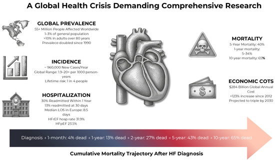
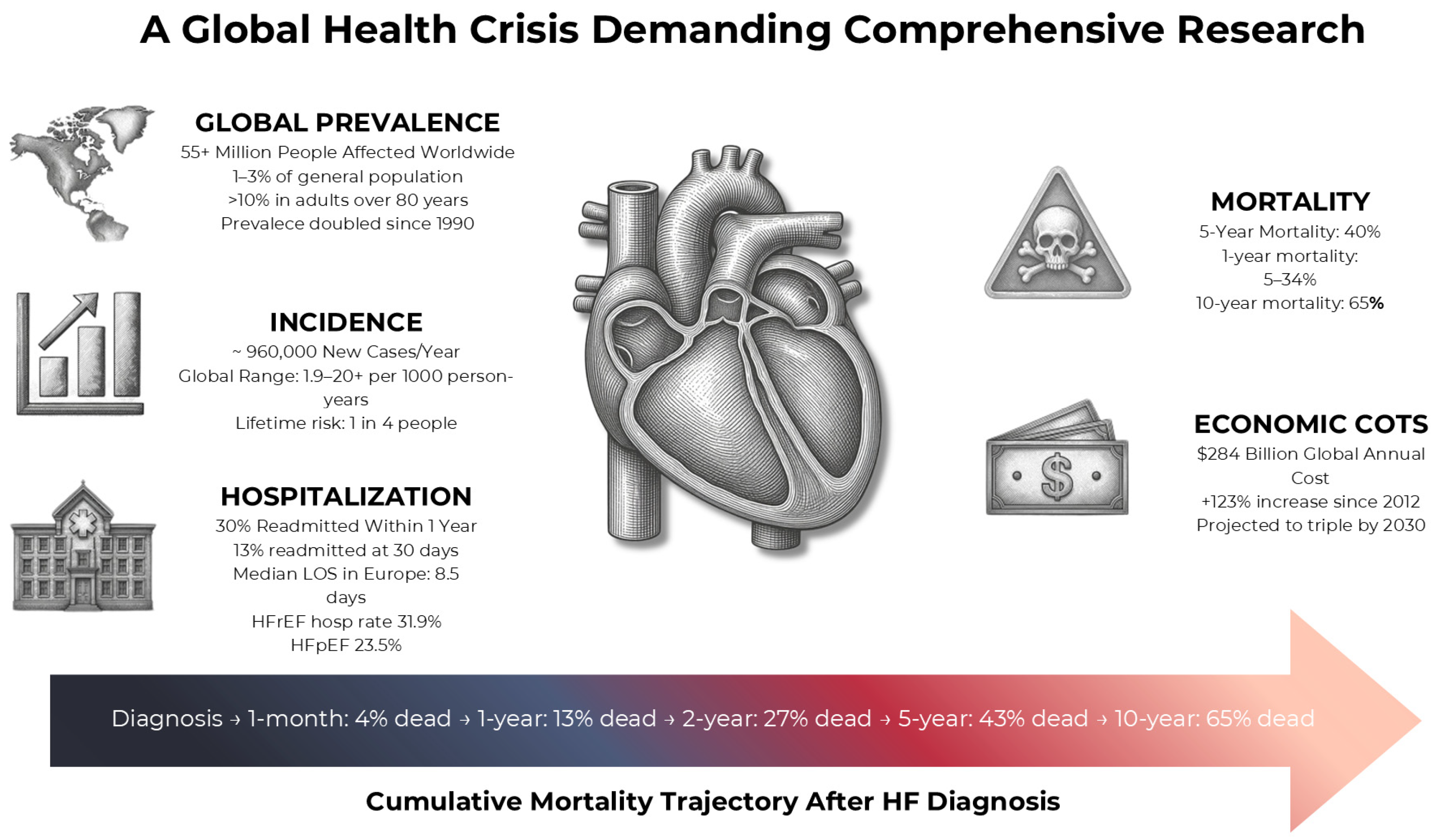
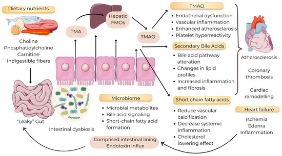
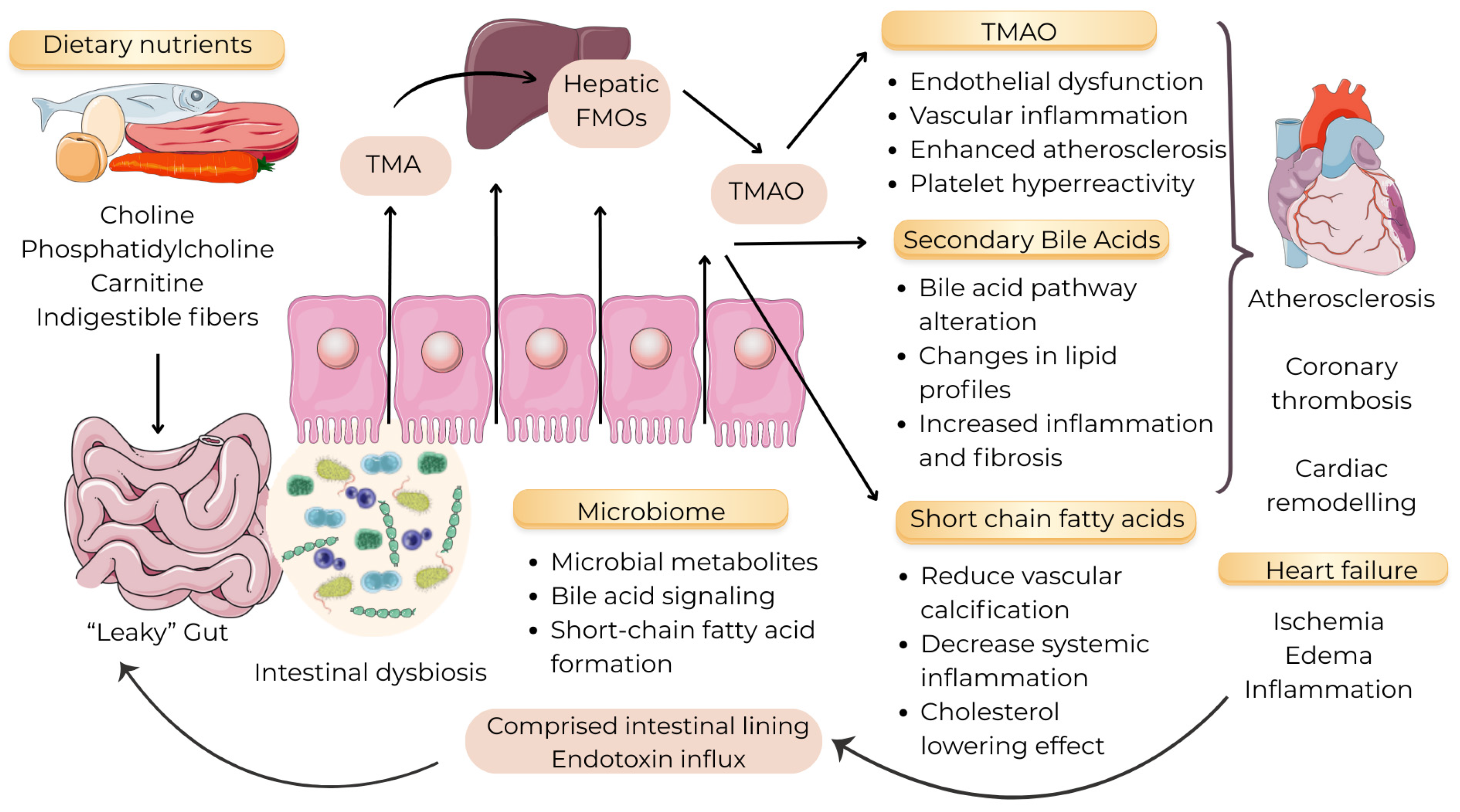
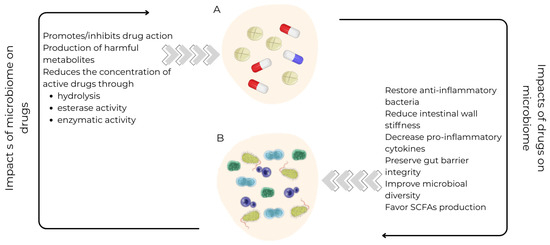
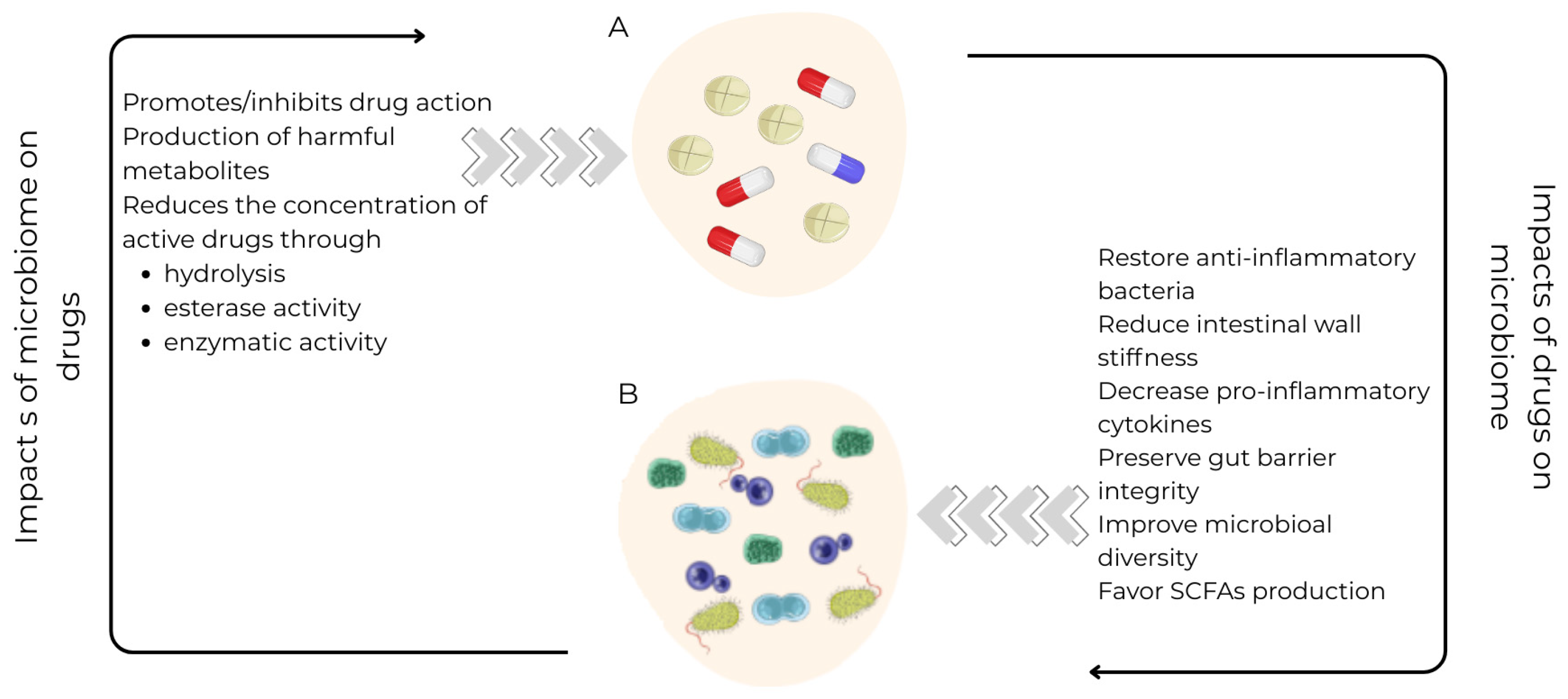

Disclaimer/Publisher’s Note: The statements, opinions and data contained in all publications are solely those of the individual author(s) and contributor(s) and not of MDPI and/or the editor(s). MDPI and/or the editor(s) disclaim responsibility for any injury to people or property resulting from any ideas, methods, instructions or products referred to in the content. |
© 2026 by the authors. Licensee MDPI, Basel, Switzerland. This article is an open access article distributed under the terms and conditions of the Creative Commons Attribution (CC BY) license.