


Abstract
Neutrophils constitute the largest fraction of total circulating leukocytes in humans and mediate early innate immune responses. Although they are often considered a uniform population of short-lived immune cells, emerging evidence from single-cell RNA sequencing and high-dimensional flow cytometry has revealed that neutrophils are functionally and phenotypically heterogeneous in both healthy and pathological conditions. However, a critical gap is how molecularly defined neutrophil states translate into distinct spatiotemporal behaviors in vivo. This review summarizes our current understanding of the molecular signatures underlying neutrophil heterogeneity and explores the functional in vivo behaviors in various diseases, including cancer, sepsis, and ischemic stroke. We also discuss the potential of intravital imaging to bridge the gap between static molecular profiling and dynamic cellular behavior, offering a comprehensive view of the functional heterogeneity of neutrophils.
1. Introduction
Neutrophils play a central role in the immediate innate immune response and rapidly migrate to sites of infection or tissue injury to perform various functions, including degranulation (granule secretion), phagocytosis, reactive oxygen species (ROS) generation and neutrophil extracellular trap (NET) formation [1,2,3]. These responses are important for host defense; however, when overactive or dysregulated, neutrophils can exacerbate inflammation and contribute to tissue damage, thereby contributing to a wide range of pathological conditions, including cancer [4,5], sepsis [6,7] and ischemic stroke [8,9,10]. Previously, neutrophils were considered to be short-lived, terminally differentiated immune cells with relatively limited phenotypic variation [11]. However, a growing body of evidence supports heterogeneity and plasticity across developmental stages, activation states, and tissue contexts. Single-cell RNA sequencing has identified transcriptionally distinct neutrophil states and maturation trajectories in both healthy and pathological conditions [12,13,14], whereas high-dimensional flow cytometry has identified disease-associated neutrophil states [15,16]. Despite these molecular insights, a critical limitation remains: the disconnection between static molecular profiles and in vivo cellular behaviors. Under inflammatory conditions, neutrophil recruitment to inflamed tissues is a tightly orchestrated multi-step process involving rolling, arrest, crawling and transendothelial migration (TEM), which are regulated by interactions with endothelial adhesion molecules [17]. Although omics approaches provide detailed snapshots of cellular states, they often fail to capture the real-time spatiotemporal dynamics in vivo.
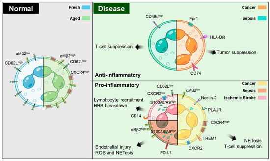
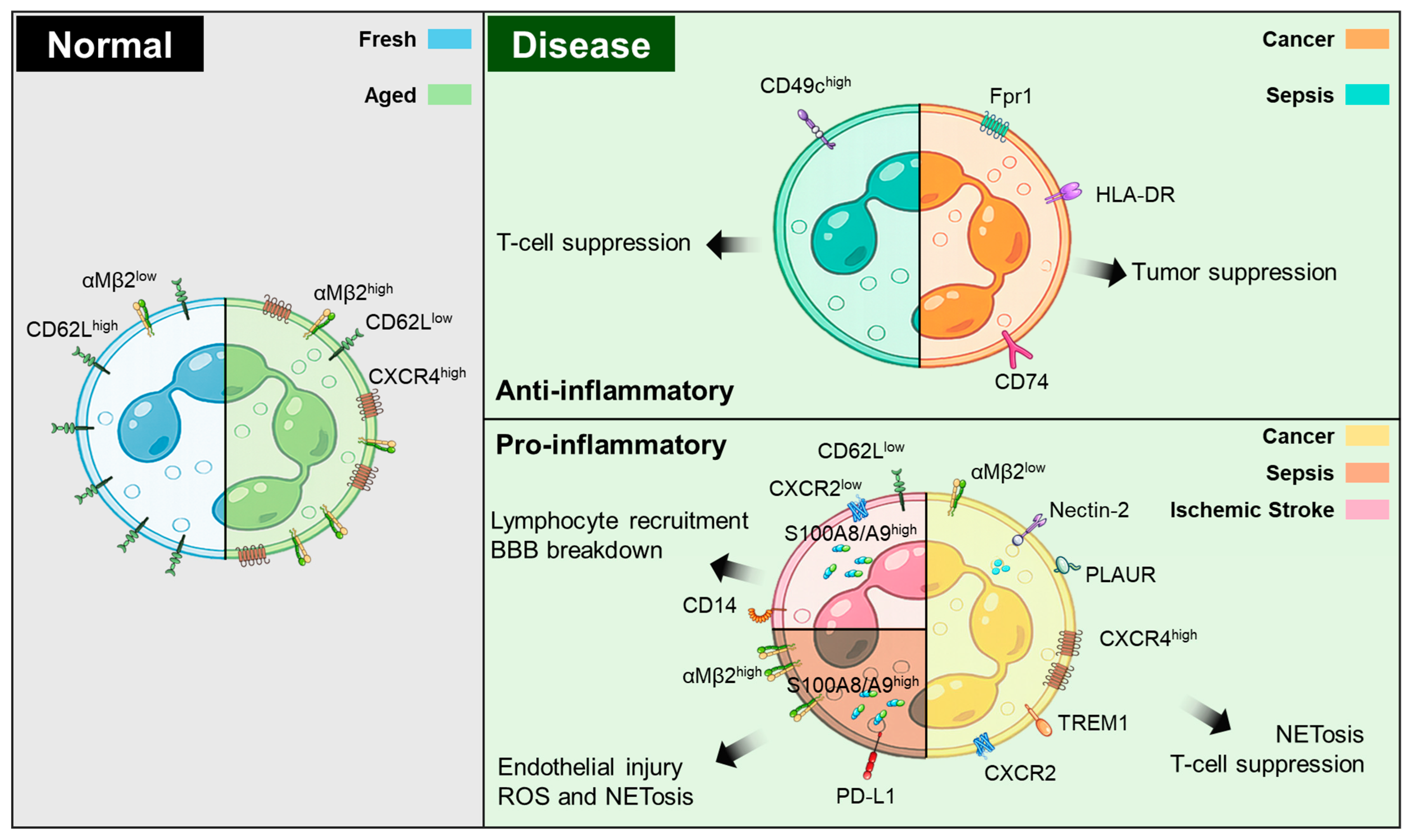
Intravital microscopy (IVM) has emerged as a critical tool for bridging this gap. Unlike static analyses from omics, IVM allows for in vivo real-time visualization and quantification of immune cell behavior under pathological conditions [18,19]. Using reporter mice or in vivo fluorescent labeling strategies, IVM enables real-time monitoring of neutrophils within living tissues at a single-cell resolution. This approach allows the quantitative analysis of spatiotemporal behaviors such as adhesion, crawling, TEM, and interactions with resident cells in the local microenvironment [20]. Recent advances in imaging analysis have enabled the linking of in vivo behavioral data to distinct cell populations, providing behavioral phenotypes at the single-cell level [21,22]. Thus, integrating single-cell molecular analysis with intravital imaging offers a robust platform for comprehensively understanding neutrophil heterogeneity in vivo (Figure 1). This review discusses the potential applications of IVM to elucidate the link between molecular heterogeneity and cellular behavior, thereby offering an integrated perspective on neutrophil function from in vitro profiling to in vivo dynamics.
Figure 1.
Neutrophil heterogeneity under physiological and pathological conditions. Under normal conditions, circulating neutrophils transition from ‘fresh’ (CD62LhighαMβ2lowCXCR4low) to ‘aged’ (CD62LlowαMβ2highCXCR4high) populations. Under pathological conditions such as cancer, sepsis, and ischemic stroke, neutrophils further diversify into disease-associated states defined by distinct transcriptional and surface-marker signatures. Anti-inflammatory or protective neutrophils in cancer and sepsis are characterized by representative markers linked to host-protective or anti-tumor functions. In contrast, pro-inflammatory neutrophils in cancer, sepsis, and ischemic stroke are associated with representative markers linked to immunosuppression, endothelial injury, BBB breakdown, ROS production, and NETosis. Overall, this schematic highlights how neutrophil heterogeneity can be linked to either protective or pathogenic functions across physiological and pathological conditions.
2. Molecular Signatures to Cellular Behaviors Under Healthy Conditions
Granulocyte colony-stimulating factor (G-CSF) promotes bone marrow granulopoiesis, thereby facilitating the egress of mature neutrophils into peripheral blood [23,24]. The CXCL12-CXCR4 pathway functions as a major retention signal that maintains mature neutrophils within the bone marrow, whereas CXCL1 and CXCL2, acting through CXCR2, promote neutrophil egress into the blood [25,26]. G-CSF promotes neutrophil mobilization by reducing CXCR4 expression in the marrow [27]. A recent study reported that under homeostatic circadian conditions in mice, fresh neutrophils are released from zeitgeber time (ZT)17 to ZT5, and aged neutrophils are cleared from ZT5 to ZT13 [28]. Here, the ZT is defined relative to the light–dark cycle, with ZT0 indicating lights on and ZT12 indicating lights off. Because mice are nocturnal, whereas humans are diurnal, the physiological meaning of a given ZT value differs between the two species; under conventional light-dark conditions, ZT0-12 broadly corresponds to the active phase in humans but to the inactive phase in mice [29]. Phenotypically, circulating neutrophils can be defined by CD62L and CXCR4 expression: CD62LhighCXCR4low cells represent a “fresh” population released from the bone marrow, whereas CD62LlowCXCR4high cells are defined as “aged” blood neutrophils, reaching their highest proportion at ZT5 [28]. Mechanistically, circadian regulation of neutrophils depends on both external and internal cues. Freshly circulating neutrophils undergo a Bmal1-dependent intrinsic aging program in which autocrine CXCL2-CXCR2 signaling promotes the transition from fresh to aged states [29]. Notably, aged neutrophils exhibit increased surface levels of CD11b and CD49d, suggesting adhesive/trafficking features [30]. Additionally, aged neutrophils migrate back to the bone marrow via CXCL12-CXCR4 signaling, where they are subsequently engulfed and cleared by resident CD169+ macrophages, this clearance supporting circadian oscillations in neutrophil homeostasis [28].
To investigate neutrophil aging in vivo, IVM of inflamed microvessels identified a CD62Llow neutrophil state with enhanced Mac-1 activation, consistent with microbiome-driven TLR-MyD88 signals that promote the emergence and activation of aged neutrophils [31]. Recent intravital imaging in a murine stroke model showed that at ZT5 (the inactive phase), aged neutrophils were associated with increased microvascular stalling and reduced collateral perfusion, suggesting that time-of-day-dependent neutrophil-microvessel interactions impair flow and thereby worsen stroke injury [32]. Additionally, emerging evidence suggests that circadian dysregulation affects tumor-associated neutrophil populations. In experimental metastatic cancer models, CXCR4highCD62Llow aged neutrophils were enriched in association with disrupted neutrophil circadian homeostasis, indicating that tumors can perturb physiological neutrophil aging and selectively expand the metastasis-associated state [33]. In colorectal cancer, the disruption of the epithelial clock is associated with increased neutrophil recruitment and myeloid-derived suppressor cell (MDSC) accumulation. Notably, the associated neutrophilic state was characterized by the expression of S100a8, S100a9, and CXCR2, and the enrichment of MDSC-related markers, such as Ifitm1, Wfdc17, Irg1, and Arg2, together with increased ROS and PD-L1, consistent with an immunosuppressive phenotype [34].
3. Molecular Signatures to Cellular Behaviors Under Pathological Conditions
Under pathological conditions, such as cancer, sepsis, and ischemic stroke, neutrophils respond and migrate to inflamed sites, exhibiting heterogeneous phenotypes. Here, we discuss the current understanding of molecular signatures across these diseases and how functional in vivo behaviors can be investigated using intravital imaging. A deeper understanding of the relationship between the phenotypic states and in vivo behavioral characteristics of neutrophils provides a comprehensive view of neutrophil functional heterogeneity and contributes to the identification of novel therapeutic targets.
3.1. Cancer
Tumor-associated neutrophils (TANs) are broadly described in anti-tumorigenic N1 and pro-tumorigenic N2 states. TGF-β signaling in the tumor microenvironment promotes polarization toward a tumor-supportive N2 state, whereas blockade of TGF-β shifts TANs toward an N1 state. Functionally, N1 TANs exhibit enhanced cytotoxic activity against tumor cells, exerting an anti-tumorigenic function, whereas N2 TANs are immunosuppressive, thereby promoting tumor progression. In addition, N1-polarized TANs enhance CD8+ T cell activation, whereas N2-polarized TANs suppress anti-tumor T cell responses [35,36]. In parallel, pseudo-time and trajectory analyses further suggested that TGF-β/SMAD3 signaling maintains TANs in an N2-like state, whereas Smad3 deficiency or pharmacologic inhibition shifts TANs toward an N1-like state, with increased neutrophil infiltration and tumor regression [37]. Consistent with this, in lung adenocarcinoma, a miR-941/FOXN4/TGF-β feedback loop has been proposed to promote N2-like TAN polarization and tumor progression, linking TAN-associated miRNA activity to FOXN4 suppression and enhanced TGF-β signaling in tumor cells [38]. In cancer, neutrophils appear to undergo stage-dependent reprogramming, transitioning from early polymorphonuclear myeloid-derived suppressor cells (PMN-MDSC)-like neutrophils, characterized by enhanced migratory capacity and metabolic activation, to later PMN-MDSCs/activated PMN-MDSCs with enhanced immunosuppressive activity [39,40]. Moreover, lineage-defining transcription factors, such as SOX2 and NKX2-1 can shape the tumor immune microenvironment in non-small cell lung cancer (NSCLC) by regulating TAN recruitment. In this context, TANs exhibit N2-like features, including SiglecF expression and increased ROS-related signatures, and neutrophil depletion reduces squamous tumor burden, suggesting that TANs contribute to squamous tumor progression [41]. Interestingly, the modulation of neutrophil states in cancer may be a potential complementary strategy for strengthening anti-cancer immunotherapy. For example, trained innate immunity can reprogram granulopoiesis, thereby shifting neutrophils toward an anti-tumor phenotype. In particular, β-glucan preconditioning induces transcriptional and epigenetic remodeling at the level of granulocyte-monocyte progenitors, generating neutrophils with enhanced ROS-dependent tumor-killing activity that suppresses tumor growth [42]. In resistant head and neck squamous cell carcinoma (HNSCC), Sox9+ tumor cells were enriched and promoted the apoptosis of Fpr1+ neutrophils. This loss of Fpr1+ neutrophils was associated with reduced infiltration and cytotoxic activity of CD8+ and γδ T cells, ultimately contributing to resistance to combined anti-LAG-3 and anti-PD-1 immunotherapy [43].
In addition to the conventional N1/N2 TAN concept, recent single-cell transcriptomic and proteomic studies have revealed the cancer-specific neutrophil states. Single-cell transcriptomic profiling of human and mouse lung cancers revealed that TANs represent a heterogeneous population, including canonical neutrophil-like states, tumor-enriched pro-tumor states, and distinct type I interferon-responsive neutrophil states, supporting the state-based classification of TANs [44]. Integrated single-cell analysis across 17 cancer types identified 10 distinct neutrophil states [45]. Among these, anti-tumor or protective states include HLA-DR+CD74+ neutrophil states, which exhibit an MHC class II/antigen-presenting signature and are associated with better clinical outcomes. In gastric cancer, single-cell transcriptomic analysis has identified CD44−CXCR2− tumor-specific neutrophils (tsNeus) as a distinct neutrophil state, including a CD54+ tsNeu state with anti-tumor activity linked to reduced tumor progression and improved survival [46]. Additionally, neutrophil-induced pyroptosis involving ELANE+ neutrophils, NETs, and CASP1-associated pyroptotic signaling is associated with increased CD8+ T cell activation [47].
In contrast, immunosuppressive or tumor-promoting states include Nectin2-expressing TAN states associated with CD8+ T-cell dysfunction/exhaustion and cell–cell adhesion, suggesting a role for this neutrophil state in an immunosuppressive tumor microenvironment [48], a CXCR4high neutrophil state in hepatoblastoma characterized by ferroptotic signatures together with T-cell suppressive features and chemotactic behavior [49], a PLAUR+ neutrophil state in hepatocellular carcinoma linked to reduced CD8+ T-cell infiltration and macrophage-driven immunosuppression, and migration [50]. Tumor-enriched CD83+ TANs characterized by the upregulation of CXCR4 and downregulation of CXCR2/SELL, consistent with a senescent-like phenotype and immunosuppressive signatures, and associated with poor prognosis across various cancer types [51]. Similarly, in patients with NSCLC, circulating neutrophil-platelet aggregates (NPAs) have been identified in peripheral blood, and a platelet gene-high neutrophil cluster is associated with degranulation, chemotaxis, and TEM, indicating an activated and tumor-promoting circulating neutrophil state [52]. Importantly, the NPA-associated neutrophil signature was correlated with poor prognosis across multiple solid tumor types, supporting its potential as a prognostic biomarker.
Pro-inflammatory states include an immature CD10− neutrophil (CD66bhighCD11blow) state enriched in the low-density neutrophil fraction in patients with glioblastoma (GBM) [53]. Single-cell proteomic analysis demonstrated that GBM TANs comprise multiple functional states, including ARG1-linked immunosuppressive/angiogenic signatures and degranulation/NETosis-like states. Additional NET-associated states include tumor-enriched PPIF+ neutrophil states with a high NET-related score, consistent with NETosis, and a potential link to colorectal cancer progression [54], and CD10+TREM1+CXCR2+ neutrophil states in multiple myeloma that exhibit T-cell suppressive activity and NETosis, and are associated with poor outcomes as well as increased T-cell exhaustion-related signatures [55].
Metabolic states represented by dcTRAIL-R1+ terminal T3 states in pancreatic ductal adenocarcinoma are characterized by inflammation-, hypoxia-, and glycolysis-associated gene signatures [56]. Functionally, T3 neutrophils exhibit pro-angiogenic activity that supports tumor growth. Single-cell transcriptomic analysis identified seven neutrophil states, among which OLR1+ and MIF+ neutrophils were classified as tumor-enriched TAN states [57]. Notably, the MIF+ TAN state was associated with hypoxia- and glycolysis-related signatures and was linked to poor clinical outcomes. During cervical cancer progression, single-cell transcriptomic analysis revealed five distinct neutrophil states (N0–N4), among which the N4 state is related to hypoxia, WNT, and inflammatory signaling, contributing to tumor growth [58]. Collectively, these studies indicate that neutrophil states in cancer are linked to distinct functional outcomes and behavioral features, including anti-tumor activity, immunosuppression, chemotaxis, TEM, NETosis, and pro-angiogenic metabolic adaptation (Table 1).
Table 1.
Surface markers and gene expression associated with neutrophil heterogeneity in cancer.
IVM has been used to observe neutrophil dynamics in tumor models in vivo. In a cutaneous melanoma model, repetitive UVB irradiation induces skin inflammation and increases neutrophil infiltration, promoting melanoma angiotropism and enhancing metastatic dissemination [59]. In an HNSCC-like murine tumor model, intravital two-photon microscopy revealed that intratumoral TANs remained within the tumor core for longer periods, whereas peritumoral TANs exhibited increased motility and migration [60]. In a transplanted murine head and neck cancer model, two-photon microscopy visualized in vivo interactions between neutrophils and T cells in tumor-draining lymph nodes (TDLNs) [61]. IFNAR1 deficiency enhanced neutrophil accumulation in the TDLN but resulted in impaired neutrophil-T cell interactions and increased tumor progression. In the subcutaneous MC38-cEGFR tumor model, intravital imaging revealed that FcαRI-TNF increased tumor-infiltrating neutrophils [62]. Overall, these IVM studies demonstrate that intravital imaging can be used to monitor neutrophil infiltration/accumulation, spatial retention versus migratory behavior within the tumor microenvironment, and neutrophil-T cell interactions, thereby highlighting in vivo behavioral features that may relate to the molecularly defined neutrophil states described above. However, the mechanism by which each transcriptionally defined neutrophil state corresponds to distinct patterns of tumor infiltration, migration, and immune cell interactions in vivo remains unclear. Further integration of intravital imaging with transcriptomic profiling is important to define how specific neutrophil states contribute to tumor progression and functions within the tumor microenvironment.
3.2. Sepsis
Sepsis is a leading cause of global mortality and is associated with organ dysfunction [63]. Septic conditions induce dynamic host inflammatory responses, typically defined as acute hyperinflammation and subsequent persistent immunosuppression, which may contribute to mortality through impaired clearance of secondary infections [64,65]. In this context, neutrophils in sepsis are increased due to delayed apoptosis and impaired clearance, as well as the release of immature cells from the bone marrow [66,67]; however, their functional capacity is markedly impaired, including impaired bacterial clearance, attenuated ROS generation, and defective recruitment to sites of infection [68]. Moreover, depending on the stage of sepsis, neutrophils exhibit either pro-inflammatory functions [66] or immunosuppressive phenotypes characterized by IL-10 production and T-cell suppressive activity [69,70]. These functional distinctions may provide a useful framework for understanding molecularly defined neutrophil states in sepsis.
Recent single-cell analyses have suggested that the molecularly defined neutrophil states in sepsis can be broadly organized into anti-inflammatory/protective, pro-inflammatory/NET-associated, and immunosuppressive states. An anti-inflammatory or protective state is represented by a C1q-upregulating CD49chigh neutrophil state associated with improved survival [71]. In contrast, pro-inflammatory states include an olfactomedin-4 (OLFM4)-positive neutrophil state [72], and flow cytometric analyses in adult patients with sepsis also identified circulating OLFM4+ neutrophils, which were associated with 60-day mortality [73]. These OLFM4-positive states are associated with poor clinical outcomes; however, their specific in vivo functions remain unclear. An animal study using a cecal ligation and puncture (CLP)-induced sepsis model revealed a pro-inflammatory Ly6G+ CD11bhigh low-density neutrophil state, which displayed enhanced production of pro-inflammatory mediators and increased NET formation, thereby contributing to inflammatory tissue injury [74]. Additionally, S100A8/A9high neutrophils promote endothelial mitochondrial dysfunction and PANoptosis [75], linking this state to endothelial injury and microvascular damage. An immunosuppressive state was defined as IL1R2-expressing neutrophils representing an immature and immunosuppressive state linked to poor clinical outcomes [76,77]. Furthermore, PD-L1-expressing neutrophils in sepsis have been reported to contribute to immunosuppression by inhibiting T-cell responses, and are linked to poor clinical prognosis [78]. Integrative analyses in human and murine models further delineated these PD-L1+ cells as an immature, transcriptionally distinct state that suppresses T-cell activation and can additionally promote endothelial injury and pyroptosis [15,79,80,81,82], consistent with the molecular state of both immunosuppressive and endothelial-injurious functions. Although these studies defined molecularly distinct neutrophil states in sepsis (Table 2), linking these states to their corresponding in vivo spatiotemporal behaviors requires approaches such as intravital microscopy.
Table 2.
Surface markers and gene expression associated with neutrophil heterogeneity in sepsis.
Recently, IVM has been widely used to visualize neutrophil responses within inflamed lung tissues in vivo. Several IVM studies using lipopolysaccharides (LPS)-induced acute lung injury or sepsis models have demonstrated pulmonary endothelial barrier disruption accompanied by functional microvascular dead space [83], accumulation of capillary-entrapped Mac-1-expressing neutrophils associated with impaired microcirculation [19], and increased neutrophil infiltration with NET formation [84,85]. Collectively, these IVM studies highlighted in vivo pathophysiological features of septic neutrophil responses, including endothelial barrier disruption, capillary entrapment with impaired microcirculation, neutrophil infiltration, and NET formation, and conceptually related these phenomena to the molecularly defined neutrophil states described above, particularly the pro-inflammatory/NET-associated and endothelial-injurious states. However, the direct link between these molecular states and distinct in vivo behaviors during sepsis remains unresolved. Further integration of IVM with transcriptomic profiling is important to determine how specific neutrophil states contribute to septic tissue injury in vivo.
3.3. Ischemic Stroke
A rapid surge in circulating neutrophils occurs within hours of ischemic stroke onset, and histological evaluations in human patients consistently demonstrate a peak in neutrophil accumulation around days 1–2 post-stroke [86,87]. Clinically, early neutrophilia and an increased neutrophil-to-lymphocyte ratio have been linked to greater stroke severity/infarct volume and poorer outcomes, including higher mortality [88,89]. Previous animal studies have identified pro-inflammatory N1 neutrophils in brain damage and anti-inflammatory N2 neutrophils in an ischemic stroke model for the resolution of inflammation and tissue recovery [90,91,92,93]. In addition, neutrophils release NETs that promote blood–brain barrier (BBB) disruption and thrombosis, thereby contributing to ischemic brain injury [94]. Patients with acute ischemic stroke presenting within 6 h of onset exhibit hyperactivated circulating neutrophils, defined by downregulated L-selectin and upregulated αMβ2 integrin expression, which is linked to stroke severity [95].
Recent studies have suggested that neutrophils exhibit distinct pro-inflammatory and tissue-damaging states under the inflammatory conditions of ischemic stroke. Transcriptomic analyses of purified neutrophils from 38 patients with ischemic stroke showed temporal transcriptional alterations, peaking at 24–48 h post-onset (TP2), including leukocyte extravasation, EIF2 signaling, and TP2-specific IL-1/4/7/8 signaling [96]. In addition, single-cell transcriptomic analysis of aged mice on day 3 post-tMCAO revealed three neutrophil states, among which Neut1 showed upregulated mitochondrial and inflammatory gene programs (including mt-Nd1-4, Il1b, Aif1, and Ly86), consistent with a highly activated neutrophil state infiltrating the ischemic brain and likely contributing to inflammatory tissue injury [97]. Gullotta et al. showed that, in an aged mouse stroke model, CD62Llow atypical neutrophils accumulated and were collectively enriched in oxidative stress, phagocytosis, and procoagulant features [98]. Consistently, in elderly patients with stroke, a CD62Llow neutrophil state is linked to worse reperfusion and poor functional outcomes. CyTOF and scRNA-seq profiling of aged mice subjected to tMCAO showed that circulating neutrophils shifted toward a late maturation/aging phenotype with decreased CD62L. A brain-enriched neutrophil state (BrNeu2), characterized by CXCR2lowCD62Llow showed senescence, degranulation, and adhesion features linked to increased BBB permeability [99], indicating a recruitment-associated and BBB-disruptive neutrophil state. Moreover, trajectory analysis integrating six blood neutrophil states and six brain granulocyte states from tMCAO-challenged mice indicated that brain granulocytes (Gran2 and Gran3) were recruited from late-stage mature blood neutrophils (Neu3) rather than differentiating locally within the brain [100]. Another study in a mouse model of ischemic stroke also identified a distinct S100A8/A9high neutrophil state, associated with BBB disruption and increased lymphocyte recruitment [101], supporting its role in amplifying vascular inflammation. Interestingly, Vázquez-Reyes et al. reported that neutrophils in the inactive phase (Zeitgeber time 5; ZT5) of a murine stroke model, including pro-inflammatory/degranulation-associated BNc1 and ISG/NETosis-prone BNc4 states, were associated with larger infarcts [32]. In patients, infarct outcomes also showed diurnal variations that correlated with oscillations in NET-related neutrophils, indicating that time-of-day-dependent neutrophils/NETs contribute to stroke outcomes. Furthermore, integrated single-cell transcriptomic analysis identified a CD14-expressing neutrophil state, and flow cytometry confirmed that these cells accounted for approximately 80% of brain neutrophils 24 h after stroke, which is consistent with an activated/mobilized mature neutrophil feature [102]. Collectively, recent single-cell transcriptomic studies have identified distinct neutrophil states characterized by CD62L, S100A8/A9, CXCR2, and CD14 in rodent ischemic stroke models and patients with stroke (Table 3). Although these states share pro-inflammatory and tissue-damaging features, they differ in their functions, including oxidative activity, BBB-disruptive adhesion, leukocyte-recruiting inflammatory signaling, and NET formation. However, the mechanism by which these molecularly defined states translate into distinct spatiotemporal behaviors in the ischemic brain remains unknown. To further investigate the in vivo spatiotemporal behavior of neutrophils, advanced approaches such as intravital imaging could be combined with transcriptomic analyses.
Table 3.
Surface markers and gene expression associated with neutrophil heterogeneity in ischemic stroke.
Although single-cell analyses provide static snapshots of neutrophils under inflammatory conditions, IVM is a powerful technique for visualizing neutrophils in vivo. Recent studies using two-photon intravital microscopy revealed increased neutrophil infiltration and reverse TEM (rTEM) in myeloid-specific LysM-GFP mice during LPS-induced neuroinflammation [103], neutrophil-mediated dynamic capillary stalls leading to microvascular flow impairment in LysM-GFP or Ly6g-Cre-tdTomato mice during photothrombosis-induced stroke [104], and NET formation in neuroinflammation [105,106]. These imaging findings broadly correspond to recruitment/infiltration, rTEM, vascular stalling, and NET formation, thereby highlighting in vivo behavioral features that may be related to the molecularly defined neutrophil states described above. Although IVM enables the visualization of neutrophil behavior in the brain in vivo, the direct integration of transcriptomic states with these behaviors at the single-cell level remains unresolved. Thus, integrated approaches that combine IVM with transcriptomic profiling are required to clarify how distinct neutrophil states drive ischemic brain injury in vivo.
4. Advanced Approaches for Bridging Molecular Signatures and Cellular Behaviors
Recent developments in machine learning have provided more comprehensive views across various research fields, and these approaches have been increasingly applied in biomedical research. Integrated scRNA-seq and machine learning-based multi-omics analyses of peripheral blood from four patients with sepsis and two healthy controls identified two neutrophil clusters (clusters 1 and 2) that were markedly expanded in patients with sepsis. These clusters exhibit distinct inflammatory transcriptomic signatures, including ALPL, CD177, S100A8, S100A9, and STXBP2, suggesting a shift toward activated and pro-inflammatory neutrophil states [107]. A recent machine learning-based transcriptomic study of patients with sepsis identified S100A12 as a robust diagnostic biomarker for sepsis associated with sepsis progression and proposed it as a putative therapeutic target [108]. Consistently, in hepatocellular carcinoma, Deep scSTAR-based analysis also identified S100A12+ tumor-associated neutrophils that correlated with poor prognosis and immunotherapy non-response [109]. Collectively, advances in single-cell analysis combined with machine learning are expected to facilitate the efficient interpretation of large-scale multidimensional datasets and accelerate the discovery of novel therapeutic targets.
With advances in machine learning-based transcriptomic studies, recent research has enabled the high-resolution identification of disease-associated immune cell states at the molecular level. However, transcriptomic signatures cannot capture the dynamic functional states of immune cells in living tissues. In addition, cell dissociation-based scRNA-seq inevitably loses spatial and temporal context, limiting its ability to directly reflect dynamic cellular behavior in vivo [110]. To overcome these limitations, single-cell behavioral analyses obtained through intravital imaging provide a complementary framework for understanding immune cell heterogeneity in vivo. For example, individual immune cell behavioral profiling combined with computational clustering has demonstrated distinct behavioral states in inflamed tissues that are functionally associated with pathological roles in inflammatory conditions [21]. In addition, four-dimensional IVM with quantitative single-cell tracking also showed the emergence of highly migratory or adhesive αMβ2high neutrophil states during ischemic stroke [22]. Nevertheless, IVM is also limited by tissue accessibility and imaging depth, as well as potential biases related to labeling strategies and imaging windows. Moreover, imaging-derived behaviors cannot be directly linked to molecular states, because these features are not measured in the same cells [111]. One complementary strategy would be to combine intravital imaging with image-guided or time- and site-matched sampling to isolate neutrophil populations enriched for tissue-damaging behaviors under inflammatory conditions, and then define their transcriptomic profiles. This strategy could help connect imaging-derived behavioral phenotypes with molecular states, thereby facilitating the identification of transcriptional features associated with pathologically relevant neutrophil populations. Collectively, the combination of machine learning-driven omics analyses with in vivo single-cell behavioral profiling offers a multidimensional approach that bridges molecular signatures and cellular behaviors, thereby enabling a better understanding of immune cell dynamics under both physiological and pathological conditions.
5. Concluding Remarks
Taken together, the advanced convergence of single-cell transcriptomic profiling with real-time intravital imaging provides a novel perspective on how neutrophil heterogeneity is conceptualized, from static molecular signatures to dynamic cellular behaviors embedded within tissue microenvironments. Furthermore, integrative approaches combining omics studies, high-resolution live imaging, and artificial intelligence-assisted behavioral quantification will be useful for delineating the associations between gene programs, cellular trajectories, and context-dependent immune functions. Such multidimensional frameworks are expected to not only refine mechanistic insights into neutrophil heterogeneity across physiological and pathological conditions but also enable predictive and state-specific intervention strategies. Although these approaches remain speculative, integrated molecular and behavioral analyses may help identify transcriptionally defined neutrophil states linked to pathogenic in vivo behaviors. This may support more precise immunomodulatory strategies that selectively target tissue-damaging neutrophils while preserving protective host defense functions. Ultimately, bridging molecular signatures with spatiotemporal behavior is expected to advance neutrophil research from descriptive heterogeneity to precise immunomodulation and the translational discovery of therapeutic targets under inflammatory conditions.
Author Contributions
J.L. (Jonghee Lee) and J.L. (Jingu Lee) wrote the manuscript. All authors have read and agreed to the published version of the manuscript.
Funding
This work was supported by a 2-Year Research Grant of Pusan National University.
Institutional Review Board Statement
Not applicable.
Informed Consent Statement
Not applicable.
Data Availability Statement
No new data were created or analyzed in this study. Data sharing is not applicable to this article.
Conflicts of Interest
The authors declare no conflicts of interest.
Abbreviations
The following abbreviations are used in this manuscript:
| ARG1 | Arginase 1 |
| BBB | Blood–brain barrier |
| CASP1 | Caspase-1 |
| CLP | Cecal ligation and puncture |
| CyTOF | Cytometry by time-of-flight |
| G-CSF | Granulocyte colony-stimulating factor |
| GBM | Glioblastoma |
| HCC | Hepatocellular carcinoma |
| HNSCC | Head and neck squamous cell carcinoma |
| IFNAR1 | Interferon-α/β receptor 1 |
| IL | Interleukin |
| ISG | Interferon-stimulated genes |
| IVM | Intravital microscopy |
| LAG-3 | Lymphocyte activation gene 3 |
| LPS | Lipopolysaccharide |
| Mac-1 | Macrophage-1 antigen (integrin αMβ2; CD11b/CD18) |
| MHC | Major histocompatibility complex |
| miR | microRNA |
| MyD88 | Myeloid differentiation primary response 88 |
| NETs | Neutrophil extracellular traps |
| NPA | Neutrophil-platelet aggregates |
| NSCLC | Non-small cell lung cancer |
| OLFM4 | Olfactomedin 4 |
| PANoptosis | Inflammatory cell death pathway integrating pyroptosis/apoptosis/necroptosis |
| PD-1 | Programmed cell death protein 1 |
| PD-L1 | Programmed death-ligand 1 |
| PMN-MDSC | Polymorphonuclear myeloid-derived suppressor cells |
| rTEM | Reverse transendothelial migration |
| ROS | Reactive oxygen species |
| scRNA-seq | Single-cell RNA sequencing |
| TANs | Tumor-associated neutrophils |
| TDLN | Tumor-draining lymph nodes |
| TEM | Transendothelial migration |
| tMCAO | Transient middle cerebral artery occlusion |
| TLR | Toll-like receptor |
| UVB | Ultraviolet B |
| WNT | Wingless/Integrated signaling pathway |
| YAP/TAZ | Yes-associated protein / transcriptional coactivator with PDZ-binding motif |
| ZT | Zeitgeber time |
References
- Brinkmann, V.; Reichard, U.; Goosmann, C.; Fauler, B.; Uhlemann, Y.; Weiss, D.S.; Weinrauch, Y.; Zychlinsky, A. Neutrophil extracellular traps kill bacteria. Science 2004, 303, 1532–1535. [Google Scholar] [CrossRef] [Scilit] [PubMed]
- Segal, A.W. How neutrophils kill microbes. Annu. Rev. Immunol. 2005, 23, 197–223. [Google Scholar] [CrossRef] [Scilit] [PubMed]
- Lacy, P. Mechanisms of degranulation in neutrophils. Allergy Asthma Clin. Immunol. 2006, 2, 98–108. [Google Scholar] [CrossRef] [Scilit]
- Spiegel, A.; Brooks, M.W.; Houshyar, S.; Reinhardt, F.; Ardolino, M.; Fessler, E.; Chen, M.B.; Krall, J.A.; DeCock, J.; Zervantonakis, I.K.; et al. Neutrophils suppress intraluminal NK cell-mediated tumor cell clearance and enhance extravasation of disseminated carcinoma cells. Cancer Discov. 2016, 6, 630–649. [Google Scholar] [CrossRef] [Scilit]
- Albrengues, J.; Shields, M.A.; Ng, D.; Park, C.G.; Ambrico, A.; Poindexter, M.E.; Upadhyay, P.; Uyeminami, D.L.; Pommier, A.; Kuttner, V.; et al. Neutrophil extracellular traps produced during inflammation awaken dormant cancer cells in mice. Science 2018, 361, eaao4227. [Google Scholar] [CrossRef] [Scilit]
- Chignard, M.; Balloy, V. Neutrophil recruitment and increased permeability during acute lung injury induced by lipopolysaccharide. Am. J. Physiol. Lung Cell. Mol. Physiol. 2000, 279, L1083–L1090. [Google Scholar] [CrossRef] [Scilit]
- Zhang, H.; Wang, Y.; Qu, M.; Li, W.; Wu, D.; Cata, J.P.; Miao, C. Neutrophil, neutrophil extracellular traps and endothelial cell dysfunction in sepsis. Clin. Transl. Med. 2023, 13, e1170. [Google Scholar] [CrossRef] [Scilit] [PubMed]
- Kang, L.; Yu, H.; Yang, X.; Zhu, Y.; Bai, X.; Wang, R.; Cao, Y.; Xu, H.; Luo, H.; Lu, L.; et al. Neutrophil extracellular traps released by neutrophils impair revascularization and vascular remodeling after stroke. Nat. Commun. 2020, 11, 2488. [Google Scholar] [CrossRef] [Scilit]
- El Amki, M.; Gluck, C.; Binder, N.; Middleham, W.; Wyss, M.T.; Weiss, T.; Meister, H.; Luft, A.; Weller, M.; Weber, B.; et al. Neutrophils obstructing brain capillaries are a major cause of no-reflow in ischemic stroke. Cell Rep. 2020, 33, 108260. [Google Scholar] [CrossRef] [Scilit]
- Denorme, F.; Portier, I.; Rustad, J.L.; Cody, M.J.; de Araujo, C.V.; Hoki, C.; Alexander, M.D.; Grandhi, R.; Dyer, M.R.; Neal, M.D.; et al. Neutrophil extracellular traps regulate ischemic stroke brain injury. J. Clin. Investig. 2022, 132, e154225. [Google Scholar] [CrossRef] [Scilit]
- Jablonska, J.; Granot, Z. Neutrophil, quo vadis? J. Leukoc. Biol. 2017, 102, 685–688. [Google Scholar] [CrossRef] [Scilit]
- Evrard, M.; Kwok, I.W.H.; Chong, S.Z.; Teng, K.W.W.; Becht, E.; Chen, J.; Sieow, J.L.; Penny, H.L.; Ching, G.C.; Devi, S.; et al. Developmental analysis of bone marrow neutrophils reveals populations specialized in expansion, trafficking, and effector functions. Immunity 2018, 48, 364–379.e8. [Google Scholar] [CrossRef] [Scilit]
- Xie, X.; Shi, Q.; Wu, P.; Zhang, X.; Kambara, H.; Su, J.; Yu, H.; Park, S.Y.; Guo, R.; Ren, Q.; et al. Single-cell transcriptome profiling reveals neutrophil heterogeneity in homeostasis and infection. Nat. Immunol. 2020, 21, 1119–1133. [Google Scholar] [CrossRef] [Scilit]
- Grieshaber-Bouyer, R.; Radtke, F.A.; Cunin, P.; Stifano, G.; Levescot, A.; Vijaykumar, B.; Nelson-Maney, N.; Blaustein, R.B.; Monach, P.A.; Nigrovic, P.A.; et al. The neutrotime transcriptional signature defines a single continuum of neutrophils across biological compartments. Nat. Commun. 2021, 12, 2856. [Google Scholar] [CrossRef] [Scilit]
- Meghraoui-Kheddar, A.; Chousterman, B.G.; Guillou, N.; Barone, S.M.; Granjeaud, S.; Vallet, H.; Corneau, A.; Guessous, K.; de Roquetaillade, C.; Boissonnas, A.; et al. Two new neutrophil subsets define a discriminating sepsis signature. Am. J. Respir. Crit. Care Med. 2022, 205, 46–59. [Google Scholar] [CrossRef] [Scilit] [PubMed]
- Parthasarathy, U.; Kuang, Y.; Thakur, G.; Hogan, J.D.; Wyche, T.P.; Norton, J.E., Jr.; Killough, J.R.; Sana, T.R.; Beakes, C.; Shyong, B.; et al. Distinct subsets of neutrophils crosstalk with cytokines and metabolites in patients with sepsis. iScience 2023, 26, 105948. [Google Scholar] [CrossRef] [Scilit] [PubMed]
- Phillipson, M.; Kubes, P. The neutrophil in vascular inflammation. Nat. Med. 2011, 17, 1381–1390. [Google Scholar] [CrossRef] [Scilit]
- Miller, M.J.; Wei, S.H.; Parker, I.; Cahalan, M.D. Two-photon imaging of lymphocyte motility and antigen response in intact lymph node. Science 2002, 296, 1869–1873. [Google Scholar] [CrossRef] [Scilit]
- Park, I.; Kim, M.; Choe, K.; Song, E.; Seo, H.; Hwang, Y.; Ahn, J.; Lee, S.H.; Lee, J.H.; Jo, Y.H.; et al. Neutrophils disturb pulmonary microcirculation in sepsis-induced acute lung injury. Eur. Respir. J. 2019, 53, 1800786. [Google Scholar] [CrossRef] [Scilit]
- Pittet, M.J.; Weissleder, R. Intravital imaging. Cell 2011, 147, 983–991. [Google Scholar] [CrossRef] [Scilit] [PubMed]
- Crainiciuc, G.; Palomino-Segura, M.; Molina-Moreno, M.; Sicilia, J.; Aragones, D.G.; Li, J.L.Y.; Madurga, R.; Adrover, J.M.; Aroca-Crevillen, A.; Martin-Salamanca, S.; et al. Behavioural immune landscapes of inflammation. Nature 2022, 601, 415–421. [Google Scholar] [CrossRef] [Scilit] [PubMed]
- Lee, J.; Balzraine, B.; Schweizer, A.; Kuzmanova, V.; Gwack, Y.; Razani, B.; Lee, J.M.; Mosher, D.F.; Cho, J. Neutrophil CRACR2A promotes neutrophil recruitment in sterile inflammation and ischemic stroke. Circulation 2025, 151, 696–715. [Google Scholar] [CrossRef] [Scilit] [PubMed]
- Lord, B.I.; Bronchud, M.H.; Owens, S.; Chang, J.; Howell, A.; Souza, L.; Dexter, T.M. The kinetics of human granulopoiesis following treatment with granulocyte colony-stimulating factor in vivo. Proc. Natl. Acad. Sci. USA 1989, 86, 9499–9503. [Google Scholar] [CrossRef] [Scilit]
- Semerad, C.L.; Liu, F.; Gregory, A.D.; Stumpf, K.; Link, D.C. G-CSF is an essential regulator of neutrophil trafficking from the bone marrow to the blood. Immunity 2002, 17, 413–423. [Google Scholar] [CrossRef] [Scilit]
- Suratt, B.T.; Petty, J.M.; Young, S.K.; Malcolm, K.C.; Lieber, J.G.; Nick, J.A.; Gonzalo, J.A.; Henson, P.M.; Worthen, G.S. Role of the CXCR4/SDF-1 chemokine axis in circulating neutrophil homeostasis. Blood 2004, 104, 565–571. [Google Scholar] [CrossRef] [Scilit]
- Eash, K.J.; Greenbaum, A.M.; Gopalan, P.K.; Link, D.C. CXCR2 and CXCR4 antagonistically regulate neutrophil trafficking from murine bone marrow. J. Clin. Investig. 2010, 120, 2423–2431. [Google Scholar] [CrossRef] [Scilit]
- Kim, H.K.; De La Luz Sierra, M.; Williams, C.K.; Gulino, A.V.; Tosato, G. G-CSF down-regulation of CXCR4 expression identified as a mechanism for mobilization of myeloid cells. Blood 2006, 108, 812–820. [Google Scholar] [CrossRef] [Scilit]
- Casanova-Acebes, M.; Pitaval, C.; Weiss, L.A.; Nombela-Arrieta, C.; Chevre, R.; A-González, N.; Kunisaki, Y.; Zhang, D.; van Rooijen, N.; Silberstein, L.E.; et al. Rhythmic modulation of the hematopoietic niche through neutrophil clearance. Cell 2013, 153, 1025–1035. [Google Scholar] [CrossRef] [Scilit]
- Ovadia, S.; Ozcan, A.; Hidalgo, A. The circadian neutrophil, inside-out. J. Leukoc. Biol. 2023, 113, 555–566. [Google Scholar] [CrossRef] [Scilit]
- Adrover, J.M.; Del Fresno, C.; Crainiciuc, G.; Cuartero, M.I.; Casanova-Acebes, M.; Weiss, L.A.; Huerga-Encabo, H.; Silvestre-Roig, C.; Rossaint, J.; Cossio, I.; et al. A neutrophil timer coordinates immune defense and vascular protection. Immunity 2019, 51, 966–967. [Google Scholar] [CrossRef] [Scilit] [PubMed]
- Zhang, D.; Chen, G.; Manwani, D.; Mortha, A.; Xu, C.; Faith, J.J.; Burk, R.D.; Kunisaki, Y.; Jang, J.E.; Scheiermann, C.; et al. Neutrophil ageing is regulated by the microbiome. Nature 2015, 525, 528–532. [Google Scholar] [CrossRef] [Scilit]
- Vazquez-Reyes, S.; Garcia-Culebras, A.; Di, G.; De Castro-Millan, F.J.; Diaz-Benito, B.; Nieto-Vaquero, C.; Ruiz-Sanchez, A.; Merino-Casamayor, E.; Parra-Perez, C.; Moraga, A.; et al. Circadian control of neutrophils drives collateral perfusion and stroke outcome. Circ. Res. 2026, 138, e326438. [Google Scholar] [CrossRef] [Scilit] [PubMed]
- Peng, Z.; Liu, C.; Victor, A.R.; Cao, D.Y.; Veiras, L.C.; Bernstein, E.A.; Khan, Z.; Giani, J.F.; Cui, X.; Bernstein, K.E.; et al. Tumors exploit CXCR4(hi)CD62L(lo) aged neutrophils to facilitate metastatic spread. Oncoimmunology 2021, 10, 1870811. [Google Scholar] [CrossRef] [Scilit]
- Fortin, B.M.; Pfeiffer, S.M.; Insua-Rodriguez, J.; Alshetaiwi, H.; Moshensky, A.; Song, W.A.; Mahieu, A.L.; Chun, S.K.; Lewis, A.N.; Hsu, A.; et al. Circadian control of tumor immunosuppression affects efficacy of immune checkpoint blockade. Nat. Immunol. 2024, 25, 1257–1269. [Google Scholar] [CrossRef] [Scilit] [PubMed]
- Fridlender, Z.G.; Sun, J.; Kim, S.; Kapoor, V.; Cheng, G.; Ling, L.; Worthen, G.S.; Albelda, S.M. Polarization of tumor-associated neutrophil phenotype by TGF-beta: “N1” versus “N2” TAN. Cancer Cell 2009, 16, 183–194. [Google Scholar] [CrossRef] [Scilit]
- Li, K.; Gong, L.; Zhang, Y.; Yang, L.; Xu, D.; Wang, M.; Hu, Y. Identification of tumor associated neutrophils-related genes in triple-negative breast cancer for predicting prognosis and therapeutic response through integrated single-cell analysis. Front. Immunol. 2025, 16, 1613529. [Google Scholar] [CrossRef] [Scilit]
- Chung, J.Y.; Tang, P.C.; Chan, M.K.; Xue, V.W.; Huang, X.R.; Ng, C.S.; Zhang, D.; Leung, K.T.; Wong, C.K.; Lee, T.L.; et al. Smad3 is essential for polarization of tumor-associated neutrophils in non-small cell lung carcinoma. Nat. Commun. 2023, 14, 1794. [Google Scholar] [CrossRef] [Scilit] [PubMed]
- Zhang, X.; Huang, X.; Zhang, X.; Lai, L.; Zhu, B.; Lin, P.; Kang, Z.; Yin, D.; Tian, D.; Chen, Z.; et al. The miR-941/FOXN4/TGF-beta feedback loop induces N2 polarization of neutrophils and enhances tumor progression of lung adenocarcinoma. Front. Immunol. 2025, 16, 1561081. [Google Scholar] [CrossRef] [Scilit]
- Patel, S.; Fu, S.; Mastio, J.; Dominguez, G.A.; Purohit, A.; Kossenkov, A.; Lin, C.; Alicea-Torres, K.; Sehgal, M.; Nefedova, Y.; et al. Unique pattern of neutrophil migration and function during tumor progression. Nat. Immunol. 2018, 19, 1236–1247. [Google Scholar] [CrossRef] [Scilit]
- Veglia, F.; Hashimoto, A.; Dweep, H.; Sanseviero, E.; De Leo, A.; Tcyganov, E.; Kossenkov, A.; Mulligan, C.; Nam, B.; Masters, G.; et al. Analysis of classical neutrophils and polymorphonuclear myeloid-derived suppressor cells in cancer patients and tumor-bearing mice. J. Exp. Med. 2021, 218, e20201803. [Google Scholar] [CrossRef] [Scilit]
- Mollaoglu, G.; Jones, A.; Wait, S.J.; Mukhopadhyay, A.; Jeong, S.; Arya, R.; Camolotto, S.A.; Mosbruger, T.L.; Stubben, C.J.; Conley, C.J.; et al. The Lineage-Defining Transcription Factors SOX2 and NKX2-1 Determine Lung Cancer Cell Fate and Shape the Tumor Immune Microenvironment. Immunity 2018, 49, 764–779.e9. [Google Scholar] [CrossRef] [Scilit]
- Kalafati, L.; Kourtzelis, I.; Schulte-Schrepping, J.; Li, X.; Hatzioannou, A.; Grinenko, T.; Hagag, E.; Sinha, A.; Has, C.; Dietz, S.; et al. Innate immune training of granulopoiesis promotes anti-tumor activity. Cell 2020, 183, 771–785.e2. [Google Scholar] [CrossRef] [Scilit]
- Wang, X.; Cheng, M.; Chen, S.; Zhang, C.; Ling, R.; Qiu, S.; Chen, K.; Zhou, B.; Li, Q.; Lei, W.; et al. Resistance to anti-LAG-3 plus anti-PD-1 therapy in head and neck cancer is mediated by Sox9+ tumor cells interaction with Fpr1+ neutrophils. Nat. Commun. 2025, 16, 3975. [Google Scholar] [CrossRef] [Scilit]
- Zilionis, R.; Engblom, C.; Pfirschke, C.; Savova, V.; Zemmour, D.; Saatcioglu, H.D.; Krishnan, I.; Maroni, G.; Meyerovitz, C.V.; Kerwin, C.M.; et al. Single-cell transcriptomics of human and mouse lung cancers reveals conserved myeloid populations across individuals and species. Immunity 2019, 50, 1317–1334.e10. [Google Scholar] [CrossRef] [Scilit] [PubMed]
- Wu, Y.; Ma, J.; Yang, X.; Nan, F.; Zhang, T.; Ji, S.; Rao, D.; Feng, H.; Gao, K.; Gu, X.; et al. Neutrophil profiling illuminates anti-tumor antigen-presenting potency. Cell 2024, 187, 1422–1439.e24. [Google Scholar] [CrossRef] [Scilit]
- Nie, P.; Zhang, W.; Meng, Y.; Lin, M.; Guo, F.; Zhang, H.; Tong, Z.; Wang, M.; Chen, F.; An, L.; et al. A YAP/TAZ-CD54 axis is required for CXCR2−CD44− tumor-specific neutrophils to suppress gastric cancer. Protein Cell 2023, 14, 513–531. [Google Scholar] [CrossRef] [Scilit] [PubMed]
- Fu, T.; Zhou, J.; Yang, L.; Wang, J.; Li, H.; Shan, Y.; Gao, H.; Xie, C.; Jiang, D.; Zhang, L.; et al. Neutrophil-induced pyroptosis promotes survival in patients with hepatoblastoma. Cancer Immunol. Immunother. 2025, 74, 106. [Google Scholar] [CrossRef] [Scilit] [PubMed]
- Luo, H.; Ikenaga, N.; Nakata, K.; Higashijima, N.; Zhong, P.; Kubo, A.; Wu, C.; Tsutsumi, C.; Shimada, Y.; Hayashi, M.; et al. Tumor-associated neutrophils upregulate Nectin2 expression, creating the immunosuppressive microenvironment in pancreatic ductal adenocarcinoma. J. Exp. Clin. Cancer Res. 2024, 43, 258. [Google Scholar] [CrossRef] [Scilit]
- Lu, Z.; Wang, X.; Feng, J.; Chai, W.; Wang, W.; Wang, Q.; Yang, S.; Yang, W.; Su, Y.; Mou, W.; et al. Intratumoral CXCR4(hi) neutrophils display ferroptotic and immunosuppressive signatures in hepatoblastoma. Front. Immunol. 2024, 15, 1363454. [Google Scholar] [CrossRef] [Scilit]
- Liu, S.; Zhou, Y.; Li, G.; Zhu, B.; Wu, F.; Zhou, J.; Chen, X.; Qin, B.; Gao, Y.; Wang, F.; et al. PLAUR(+) Neutrophils Drive Anti-PD-1 Therapy Resistance in Patients with Hepatocellular Carcinoma by Shaping an Immunosuppressive Microenvironment. Adv. Sci. 2025, 12, e07167. [Google Scholar] [CrossRef] [Scilit]
- Wang, Y.; Meng, Y.; Chen, K.; Li, A.; Yan, J.; Ding, J.; Yang, B.; Tang, L.; Ding, B.; Chen, S.; et al. Pan-cancer single-cell transcriptomic analysis reveals CD83 as a hallmark of tumor-associated neutrophils with senescent and pro-tumor properties. Comput. Struct. Biotechnol. J. 2025, 27, 4615–4632. [Google Scholar] [CrossRef] [Scilit]
- Lecot, P.; Ardin, M.; Dussurgey, S.; Alcazer, V.; Moudombi, L.; Pereira Abrantes, M.; Hubert, M.; Swalduz, A.; Hernandez-Vargas, H.; Viari, A.; et al. Gene signature of circulating platelet-bound neutrophils is associated with poor prognosis in cancer patients. Int. J. Cancer 2022, 151, 138–152. [Google Scholar] [CrossRef] [Scilit] [PubMed]
- Sadiku, P.; Brenes, A.J.; Mayer, R.L.; Reyes, L.; Coelho, P.; van Stralen, G.; Zhang, A.; Sanchez-Garcia, M.A.; Watts, E.R.; Liaquat, I.; et al. Single cell proteomic analysis defines discrete neutrophil functional states in human glioblastoma. Nat. Commun. 2025, 17, 621. [Google Scholar] [CrossRef] [Scilit] [PubMed]
- Ying, Y.; Wang, H.; Wang, Y.; Huang, J.; Wu, Z.; Qiu, B.; Chen, H.; Long, M.; Mo, K.; Cui, C. PPIF(+) neutrophils promote mtROS driven NETosis mediated progression of colorectal cancer. J. Transl. Med. 2025, 23, 1259. [Google Scholar] [CrossRef] [Scilit]
- Rivera, J.; Yan, Q.; Daneshmandi, S.; Lannes, R.; Katsuta, E.; Choi, J.; Singh, P.; Belal, A.; Alberico, R.; Lund, I.; et al. Deciphering neutrophil dynamics in the focal lesion tumor microenvironment to overcome immunosuppression in multiple myeloma. Blood 2025, 146, 2392–2405. [Google Scholar] [CrossRef] [Scilit]
- Ng, M.S.F.; Kwok, I.; Tan, L.; Shi, C.; Cerezo-Wallis, D.; Tan, Y.; Leong, K.; Calvo, G.F.; Yang, K.; Zhang, Y.; et al. Deterministic reprogramming of neutrophils within tumors. Science 2024, 383, eadf6493. [Google Scholar] [CrossRef] [Scilit] [PubMed]
- Zeng, Y.; Yu, J.; Chen, Y.; Zhuang, J.; Liang, X.; Li, Y.; Chen, S.; Pang, W.; Zeng, L. Single-cell transcriptome analysis identifies MIF as a novel tumor-associated neutrophil marker for pancreatic ductal adenocarcinoma. npj Precis. Oncol. 2025, 9, 293. [Google Scholar] [CrossRef] [Scilit]
- Wang, Z.; Zhuang, L.; Liu, S.; Cao, C.; Chen, S.; Yu, N.; Huang, X.; Zhang, T. Single-cell and multiomic analysis reveals neutrophil heterogeneity and prognostic value in cervical lesions. Cancer Sci. 2025, 117, 597–612. [Google Scholar] [CrossRef] [Scilit]
- Bald, T.; Quast, T.; Landsberg, J.; Rogava, M.; Glodde, N.; Lopez-Ramos, D.; Kohlmeyer, J.; Riesenberg, S.; van den Boorn-Konijnenberg, D.; Homig-Holzel, C.; et al. Ultraviolet-radiation-induced inflammation promotes angiotropism and metastasis in melanoma. Nature 2014, 507, 109–113. [Google Scholar] [CrossRef] [Scilit]
- Sody, S.; Uddin, M.; Gruneboom, A.; Gorgens, A.; Giebel, B.; Gunzer, M.; Brandau, S. Distinct spatio-temporal dynamics of tumor-associated neutrophils in small tumor lesions. Front. Immunol. 2019, 10, 1419. [Google Scholar] [CrossRef] [Scilit]
- Hussain, T.; Domnich, M.; Bordbari, S.; Pylaeva, E.; Siakaeva, E.; Spyra, I.; Ozel, I.; Droege, F.; Squire, A.; Lienenklaus, S.; et al. IFNAR1 deficiency impairs immunostimulatory properties of neutrophils in tumor-draining lymph nodes. Front. Immunol. 2022, 13, 878959. [Google Scholar] [CrossRef] [Scilit]
- Gout, D.Y.; Sewnath, C.A.N.; Duru, G.; van der Mast, R.; Tuk, C.W.; Bentlage, A.E.H.; Gruijs, M.; Vidarsson, G.; Heemskerk, N.; van Egmond, M. TNF-alpha x FcalphaRI bi-specific antibody potentiates neutrophil-mediated anti-tumor effects. Sci. Rep. 2025, 15, 34611. [Google Scholar] [CrossRef] [Scilit]
- Angus, D.C. The lingering consequences of sepsis: A hidden public health disaster? JAMA 2010, 304, 1833–1834. [Google Scholar] [CrossRef] [Scilit]
- Hotchkiss, R.S.; Monneret, G.; Payen, D. Sepsis-induced immunosuppression: From cellular dysfunctions to immunotherapy. Nat. Rev. Immunol. 2013, 13, 862–874. [Google Scholar] [CrossRef] [Scilit]
- Tsirigotis, P.; Chondropoulos, S.; Gkirkas, K.; Meletiadis, J.; Dimopoulou, I. Balanced control of both hyper and hypo-inflammatory phases as a new treatment paradigm in sepsis. J. Thorac. Dis. 2016, 8, E312–E316. [Google Scholar] [CrossRef] [Scilit]
- Tamayo, E.; Gomez, E.; Bustamante, J.; Gomez-Herreras, J.I.; Fonteriz, R.; Bobillo, F.; Bermejo-Martin, J.F.; Castrodeza, J.; Heredia, M.; Fierro, I.; et al. Evolution of neutrophil apoptosis in septic shock survivors and nonsurvivors. J. Crit. Care 2012, 27, 415.e1–415.e11. [Google Scholar] [CrossRef] [Scilit]
- Drifte, G.; Dunn-Siegrist, I.; Tissieres, P.; Pugin, J. Innate immune functions of immature neutrophils in patients with sepsis and severe systemic inflammatory response syndrome. Crit. Care Med. 2013, 41, 820–832. [Google Scholar] [CrossRef] [Scilit] [PubMed]
- Kovach, M.A.; Standiford, T.J. The function of neutrophils in sepsis. Curr. Opin. Infect. Dis. 2012, 25, 321–327. [Google Scholar] [CrossRef] [Scilit]
- Kasten, K.R.; Muenzer, J.T.; Caldwell, C.C. Neutrophils are significant producers of IL-10 during sepsis. Biochem. Biophys. Res. Commun. 2010, 393, 28–31. [Google Scholar] [CrossRef] [Scilit] [PubMed]
- Pillay, J.; Kamp, V.M.; van Hoffen, E.; Visser, T.; Tak, T.; Lammers, J.W.; Ulfman, L.H.; Leenen, L.P.; Pickkers, P.; Koenderman, L. A subset of neutrophils in human systemic inflammation inhibits T cell responses through Mac-1. J. Clin. Investig. 2012, 122, 327–336. [Google Scholar] [CrossRef] [Scilit] [PubMed]
- Trzeciak, A.; Mongre, R.K.; Kim, M.R.; Lim, K.; Madero, R.A.; Parkhurst, C.N.; Pietropaoli, A.P.; Kim, M. Neutrophil heterogeneity in complement C1q expression associated with sepsis mortality. Front. Immunol. 2022, 13, 965305. [Google Scholar] [CrossRef] [Scilit]
- Alder, M.N.; Opoka, A.M.; Lahni, P.; Hildeman, D.A.; Wong, H.R. Olfactomedin-4 is a candidate marker for a pathogenic neutrophil subset in septic shock. Crit. Care Med. 2017, 45, e426–e432. [Google Scholar] [CrossRef] [Scilit]
- Kangelaris, K.N.; Clemens, R.; Fang, X.; Jauregui, A.; Liu, T.; Vessel, K.; Deiss, T.; Sinha, P.; Leligdowicz, A.; Liu, K.D.; et al. A neutrophil subset defined by intracellular olfactomedin 4 is associated with mortality in sepsis. Am. J. Physiol. Lung Cell. Mol. Physiol. 2021, 320, L892–L902. [Google Scholar] [CrossRef] [Scilit]
- Takizawa, S.; Murao, A.; Ochani, M.; Aziz, M.; Wang, P. Frontline Science: Extracellular CIRP generates a proinflammatory Ly6G(+) CD11b(hi) subset of low-density neutrophils in sepsis. J. Leukoc. Biol. 2021, 109, 1019–1032. [Google Scholar] [CrossRef] [Scilit]
- Wang, Y.; Shi, Y.; Shao, Y.; Lu, X.; Zhang, H.; Miao, C. S100A8/A9(hi) neutrophils induce mitochondrial dysfunction and PANoptosis in endothelial cells via mitochondrial complex I deficiency during sepsis. Cell Death Dis. 2024, 15, 462. [Google Scholar] [CrossRef] [Scilit]
- Kwok, A.J.; Allcock, A.; Ferreira, R.C.; Cano-Gamez, E.; Smee, M.; Burnham, K.L.; Zurke, Y.X.; Emergency Medicine Research, O.; McKechnie, S.; Mentzer, A.J.; et al. Neutrophils and emergency granulopoiesis drive immune suppression and an extreme response endotype during sepsis. Nat. Immunol. 2023, 24, 767–779. [Google Scholar] [CrossRef] [Scilit]
- Chen, J.; Zhang, J.; Chen, S.; Qin, J.; Hu, X.; Xi, S.; Zhang, L.; Zhou, M.; Zhou, Y.; Fu, B.; et al. CD121b-positive neutrophils predict immunosuppression in septic shock. Front. Immunol. 2025, 16, 1565797. [Google Scholar] [CrossRef] [Scilit]
- Qin, W.; Hu, L.; Zhang, X.; Jiang, S.; Li, J.; Zhang, Z.; Wang, X. The diverse function of PD-1/PD-L pathway beyond cancer. Front. Immunol. 2019, 10, 2298. [Google Scholar] [CrossRef] [Scilit]
- Qi, X.; Yu, Y.; Sun, R.; Huang, J.; Liu, L.; Yang, Y.; Rui, T.; Sun, B. Identification and characterization of neutrophil heterogeneity in sepsis. Crit. Care 2021, 25, 50. [Google Scholar] [CrossRef] [Scilit]
- Martelossi Cebinelli, G.C.; de Oliveira Leandro, M.; Rocha Oliveira, A.E.; Alves de Lima, K.; Donate, P.B.; da Cruz Oliveira Barros, C.; Ramos, A.D.S.; Costa, V.; Bernardo Nascimento, D.C.; Alves Damasceno, L.E.; et al. CXCR4(+) PD-L1(+) neutrophils are increased in non-survived septic mice. iScience 2025, 28, 112083. [Google Scholar] [CrossRef] [Scilit]
- Ma, C.; Wang, L.; Wang, X.; Zhu, X.; Chen, Y. PD-L1(+) neutrophils trigger pulmonary endothelial pyroptosis via an oxidative phosphorylation-dependent mechanism in sepsis. Respir. Res. 2025, 26, 327. [Google Scholar] [CrossRef] [Scilit]
- Shen, R.; Jiang, Y.; Liu, G.; Gao, S.; Sun, H.; Wu, X.; Gu, J.; Wu, H.; Mo, K.; Niu, X.; et al. Single-cell landscape of bronchoalveolar lavage fluid identifies specific neutrophils during septic immunosuppression. Adv. Sci. 2025, 12, e2406218. [Google Scholar] [CrossRef] [Scilit]
- Park, I.; Choe, K.; Seo, H.; Hwang, Y.; Song, E.; Ahn, J.; Hwan Jo, Y.; Kim, P. Intravital imaging of a pulmonary endothelial surface layer in a murine sepsis model. Biomed. Opt. Express 2018, 9, 2383–2393. [Google Scholar] [CrossRef] [Scilit]
- Jeong, S.; Kim, B.; Byun, D.J.; Jin, S.; Seo, B.S.; Shin, M.H.; Leem, A.Y.; Choung, J.J.; Park, M.S.; Hyun, Y.M. Lysophosphatidylcholine alleviates acute lung injury by regulating neutrophil motility and neutrophil extracellular trap formation. Front. Cell Dev. Biol. 2022, 10, 941914. [Google Scholar] [CrossRef] [Scilit]
- Conrad, C.; Yildiz, D.; Cleary, S.J.; Margraf, A.; Cook, L.; Schlomann, U.; Panaretou, B.; Bowser, J.L.; Karmouty-Quintana, H.; Li, J.; et al. ADAM8 signaling drives neutrophil migration and ARDS severity. JCI Insight 2022, 7, e149870. [Google Scholar] [CrossRef] [Scilit]
- Jickling, G.C.; Liu, D.; Ander, B.P.; Stamova, B.; Zhan, X.; Sharp, F.R. Targeting neutrophils in ischemic stroke: Translational insights from experimental studies. J. Cereb. Blood Flow. Metab. 2015, 35, 888–901. [Google Scholar] [CrossRef] [Scilit]
- Beuker, C.; Strecker, J.K.; Rawal, R.; Schmidt-Pogoda, A.; Ruck, T.; Wiendl, H.; Klotz, L.; Schabitz, W.R.; Sommer, C.J.; Minnerup, H.; et al. Immune cell infiltration into the brain after ischemic stroke in humans compared to mice and rats: A systematic review and meta-analysis. Transl. Stroke Res. 2021, 12, 976–990. [Google Scholar] [CrossRef] [Scilit]
- Maestrini, I.; Strbian, D.; Gautier, S.; Haapaniemi, E.; Moulin, S.; Sairanen, T.; Dequatre-Ponchelle, N.; Sibolt, G.; Cordonnier, C.; Melkas, S.; et al. Higher neutrophil counts before thrombolysis for cerebral ischemia predict worse outcomes. Neurology 2015, 85, 1408–1416. [Google Scholar] [CrossRef] [Scilit]
- Buck, B.H.; Liebeskind, D.S.; Saver, J.L.; Bang, O.Y.; Yun, S.W.; Starkman, S.; Ali, L.K.; Kim, D.; Villablanca, J.P.; Salamon, N.; et al. Early neutrophilia is associated with volume of ischemic tissue in acute stroke. Stroke 2008, 39, 355–360. [Google Scholar] [CrossRef] [Scilit]
- Hou, Y.; Yang, D.; Xiang, R.; Wang, H.; Wang, X.; Zhang, H.; Wang, P.; Zhang, Z.; Che, X.; Liu, Y.; et al. N2 neutrophils may participate in spontaneous recovery after transient cerebral ischemia by inhibiting ischemic neuron injury in rats. Int. Immunopharmacol. 2019, 77, 105970. [Google Scholar] [CrossRef] [Scilit]
- Cai, W.; Liu, S.; Hu, M.; Huang, F.; Zhu, Q.; Qiu, W.; Hu, X.; Colello, J.; Zheng, S.G.; Lu, Z. Functional dynamics of neutrophils after ischemic stroke. Transl. Stroke Res. 2020, 11, 108–121. [Google Scholar] [CrossRef] [Scilit]
- Cuartero, M.I.; Ballesteros, I.; Moraga, A.; Nombela, F.; Vivancos, J.; Hamilton, J.A.; Corbi, A.L.; Lizasoain, I.; Moro, M.A. N2 neutrophils, novel players in brain inflammation after stroke: Modulation by the PPARgamma agonist rosiglitazone. Stroke 2013, 44, 3498–3508. [Google Scholar] [CrossRef] [Scilit]
- Garcia-Culebras, A.; Duran-Laforet, V.; Pena-Martinez, C.; Moraga, A.; Ballesteros, I.; Cuartero, M.I.; de la Parra, J.; Palma-Tortosa, S.; Hidalgo, A.; Corbi, A.L.; et al. Role of TLR4 (Toll-Like Receptor 4) in N1/N2 Neutrophil Programming After Stroke. Stroke 2019, 50, 2922–2932. [Google Scholar] [CrossRef] [Scilit]
- Li, C.; Xing, Y.; Zhang, Y.; Hua, Y.; Hu, J.; Bai, Y. Neutrophil extracellular traps exacerbate ischemic brain damage. Mol. Neurobiol. 2022, 59, 643–656. [Google Scholar] [CrossRef] [Scilit]
- Weisenburger-Lile, D.; Dong, Y.; Yger, M.; Weisenburger, G.; Polara, G.F.; Chaigneau, T.; Ochoa, R.Z.; Marro, B.; Lapergue, B.; Alamowitch, S.; et al. Harmful neutrophil subsets in patients with ischemic stroke: Association with disease severity. Neurol. Neuroimmunol. Neuroinflamm. 2019, 6, e571. [Google Scholar] [CrossRef] [Scilit]
- Carmona-Mora, P.; Knepp, B.; Jickling, G.C.; Zhan, X.; Hakoupian, M.; Hull, H.; Alomar, N.; Amini, H.; Sharp, F.R.; Stamova, B.; et al. Monocyte, neutrophil, and whole blood transcriptome dynamics following ischemic stroke. BMC Med. 2023, 21, 65. [Google Scholar] [CrossRef] [Scilit]
- Li, X.; Lyu, J.; Li, R.; Jain, V.; Shen, Y.; Del Aguila, A.; Hoffmann, U.; Sheng, H.; Yang, W. Single-cell transcriptomic analysis of the immune cell landscape in the aged mouse brain after ischemic stroke. J. Neuroinflamm. 2022, 19, 83. [Google Scholar] [CrossRef] [Scilit]
- Gullotta, G.S.; De Feo, D.; Friebel, E.; Semerano, A.; Scotti, G.M.; Bergamaschi, A.; Butti, E.; Brambilla, E.; Genchi, A.; Capotondo, A.; et al. Age-induced alterations of granulopoiesis generate atypical neutrophils that aggravate stroke pathology. Nat. Immunol. 2023, 24, 925–940. [Google Scholar] [CrossRef] [Scilit]
- Lu, J.; Li, H.; Zhang, G.; Yang, F.; Zhang, X.; Ping, A.; Xu, Z.; Gu, Y.; Wang, R.; Ying, D.; et al. Age-related alterations in peripheral immune landscape with magnified impact on post-stroke brain. Research 2023, 6, 0287. [Google Scholar] [CrossRef] [Scilit]
- Garcia-Bonilla, L.; Shahanoor, Z.; Sciortino, R.; Nazarzoda, O.; Racchumi, G.; Iadecola, C.; Anrather, J. Analysis of brain and blood single-cell transcriptomics in acute and subacute phases after experimental stroke. Nat. Immunol. 2024, 25, 357–370. [Google Scholar] [CrossRef] [Scilit]
- Wang, S.; Wang, Y.; Wang, S.; Sun, Y.; Du, Y.; Zhang, S.; Yao, J.; Wu, J.; Xie, D. Single-cell RNA sequencing reveals S100A8/A9(hi) neutrophils-induced endothelial cell death and lymphocyte infiltration after ischemic stroke. Biochem. Biophys. Res. Commun. 2024, 741, 151023. [Google Scholar] [CrossRef] [Scilit]
- Yang, M.; Li, Y.; Shi, K.; Wang, X.; Liu, X.; Huang, X.; Shi, F.D.; Ma, S.; Li, M.; Wang, Y. Single-cell transcriptomes of immune cells from multiple compartments redefine the ontology of myeloid subtypes post-stroke. Adv. Sci. 2025, 12, e2408722. [Google Scholar] [CrossRef] [Scilit]
- Kim, Y.R.; Kim, Y.M.; Lee, J.; Park, J.; Lee, J.E.; Hyun, Y.M. Neutrophils return to bloodstream through the brain blood vessel after crosstalk with microglia during LPS-induced neuroinflammation. Front. Cell Dev. Biol. 2020, 8, 613733. [Google Scholar] [CrossRef] [Scilit]
- Rolfes, L.; Riek-Burchardt, M.; Pawlitzki, M.; Minnerup, J.; Bock, S.; Schmidt, M.; Meuth, S.G.; Gunzer, M.; Neumann, J. Neutrophil granulocytes promote flow stagnation due to dynamic capillary stalls following experimental stroke. Brain Behav. Immun. 2021, 93, 322–330. [Google Scholar] [CrossRef] [Scilit]
- Byun, D.J.; Lee, J.; Yu, J.W.; Hyun, Y.M. NLRP3 Exacerbate NETosis-Associated Neuroinflammation in an LPS-Induced Inflamed Brain. Immune Netw. 2023, 23, e27. [Google Scholar] [CrossRef] [Scilit]
- Nakagawa, R.; Itokazu, T.; Shibuya, N.; Kishima, H.; Yamashita, T. Perivascular neutrophil extracellular traps exacerbate microvasospasm after experimental subarachnoid hemorrhage. Stroke 2024, 55, 2872–2881. [Google Scholar] [CrossRef] [Scilit]
- Xu, P.; Tao, Z.; Zhang, C. Integrated multi-omics and artificial intelligence to explore new neutrophils clusters and potential biomarkers in sepsis with experimental validation. Front. Immunol. 2024, 15, 1377817. [Google Scholar] [CrossRef] [Scilit]
- Huang, X.; Yu, Z.; Zhuo, Z. S100A12 as a key biomarker in a neutrophil-associated gene prediction model for sepsis diagnosis. Medicine 2025, 104, e46140. [Google Scholar] [CrossRef] [Scilit] [PubMed]
- Gao, L.; Liu, Y.; Zou, J.; Deng, F.; Liu, Z.; Zhang, Z.; Zhao, X.; Chen, L.; Tong, H.H.Y.; Ji, Y.; et al. Deep scSTAR: Leveraging deep learning for the extraction and enhancement of phenotype-associated features from single-cell RNA sequencing and spatial transcriptomics data. Brief. Bioinform. 2025, 26, bbaf160. [Google Scholar] [CrossRef] [Scilit] [PubMed]
- Li, X.; Wang, C.Y. From bulk, single-cell to spatial RNA sequencing. Int. J. Oral Sci. 2021, 13, 36. [Google Scholar] [CrossRef] [Scilit] [PubMed]
- Hor, J.L.; Germain, R.N. Intravital and high-content multiplex imaging of the immune system. Trends Cell Biol. 2022, 32, 406–420. [Google Scholar] [CrossRef] [Scilit] [PubMed]

Disclaimer/Publisher’s Note: The statements, opinions and data contained in all publications are solely those of the individual author(s) and contributor(s) and not of MDPI and/or the editor(s). MDPI and/or the editor(s) disclaim responsibility for any injury to people or property resulting from any ideas, methods, instructions or products referred to in the content. |
© 2026 by the authors. Licensee MDPI, Basel, Switzerland. This article is an open access article distributed under the terms and conditions of the Creative Commons Attribution (CC BY) license.