


Abstract
The initial effectiveness of anticancer treatment is often undermined by tumor cell adaptation to therapeutic pressure. Epigallocatechin-3-gallate (EGCG), the principal catechin in green tea, has shown potential to restore drug sensitivity in resistant cancer cells. Previous reviews have broadly described its anticancer activities. Here, we focus on experimentally established acquired drug-resistant cancer models, distinguishing evidence for resistance reversal from general enhancement of anticancer drug activity. We also examine EGCG derivatives and nanoplatform-based delivery systems, alongside epidemiological and early clinical findings. Studies in resistant models indicate that EGCG can reduce drug efflux, inhibit pro-survival signaling, promote apoptosis, alter epigenetic regulation, and enhance drug-induced DNA damage. EGCG can also suppress epithelial–mesenchymal transition, cancer stem cell-like properties, and invasive phenotypes. Structural modification and delivery systems may improve the stability, bioavailability, tumor targeting, and therapeutic efficacy. However, evidence for reversal remains largely preclinical, and direct clinical validation in patients with treatment-resistant cancers is lacking. Further development requires formulations that achieve adequate tumor exposure and studies that establish whether the effects observed in experimental models translate into improved treatment responses in drug-resistant cancers.
1. Introduction
Cancer remains a leading cause of mortality worldwide despite substantial advances in surgery, radiotherapy, chemotherapy, and targeted therapies. A major obstacle to successful treatment is the development of therapeutic resistance, which arises through diverse mechanisms including enhanced drug efflux, activation of pro-survival signaling pathways, evasion of apoptosis, metabolic reprogramming, and acquisition of cancer stem cell-like properties. These adaptive changes not only reduce treatment efficacy but also facilitate tumor progression and metastasis, ultimately contributing to poor clinical outcomes. Accordingly, identifying strategies capable of overcoming drug resistance remains a central challenge in oncology [1,2].
Phytochemicals have attracted considerable attention as potential adjuncts to conventional cancer therapy because they can modulate multiple cellular processes. Among these compounds, catechins, a class of flavan-3-ols, have been extensively investigated for their anti-inflammatory, antioxidant, and anticancer properties. The major catechins in green tea include epigallocatechin-3-gallate (EGCG), epicatechin-3-gallate, epigallocatechin, and epicatechin [3]. EGCG is the most abundant of these catechins in green tea and is also found in oolong and black teas [4]. Structurally, EGCG is a flavan-3-ol with a galloyl group esterified to the hydroxyl group at C-3 of epigallocatechin [5].
Preclinical studies have documented the effects of EGCG on tumor growth [3] and treatment response [6]. Epidemiological and clinical studies have explored its relevance to cancer prevention and potential clinical applications [7,8,9]. At the cellular level, EGCG has been reported to modulate pathways involved in cell survival [10], oxidative stress responses [3], and apoptosis [11]. For example, studies in cultured prostate cancer cells have reported reduced Akt phosphorylation [12], altered cell-cycle progression, and induction of apoptosis following EGCG treatment [13].
Beyond its effects in treatment-sensitive cancer models, EGCG has also been investigated in cells with acquired drug resistance. In these models, EGCG has been reported to downregulate drug efflux transporters [14], modulate pro-survival signaling [15], inhibit epithelial–mesenchymal transition (EMT)-associated invasion [16], and suppress cancer stem cell (CSC)-like properties [6]. The evidence accumulated across models of acquired drug resistance warrants a focused assessment of EGCG. The chemical instability and limited oral bioavailability have also motivated research on its derivatives and delivery systems [10].
Previous reviews have broadly addressed the cancer-preventive effects [3], anticancer mechanisms [11], and pharmacological properties of EGCG [10]. Here, we focus on experimental evidence from established acquired drug-resistant cancer models, covering both restoration of drug sensitivity and the effects of EGCG on resistance-associated processes. We also examine EGCG derivatives and delivery systems and discuss the relevance and limitations of available human evidence for overcoming acquired drug resistance.
Literature search and selection: We searched MEDLINE for original studies published from January 2000 to October 2026 examining EGCG, its analogs, or EGCG-based delivery systems in established models of acquired drug resistance in cancer. Studies assessing drug sensitivity or resistance-related cellular and molecular effects were included; those using only treatment-sensitive models were excluded from the core analysis.
2. Molecular Mechanisms of Catechins in Drug-Resistant Cancer Cells
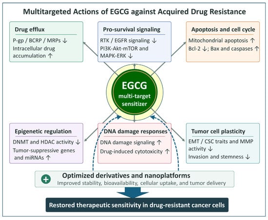
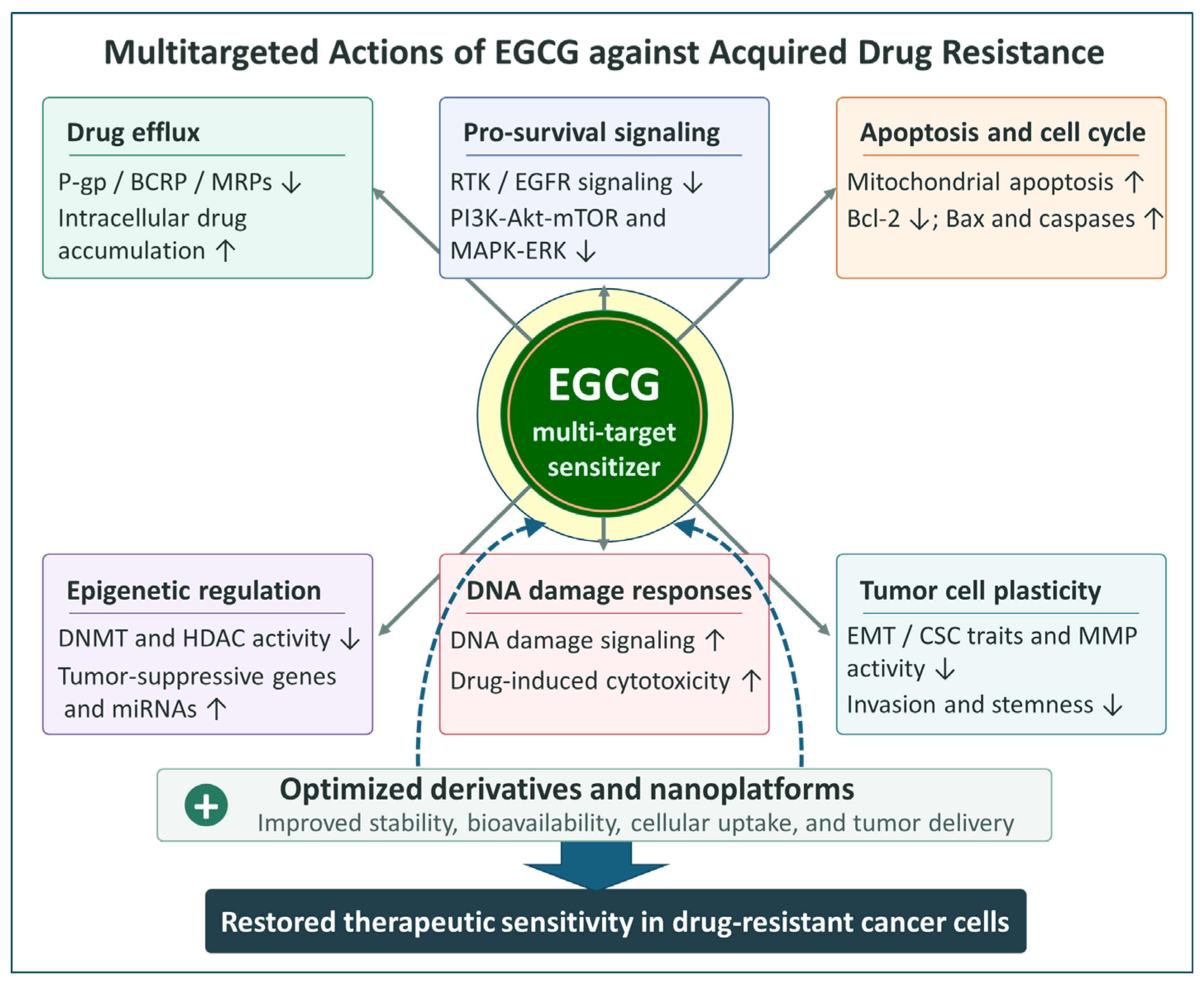
Preclinical studies have examined the effects of EGCG on multiple processes that contribute to acquired drug resistance. Figure 1 summarizes the proposed mechanisms discussed in this section.
Figure 1.
Multi-targeted mechanisms by which EGCG attenuates acquired drug resistance in cancer cells. EGCG modulates multiple resistance-associated processes, including drug efflux, pro-survival signaling, apoptosis and cell-cycle regulation, epigenetic regulation, DNA damage responses, and tumor cell plasticity. Optimized EGCG derivatives and nanoplatforms may improve stability, bioavailability, cellular uptake, and tumor delivery, thereby supporting the restoration of therapeutic sensitivity in drug-resistant cancer cells. The arrows at the bottom of the figure indicate that optimized EGCG derivatives and nanoplatforms may help restore drug sensitivity in resistant cancer cells. The arrows ↑ and ↓ indicate an increase and a decrease, respectively
2.1. Inhibition of Drug Efflux and Enhancement of Drug Accumulation
A major mechanism underlying acquired resistance to chemotherapy is the overexpression of ATP-binding cassette (ABC) transporters, which actively export cytotoxic agents from cancer cells and reduce intracellular drug accumulation [17]. Among these transporters, P-glycoprotein (P-gp), encoded by the ABCB1 (MDR1) gene, breast cancer resistance protein (BCRP), encoded by ABCG2, and multidrug resistance-associated proteins play central roles in the multidrug resistance (MDR) phenotype [17]. Increasing evidence indicates that EGCG can counteract this mechanism by modulating the expression and functional activity of these efflux pumps, thereby enhancing intracellular drug retention [14,18].
2.1.1. Downregulation of MDR Transporter Expression
Several studies have demonstrated that EGCG suppresses the expression of key MDR-associated transporters. In tamoxifen-resistant breast cancer models, EGCG reduced P-gp protein levels to approximately half of control levels without altering ABCB1 mRNA expression, suggesting post-transcriptional regulation [14]. In 5-fluorouracil-resistant SGC-7901/FU and MGC-803/FU cells, EGCG downregulated ABCB1 mRNA and P-gp protein levels, with stronger effects in resistant cells than in their parental counterparts [19]. This suppression was also confirmed in xenograft tumors in vivo [19]. Consistently, in 5-fluorouracil- and Adriamycin-resistant carcinoma cells, EGCG decreased the expression of efflux-associated genes such as ABCB1 and lung resistance protein [20]. In cisplatin-resistant CAR cells, EGCG reduced ABCB1 expression, accompanied by decreased phosphorylation of Akt and STAT3 [15].
2.1.2. Functional Inhibition of Efflux Transporter Activity
In addition to transcriptional regulation, EGCG can interfere with the functional activity of efflux transporters. In P-gp-overexpressing multidrug-resistant KB-A-1 cells, EGCG significantly inhibited P-gp-mediated efflux of doxorubicin, resulting in a concentration-dependent increase in intracellular drug accumulation without altering MDR1 mRNA expression [18], suggesting that resistance reversal in this model occurs primarily through functional inhibition of transporter activity. Consistent with this observation, EGCG and tea polyphenols enhanced intracellular doxorubicin levels in resistant KB-A-1 cells by increasing drug influx, reducing efflux, and inhibiting P-gp ATPase activity [21]. Notably, these resistance-reversal effects were largely restricted to MDR cells, as EGCG markedly reduced the IC50 of doxorubicin in resistant KB-A-1 cells but had little effect in parental KB-3-1 cells [22].
Additional studies further support the ability of EGCG to enhance intracellular drug retention through functional modulation of efflux transporters without altering transporter expression. In multidrug-resistant cells MCF-7/ADR, EGCG alone or in combination with curcumin did not affect P-gp protein levels, yet combination treatment markedly increased intracellular accumulation of rhodamine-123, curcumin, and doxorubicin, indicating functional modulation of transporter activity [23]. This enhanced intracellular drug retention was accompanied by increased caspase activation and PARP cleavage, together with downregulation of Bcl-2 and Survivin [23]. Similarly, although EGCG alone exerted only modest inhibition of P-gp activity in CEM/ADR5000 leukemia cells, it significantly enhanced doxorubicin-induced cytotoxicity when used in combination [24], suggesting that even modest inhibition of P-gp activity can substantially increase chemosensitivity. EGCG may also target multiple efflux transporters. For example, EGCG inhibited BCRP-mediated drug efflux, as demonstrated by increased intracellular accumulation of the BCRP substrate mitoxantrone without altering BCRP expression [14].
2.2. Suppression of Pro-Survival Signaling Pathways
In addition to modulating drug efflux systems, EGCG has been widely reported to sensitize drug-resistant cancer cells by suppressing key survival signaling pathways that support proliferation, stress adaptation, and resistance to cell death [25]. Many chemoresistant tumors exhibit hyperactivation of receptor tyrosine kinases (RTKs) and downstream signaling pathways, including the PI3K/Akt/mTOR and MAPK/ERK pathways [1]. EGCG interferes with these signaling networks at multiple levels, thereby attenuating survival signaling and restoring drug sensitivity [25].
Several studies indicate that EGCG targets RTK signaling pathways upregulated in resistant cancer cells. For example, tamoxifen-resistant MCF-7 cells display markedly elevated EGFR expression and phosphorylation compared with parental cells [16]. EGCG treatment significantly reduced EGFR levels in a dose-dependent manner, accompanied by decreased EGFR membrane localization and suppression of downstream ERK signaling. Notably, these inhibitory effects were more pronounced in resistant cells than in parental MCF-7 cells [16]. Similarly, EGCG inhibited phosphorylation of KIT, the major oncogenic driver in gastrointestinal stromal tumors, in both imatinib-sensitive and resistant cells, leading to suppression of downstream Akt and MAPK signaling pathways [26]. In non-small-cell lung cancer models, EGCG also downregulated Axl and Tyro3 expression at both the mRNA and protein levels in parental and cisplatin-resistant cell lines [27].
Beyond receptor-level inhibition, EGCG has also been shown to suppress major intracellular survival pathways. In multiple resistant cancer cell models, EGCG treatment reduced phosphorylation of Akt at Ser473 in a dose-dependent manner [28]. Inhibition of Akt signaling triggered nuclear translocation of the transcription factor FOXO3a and increased expression of the cell-cycle inhibitor p27^Kip1, ultimately contributing to growth suppression [28]. Importantly, expression of constitutively active Akt abolished the growth-inhibitory effects of EGCG, demonstrating that suppression of Akt signaling is a central mechanism underlying the growth-inhibitory effects of EGCG [28]. Consistent with this mechanism, EGCG also inhibited phosphorylation of Akt and STAT3 in cisplatin-resistant oral cancer cells [15].
EGCG further modulates downstream metabolic and growth-control pathways. In trastuzumab-resistant (SKTR) and lapatinib-resistant (SKLR) HER2-positive SK-BR-3 breast cancer models, combined treatment with EGCG and the mTOR inhibitor temsirolimus abolished mTOR expression, indicating potent inhibition of mTOR-driven growth signaling [29]. Similarly, in EGFR-inhibitor-resistant lung cancer sublines (H1975 GR and H1975 AR), EGCG combined with gefitinib or osimertinib suppressed phosphorylation of ERK, Akt, and mTOR while activating AMP-activated protein kinase signaling, suggesting metabolic regulation of survival pathways [30].
In addition to classical survival pathways, EGCG has been reported to interfere with stress-adaptation mechanisms. Transcriptomic analyses revealed downregulation of stress-response genes such as HSPA1B, HSPA1L, and USP5 in drug-resistant BEL7404/ADM and BEL7402/5-FU cells [20].
EGCG can also disrupt pro-angiogenic survival signaling. In gastric cancer cells resistant to 5-fluorouracil, EGCG suppressed phosphorylation of TFAP2A and reduced VEGF secretion without altering total TFAP2A levels, indicating inhibition of the TFAP2A/VEGF axis both in vitro and in xenograft tumors [19].
2.3. Induction of Apoptosis and Cell-Cycle Arrest
A major mechanism by which EGCG reverses drug resistance is the restoration of apoptotic responsiveness and induction of cell-cycle arrest [23]. Many resistant cancer cells evade cytotoxic therapy by suppressing apoptotic signaling [1] and upregulating anti-apoptotic proteins such as Bcl-2, Survivin, and XIAP. Evidence from multiple drug-resistant cancer models indicates that EGCG restores apoptotic responsiveness by activating mitochondrial apoptotic pathways and shifting the balance between pro- and anti-apoptotic proteins [14,15].
Activation of the intrinsic mitochondrial apoptotic pathway has been reported in multiple drug-resistant cancer models. In tamoxifen-resistant MCF-7Tam cells, EGCG induced dose-dependent apoptosis accompanied by increased Bax expression and reduced Bcl-2 levels [14]. Similar effects were reported in cisplatin-resistant CAR cells, a subline established from human tongue squamous cell carcinoma CAL27 cells by repeated cisplatin exposure [15]. In these cells, EGCG induced apoptosis through activation of caspase-9 and caspase-3, together with modulation of Bax and Bcl-2 expression [15]. EGCG also induced apoptosis in drug-sensitive H69 and drug-resistant H69VP small-cell lung cancer cells, as evidenced by DNA fragmentation and TUNEL staining [31].
EGCG-induced apoptosis is also associated with suppression of anti-apoptotic proteins. In gastrointestinal stromal tumor cells, EGCG triggered anoikis-like apoptosis accompanied by downregulation of Survivin and XIAP [26]. In HER2-targeted therapy-resistant breast cancer models, EGCG also enhanced PARP cleavage and apoptotic signaling, accompanied by increased terminal deoxynucleotidyl transferase dUTP nick-end labeling (TUNEL)-positive cells in xenograft tumors [29]. In Hep3B sublines with acquired resistance to lenvatinib or sorafenib, EGCG inhibited cell proliferation and induced apoptosis [32]. Protein array analysis after 48 h of exposure to 10 μg/mL EGCG (approximately 21.8 μM) showed reduced levels of paraoxonase-2 (PON2) and heat shock protein 27 (HSP27) in the resistant sublines [32].
Importantly, EGCG exhibits enhanced pro-apoptotic effects when combined with anticancer agents or other bioactive compounds. In multidrug-resistant MCF-7/ADR cells, 2 μM EGCG alone induced little apoptosis, but co-treatment with 2 μM EGCG and 10 μM curcumin for 48 h increased apoptosis compared with curcumin alone [23]. This finding illustrates that EGCG can enhance an apoptotic response at a concentration with little pro-apoptotic activity on its own in the same resistant model. Similar synergistic effects have been reported for combinations with 5-fluorouracil [6], cisplatin [33], EGFR inhibitors [30], and other phytochemicals [23].
EGCG also induces cell-cycle perturbations in drug-resistant cancer cells, with arrest occurring at different cell-cycle checkpoints. EGCG induced G2 arrest in resistant cells and enhanced G2 arrest when combined with 5-fluorouracil [6]. In other studies, EGCG triggered G1 arrest with upregulation of p21 and reduced expression of CDK2 and CDK6 [30]. Transcriptomic analyses further showed modulation of genes associated with cell-cycle progression and checkpoint regulation, including Cyclin G1, Cyclin G2, RB1, and GADD34 [20].
2.4. Epigenetic Modulation of Drug Resistance
Beyond its effects on drug transporters and survival signaling, EGCG has also been reported to modulate epigenetic mechanisms associated with drug resistance [6]. Epigenetic alterations—including aberrant DNA methylation, histone modifications, and dysregulation of microRNAs—are increasingly recognized as key contributors to chemoresistance [1], as they can silence tumor suppressor genes and promote CSC maintenance. Emerging evidence suggests that EGCG can modulate epigenetic regulatory mechanisms and restore the expression of tumor suppressor genes [3].
One such mechanism involves modulation of tumor-suppressive microRNAs associated with CSC regulation and drug resistance. In 5-fluorouracil-resistant cancer cells, EGCG significantly increased the expression of miR-34a, miR-145, and miR-200c, whereas treatment with 5-fluorouracil alone had little effect [6]. These microRNAs are well known to regulate pathways involved in stemness and epithelial–mesenchymal transition, suggesting that EGCG may attenuate CSC-associated programs that contribute to chemoresistance [6].
EGCG has also been shown to influence epigenetic regulators associated with drug resistance. Transcriptomic analyses revealed reduced expression of histone deacetylase 5 (HDAC5) following EGCG treatment in drug-resistant BEL7404/ADM cells [20]. More direct evidence of epigenetic regulation has been reported in cisplatin-resistant cancer cells, where EGCG inhibited both DNA methyltransferase (DNMT) and HDAC enzymatic activity, leading to decreased promoter methylation and reactivation of multiple tumor suppressor genes, including GAS1, TIMP4, ICAM1, and WISP2 [34]. These genes had previously been identified as epigenetically silenced during the development of cisplatin resistance. Notably, the epigenetic effects of EGCG were comparable to those observed with the demethylating agent 5-aza-2′-deoxycytidine (5-Aza-dC). Similarly, in paclitaxel-resistant ovarian cancer cells, co-treatment with EGCG and sulforaphane reduced DNMT1 protein expression, accompanied by suppression of hTERT expression [35].
2.5. Enhancement of DNA Damage Responses
EGCG has also been reported to induce or enhance drug-induced DNA damage in drug-resistant cancer cells. In small-cell lung carcinoma models, EGCG treatment induced significant DNA fragmentation, as detected by enzyme-linked immunosorbent assay and TUNEL assays [31].
Consistent with these findings, enhanced DNA damage signaling was observed in paclitaxel-resistant ovarian cancer models. In SKOV3 and paclitaxel-resistant SKOV3TR-ip2 cells, combined treatment with EGCG and sulforaphane markedly increased H2AX phosphorylation, a widely used marker of DNA damage [35].
In addition, sequential treatment with cisplatin followed by EGCG produced synergistic cytotoxicity in both cisplatin-sensitive and resistant ovarian cancer cells, accompanied by increased intracellular platinum accumulation and platinum–DNA binding [33], suggesting that increased platinum–DNA binding contributes to the enhanced cytotoxic response.
2.6. Suppression of EMT, Cancer Stemness, and Invasive Phenotypes
EGCG has also been reported to attenuate invasive and stem cell-like phenotypes frequently associated with chemoresistance, extending its effects beyond the regulation of drug efflux and survival signaling [6,16]. Drug-resistant cancer cells often acquire EMT features, enhanced extracellular matrix (ECM) remodeling capacity, and enrichment of CSC populations, all of which contribute to tumor progression, metastasis, and therapeutic failure. EGCG appears to interfere with these processes by suppressing matrix-degrading enzymes, inhibiting EMT-associated invasion, and modulating CSC-related signaling pathways.
EGCG modulates matrix metalloproteinases (MMPs) involved in extracellular matrix (ECM) remodeling and tumor invasion. The doxorubicin-resistant breast cancer sublines MCF-7/DOX and MCF-7/DOX500 exhibited elevated MMP-2 and MMP-9 activities, whereas neither activity was detected in parental MCF-7 cells [36]. Despite markedly different P-glycoprotein levels, both resistant sublines showed concentration-dependent reductions in MMP-2 and MMP-9 activity after 24 h of exposure to 5–20 μM EGCG [36]. In tamoxifen-resistant MCF-7Tam cells, which displayed an EMT-like phenotype, EGCG reduced Matrigel invasion and lowered the protein levels of MMP-2, MMP-9, and extracellular matrix metalloproteinase inducer while increasing TIMP-1 and TIMP-2 expression. Gelatin zymography showed that the bands corresponding to MMP-2 and MMP-9 were undetectable after 24 h of exposure to either 50 or 100 μg/mL EGCG (approximately 109 and 218 μM, respectively) [16]. In drug-resistant BEL7404/ADM and BEL7402/5-FU cells, microarray analysis showed that exposure to 20 mg/L (43.6 μM) EGCG reduced MMP15 mRNA expression [20]. Although the two breast cancer studies assessed MMP-2 and MMP-9 activity after the same exposure duration in MCF-7-derived cells, the differences in acquired resistance phenotypes limit direct comparisons of EGCG sensitivity [16,36]. The study of BEL7404/ADM and BEL7402/5-FU cells extends these observations to a different MMP at the transcript level, further illustrating why the reported concentrations should be interpreted in relation to the resistance model and the endpoint measured.
In addition to regulating ECM remodeling, EGCG suppresses cancer stem cell-like properties associated with drug resistance. In colorectal cancer models, EGCG inhibited spheroid formation in both parental and drug-resistant cell lines, and co-treatment with 5-fluorouracil synergistically reduced spheroid numbers [6]. This effect was accompanied by downregulation of CSC markers including Oct4, Nanog, CD44, ALDH1, and CD133 [6]. Similarly, cisplatin-resistant lung cancer cells exhibited increased side-population fractions and elevated expression of stemness markers such as CD44, CD133, Sox2, Nanog, and Oct4, which were attenuated by EGCG treatment [37]. Mechanistically, EGCG upregulated miR-485, which directly targets CD44, thereby suppressing CSC-like properties [37]. In xenograft models, EGCG treatment also reduced CSC marker expression and the proliferation marker Ki-67 [37].
Consistent with these observations, EGCG has been reported to interfere with signaling networks that regulate CSC maintenance and self-renewal. EGCG suppressed Notch1 signaling, as evidenced by reduced levels of cleaved Notch1 and c-Myc, accompanied by downregulation of polycomb repressive complex components including Bmi1, Ezh2, and Suz12 [6].
Table 1 summarizes the preclinical studies discussed in Section 2.1, Section 2.2, Section 2.3, Section 2.4, Section 2.5 and Section 2.6, including the models of acquired drug resistance and the reported effects of EGCG.
Table 1.
Preclinical studies evaluating EGCG in acquired drug-resistant cancer models.
2.7. EGCG Derivatives and Nanoplatform Strategies to Overcome Pharmacological Limitations
Despite its diverse anti-resistance activities, the clinical application of EGCG remains limited by poor stability, rapid oxidation, and relatively low bioavailability. To address these limitations, recent studies have focused on EGCG derivatives and EGCG-based nanoplatforms designed to improve its pharmacological properties and efficacy in drug-resistant cancer models.
One major approach involves incorporating EGCG into nanocarrier systems to enable co-delivery with conventional chemotherapeutic agents. In MCF-7/ADR cells, PEI-DOX/EGCG and its folate-targeted formulation PEI-DOX/EGCG/FA suppressed P-gp expression and enhanced intracellular doxorubicin accumulation [38]. The folate-modified nanoparticles further enhanced the inhibitory effect on cell migration [38]. These systems also mitigated doxorubicin-induced cardiotoxicity by attenuating oxidative stress, as evidenced by reduced levels of malondialdehyde, creatine kinase-MB, and lactate dehydrogenase together with restoration of superoxide dismutase activity. In xenograft models, both nanoparticle formulations suppressed tumor growth more effectively than free doxorubicin, whereas the folate-targeted system showed the greatest antitumor efficacy together with reduced cardiac doxorubicin accumulation and fibrosis [38]. These findings highlight the dual potential of EGCG-based nanoplatforms to overcome multidrug resistance while reducing doxorubicin-associated cardiotoxicity.
Similarly, an EGCG-loaded mesoporous silica–gold nanocluster formulation (EAuSSi) and its dual-drug counterpart co-encapsulating EGCG and the EGFR/VEGFR2 inhibitor ZD6474 (ZEAuSS) effectively overcame tamoxifen resistance by suppressing EGFR/VEGFR2/Akt signaling, resulting in enhanced apoptosis and cell-cycle arrest [39]. Enhanced therapeutic efficacy was associated with improved intracellular uptake and activation of mitochondrial apoptotic pathways, accompanied by downregulation of Bcl-2 and upregulation of Bax, Bak, Bim, and p53. In xenograft models, the dual-loaded nanoplatform produced the greatest tumor suppression, accompanied by reduced Ki-67 expression and increased TUNEL-positive cells [39].
In hematological malignancies, daunorubicin/EGCG nanocomplexes enhanced intracellular delivery and nuclear accumulation of daunorubicin, resulting in increased reactive oxygen species generation and activation of caspase-3 and caspase-7, thereby producing synergistic cytotoxicity accompanied by caspase-mediated apoptosis in multidrug-resistant leukemia cells [40].
EGCG-based platforms have also been extended to gene-targeting strategies. Nanoparticle-mediated delivery of siRNA targeting the resistance-associated long non-coding RNA TMEM44-AS1 reduced expression of the efflux transporters ABCC1 and ABCG2. This was accompanied by increased levels of cleaved caspase-3, Bax, p53, and AIFM2 together with reduced expression of Survivin and Bcl-2, resulting in enhanced chemosensitivity in resistant tumor models [41]. The chitosan–gelatin–EGCG nanoparticle (CGE) platform facilitated siRNA loading and lysosomal escape, leading to enhanced antitumor efficacy in 5-fluorouracil-resistant xenograft models [41].
In parallel, chemically modified EGCG derivatives have been developed to enhance etoposide intrinsic anticancer potency. The derivative G28 exhibited greater efficacy than native EGCG in suppressing cancer stem cell-like properties, achieving near-complete inhibition of mammosphere formation in parental MDA-MB-231 cells and their doxorubicin-resistant (231DXR) and paclitaxel-resistant (231PTR) derivatives [42].
The ethylated derivative Y6, developed to improve the stability and lipophilicity of native EGCG, reversed multidrug resistance in doxorubicin-resistant BEL-7404/DOX cells by downregulating ABCB1/P-gp expression and suppressing HIF-1α-mediated hypoxia signaling, thereby restoring apoptotic responsiveness [43]. In oxaliplatin-resistant SMMC-7721/OXA cells, Y6 further inhibited EMT-associated resistance in association with suppression of the HIF-1α/TWIST axis, accompanied by increased E-cadherin expression, reduced vimentin expression, and decreased migratory and invasive capacities [44].
Table 2 summarizes the preclinical studies discussed in Section 2.7, including the EGCG derivatives and nanoplatform-based delivery systems evaluated and their reported effects in models of acquired drug resistance.
Table 2.
Preclinical studies evaluating EGCG derivatives and nanoplatform-based delivery systems in acquired drug-resistant cancer models.
3. Clinical and Translational Relevance and Remaining Challenges of EGCG in Cancer
Given the extensive mechanistic evidence and recent advances in EGCG-based derivatives and delivery systems, an important question is whether these findings can be translated into clinically meaningful outcomes. Although direct clinical evidence in patients with established drug-resistant cancers remains limited, human pharmacokinetic studies confirm that orally administered EGCG is absorbed, with limited systemic exposure that varies according to dosing conditions [45].
Observational studies in Japan have associated higher green tea consumption with a lower risk of recurrence among patients with stage I-II breast cancer [46] and with later cancer onset in a population-based cohort [47]. A more recent meta-analysis reported inverse associations between green tea or EGCG intake and the risk of selected cancers [9]. These findings relate to cancer risk or recurrence and provide limited insight into therapeutic resensitization in patients with acquired drug resistance.
Trials in premalignant and high-risk populations have also evaluated green tea catechin preparations. Green tea extract supplementation was associated with fewer metachronous colorectal adenomas after polypectomy [7,8]. In men with high-grade prostatic intraepithelial neoplasia, an early trial reported fewer prostate cancer diagnoses with catechin supplementation [48], whereas a subsequent randomized trial in men with high-grade prostatic intraepithelial neoplasia and/or atypical small acinar proliferation found no statistically significant reduction in overall prostate cancer incidence [49]. In patients with high-risk oral premalignant lesions, clinical responses were more frequent at higher green tea extract doses, and reductions in stromal VEGF and cyclin D1 expression were observed among clinical responders [50].
In women with a history of hormone receptor-negative breast cancer, a phase Ib dose-escalation trial evaluated the tolerability of oral Polyphenon E and included exploratory biomarker analyses [51]. In a separate presurgical trial in patients with bladder cancer, Polyphenon E was associated with dose-dependent decreases in PCNA and clusterin expression [52]. In patients with asymptomatic, early-stage chronic lymphocytic leukemia, phase I and II studies of oral Polyphenon E reported decreases in absolute lymphocyte counts and/or lymphadenopathy [53,54]. Table 3 summarizes selected human trials of EGCG and EGCG-containing preparations, including the study populations, dosing regimens, endpoints, and outcomes.
Table 3.
Selected human intervention trials cited in Section 3.
These findings document biological effects in specific clinical settings. The ability of EGCG to restore responsiveness to a therapy after resistance has emerged remains unproven. A key question for clinical translation is whether oral EGCG or an optimized formulation can achieve sufficient exposure within tumors to reproduce the effects observed in drug-resistant models. In healthy volunteers, a single oral dose of Polyphenon E containing 800 mg of EGCG yielded mean peak plasma concentrations of unconjugated EGCG of approximately 0.64 μM with food and 3.3 μM under fasting conditions [45]. The fasting peak was in the same range as the nominal 2 μM EGCG concentration used in the 48 h curcumin co-treatment experiment [23], but below the concentrations used in the 24 h MMP-2 and MMP-9 activity experiments [16,36]. This comparison illustrates the difference between a plasma peak after oral dosing and the concentration and duration of exposure used in cell culture. Whether comparable concentrations can be achieved and maintained within human tumors remains uncertain. In the presurgical bladder cancer trial, EGCG concentrations in nonmalignant bladder tissue did not differ significantly between the combined Polyphenon E groups and the placebo group, although supplemental analyses across all three study arms showed significant dose–response relationships in both nonmalignant and tumor tissue [52]. Differences among brewed green tea, standardized extracts, and purified EGCG further complicate comparisons across studies. Chemical instability, metabolism, dose-related toxicity, and potential interactions with anticancer agents also warrant consideration. In the Minnesota Green Tea Trial, daily supplementation with green tea extract containing 843 mg EGCG for one year was associated with more frequent ALT elevations than placebo (6.7% versus 0.7%) [55]. Thus, attempts to increase systemic exposure through higher supplement doses must also account for hepatic safety. EGCG derivatives and delivery systems may improve exposure, but their ability to restore drug responsiveness requires direct evaluation.
A search of ClinicalTrials.gov on 9 November 2026 identified registered EGCG-containing combination studies, including Polyphenon E plus erlotinib in head and neck chemoprevention (NCT01116336; completed) [56] and advanced non-small-cell lung cancer (NCT00707252; terminated) [57]. A breast cancer trial combining EGCG, quercetin, zinc, and metformin with chemotherapy had an unknown recruitment status (NCT05680662) [58]. No ongoing combination trial specifically designed to demonstrate reversal of acquired drug resistance was identified.
Trials aimed at reversing acquired resistance should enroll patients with documented resistance and evaluate an EGCG formulation in combination with the therapy to which resistance has emerged. Pharmacokinetic assessments should measure systemic and, where feasible, intratumoral exposure and examine how these exposures relate to changes in resistance-associated mechanisms, such as drug efflux, survival signaling, apoptotic responsiveness, or stem cell-like properties. Assessing whether the combination restores tumor responsiveness would help determine whether these pharmacodynamic changes translate into resensitization.
4. Conclusions
Evidence from established drug-resistant cancer models indicates that EGCG can resensitize resistant cells by modulating interconnected processes, including drug efflux, pro-survival signaling, apoptotic dysfunction, epigenetic regulation, DNA damage responses, and cancer stem cell-like and invasive phenotypes. This multi-targeted activity may explain its chemosensitizing effects across different cancer types and therapeutic agents. However, the contribution of individual mechanisms varies among models, and some molecular changes remain associative rather than direct determinants of resistance reversal.
EGCG derivatives and delivery systems may overcome the limited stability, bioavailability, and tumor exposure of native EGCG. By improving cellular uptake and delivery to tumor tissues [38,40], these approaches may enhance the modulation of resistance-associated pathways and provide a stronger pharmacological basis for EGCG-mediated chemosensitization. Further validation in clinically relevant resistant models, together with assessment of systemic and intratumoral exposure, will help determine the extent to which these advances can improve therapeutic responses.
The principal translational challenge is to determine whether EGCG can achieve sufficient exposure to reverse defined resistance mechanisms while maintaining an acceptable safety profile and compatibility with concurrently administered anticancer drugs. Future studies should combine optimized formulations with therapies to which resistance has developed and incorporate pharmacokinetic, pharmacodynamic, and resistance-associated biomarkers. Ultimately, trials in molecularly characterized drug-resistant cancers are needed to establish whether the multi-targeted activity of EGCG can produce a clinically meaningful restoration of therapeutic sensitivity.
Author Contributions
Conceptualization, S.I.H. and T.-B.L.; methodology, S.-C.L. and T.-B.L.; data curation, S.I.H. and C.Y.C.; formal analysis, S.-C.L. and T.-B.L.; writing—original draft preparation, T.-B.L.; writing—review and editing, S.I.H. and S.-C.L.; visualization, T.-B.L. and C.Y.C.; project administration, T.-B.L.; funding acquisition, S.I.H. All authors have read and agreed to the published version of the manuscript.
Funding
This research was supported by the Basic Science Research Program through the National Research Foundation of Korea funded by the Ministry of Education (NRF-2022R1I1A3065349).
Institutional Review Board Statement
Not applicable.
Informed Consent Statement
Not applicable.
Data Availability Statement
No new data were created or analyzed in this study. Data sharing is not applicable to this article.
Acknowledgments
During the preparation of this manuscript, the authors used ChatGPT (OpenAI, GPT-5 series) to improve the language and clarity of the manuscript. The authors have reviewed and edited the output and take full responsibility for the content of this publication.
Conflicts of Interest
The authors declare no conflicts of interest in relation to this study.
Abbreviations
The following abbreviations are used in this manuscript:
| 5-FU | 5-fluorouracil |
| BCRP | breast cancer resistance protein |
| CGE | chitosan–gelatin–epigallocatechin-3-gallate |
| CSC | cancer stem cell |
| DNMT | DNA methyltransferase |
| DOX | doxorubicin |
| EAuSSi | EGCG-loaded mesoporous silica–gold nanoclusters |
| ECM | extracellular matrix |
| EGCG | epigallocatechin-3-gallate |
| EGFR | epidermal growth factor receptor |
| EMT | epithelial–mesenchymal transition |
| ERK | extracellular signal-regulated kinase |
| FA | folic acid |
| HDAC | histone deacetylase |
| HIF-1α | hypoxia-inducible factor 1 alpha |
| MAPK | mitogen-activated protein kinase |
| MDR | multidrug resistance |
| mTOR | mechanistic target of rapamycin |
| P-gp | P-glycoprotein |
| PEI | polyethyleneimine |
| RTK | receptor tyrosine kinase |
| siRNA | small interfering RNA |
| TUNEL | terminal deoxynucleotidyl transferase dUTP nick-end labeling |
| VEGF | vascular endothelial growth factor |
| ZEAuSS | ZD6474- and EGCG-loaded mesoporous silica–gold nanoclusters |
References
- Holohan, C.; Van Schaeybroeck, S.; Longley, D.B.; Johnston, P.G. Cancer drug resistance: An evolving paradigm. Nat. Rev. Cancer 2013, 13, 714–726. [Google Scholar] [CrossRef] [Scilit] [PubMed]
- Vasan, N.; Baselga, J.; Hyman, D.M. A view on drug resistance in cancer. Nature 2019, 575, 299–309. [Google Scholar] [CrossRef] [Scilit] [PubMed]
- Yang, C.S.; Wang, X.; Lu, G.; Picinich, S.C. Cancer prevention by tea: Animal studies, molecular mechanisms and human relevance. Nat. Rev. Cancer 2009, 9, 429–439. [Google Scholar] [CrossRef] [Scilit] [PubMed]
- Lin, Y.S.; Tsai, Y.J.; Tsay, J.S.; Lin, J.K. Factors affecting the levels of tea polyphenols and caffeine in tea leaves. J. Agric. Food Chem. 2003, 51, 1864–1873. [Google Scholar] [CrossRef] [Scilit] [PubMed]
- National Center for Biotechnology Information. Pubchem Compound Summary for Cid 65064, Epigallocatechin Gallate. Available online: https://pubchem.ncbi.nlm.nih.gov/compound/65064 (accessed on 12 November 2026).
- Toden, S.; Tran, H.M.; Tovar-Camargo, O.A.; Okugawa, Y.; Goel, A. Epigallocatechin-3-gallate targets cancer stem-like cells and enhances 5-fluorouracil chemosensitivity in colorectal cancer. Oncotarget 2016, 7, 16158–16171. [Google Scholar] [CrossRef] [Scilit] [PubMed]
- Shimizu, M.; Fukutomi, Y.; Ninomiya, M.; Nagura, K.; Kato, T.; Araki, H.; Suganuma, M.; Fujiki, H.; Moriwaki, H. Green tea extracts for the prevention of metachronous colorectal adenomas: A pilot study. Cancer Epidemiol. Biomark. Prev. 2008, 17, 3020–3025. [Google Scholar] [CrossRef] [Scilit] [PubMed]
- Shin, C.M.; Lee, D.H.; Seo, A.Y.; Lee, H.J.; Kim, S.B.; Son, W.C.; Kim, Y.K.; Lee, S.J.; Park, S.H.; Kim, N.; et al. Green tea extracts for the prevention of metachronous colorectal polyps among patients who underwent endoscopic removal of colorectal adenomas: A randomized clinical trial. Clin. Nutr. 2018, 37, 452–458. [Google Scholar] [CrossRef] [Scilit] [PubMed]
- Zhang, Y.; Xu, Q.; Hu, J.; Zhang, F.; Yu, Y.; Ma, L. Green tea and epigallocatechin gallate (egcg) for cancer prevention: A systematic review and meta-analysis. Am. J. Chin. Med. 2025, 53, 1755–1784. [Google Scholar] [CrossRef] [Scilit] [PubMed]
- Gan, R.Y.; Li, H.B.; Sui, Z.Q.; Corke, H. Absorption, metabolism, anti-cancer effect and molecular targets of epigallocatechin gallate (egcg): An updated review. Crit. Rev. Food Sci. Nutr. 2018, 58, 924–941. [Google Scholar] [CrossRef] [Scilit] [PubMed]
- Singh, B.N.; Shankar, S.; Srivastava, R.K. Green tea catechin, epigallocatechin-3-gallate (egcg): Mechanisms, perspectives and clinical applications. Biochem. Pharmacol. 2011, 82, 1807–1821. [Google Scholar] [CrossRef] [Scilit] [PubMed]
- Siddiqui, I.A.; Adhami, V.M.; Afaq, F.; Ahmad, N.; Mukhtar, H. Modulation of phosphatidylinositol-3-kinase/protein kinase b- and mitogen-activated protein kinase-pathways by tea polyphenols in human prostate cancer cells. J. Cell. Biochem. 2004, 91, 232–242. [Google Scholar] [CrossRef] [Scilit] [PubMed]
- Gupta, S.; Ahmad, N.; Nieminen, A.L.; Mukhtar, H. Growth inhibition, cell-cycle dysregulation, and induction of apoptosis by green tea constituent (-)-epigallocatechin-3-gallate in androgen-sensitive and androgen-insensitive human prostate carcinoma cells. Toxicol. Appl. Pharmacol. 2000, 164, 82–90. [Google Scholar] [CrossRef] [Scilit] [PubMed]
- Farabegoli, F.; Papi, A.; Bartolini, G.; Ostan, R.; Orlandi, M. (-)-Epigallocatechin-3-gallate downregulates pg-p and bcrp in a tamoxifen resistant mcf-7 cell line. Phytomedicine 2010, 17, 356–362. [Google Scholar] [CrossRef] [Scilit] [PubMed]
- Yuan, C.H.; Horng, C.T.; Lee, C.F.; Chiang, N.N.; Tsai, F.J.; Lu, C.C.; Chiang, J.H.; Hsu, Y.M.; Yang, J.S.; Chen, F.A. Epigallocatechin gallate sensitizes cisplatin-resistant oral cancer car cell apoptosis and autophagy through stimulating akt/stat3 pathway and suppressing multidrug resistance 1 signaling. Environ. Toxicol. 2017, 32, 845–855. [Google Scholar] [CrossRef] [Scilit] [PubMed]
- Farabegoli, F.; Papi, A.; Orlandi, M. (-)-Epigallocatechin-3-gallate down-regulates egfr, mmp-2, mmp-9 and emmprin and inhibits the invasion of mcf-7 tamoxifen-resistant cells. Biosci. Rep. 2011, 31, 99–108. [Google Scholar] [CrossRef] [Scilit] [PubMed]
- Robey, R.W.; Pluchino, K.M.; Hall, M.D.; Fojo, A.T.; Bates, S.E.; Gottesman, M.M. Revisiting the role of abc transporters in multidrug-resistant cancer. Nat. Rev. Cancer 2018, 18, 452–464. [Google Scholar] [CrossRef] [Scilit] [PubMed]
- Qian, F.; Wei, D.; Zhang, Q.; Yang, S. Modulation of p-glycoprotein function and reversal of multidrug resistance by (-)-epigallocatechin gallate in human cancer cells. Biomed. Pharmacother. 2005, 59, 64–69. [Google Scholar] [CrossRef] [Scilit] [PubMed]
- Tang, H.; Zeng, L.; Wang, J.; Zhang, X.; Ruan, Q.; Wang, J.; Cui, S.; Yang, D. Reversal of 5-fluorouracil resistance by egcg is mediate by inactivation of tfap2a/vegf signaling pathway and down-regulation of mdr-1 and p-gp expression in gastric cancer. Oncotarget 2017, 8, 82842–82853. [Google Scholar] [CrossRef] [Scilit] [PubMed]
- Tang, H.H.; Zhou, M.; Liang, G. Impact of epigallocatechin gallate on gene expression profiles of human hepatocellular carcinoma cell lines bel7404/adm and bel7402/5-fu. Pharmacol. Clin. Chin. Mater. Medica 2008, 27, 1056–1064. [Google Scholar]
- Mei, Y.; Qian, F.; Wei, D.; Liu, J. Reversal of cancer multidrug resistance by green tea polyphenols. J. Pharm. Pharmacol. 2004, 56, 1307–1314. [Google Scholar] [CrossRef] [Scilit] [PubMed]
- Mei, Y.; Wei, D.; Liu, J. Reversal of multidrug resistance in kb cells with tea polyphenol antioxidant capacity. Cancer Biol. Ther. 2005, 4, 468–473. [Google Scholar] [CrossRef] [Scilit] [PubMed]
- Wang, S.; Chen, R.; Zhong, Z.; Shi, Z.; Chen, M.; Wang, Y. Epigallocatechin-3-gallate potentiates the effect of curcumin in inducing growth inhibition and apoptosis of resistant breast cancer cells. Am. J. Chin. Med. 2014, 42, 1279–1300. [Google Scholar] [CrossRef] [Scilit] [PubMed]
- Li, H.; Krstin, S.; Wink, M. Modulation of multidrug resistant in cancer cells by egcg, tannic acid and curcumin. Phytomedicine 2018, 50, 213–222. [Google Scholar] [CrossRef] [Scilit] [PubMed]
- Meng, J.; Chang, C.; Chen, Y.; Bi, F.; Ji, C.; Liu, W. Egcg overcomes gefitinib resistance by inhibiting autophagy and augmenting cell death through targeting erk phosphorylation in nsclc. Onco Targets Ther. 2019, 12, 6033–6043. [Google Scholar] [CrossRef] [Scilit] [PubMed]
- Chi, H.T.; Vu, H.A.; Iwasaki, R.; Thao, L.B.; Hara, Y.; Taguchi, T.; Watanabe, T.; Sato, Y. Green tea (-)-epigalocatechin-3-gallate inhibits kit activity and causes caspase-dependent cell death in gastrointestinal stromal tumor including imatinib-resistant cells. Cancer Biol. Ther. 2009, 8, 1934–1939. [Google Scholar] [CrossRef] [Scilit] [PubMed]
- Kim, K.C.; Lee, C. Reversal of cisplatin resistance by epigallocatechin gallate is mediated by downregulation of axl and tyro 3 expression in human lung cancer cells. Korean J. Physiol. Pharmacol. 2014, 18, 61–66. [Google Scholar] [CrossRef] [Scilit] [PubMed]
- Eddy, S.F.; Kane, S.E.; Sonenshein, G.E. Trastuzumab-resistant her2-driven breast cancer cells are sensitive to epigallocatechin-3 gallate. Cancer Res. 2007, 67, 9018–9023. [Google Scholar] [CrossRef] [Scilit] [PubMed]
- Blancafort, A.; Giro-Perafita, A.; Oliveras, G.; Palomeras, S.; Turrado, C.; Campuzano, O.; Carrion-Salip, D.; Massaguer, A.; Brugada, R.; Palafox, M.; et al. Dual fatty acid synthase and her2 signaling blockade shows marked antitumor activity against breast cancer models resistant to anti-her2 drugs. PLoS ONE 2015, 10, e0131241. [Google Scholar] [CrossRef] [Scilit] [PubMed]
- Zhou, Y.; Huang, S.; Guo, Y.; Ran, M.; Shan, W.; Chen, W.H.; Tam, K.Y. Epigallocatechin gallate circumvents drug-induced resistance in non-small-cell lung cancer by modulating glucose metabolism and ampk/akt/mapk axis. Phytother. Res. 2023, 37, 5837–5853. [Google Scholar] [CrossRef] [Scilit] [PubMed]
- Sadava, D.; Whitlock, E.; Kane, S.E. The green tea polyphenol, epigallocatechin-3-gallate inhibits telomerase and induces apoptosis in drug-resistant lung cancer cells. Biochem. Biophys. Res. Commun. 2007, 360, 233–237. [Google Scholar] [CrossRef] [Scilit] [PubMed]
- Kitaoka, A.; Oura, K.; Fujita, K.; Sasaki, K.; Suetsugu, F.; Nakahara, M.; Tadokoro, T.; Mimura, S.; Tani, J.; Morishita, A.; et al. Antitumor potential of epigallocatechin gallate in overcoming molecular therapy resistance in hepatocellular carcinoma. Anticancer Res. 2025, 45, 4211–4229. [Google Scholar] [CrossRef] [Scilit] [PubMed]
- Mazumder, M.E.; Beale, P.; Chan, C.; Yu, J.Q.; Huq, F. Epigallocatechin gallate acts synergistically in combination with cisplatin and designed trans-palladiums in ovarian cancer cells. Anticancer Res. 2012, 32, 4851–4860. [Google Scholar] [PubMed]
- Zhang, Y.; Wang, X.; Han, L.; Zhou, Y.; Sun, S. Green tea polyphenol egcg reverse cisplatin resistance of a549/ddp cell line through candidate genes demethylation. Biomed. Pharmacother. 2015, 69, 285–290. [Google Scholar] [CrossRef] [Scilit] [PubMed]
- Chen, H.; Landen, C.N.; Li, Y.; Alvarez, R.D.; Tollefsbol, T.O. Epigallocatechin gallate and sulforaphane combination treatment induce apoptosis in paclitaxel-resistant ovarian cancer cells through htert and bcl-2 down-regulation. Exp. Cell Res. 2013, 319, 697–706. [Google Scholar] [CrossRef] [Scilit] [PubMed]
- Nowakowska, A.; Tarasiuk, J. Comparative effects of selected plant polyphenols, gallic acid and epigallocatechin gallate, on matrix metalloproteinases activity in multidrug resistant mcf7/dox breast cancer cells. Acta Biochim. Pol. 2016, 63, 571–575. [Google Scholar] [CrossRef] [Scilit] [PubMed]
- Jiang, P.; Xu, C.; Chen, L.; Chen, A.; Wu, X.; Zhou, M.; Haq, I.U.; Mariyam, Z.; Feng, Q. Egcg inhibits csc-like properties through targeting mir-485/cd44 axis in a549-cisplatin resistant cells. Mol. Carcinog. 2018, 57, 1835–1844. [Google Scholar] [CrossRef] [Scilit] [PubMed]
- Zhang, T.; Li, N.; Wang, R.; Sun, Y.; He, X.; Lu, X.; Chu, L.; Sun, K. Enhanced therapeutic efficacy of doxorubicin against multidrug-resistant breast cancer with reduced cardiotoxicity. Drug Deliv. 2023, 30, 2189118. [Google Scholar] [CrossRef] [Scilit] [PubMed]
- Kumar, B.N.P.; Puvvada, N.; Rajput, S.; Sarkar, S.; Mahto, M.K.; Yallapu, M.M.; Pathak, A.; Emdad, L.; Das, S.K.; Reis, R.L.; et al. Targeting of egfr, vegfr2, and akt by engineered dual drug encapsulated mesoporous silica-gold nanoclusters sensitizes tamoxifen-resistant breast cancer. Mol. Pharm. 2018, 15, 2698–2713. [Google Scholar] [CrossRef] [Scilit] [PubMed]
- Bae, K.H.; Lai, F.; Oruc, B.; Osato, M.; Chen, Q.; Kurisawa, M. Self-assembled daunorubicin/epigallocatechin gallate nanocomplex for synergistic reversal of chemoresistance in leukemia. Int. J. Mol. Sci. 2023, 24, 381. [Google Scholar] [CrossRef] [Scilit] [PubMed]
- Zhou, M.; Dong, J.; Huang, J.; Ye, W.; Zheng, Z.; Huang, K.; Pan, Y.; Cen, J.; Liang, Y.; Shu, G.; et al. Chitosan-gelatin-egcg nanoparticle-meditated lncrna tmem44-as1 silencing to activate the p53 signaling pathway for the synergistic reversal of 5-fu resistance in gastric cancer. Adv. Sci. 2022, 9, e2105077. [Google Scholar] [CrossRef] [Scilit] [PubMed]
- Giro-Perafita, A.; Rabionet, M.; Planas, M.; Feliu, L.; Ciurana, J.; Ruiz-Martinez, S.; Puig, T. Egcg-derivative g28 shows high efficacy inhibiting the mammosphere-forming capacity of sensitive and resistant tnbc models. Molecules 2019, 24, 1027. [Google Scholar] [CrossRef] [Scilit] [PubMed]
- Wen, Y.; Zhao, R.Q.; Zhang, Y.K.; Gupta, P.; Fu, L.X.; Tang, A.Z.; Liu, B.M.; Chen, Z.S.; Yang, D.H.; Liang, G. Effect of y6, an epigallocatechin gallate derivative, on reversing doxorubicin drug resistance in human hepatocellular carcinoma cells. Oncotarget 2017, 8, 29760–29770. [Google Scholar] [CrossRef] [Scilit] [PubMed]
- Huang, C.; Wang, S.H.; Liu, T.T.; Wu, M.Y.; Cai, K.N.; Xie, Z.H.; Zhao, R.Q.; Wen, Y. Epigallocatechin gallate derivative y6 reverses oxaliplatin resistance in hepatocellular carcinoma via targeting the mir-338-3p/hif-1alpha/twist axis to inhibit emt. Recent Pat. Anti-Cancer Drug Discov. 2025, 20, 753–764. [Google Scholar] [CrossRef] [Scilit] [PubMed]
- Chow, H.H.; Hakim, I.A.; Vining, D.R.; Crowell, J.A.; Ranger-Moore, J.; Chew, W.M.; Celaya, C.A.; Rodney, S.R.; Hara, Y.; Alberts, D.S. Effects of dosing condition on the oral bioavailability of green tea catechins after single-dose administration of polyphenon e in healthy individuals. Clin. Cancer Res. 2005, 11, 4627–4633. [Google Scholar] [CrossRef] [Scilit] [PubMed]
- Nakachi, K.; Suemasu, K.; Suga, K.; Takeo, T.; Imai, K.; Higashi, Y. Influence of drinking green tea on breast cancer malignancy among japanese patients. Jpn. J. Cancer Res. 1998, 89, 254–261. [Google Scholar] [CrossRef] [Scilit] [PubMed]
- Fujiki, H.; Watanabe, T.; Sueoka, E.; Rawangkan, A.; Suganuma, M. Cancer prevention with green tea and its principal constituent, egcg: From early investigations to current focus on human cancer stem cells. Mol. Cells 2018, 41, 73–82. [Google Scholar] [CrossRef] [Scilit] [PubMed]
- Bettuzzi, S.; Brausi, M.; Rizzi, F.; Castagnetti, G.; Peracchia, G.; Corti, A. Chemoprevention of human prostate cancer by oral administration of green tea catechins in volunteers with high-grade prostate intraepithelial neoplasia: A preliminary report from a one-year proof-of-principle study. Cancer Res. 2006, 66, 1234–1240. [Google Scholar] [CrossRef] [Scilit] [PubMed]
- Kumar, N.B.; Pow-Sang, J.; Egan, K.M.; Spiess, P.E.; Dickinson, S.; Salup, R.; Helal, M.; McLarty, J.; Williams, C.R.; Schreiber, F.; et al. Randomized, placebo-controlled trial of green tea catechins for prostate cancer prevention. Cancer Prev. Res. 2015, 8, 879–887. [Google Scholar] [CrossRef] [Scilit] [PubMed]
- Tsao, A.S.; Liu, D.; Martin, J.; Tang, X.M.; Lee, J.J.; El-Naggar, A.K.; Wistuba, I.; Culotta, K.S.; Mao, L.; Gillenwater, A.; et al. Phase ii randomized, placebo-controlled trial of green tea extract in patients with high-risk oral premalignant lesions. Cancer Prev. Res. 2009, 2, 931–941. [Google Scholar] [CrossRef] [Scilit] [PubMed]
- Crew, K.D.; Brown, P.; Greenlee, H.; Bevers, T.B.; Arun, B.; Hudis, C.; McArthur, H.L.; Chang, J.; Rimawi, M.; Vornik, L.; et al. Phase ib randomized, double-blinded, placebo-controlled, dose escalation study of polyphenon e in women with hormone receptor-negative breast cancer. Cancer Prev. Res. 2012, 5, 1144–1154. [Google Scholar] [CrossRef] [Scilit] [PubMed]
- Gee, J.R.; Saltzstein, D.R.; Kim, K.; Kolesar, J.; Huang, W.; Havighurst, T.C.; Wollmer, B.W.; Stublaski, J.; Downs, T.; Mukhtar, H.; et al. A phase ii randomized, double-blind, presurgical trial of polyphenon e in bladder cancer patients to evaluate pharmacodynamics and bladder tissue biomarkers. Cancer Prev. Res. 2017, 10, 298–307. [Google Scholar] [CrossRef] [Scilit] [PubMed]
- Shanafelt, T.D.; Call, T.G.; Zent, C.S.; LaPlant, B.; Bowen, D.A.; Roos, M.; Secreto, C.R.; Ghosh, A.K.; Kabat, B.F.; Lee, M.J.; et al. Phase i trial of daily oral polyphenon e in patients with asymptomatic rai stage 0 to ii chronic lymphocytic leukemia. J. Clin. Oncol. 2009, 27, 3808–3814. [Google Scholar] [CrossRef] [Scilit] [PubMed]
- Shanafelt, T.D.; Call, T.G.; Zent, C.S.; Leis, J.F.; LaPlant, B.; Bowen, D.A.; Roos, M.; Laumann, K.; Ghosh, A.K.; Lesnick, C.; et al. Phase 2 trial of daily, oral polyphenon e in patients with asymptomatic, rai stage 0 to ii chronic lymphocytic leukemia. Cancer 2013, 119, 363–370. [Google Scholar] [CrossRef] [Scilit] [PubMed]
- Dostal, A.M.; Samavat, H.; Bedell, S.; Torkelson, C.; Wang, R.; Swenson, K.; Le, C.; Wu, A.H.; Ursin, G.; Yuan, J.M.; et al. The safety of green tea extract supplementation in postmenopausal women at risk for breast cancer: Results of the minnesota green tea trial. Food Chem. Toxicol. 2015, 83, 26–35. [Google Scholar] [CrossRef] [Scilit] [PubMed]
- ClinicalTrials.gov. Phase i Chemoprevention Trial with Green Tea Polyphenon e & Erlotinib in Patients with Premalignant Lesions of the Head & Neck. NCT01116336. Available online: https://clinicaltrials.gov/study/NCT01116336 (accessed on 9 November 2026).
- ClinicalTrials.gov. Study of Polyphenon e in Addition to Erlotinib in Advanced Non Small Cell Lung Cancer. NCT00707252. Available online: https://clinicaltrials.gov/study/NCT00707252 (accessed on 9 November 2026).
- ClinicalTrials.gov. The Study of Quadruple Therapy Quercetin, Zinc, Metformin, and EGCG as Adjuvant Therapy for Early, Metastatic Breast Cancer and Triple-Negative Breast Cancer, a Novel Mechanism. NCT05680662. Available online: https://clinicaltrials.gov/study/NCT05680662 (accessed on 9 November 2026).

Disclaimer/Publisher’s Note: The statements, opinions and data contained in all publications are solely those of the individual author(s) and contributor(s) and not of MDPI and/or the editor(s). MDPI and/or the editor(s) disclaim responsibility for any injury to people or property resulting from any ideas, methods, instructions or products referred to in the content. |
© 2026 by the authors. Licensee MDPI, Basel, Switzerland. This article is an open access article distributed under the terms and conditions of the Creative Commons Attribution (CC BY) license.