


Abstract
The dromedary camel (Camelus dromedarius) is adapted to arid environments, yet the integrated cellular and vascular organization of its lymph nodes remains incompletely characterized. This study examined femoral lymph nodes from ten clinically healthy adult male camels using semithin histology, immunohistochemistry for CD3, CD163, and major histocompatibility complex class II (MHC-II), and transmission electron microscopy (TEM). The nodes showed cortical and medullary compartments, with lymphoid nodules containing germinal centers and mantle zones. CD3-immunoreactive T lymphocytes were concentrated mainly in interfollicular regions and around lymphoid follicles, with fewer positive cells in germinal centers. CD163-immunoreactive macrophage-like cells occurred mainly in medullary, interfollicular, and perivascular regions, whereas MHC-II-immunoreactive cells were distributed across follicular, interfollicular, and medullary compartments. The estimated densities were 185 ± 13 CD163-positive cells/mm2 and 268 ± 9.0 MHC-II-positive cells/mm2; CD3-positive cell density was 497 ± 28 cells/mm2. TEM revealed lymphocytes, plasma cells, granulocytes, and stromal reticular cells. Semithin sections also showed profiles interpreted morphologically as high endothelial venules (HEVs), Vascular profiles morphologically compatible with AVAs, and glomus-like vascular profiles. Collectively, these findings provide a morphological description of the lymphoid, stromal, vascular, and immune-cell organization of the dromedary camel femoral lymph nodes, offering a baseline for future investigations into their potential roles in immune-cell organization, antigenic surveillance, and lymphocyte trafficking.
Keywords:
CD3; CD163; MHC class II; HEVs; glomus vessels; reticular cells; plasma cells; granulocytes; antigen-presenting cells 1. Introduction
Lymph nodes are highly organized secondary lymphoid organs strategically distributed along the lymphatic vasculature, where they serve as major sites for lymph filtration, immune surveillance, antigen recognition, and initiation of adaptive immune responses [1]. Their strategic anatomical distribution enables lymph draining from peripheral tissues to encounter a diverse population of immune cells within a highly specialized microenvironment. This organization facilitates the efficient interaction of antigens with lymphocytes and antigen-presenting cells and thereby contributes to the development and regulation of both cellular and humoral immune responses [2].
Histologically, camel lymph nodes are surrounded by a connective-tissue capsule that extends into the lymphoid parenchyma as trabeculae and is associated with a specialized reticular fiber framework that provides structural support for the lymphoid tissue [3]. In most mammals, the lymphoid parenchyma is classically organized into distinct but functionally interconnected compartments, including the cortex, paracortex, medulla, and hilus [2]. The cortex typically contains primary and secondary lymphoid follicles, including germinal centers, and represents a predominantly B-cell-rich compartment. The paracortex constitutes the T-cell-dependent zone and contains abundant T lymphocytes and antigen-presenting cells, whereas the medulla is composed mainly of medullary cords and sinuses containing variable populations of immune cells [4].
However, the structural organization of dromedary camel lymph nodes appears to differ from this conventional mammalian pattern. Ref. [5] reported that the parenchyma of dromedary camel lymph nodes was organized into lymphatic nodules, nodular dense lymphoid tissue, and diffuse lymphatic tissue. This distinctive arrangement suggests that, although the fundamental immunological functions of lymph nodes are conserved, their microscopic organization may exhibit species-specific adaptations in the dromedary camel.
The functional organization of lymph nodes depends not only on lymphoid cells but also on a complex vascular and stromal microenvironment. Fibroblastic reticular cells form interconnected networks within the T-cell zone and provide structural pathways for lymphocyte migration. Follicular dendritic cells are associated with B-cell follicles and contribute to the organization of germinal centers, whereas other specialized stromal populations occupy perifollicular and medullary regions. Together with lymphatic endothelial cells and vascular endothelial cells, these stromal populations establish distinct cellular niches that regulate immune-cell positioning, migration, and communication [6].
Dendritic cells (DCs) are specialized professional antigen-presenting cells that occupy important functional niches within lymph nodes. They capture, process, and present antigen-derived peptides to T lymphocytes, thereby contributing to the initiation and regulation of adaptive immune responses [7]. Following antigen acquisition, DCs migrate to draining lymph nodes and localize predominantly within the paracortical and interfollicular regions, where their strategic distribution facilitates interactions with antigen-specific T lymphocytes [8]. DCs are derived from hematopoietic progenitors and characteristically express high levels of major histocompatibility complex class II (MHC II) molecules, which facilitate the presentation of processed antigens to CD4+ T lymphocytes [9]. Macrophages represent another important cellular population and are distributed within lymphatic sinuses and lymphoid compartments, where they participate in the capture and processing of particulate antigens, clearance of cellular debris, and regulation of local immune responses. Together with B and T lymphocytes, these cells form a highly organized cellular network that underlies the functional specialization of different lymph-node compartments [10].
The histological architecture of lymph nodes has been investigated in a wide range of mammalian species, including humans [11], cattle [4], goats [12], mice [13], and water buffaloes [14].
Despite previous investigations of dromedary camel lymph nodes, important aspects of their cellular and vascular organization remain insufficiently characterized. These previous studies [5,15] established an important foundation for understanding camel lymph-node organization. However, the regional distribution of CD163-immunoreactive macrophage-associated cells and the quantitative distribution of selected immune-cell populations in relation to lymph-node compartments remain insufficiently documented. Furthermore, specialized vascular profiles, particularly glomus-type vessels and arteriovenous anastomoses, have not been specifically characterized in the previously reported camel lymph-node studies. The present investigation therefore extends existing knowledge by documenting these vascular profiles, quantifying the overall densities of CD3-, CD163-, and MHC-II-immunoreactive cells, and describing their regional distribution within the lymph-node compartments, and correlating their tissue distribution with detailed ultrastructural observations of immune and stromal populations. Particular attention is given to the vascular–stromal relationships that may contribute to the structural organization of the camel femoral lymph-node microenvironment.
Therefore, the present study aimed to characterize the structural organization, cellular composition, immunophenotypic distribution of selected immune-cell populations, and vascular–stromal features of subcapsular and deep femoral lymph nodes in adult dromedary camels. Particular emphasis was placed on the overall quantitative assessment of CD3-, CD163-, and MHC-II-immunoreactive cell densities, the descriptive evaluation of their distribution within the lymph-node compartments, the ultrastructural characteristics of selected immune and stromal elements, and the identification of specialized vascular profiles. This investigation provides additional morphological information on camel femoral lymph nodes and establishes a basis for future studies examining the functional significance of their vascular and immune-cell organization.
2. Materials and Methods
2.1. Ethical Statement
The experimental procedures performed in accordance with the ARRIVE guidelines and carried out in accordance with the U.K. Animals (Scientific Procedures) Act, 1986 and associated guidelines, EU Directive 2010/63/EU for animal experiments, the National Institutes of Health guide for the care and use of Laboratory animals (NIH Publications No. 8023, revised 1978), and the guidelines of the Egyptian animal law, approved by the Ethics Committee of the Faculty of Veterinary Medicine in Assiut University, Egypt and by the Institutional Animal Care and Use Committee (IACUC) of Assiut University, Egypt (Ethical approval number: 06/2026/0448). The animal study protocol was approved by the Institutional Animal Care and Use Committee on 20 May 2026. All samples were collected in 2026 in accordance with the approved protocol and relevant institutional guidelines.
2.2. Specimen Collection
Subcapsular and deep femoral lymph nodes were collected from ten clinically healthy adult male dromedary camels (Camelus dromedarius), aged 4–5 years, at the Baniadie slaughterhouse in Assiut Governorate, Egypt, during May. Both lymph-node locations were sampled from each animal. The two locations from a given camel were treated as subsamples, not as independent experimental units. Immediately after collection, the nodes were examined macroscopically, and specimens with obvious pathological changes were excluded. Representative portions were allocated to histology, immunohistochemistry, and ultrastructural examination. For routine histology and immunohistochemistry, tissues were fixed in Bouin’s fluid for 22 h, then dehydrated, embedded in paraffin, and sectioned.
Immediately after collection, the lymph nodes were examined grossly to exclude specimens showing obvious pathological alterations. Subcapsular and deep femoral lymph nodes were collected from each animal and processed using the same histological, immunohistochemical, and ultrastructural procedures. As no comparative analysis between the two anatomical locations was intended, samples from both locations were considered together for the descriptive assessment and morphometrical analysis. For routine histological and immunohistochemical procedures, tissue samples were fixed in Bouin’s fluid for 22 h, followed by routine dehydration, paraffin embedding, and sectioning.
2.3. Immunohistochemistry
Paraffin sections were processed using the UltraTek HRP Anti-Polyvalent (DAB) Staining System (ScyTek Laboratories, West Logan, UT, USA; AMF080). Sections were deparaffinized in xylene, rehydrated through graded ethanol, and rinsed in distilled water. Heat-induced antigen retrieval was performed in 0.01 M sodium citrate buffer (pH 6.0) in a microwave oven for 15 min. After cooling for 30 min at room temperature, sections were rinsed with phosphate-buffered saline (PBS). Endogenous peroxidase activity was blocked with 3% hydrogen peroxide for 15 min, followed by two 5 min PBS washes. The kit blocking solution was applied for 5 min at room temperature. Sections were incubated overnight at 4 °C with rabbit polyclonal anti-CD3 (1:50; Dako, Glostrup, Denmark), mouse monoclonal anti-CD163 (clone LND68A; Kingfisher; 1:200), and mouse monoclonal anti-MHC class II (clone TH81A5; Kingfisher; 1:400). These antibodies have previously been reported to react with camel leukocyte populations, supporting their use for immunophenotyping in dromedary camel tissues [16]. To assess background staining, negative controls were prepared for all three markers (CD3, CD163, and MHC-II) by omitting the primary antibody and following the remaining immunohistochemical procedures unchanged (Figure S1). In addition, spleen tissue obtained from dromedary camels was used as the positive control tissue for all three markers. These positive controls were processed using the same immunohistochemical protocols as the study samples and are presented in Figure S2. The secondary reagent supplied with the kit was applied for 15 min, followed by the kit HRP reagent for 15 min, with buffer washes between steps. DAB was used as chromogen for 10–15 min, with reaction development monitored microscopically. Sections were counterstained with Harris hematoxylin and mounted in DPX. Immunoreactivity was assessed by staining pattern, cellular morphology, and location within cortical, interfollicular, and medullary compartments.
2.4. Semithin Sections and Transmission Electron Microscopy
Small tissue pieces were fixed in Karnovsky fixative (2.5% paraformaldehyde–glutaraldehyde in 0.1 M phosphate buffer, pH 7.2) at 4 °C for 48 h and post-fixed in 1% osmium tetroxide in 0.1 M phosphate buffer for 2 h at room temperature. After dehydration through graded ethanol and propylene oxide, samples were embedded in an Araldite–Epon mixture. Semithin sections (1 µm) were cut with a Reichert Ultracut ultramicrotome (Leica, Germany), stained with toluidine blue, and examined using a Leitz Dialux 20 microscope equipped with a Canon PowerShot A95 camera. Ultrathin sections (approximately 70 nm) were stained with uranyl acetate and lead citrate [17] and examined using a JEOL 100CX II transmission electron microscope (JEOL, Tokyo, Japan) at the Electron Microscopy Unit, Assiut University. Selected TEM micrographs were digitally colorized for illustration.
2.5. Morphometric Analysis
Morphometric measurements were made using ImageJ v.1.54p (National Institutes of Health, Bethesda, MD, USA). For each camel, 15 randomly selected sections were examined, with five randomly selected fields evaluated per section. Measurements from sections and fields were averaged to obtain one representative value per animal. Two investigators performed measurements independently while blinded to specimen identity. Parameters included maximum lymphoid-nodule diameter, maximum germinal-center diameter, the germinal-center-to-nodule diameter ratio, and approximate mantle-zone radial thickness. Measurements were made on calibrated toluidine-blue-stained semithin sections. Maximum diameters were measured along the largest visible axis. The diameter ratio was calculated as germinal-center diameter divided by lymphoid-nodule diameter and expressed as a percentage. Mantle-zone thickness was estimated from the radial difference between the nodule boundary and germinal-center boundary; because follicles were not perfectly circular, this value is approximate.
For immunohistochemical morphometric analysis, 15 representative sections were examined from each animal. In each section, five microscopic fields were selected for cell counting. Fields were chosen from well-preserved tissue regions with adequate staining quality, minimal tissue artifacts, and clear histological organization. Immunoreactive cells were identified by their characteristic brown staining and counted within the examined fields. Cell densities for CD3, CD163- and MHC-II-immunoreactive cells were calculated by dividing the total number of immunoreactive cells counted by the corresponding examined tissue area and were expressed as cells/mm2. The reported values represent overall cell densities obtained from the examined lymph-node tissue fields. The regional distribution of immunoreactive cells within the cortical, interfollicular lymphoid tissue, and medullary compartments was assessed descriptively rather than through separate compartment-specific quantitative measurements.
All quantitative data are expressed as mean ± standard error of the mean (SEM), calculated from the measurements obtained from the 10 animals (n = 10), with each animal considered an independent experimental unit. Because there were no experimental groups, no inferential comparison was planned.
3. Results
3.1. Histological Analysis
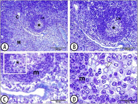
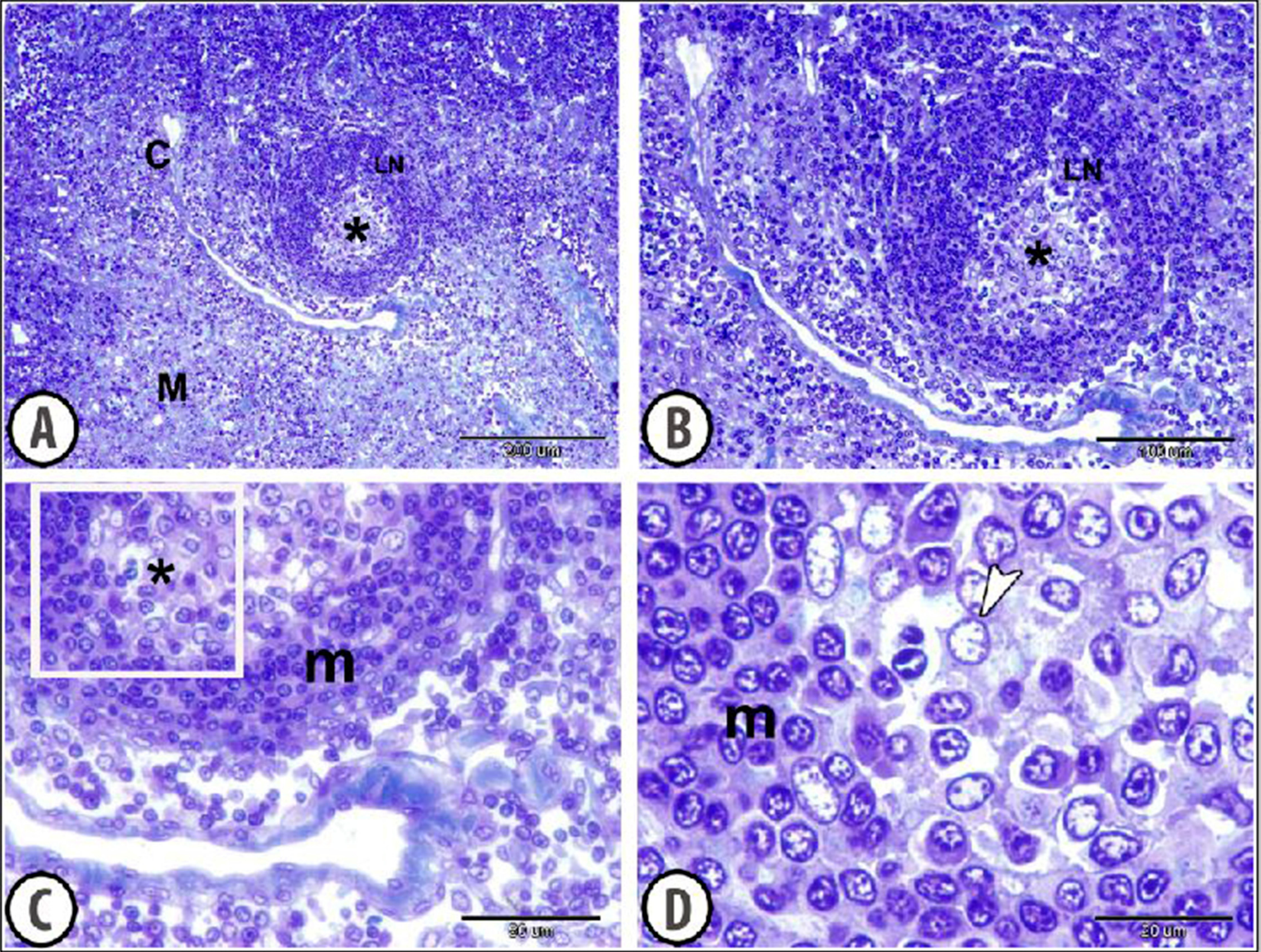
Semithin sections of the camel lymph node revealed distinct compartmental organization into a peripheral cortex and an inner medulla (Figure 1A). The cortex was characterized by densely packed lymphoid tissue containing numerous lymphatic nodules of variable size and shape. The lymphatic nodules consisted of closely aggregated lymphoid cells, with some nodules showing a relatively pale central region corresponding to a germinal center surrounded by a more densely cellular mantle zone (Figure 1B). The germinal centers contained predominantly larger lymphoid cells with relatively pale nuclei, whereas the surrounding mantle displayed smaller, more densely packed lymphocytes. The interfollicular cortical tissue was also densely populated by lymphoid cells and contained small vascular channels (Figure 1C). At higher magnification, the lymphoid cells showed distinct round to oval nuclei with variable degrees of chromatin condensation, and occasional larger cells were observed within the lymphoid tissue (Figure 1D).
Figure 1.
Toluidine-blue-stained semithin sections of the camel lymph node. (A) Overview showing the peripheral cortex (C) and inner medulla (M). (B) Lymphoid nodule (LN) with a pale-staining germinal center (asterisk) surrounded by a more densely cellular mantle zone (m). (C,D) Higher-magnification views of the germinal center and surrounding lymphocytes.
Measurements of lymphoid-nodule diameter, germinal-center diameter, the germinal-center-to-nodule diameter ratio, and approximate mantle-zone thickness are summarized in Table 1.
Table 1.
Morphometric characteristics and immunoreactive-cell densities in dromedary camel femoral lymph nodes.
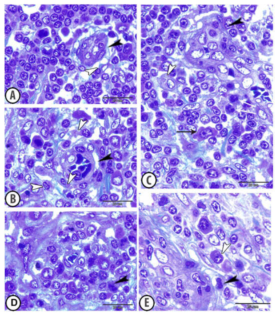
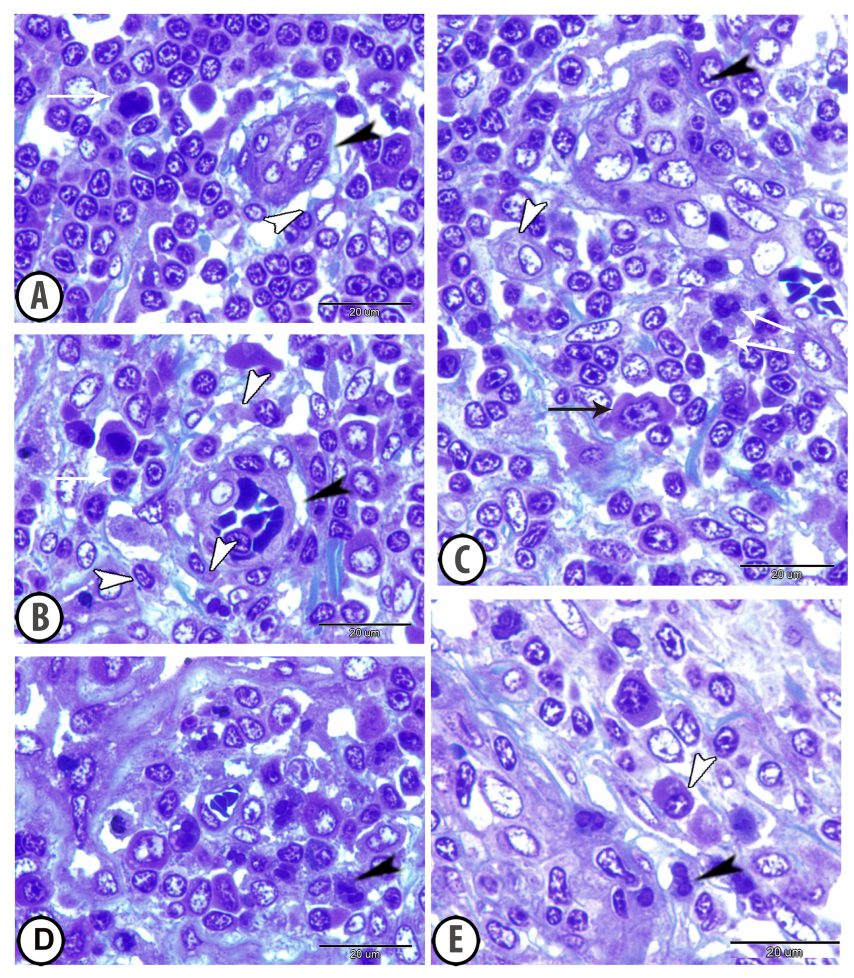
The medulla was less densely organized than the cortex and contained numerous immune cells within medullary cords and intervening sinus-like spaces. Irregular vascular and sinusoidal profiles separated cellular aggregates (Figure 2A–C). Reticular cells with cytoplasmic processes were observed, together with granular and agranular leukocytes (Figure 2A–E). Plasma-cell-like profiles occurred among the medullary cords (Figure 2E), while macrophage-like cells were observed in sinus-associated and interstitial regions (Figure 2C).
Figure 2.
Semithin sections of the lymph node medullary tissue stained with toluidine blue. (A–C) The medullary tissue contains numerous irregularly shaped vascular and sinusoidal spaces (black arrowheads). Reticular cells with prominent cytoplasmic processes (white arrowheads). (C) Neutrophils exhibiting distinctly segmented nuclei (white arrows) and macrophage with prominent cytoplasmic pseudopodia (black arrow). (D,E) Numerous granular leukocytes (black arrowheads) and plasma cells (white arrowheads) are scattered throughout the medullary tissue.
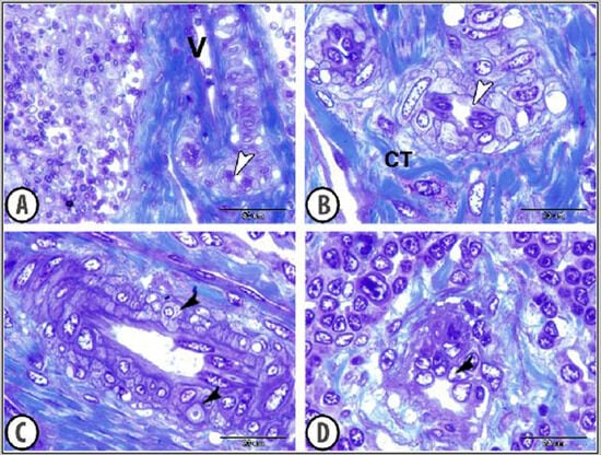
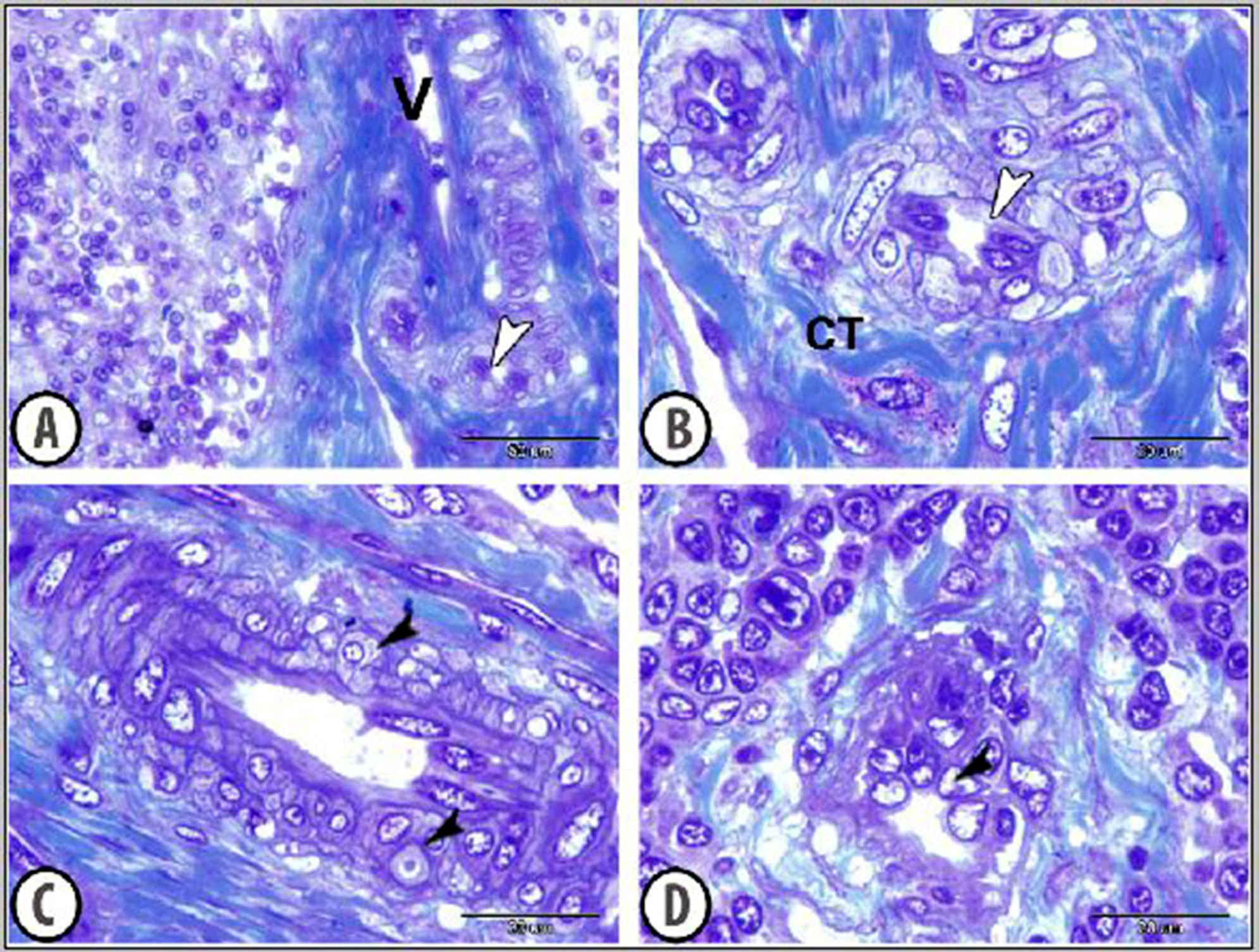
In addition to the conventional vascular profiles, some specialized vascular structures were observed within the lymph-node tissue. These included vascular profiles morphologically compatible with arteriovenous anastomoses (AVAs), characterized by relatively thick muscular walls and irregular lumina and were associated with connective-tissue fibers and nearby immune cells (Figure 3A,B). The glomus-like vascular structure was composed of endothelial-lined lumen surrounded by several layers of epithelioid-appearing cells with round to oval nuclei and pale cytoplasm (Figure 3C). However, because direct communication between arterial and venous components could not be demonstrated, these structures are described according to their morphological resemblance rather than as definitively identified arteriovenous anastomoses or glomus bodies. Vessels lined by cuboidal endothelial cells with prominent nuclei, morphologically consistent with high endothelial venules (HEVs), were observed near lymphoid nodules and dense lymphocyte aggregates (Figure 3D).
Figure 3.
Semithin sections of the camel lymph node stained with toluidine blue showing specialized vascular structures. (A,B) Vascular profiles morphologically compatible with AVAs were identified as direct communication between small arteries (white arrowheads) and veins (V) and were surrounded by connective-tissue fibers (CT). (C) Glomus-like vascular profiles surrounded by several layers of epithelioid glomus cells (black arrowheads). (D) High endothelial venules (HEVs) lined by characteristic cuboidal endothelial cells (black arrowhead).
3.2. Immunohistochemistry
Immunohistochemistry showed different distributions of CD3, CD163- and MHC class II (MHC-II)-immunoreactive cells in the camel lymph node.
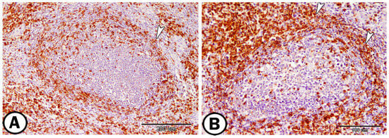
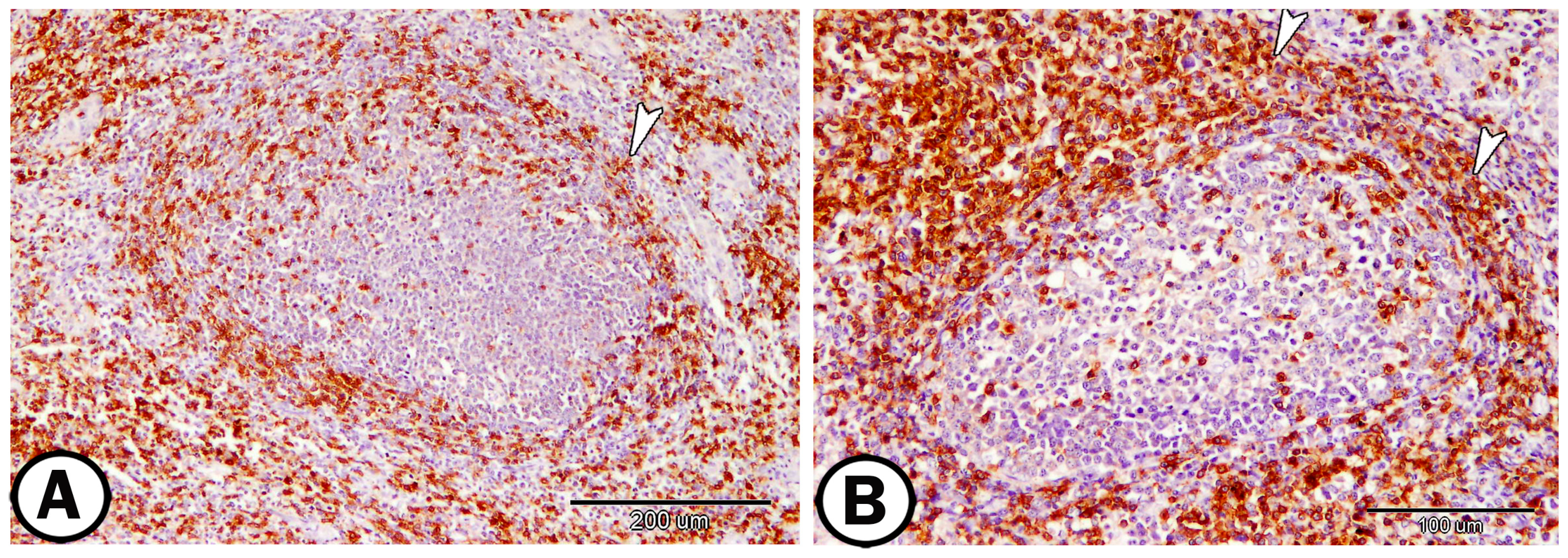
CD3 immunoreactivity was mainly associated with T lymphocytes and was distributed predominantly within the interfollicular regions of the lymph node. Positive cells appeared as small to medium-sized lymphoid cells with distinct cytoplasmic immunoreactivity. CD3-positive lymphocytes were also observed around lymphoid follicles, whereas the germinal centers of lymphatic nodules contained comparatively fewer positive cells (Figure 4).
Figure 4.
Immunohistochemical localization of CD3 in the camel lymph node. (A,B) Low and high magnification showing CD3-positive T-lymphocytes that were observed in interfollicular region of lymphatic nodules (arrowheads).
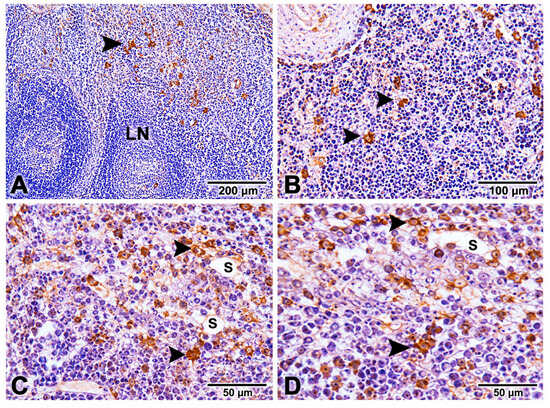
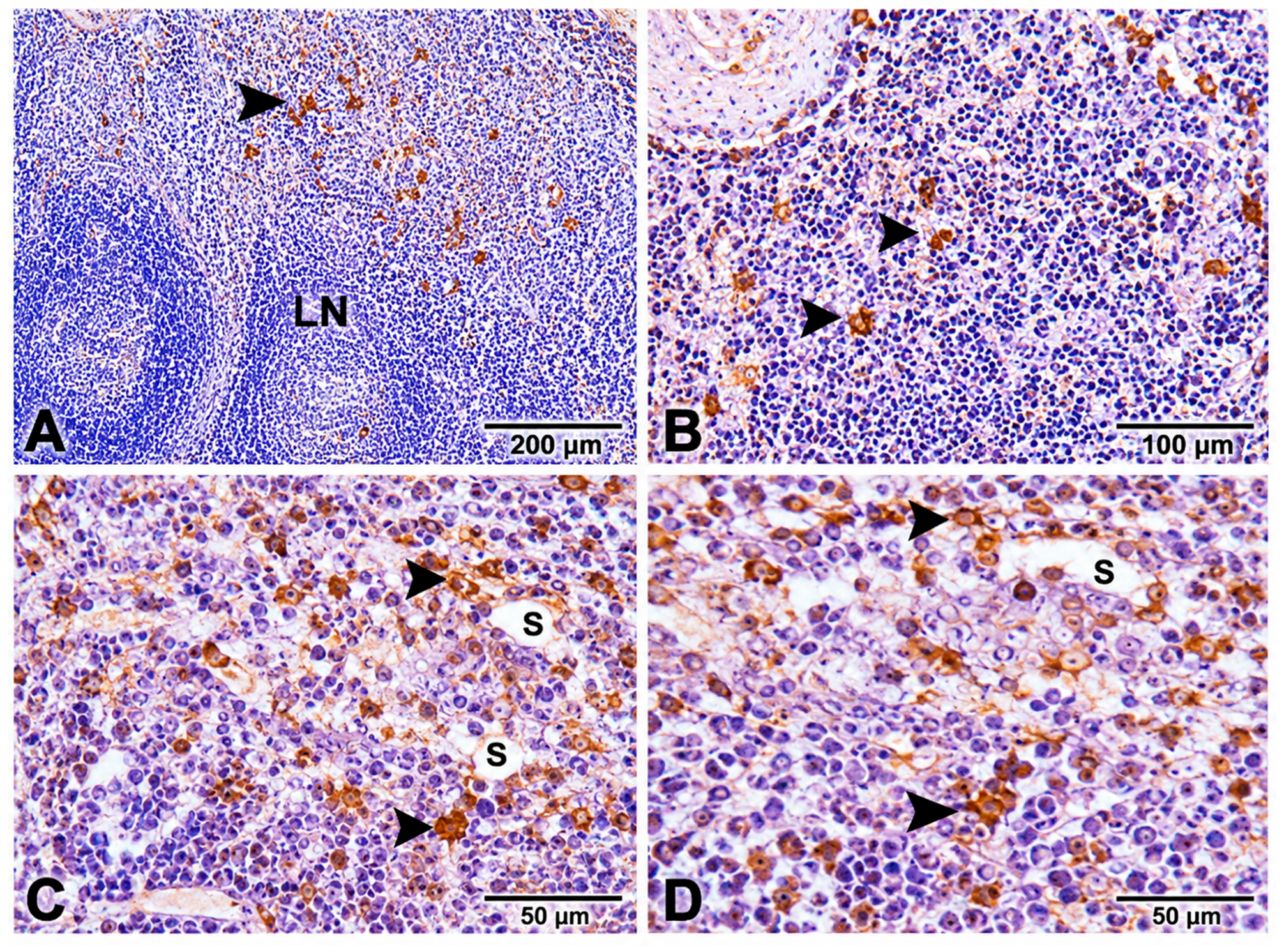
CD163 immunoreactivity was detected in cells morphologically consistent with monocyte–macrophage-lineage cells, mainly in medullary sinuses, perivascular regions, and interfollicular compartments (Figure 5A–D). Positive profiles occurred singly or in small groups among lymphocytes and stromal elements. These cells are described here as CD163-immunoreactive or macrophage-like cells.
Figure 5.
Immunohistochemical localization of CD163 in the camel lymph node. (A–D) CD163-immunoreactive cells of the monocyte–macrophage lineage (arrowheads) were predominantly distributed within the medullary sinuses (S) and perivascular regions. Note: the absence of CD163 immunoreactivity within the cortical lymphoid nodule (LN).
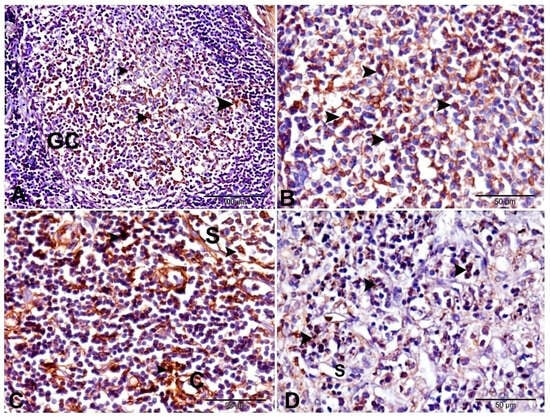
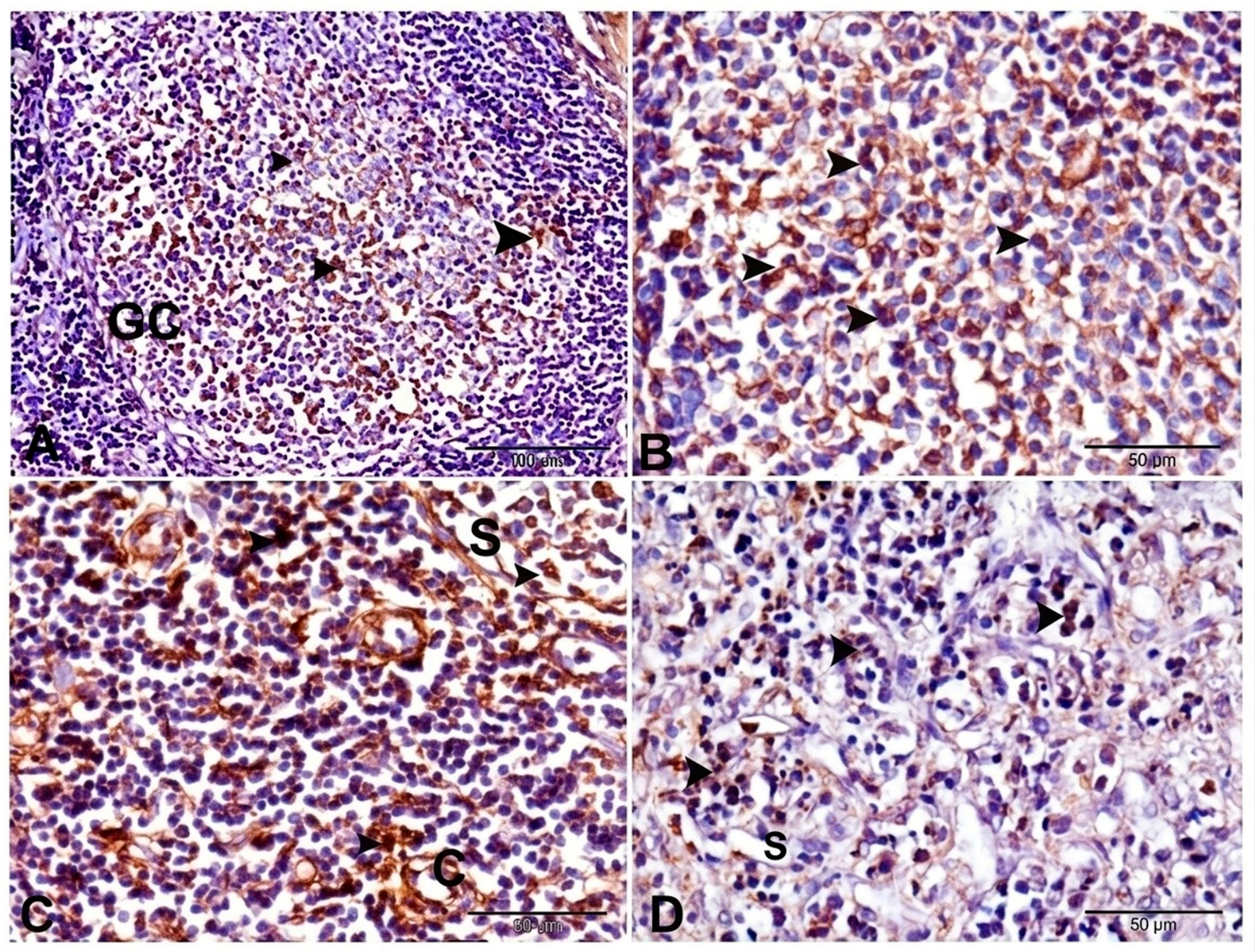
MHC-II immunoreactivity was widely distributed in the lymph node, including follicular, interfollicular, and medullary compartments. Strongly stained cells were present among larger cells in interfollicular and medullary regions, and some had morphology consistent with antigen-presenting cells. MHC-II-positive profiles were also observed within lymphoid follicles (Figure 6A–D). The estimated densities of CD3, CD163- and MHC-II-immunoreactive cells are summarized in Table 1.
Figure 6.
Immunohistochemical localization of MHC-II in the camel lymph node. (A,B) MHC-II-positive cells with macrophage-like and other antigen-presenting-cell morphology (arrowheads) were distributed within the germinal centers (GCs) of the lymphoid follicles. (C,D) Numerous strongly MHC-II-positive cells (arrowheads) were detected in the interfollicular areas and medullary sinuses (S). C: blood capillary.
3.3. Transmission Electron Microscopy (TEM)
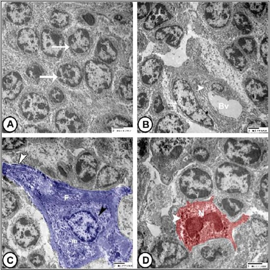
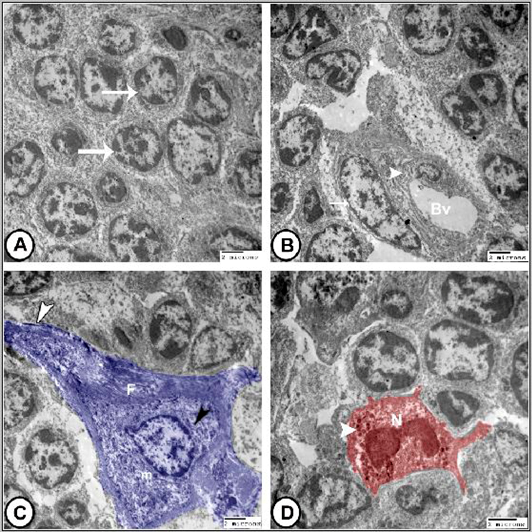
TEM revealed heterogeneous lymphoid-cell populations. Small lymphocytes had round to oval nuclei occupying most of the cellular profile, condensed peripheral heterochromatin, and scant cytoplasm. Other lymphoid cells had more abundant cytoplasm and less condensed chromatin, consistent with a more active cellular appearance (Figure 7A). At vascular interfaces, endothelial cells contained elongated nuclei, mitochondria, pinocytotic vesicles, and other cytoplasmic organelles. In some profiles, endothelial cells were closely associated with perivascular reticular cells, forming an organized interface between the vascular compartment and adjacent lymphoid tissue (Figure 7B).
Figure 7.
Digitally colorized transmission electron micrographs illustrate the ultrastructural features of the camel lymph node. (A) Numerous small lymphocytes (arrows) displaying relatively large, round to oval nuclei with condensed peripheral heterochromatin. (B) A blood vessel (Bv) lined by endothelial cells containing pinocytotic vesicles and various cytoplasmic organelles (arrowhead). The endothelial cells were closely associated with perivascular reticular cells (arrow). (C) A reticular cell (blue) showing a relatively large, irregularly shaped nucleus and prominent cytoplasmic processes (white arrowhead). Its cytoplasm contained abundant rough endoplasmic reticulum (black arrowhead), mitochondria (m), and cytoplasmic filaments (F). (D) An eosinophil (red) characterized by numerous distinctive cytoplasmic granules (arrowhead) and a relatively large, bilobed nucleus (N).
Reticular cells were distributed among lymphoid cells and had irregular nuclei, relatively electron-lucent nucleoplasm, and cytoplasmic processes extending between adjacent lymphocytes. Their cytoplasm contained rough endoplasmic reticulum, ribosomes, mitochondria, and filaments. Lymphocytes were sometimes closely apposed to these processes (Figure 7C). Eosinophils were identified by membrane-bound, electron-dense granules and relatively large, often bilobed or irregular nuclei. Granules varied in electron density; some showed a dense central region surrounded by a less electron-dense matrix. Peripheral nuclear heterochromatin was also evident (Figure 7D).
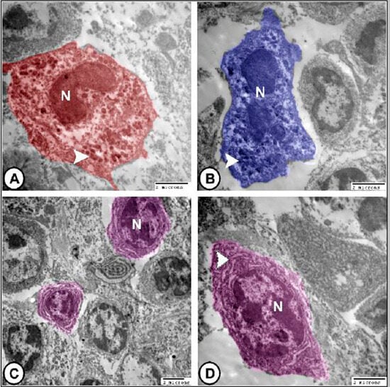
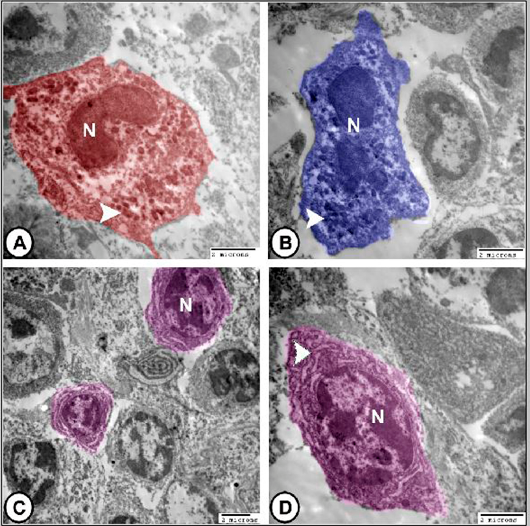
Neutrophils had irregular, indented, or segmented nuclei with peripheral heterochromatin. Their cytoplasm contained membrane-bound granules of variable size and electron density, together with mitochondria, ribosomes, and limited rough endoplasmic reticulum (Figure 8A).
Figure 8.
Digitally colorized transmission electron micrographs depicting the ultrastructural characteristics of the camel lymph node. (A) Neutrophil (red) exhibiting a segmented or indented nucleus (N) and numerous membrane-bound cytoplasmic granules (arrowhead). (B) Basophil (blue) showing a round to oval, slightly indented nucleus (N) and prominent electron-dense cytoplasmic granules (arrowhead). (C,D) Plasma cells (violet) displaying coarse aggregates of peripheral heterochromatin in a characteristic “clockface” and abundant rough endoplasmic reticulum organized into parallel arrays (arrowhead).
Basophil-like cells contained numerous membrane-bound, electron-dense granules of variable internal appearance. Their nuclei were round to oval or slightly indented and were sometimes displaced by the granular cytoplasm (Figure 8B).
Plasma cells were recognized by eccentric nuclei, coarse peripheral aggregates of heterochromatin, and abundant cytoplasm containing extensive parallel arrays of rough endoplasmic reticulum and numerous ribosomes (Figure 8C,D). These cell identities were based on ultrastructural morphology.
4. Discussion
This study provides an integrated histological, immunohistochemical, and ultrastructural description of adult dromedary camel femoral lymph nodes. The observed cortical and medullary organization, lymphoid follicles, and heterogeneous immune and stromal cell populations are broadly consistent with the established architecture of mammalian lymph nodes [2,18]. The present work extends the morphological description by documenting the distribution of CD163- and MHC-II-immunoreactive cells and describing selected vascular profiles. The addition of CD3 immunolabeling provides a qualitative and quantitative map of T-lymphocyte distribution alongside the CD163- and MHC-II-immunoreactive populations.
CD3-positive lymphocytes were concentrated mainly in interfollicular regions and around lymphoid follicles, while germinal centers contained fewer positive cells. This distribution is consistent with the recognized compartmentalization of T lymphocytes in mammalian lymph nodes, where T-cell-rich paracortical areas support interactions between T cells and antigen-presenting cells, while follicles are predominantly B-cell-rich regions [2,18].
CD163-immunoreactive cells were concentrated mainly in medullary, interfollicular, and perivascular regions. This distribution is compatible with the known role of lymph-node macrophage populations in capturing lymph-borne material and clearing particulate antigens, particularly near sinus-associated compartments [10,19]. Nevertheless, CD163 staining identifies a macrophage-associated population rather than all macrophages or a single functional state. The present data therefore support a regional association, but do not directly demonstrate antigen uptake, inflammatory activity, or a particular macrophage polarization state. MHC-II-immunoreactive cells were observed in follicular, interfollicular, and medullary regions. This broad distribution is consistent with the presence of antigen-presentation-associated cells in multiple lymph-node compartments.
TEM complemented the light-microscopic observations by revealing lymphocytes, plasma cells, macrophage-like cells, granulocytes, and stromal cells. The characteristic nuclear and cytoplasmic features of plasma cells, including eccentric nuclei and extensive rough endoplasmic reticulum, were consistent with their protein-secretory specialization. Reticular-cell-like profiles extended processes among lymphocytes, supporting a structural association between stromal elements and lymphoid cells. However, TEM morphology alone does not establish the molecular identity or functional activity of the observed cells.
The reticular cell network observed ultrastructurally parallels the fibroblastic reticular cell systems described in murine and human lymph nodes [20,21], although the prominent cytoplasmic processes and extensive rough endoplasmic reticulum observed in camel reticular cells suggest potentially high synthetic activity. Reticular cells and their extracellular-matrix network contribute to the structural organization of the lymph node and regulate interactions between immune cells and vascular structures [20]. Rather than functioning merely as passive supporting cells, lymph-node reticular cells form an organized stromal network that influences immune-cell positioning, migration, and communication [22].
The features of reticular cells observed by TEM are compatible with the general ultrastructural organization expected for stromal reticular cells, although definitive identification of a particular fibroblastic reticular-cell subset would require additional immunohistochemical or molecular markers. Current knowledge indicates that fibroblastic reticular cells are heterogeneous and include distinct populations associated with T-cell zones, B-cell follicles, marginal regions, and HEVs [23]. They form a three-dimensional stromal network that provides structural support while also producing extracellular matrix and signaling molecules involved in lymphocyte organization [21]. Thus, the reticular cells observed in the camel lymph node may contribute to the maintenance of the cellular compartments and the organization of immune-cell interactions.
The medullary cords constitute an important site for mature plasma cells in lymph nodes, and antibodies produced by these cells can enter the lymphatic circulation [2]. In addition, the frequent occurrence of plasma cells within the medullary cords observed ultrastructurally is compatible with migration of differentiated B-cell progeny from activated follicles toward the medulla, where plasma cells can secrete immunoglobulins into the lymphatic circulation. Similar relationships between germinal-center activity and medullary plasmacytosis have been described in mammalian lymph nodes [24].
The detection of eosinophils, neutrophils, and basophils by TEM adds another dimension to the cellular composition of the camel lymph node. These granulocytes exhibited characteristic nuclear and cytoplasmic features that permitted their ultrastructural identification. Although granulocytes are generally less dominant than lymphocytes in normal lymphoid tissue, their presence reflects the participation of innate immune mechanisms within the lymph node. Neutrophils and eosinophils can accumulate in lymphoid tissues under particular inflammatory or antigenic conditions [25,26], whereas basophils may participate in immune regulation through the release of preformed mediators and cytokines [27]. The identification of these granulocytes demonstrates that, in addition to lymphoid and mononuclear phagocyte populations, innate immune cells are represented within the camel lymph-node microenvironment.
An additional point of interest is the possible relevance of the specialized vascular profiles observed in the present study to the broader vascular adaptations of the dromedary camel. The identification of high endothelial venules, Vascular profiles morphologically compatible with AVAs, and glomus-like vascular profiles further highlights the organization of the camel lymph node. HEVs are specialized postcapillary venules that provide the principal route through which circulating naïve lymphocytes enter lymph nodes [28]. HEVs have been extensively characterized in human [28], mouse [29], and rat lymph nodes [30]. In the present study, HEVs were identified in cortical and interfollicular lymphoid tissue. Their close spatial relationship with lymphoid and stromal compartments supports the concept of an integrated vascular–immune microenvironment. Thus, the significance of these findings extends beyond the description of individual structures, as their organization within the lymph node provides a morphological basis for maintenance of the lymphoid microenvironment.
The vascular findings were not limited to conventional lymph-node microvessels and HEVs. Vascular profiles morphologically compatible with AVAs and glomus vessels were occasionally identified within the connective-tissue framework. The identification of glomus-like vascular profiles and arteriovenous anastomoses within camel lymph nodes represents a particularly novel finding, as such specialized vascular structures are rarely reported in mammalian lymph nodes. and are more commonly associated with thermoregulatory tissues such as skin, nasal mucosa, ovary and the spleen [31,32]. Some of these structures have been proposed to participate in local regulation of vascular resistance, blood distribution, and thermoregulatory processes. In this context, the occurrence of glomus-type vessels and arteriovenous anastomoses within the femoral lymph nodes is noteworthy and raises the possibility that local vascular regulation may contribute to maintaining the lymph-node microenvironment under variable physiological and environmental conditions. However, the present study did not investigate vascular responses to temperature changes, dehydration, or seasonal variation. Therefore, a direct relationship between these vascular structures and desert adaptation cannot be established from the current morphological observations.
The association between specialized vessels and immune-cell-rich regions may be functionally significant. Lymph nodes must continuously balance blood flow, lymphatic drainage, antigen delivery, and immune-cell recruitment [33]. HEVs provide a regulated pathway for lymphocyte entry from the blood, while lymphatic channels facilitate antigen and dendritic-cell entry and lymphocyte exit [34]. Stromal and vascular components therefore operate as an integrated system rather than as independent structures. The coexistence of HEVs, specialized vascular profiles, reticular cells, and abundant immune cells in the camel lymph node supports the concept of a highly organized vascular–stromal niche specialized for immune surveillance [18].
The present findings also demonstrate a close relationship between vascular structures and antigen-presenting cells. CD163-positive macrophage-like cells were frequently associated with vascular and sinus regions, while MHC II-positive cells were broadly distributed through the lymphoid tissue. Dendritic cells located within lymph nodes are strategically positioned to interact with T lymphocytes and participate in antigen presentation [1], while macrophages contribute to antigen capture and clearance [19]. The simultaneous presence of these cell populations and HEVs provides the anatomical basis for efficient delivery, processing, presentation, and recognition of antigens within the camel lymph node.
Comparative studies of lymph-node architecture across mammals indicate that the general organization observed in the dromedary camel shares several features with that described in other domestic and laboratory species, although some species-related variations are evident. In Egyptian water buffaloes, lymph nodes exhibit a conventional cortical organization containing primary and secondary lymphoid follicles, interfollicular lymphocytes, and medullary cords separated by lymphatic sinuses, with high endothelial venules located in the interfollicular region [15]. Similarly, studies of caprine superficial lymph nodes have demonstrated a complex organization of lymphoid and stromal components [12], while investigations in cattle have documented distinct lymphoid and vascular compartments [4]. In contrast, previous investigations of dromedary camel lymph nodes have described a characteristic arrangement of lymphoid follicles and diffuse interfollicular tissue, with variations in the degree of cortical, medullary, and lobular organization among different anatomical lymph-node locations [5,15]. The present observations of cortical and medullary compartments, lymphoid nodules with germinal centers, and specialized vascular profiles therefore demonstrate both similarities to the general mammalian lymph-node pattern and distinctive vascular–stromal features in the camel. These comparisons highlight the importance of considering species and anatomical location when interpreting lymph-node architecture.
The findings of the present study may also have practical relevance to veterinary immunology and camel health. Dromedary camels are exposed to several important infectious diseases, including Middle East respiratory syndrome coronavirus (MERS-CoV), brucellosis, and trypanosomiasis, which involve complex innate and adaptive immune responses [35]. In this context, understanding the distribution of T lymphocytes, macrophage-associated cells, antigen-presenting cells, and specialized vascular structures within camel lymph nodes provides a useful morphological basis for future investigations of local immune-cell organization during infection and following vaccination. Furthermore, camelids possess a distinctive antibody system characterized by conventional immunoglobulins and heavy-chain-only antibodies (HCAbs), which lack light chains and provide the basis for the development of single-domain antibodies (nanobodies) [36]. Although the present study did not investigate pathogen-specific responses, antibody production, or vaccine efficacy, the described lymphoid and vascular–stromal architecture provides a baseline for future studies examining the cellular organization of camel immune responses and their relationship to these distinctive immunological characteristics.
Although the present findings provide detailed morphological and immunohistochemical information regarding the organization of dromedary camel femoral lymph nodes, their interpretation should be considered within the descriptive scope of this investigation. The distribution of CD3-, CD163-, and MHC-II-immunoreactive cells, together with the observed vascular and stromal profiles, provides morphological evidence of the cellular and structural organization of these lymph nodes. However, the present study did not directly investigate lymphocyte migration, antigen uptake and presentation, immune-cell interactions, or vascular responses under physiological or pathological conditions. Therefore, possible roles of these components in lymphocyte trafficking, antigenic surveillance, and local vascular regulation are discussed in the context of established knowledge of lymph-node biology rather than as functions demonstrated experimentally in the present study. Further functional and molecular investigations will be necessary to clarify these potential roles.
A limitation of the present study is that specific immunophenotypic identification of B-lymphocyte populations using lineage-associated markers such as CD20 was not performed. Consequently, the present findings should not be interpreted as a quantitative characterization of T- versus B-cell compartments. Instead, the study was designed to provide an integrated description of lymph-node microarchitecture, T-lymphocytes, macrophage-associated cells, MHC-II-expressing cells, ultrastructural cellular diversity, and specialized vascular structures. Although negative controls (primary antibody omission) and positive controls were included to support the interpretation of the immunohistochemical findings, these controls do not independently confirm the specificity of the antibodies for their respective antigens in dromedary camel tissues. In particular, the cross-reactivity of antibodies developed against other species remains a consideration when interpreting immunoreactivity in camel tissues. Further validation using species-specific antibodies or independent approaches, such as Western blotting, would be valuable to confirm antigen specificity in dromedary camel tissues. Future studies incorporating additional immunohistochemical and molecular markers, together with comparative sampling across seasons and age groups, would help clarify the functional significance of many immune cells.
5. Conclusions
Adult dromedary camel femoral lymph nodes displayed organized cortical and medullary compartments, lymphoid nodules with germinal centers, and diverse immune and stromal cell populations. CD3-positive T lymphocytes were distributed mainly in interfollicular areas and around follicles, with fewer positive cells in germinal centers. CD163-immunoreactive macrophage-like cells were concentrated mainly in medullary, interfollicular, and perivascular regions, whereas MHC-II-immunoreactive cells occurred across several compartments. TEM further characterized lymphocytes, plasma cells, and granulocytes, providing additional ultrastructural information on the cellular composition of these lymph nodes. Vascular profiles morphologically compatible with high endothelial venules, arteriovenous anastomoses, and glomus-like vascular profiles were also observed. Overall, this study provides a morphological and immunohistochemical baseline for understanding the structural organization of camel femoral lymph nodes. Further investigations using specific vascular markers, additional immunophenotypic characterization, and broader sampling are needed to confirm the identity of these vascular structures and clarify their potential functional relationships with the lymphoid and stromal compartments.
Supplementary Materials
The following supporting information can be downloaded at https://www.mdpi.com/article/10.3390/life16111936/s1. Figure S1: Negative controls for CD163 (A), MHCII (B), and CD3 (C) immunostaining performed on camel lymph nodes sections processed without primary antibodies. No specific immunoreactivity was detected. Figure S2. Positive controls for CD3 (A), MHC-II (B), and CD163 (C) immunostaining in dromedary camel spleen, showing positive immunoreactivity in T lymphocytes (A, arrowhead around the follicular artery) and macrophage-like and other immune-associated cells (B, C; arrowheads in red pulp).
Author Contributions
Conceptualization, D.M.M., E.F., M.A.A.-M., M.H., N.M.M., H.I.E.-M. and S.H.Q.; methodology, D.M.M. and M.T.H.; software, E.F. and M.T.H.; validation, N.M.M.; formal analysis, M.A.A.-M.; investigation, D.M.M.; resources, H.I.E.-M. and M.H.; data curation, S.H.Q.; writing—original draft preparation, D.M.M.; writing—review and editing, M.T.H.; visualization, M.T.H. and H.I.E.-M.; supervision, D.M.M. and J.C.C.-S.; project administration, D.M.M. and G.Z.; funding acquisition, E.F., M.A.A.-M., N.M.M. and H.I.E.-M. All authors have read and agreed to the published version of the manuscript.
Funding
The authors extend their appreciation to the Deanship of Scientific Research and Graduate Studies at King Khalid University for funding this work through Large Research Project under grant number RGP2/337/47. The authors extend their appreciation to Princess Nourah bint Abdulrahman University Researchers Supporting Project number (PNURSP2026R768), Princess Nourah bint Abdulrahman University, Riyadh, Saudi Arabia.
Institutional Review Board Statement
The study protocol was conducted in strict accordance with the relevant guidelines and ethical regulations and was approved by the Ethics Committee of Assiut University in Egypt (ethical number: 06/2026/0448, approved on 20 May 2026) on the use of animals for scientific purposes. This study is reported in accordance with ARRIVE guidelines.
Informed Consent Statement
Not applicable.
Data Availability Statement
The original contributions presented in this study are included in the article/Supplementary Material. Further inquiries can be directed to the corresponding author.
Acknowledgments
The authors extend their appreciation to the Deanship of Scientific Research and Graduate Studies at King Khalid University for funding this work through Large Research Project under grant number RGP2/337/47. The authors extend their appreciation to Princess Nourah bint Abdulrahman University Researchers Supporting Project number (PNURSP2026R768), Princess Nourah bint Abdulrahman University, Riyadh, Saudi Arabia.
Conflicts of Interest
The authors declare no conflicts of interest.
References
- Huang, J.Y.; Gerner, M.Y. Dendritic Cell Organization and Function in Innate and Adaptive Immune Defense Within Lymph Nodes. Immunol. Rev. 2026, 337, e70071. [Google Scholar] [CrossRef] [Scilit] [PubMed]
- Grant, S.M.; Lou, M.; Yao, L.; Germain, R.N.; Radtke, A.J. The lymph node at a glance—How spatial organization optimizes the immune response. J. Cell Sci. 2020, 133, jcs241828. [Google Scholar] [CrossRef] [Scilit] [PubMed]
- Fares, M.A.; Khenenou, T.; Rahmoun, D.E. Cytological and Immunohistochemical Study of the Dromedary Iymph Nodes in Algeria. Biol. Life Sci. Forum 2023, 22, 8. [Google Scholar] [CrossRef] [Scilit]
- Christophe, R.C.; Breugelmans, S.; Simoens, P.; Van den Broeck, W. Morphological and immunological characteristics of the bovine temporal lymph node and hemal node. Vet. Immunol. Immunopathol. 2008, 126, 339–350. [Google Scholar] [CrossRef] [Scilit] [PubMed]
- Abdel-Magied, E.M.; Taha, A.M.; Al-Qarawi, A.A.; Elfaki, M.G. The parotid, mandibular and lateral retropharyngeal lymph nodes of the camel (Camelus dromedarius). Anat. Histol. Embryol. 2001, 30, 199–203. [Google Scholar] [CrossRef] [Scilit] [PubMed]
- Dasoveanu, D.C.; Shipman, W.D.; Chia, J.J.; Chyou, S.; Lu, T.T. Regulation of Lymph Node Vascular-Stromal Compartment by Dendritic Cells. Trends Immunol. 2016, 37, 764–777. [Google Scholar] [CrossRef] [Scilit] [PubMed]
- Olson, E.C.; Williams, A.; Eisenbarth, S.C. Dendritic Cell Migration: An Essential Step in Initiating Adaptive Immunity Across Tissues. Immunol. Rev. 2026, 337, e70080. [Google Scholar] [CrossRef] [Scilit] [PubMed]
- Abbaszadeh, M.; Naseri, B.; Taghizadeh-Teymorloei, M.; Mardi, A.; Javan, M.R.; Masoumi, J.; Ghorbaninezhad, F.; Hatami-Sadr, A.; Tural, Ş.; Baradaran, B.; et al. Overview of dendritic cells subsets and their involvement in immune-related pathological disease. Bioimpacts 2025, 29, e70071. [Google Scholar] [CrossRef] [Scilit] [PubMed]
- Mokhtar, D.M.; Hussein, M.M. Morphological characteristic and functional dependencies of dendritic cell in developing rabbit lung during fetal and neonatal life. Dev. Biol. 2019, 454, 29–43. [Google Scholar] [CrossRef] [Scilit] [PubMed]
- Gray, E.E.; Cyster, J.G. Lymph node macrophages. J. Innate Immun. 2012, 4, 424–436. [Google Scholar] [CrossRef] [Scilit] [PubMed]
- Hadamitzky, C.; Spohr, H.; Debertin, A.S.; Guddat, S.; Tsokos, M.; Pabst, R. Age-dependent histoarchitectural changes in human lymph nodes: An underestimated process with clinical relevance? J. Anat. 2010, 216, 556–562. [Google Scholar] [CrossRef] [Scilit] [PubMed]
- Balasundaram, K.; Sivagnanam, S. Ultrastructural Study of the Outer Cortex, Deep Cortex and Medulla of Mesenteric Lymph Node in Adult Goats. Indian J. Anim. Res. 2021, 55, 1189–1191. [Google Scholar] [CrossRef] [Scilit]
- Takeda, A.; Salmi, M.; Jalkanen, S. Lymph node lymphatic endothelial cells as multifaceted gatekeepers in the immune system. Trends Immunol. 2023, 44, 72–86. [Google Scholar] [CrossRef] [Scilit] [PubMed]
- Hussen, J. Changes in Cell Vitality, Phenotype, and Function of Dromedary Camel Leukocytes after Whole Blood Exposure to Heat Stress In Vitro. Front. Vet. Sci. 2021, 8, 647609. [Google Scholar] [CrossRef] [Scilit] [PubMed]
- Zidan, M.; Pabst, R. Histological, histochemical and immunohistochemical study of the lymph nodes of the one-humped camel (Camelus dromedarius). Vet. Immunol. Immunopathol. 2012, 145, 191–198. [Google Scholar] [CrossRef] [Scilit] [PubMed]
- Alhafiz, G.A.; Alghatam, F.H.; Almohammed, H.; Hussen, J. Milk Immune Cell Composition in Dromedary Camels with Subclinical Mastitis. Front. Vet. Sci. 2022, 9, 885523. [Google Scholar] [CrossRef] [Scilit] [PubMed]
- Reynolds, E.S. The use of lead citrate at high pH as an electron-opaque stain in electron microscopy. J. Cell Biol. 1963, 17, 208–212. [Google Scholar] [CrossRef] [Scilit] [PubMed]
- Mueller, S.N.; Germain, R.N. Stromal cell contributions to the homeostasis and functionality of the immune system. Nat. Rev. Immunol. 2009, 9, 618–629. [Google Scholar] [CrossRef] [Scilit] [PubMed]
- Louie, D.A.P.; Liao, S. Lymph Node Subcapsular Sinus Macrophages as the Frontline of Lymphatic Immune Defense. Front. Immunol. 2019, 10, 347. [Google Scholar] [CrossRef] [Scilit] [PubMed]
- Fletcher, A.L.; Acton, S.E.; Knoblich, K. Lymph node fibroblastic reticular cells in health and disease. Nat. Rev. Immunol. 2015, 15, 350–361. [Google Scholar] [CrossRef] [Scilit] [PubMed]
- Martinez, V.G.; Pankova, V.; Krasny, L.; Singh, T.; Makris, S.; White, I.J.; Benjamin, A.C.; Dertschnig, S.; Horsnell, H.L.; Kriston-Vizi, J.; et al. Fibroblastic Reticular Cells Control Conduit Matrix Deposition during Lymph Node Expansion. Cell Rep. 2019, 26, 2810–2822.e5. [Google Scholar] [CrossRef] [Scilit] [PubMed]
- Li, L.; Wu, J.; Abdi, R.; Jewell, C.M.; Bromberg, J.S. Lymph node fibroblastic reticular cells steer immune responses. Trends Immunol. 2021, 42, 723–734. [Google Scholar] [CrossRef] [Scilit] [PubMed]
- Perez-Shibayama, C.; Gil-Cruz, C.; Ludewig, B. Fibroblastic reticular cells at the nexus of innate and adaptive immune responses. Immunol. Rev. 2019, 289, 31–41. [Google Scholar] [CrossRef] [Scilit] [PubMed]
- Haberman, A.M.; Gonzalez, D.G.; Wong, P.; Zhang, T.T.; Kerfoot, S.M. Germinal center B cell initiation, GC maturation, and the coevolution of its stromal cell niches. Immunol. Rev. 2019, 288, 10–27. [Google Scholar] [CrossRef] [Scilit] [PubMed]
- Voisin, M.B.; Nourshargh, S. Neutrophil trafficking to lymphoid tissues: Physiological and pathological implications. J. Pathol. 2019, 247, 662–671. [Google Scholar] [CrossRef] [Scilit] [PubMed]
- Berek, C. Eosinophils: Important players in humoral immunity. Clin. Exp. Immunol. 2016, 183, 57–64. [Google Scholar] [CrossRef] [Scilit] [PubMed]
- Chirumbolo, S.; Bjørklund, G.; Sboarina, A.; Vella, A. The role of basophils as innate immune regulatory cells in allergy and immunotherapy. Hum. Vaccines Immunother. 2018, 14, 815–831. [Google Scholar] [CrossRef] [Scilit] [PubMed]
- Blanchard, L.; Girard, J.P. High endothelial venules (HEVs) in immunity, inflammation and cancer. Angiogenesis 2021, 24, 719–753. [Google Scholar] [CrossRef] [Scilit] [PubMed]
- Ager, A. High Endothelial Venules and Other Blood Vessels: Critical Regulators of Lymphoid Organ Development and Function. Front. Immunol. 2017, 8, 45. [Google Scholar] [CrossRef] [Scilit] [PubMed]
- Miyasaka, M.; Tanaka, T. Lymphocyte trafficking across high endothelial venules: Dogmas and enigmas. Nat. Rev. Immunol. 2004, 4, 360–370. [Google Scholar] [CrossRef] [Scilit] [PubMed]
- Mokhtar, D.M.; Abd-Elhafez, E.A. Morphological Studies on the Peripheral Circulation of the Ovary in One-Humped Camel (Camelus dromedarius). Anat. Histol. Embryol. 2016, 45, 319–328. [Google Scholar] [CrossRef] [Scilit] [PubMed]
- Taher, N.A.; Hussein, M.M.; AbdElhafez, E.A.; Mokhtar, D.M. Seasonal Modulation of Splenic Immune Architecture and Proliferative Activity in the Dromedary Camel (Camelus dromedarius): An Ecological Perspective. J. Exp. Zool. Part A Ecol. Integr. Physiol. 2026, 345, 591–600. [Google Scholar] [CrossRef] [Scilit] [PubMed]
- Johnson, L.A. In Sickness and in Health: The Immunological Roles of the Lymphatic System. Int. J. Mol. Sci. 2021, 22, 4458. [Google Scholar] [CrossRef] [Scilit] [PubMed]
- Arasa, J.; Collado-Diaz, V.; Halin, C. Structure and Immune Function of Afferent Lymphatics and Their Mechanistic Contribution to Dendritic Cell and T Cell Trafficking. Cells 2021, 10, 1269. [Google Scholar] [CrossRef] [Scilit] [PubMed]
- Shah, S.F.A.; Tharwat, M.; Rehman, A.; Alshanbari, F.A. The Impact of Bacterial and Viral Diseases on Dromedary Camel (Camelus dromedarius) Welfare: A Comprehensive Review. Front. Vet. Sci. 2026, 13, 1795334. [Google Scholar] [CrossRef] [Scilit] [PubMed]
- Mufrrih, M.; Alandijany, T.A. Camelid VHHs as a Versatile Platform for Combating Viral Infections. Pharmaceuticals 2026, 19, 1408. [Google Scholar] [CrossRef] [Scilit] [PubMed]

Disclaimer/Publisher’s Note: The statements, opinions and data contained in all publications are solely those of the individual author(s) and contributor(s) and not of MDPI and/or the editor(s). MDPI and/or the editor(s) disclaim responsibility for any injury to people or property resulting from any ideas, methods, instructions or products referred to in the content. |
© 2026 by the authors. Licensee MDPI, Basel, Switzerland. This article is an open access article distributed under the terms and conditions of the Creative Commons Attribution (CC BY) license.