


Abstract
(1) Background: Bacterial infections are frequently encountered in liver cirrhosis and may be associated with a dismal prognosis. Over recent decades, antibiotic resistance has increased, which may be linked to higher mortality. (2) Methods: We conducted a retrospective study between 2018 and 2024, evaluating all patients with cirrhosis and ascites admitted to our hospital. Antimicrobial susceptibility testing was performed on all bacterial isolates. Multivariate analysis was used to identify predictive risk factors for in-hospital death. (3) Results: In total, 806 patients were identified during the study period; 78 had spontaneous bacterial peritonitis (SBP), 6 had bacterascites, 74 had Clostridioides difficile infection (CDI), 27 had culture-positive pneumonia, 24 had urinary tract infection, and 20 had bloodstream infection (BSI). Antibiotic resistance rates were high, with multidrug-resistant (MDR) strains detected in 70.3% of cases. The isolated pathogens included MRSA (65.9%), ESBL-producing Gram-negative bacilli (18.5%), and carbapenem-resistant Gram-negative strains (31.1%); only one case of vancomycin-resistant Enterococcus (VRE) was recorded. In multivariate analysis, liver disease severity; AKI and ACLF presence; multiple infection sites; and culture-positive pneumonia, BSI, and SBP were associated with higher mortality. Gram-negative infections were also associated with higher mortality rates. (4) Conclusions: In total, 22.1% of patients hospitalised with cirrhosis and ascites had at least one confirmed bacterial infection. Increased antibiotic resistance and a high proportion of MDR were recorded. The severity of liver disease, AKI and ACLF presence, the presence of pneumonia, BSI, and SBP, Gram-negative and multiple infection sites were associated with higher mortality.
1. Introduction
Infections in liver cirrhosis are frequent and associated with significant short- and long-term mortality [1,2,3,4,5,6,7,8,9,10]; hospitalized patients with cirrhosis experience bacterial infections at rates up to 60–70%, exceeding general hospital infection rates by 4- to 5-fold [9]. Cirrhosis predisposes patients to infection through several synergistic mechanisms: immune dysfunction, altered enteric flora, increased bacterial translocation, and portal hypertension-driven impairments in gut motility and intestinal barrier integrity [3,4,11]. The most frequent infections in liver cirrhosis are spontaneous bacterial peritonitis, pneumonia, urinary tract infection, and soft tissue infection [2,3,12].
Spontaneous bacterial peritonitis (SBP) is the most prevalent infection in cirrhosis, complicating 1.5–3.5% of outpatients and 10–30% of hospitalized individuals with ascites [13,14,15]. SBP diagnosis relies on an ascitic neutrophil count ≥250/mm3, with fluid cultures yielding positive results in less than 50% of cases [1,2,13]. Cases with elevated polymorphonuclear (PMN) counts and negative cultures are classified as culture-negative SBP, while those with positive cultures but <250 PMN/mm3 are defined as bacterascites [1,16]. Most studies have stated Gram-negative bacteria (GNB) as the main etiological factor for SBP [1,2,17,18,19]; however, a microbial shift was noted in recent decades in many studies [19,20,21,22,23,24], with a better prognosis for Gram-positive bacterial (GPB) SBP compared to Gram-negative SBP in some studies [24]. Spontaneous fungal peritonitis is rarer but may be associated with a worse prognosis [20,25,26]. Extraperitoneal infections (respiratory, urinary, and bloodstream) are also common [5,6,7,8,9,10], with worse survival rates in SBP, respiratory, and bloodstream infections.
A high prevalence of multidrug-resistant (MDR) infections was noted in cirrhosis, and it can lead to significant mortality [1,2,6,8,9,27,28,29]. Because of the high prevalence of infections in advanced cirrhosis, many guidelines recommend prophylactic antibiotherapy in particular cases such as gastrointestinal bleeding, encephalopathy, high risk for SBP, and acute-on-chronic liver failure (ACLF). As a result of prophylactic or curative antibiotherapy in patients with cirrhosis, a higher proportion of Clostridioides difficile infection (CDI) may occur in cirrhosis and may significantly impact the prognosis [11,30].
The epidemiology and multidrug resistance (MDR) profiles of bacterial infections in cirrhosis exhibit significant geographic variation, requiring local data to tailor prophylactic and therapeutic guidelines. In Romania, existing studies have evaluated infection prevalence in cirrhotic cohorts [11,31,32,33], but data regarding antimicrobial susceptibility remain scarce. Therefore, this study aimed to characterize the etiological spectrum of culture-confirmed infections, determine the prevalence of MDR pathogens, and evaluate mortality rates among cirrhotic patients within our region.
2. Materials and Methods
2.1. Clinical Data
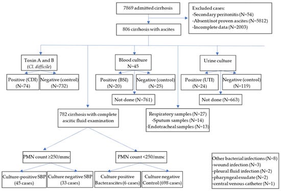
We performed a retrospective cohort study of patients with cirrhosis and ascites at the Emergency Clinical Hospital Craiova (2018–2024) to evaluate all bacterial infections (Figure 1). Patients with secondary peritonitis, recent surgery, or peritoneal dialysis were excluded. SBP was defined by an ascitic PMN count of ≥250/mm3 (subdivided by culture positivity), while bacterascites represented a positive culture with <250 PMN/mm3; during diagnostic paracentesis, at least 500 mL of ascitic fluid was collected and inoculated on specialized culture media. The diagnosis of pneumonia was based on clinical and imaging criteria combined with positive sputum or endotracheal cultures. CDI and urinary tract infections were confirmed via fecal toxin A/B assays and positive urocultures, respectively. Other recorded sites included pleural fluid, wounds, pharyngeal swabs, and central venous catheters. All cases were logged into an Excel database based on culture status.
Figure 1.
The flowchart for patients enrolled in our study.
To assess infection and mortality risks, we established a specific control group for each type of bacterial infection. We compared spontaneous bacterial peritonitis (SBP) cases against cirrhotic patients with ascites and negative peritoneal cultures. For Clostridioides difficile infection (CDI), the control group consisted of patients without documented CDI. For urinary tract and bloodstream infections, we used patients with negative urine or blood cultures as controls. Finally, for pneumonia, we used patients with either negative samples or no samples collected as the comparison group. For prognosis assessment, the main outcome analyzed was in-hospital mortality; Child–Pugh–Turcotte (CTP), creatinine-CTP, MELD-3, and MELD-Na scores were calculated and used for prognostic stratification.
2.2. Materials and Susceptibility Testing
Sample collection and bacteriological analysis adhered to institutional protocols. During paracentesis, at least 500 mL of ascitic fluid was aseptically aspirated for cytological and bacteriological analysis. Bacteriological examination involved the immediate inoculation of fluid into blood culture bottles (or blood broth media), followed by an 18-h incubation at 37 °C. Subcultures were subsequently performed on blood agar, chocolate agar, and Drigalski agar media supplied by BioMérieux (Marcy-l’Étoile, France/Salt Lake City, UT, USA). Clostridioides difficile infection was diagnosed by clinical presence of acute diarrhea (≥3 loose stools in 24 h) and confirmed by positive stool toxin A or B following initial GDH screening. Bloodstream infections (BSIs) were defined based on modified CDC/NHSN guidelines; BSI diagnosis was strictly confirmed only if the same organism was isolated from two or more independent blood cultures drawn within 24 h, accompanied by at least one clinical sign of sepsis (temperature > 38.0 °C or <36.0, chills, or hypotension); multiple positive cultures yielding identical strains within the same hospital admission were deduplicated and analyzed as a single unique isolate. Blood cultures (aerobic and anaerobic) were performed under strict aseptic conditions for febrile patients with clinical or biochemical signs of sepsis; common skin contaminants in blood cultures (e.g., coagulase-negative Staphylococci) were excluded unless isolated from at least two separate blood samples with clinical signs of sepsis. The diagnosis of pneumonia required radiographic confirmation (new or progressive pulmonary infiltrates) plus at least two clinical criteria (fever > 38 °C, purulent sputum/tracheal secretions, or leukocytosis/leukopenia), excluding asymptomatic upper respiratory tract colonization; respiratory samples were obtained via sputum collection in awake patients and endotracheal aspiration in intubated individuals. Patients with clinical suspicion of urinary tract infection were evaluated using standard mid-stream clean-catch urine cultures processed within 2 h, with standard thresholds (at least 100,000 CFU/mL) and strict aseptic techniques used for the diagnosis. In cases with multiple hospital admissions, only one hospitalisation was retained for analysis.
Definitive identification and antimicrobial susceptibility testing of individual pure strains were performed according to EUCAST version 14.0 (2024) standards [34] using the automated VITEK 2 system (BioMérieux), utilizing identification cards alongside AST-XN26, AST-N438, AST-GP67, AST-P592, and AST-ST03 susceptibility cards. In parallel, standard phenotypic antibiotic testing was performed with a Kirby-Bauer disk diffusion test using antibiotic disks (Biomaxima S.A., Cluj-Napoca, Romania). The cefoxitin disk diffusion test was used for MRSA detection. To phenotypically detect ESBL and carbapenemase production in Enterobacteriaceae strains, we used the double-disk diffusion method with Rosco Diagnostica kits (Albertslund, Denmark). A 0.5 McFarland inoculum was plated on Mueller–Hinton agar alongside five Rosco tablets: meropenem alone, meropenem with three inhibitors, and temocillin. Following overnight incubation, zone size differences were measured according to the manufacturer’s guidelines to determine the presence of KPC, MBL, AmpC-porin, or OXA-48 carbapenemases [35].
A separate Excel database was created to record every isolated pathogen and its resistance profile, specifically tracking multidrug-resistant (MDR) and extensively drug-resistant (XDR) strains, as well as the proportions of MRSA, ESBL, VRE, and carbapenem-resistant bacteria. MDR and XDR strains were defined according to the joint consensus guidelines of the European Centre for Disease Prevention and Control (ECDC) and the Centers for Disease Control and Prevention (CDC). Accordingly, MDR was defined as non-susceptibility to at least one agent in three or more antimicrobial categories, while XDR was defined as non-susceptibility to at least one agent in all but two or fewer antimicrobial categories [36]. The Multiple Antibiotic Resistance (MAR) index was calculated for each bacterial isolate using the standard formula (MAR = a/b), where “a” is the number of antimicrobial agents to which the isolate showed non-susceptibility, and “b” is the total number of antimicrobial agents evaluated against that specific isolate.
2.3. Outcomes
The outcomes followed were: the proportion of cirrhotic patients with bacterial infection demonstrated by laboratory (bacteriology or toxin detection for CDI), the characteristics of patients with and without bacterial infection, in-hospital mortality rates in patients with and without infection, and the etiology and antibiotic susceptibility for specific infections and the most frequent etiological factors.
2.4. Statistical Analysis
Statistical analysis was conducted using Microsoft Excel (Microsoft Corp., Redmond, WA, USA) utilizing the XLSTAT 2016 add-on (Addinsoft SARL, Paris, France). Descriptive statistics included percentages for categorical data and medians with interquartile ranges for non-normally distributed continuous variables, verified via the Shapiro–Wilk test. Group comparisons were executed using the Mann–Whitney test for continuous metrics and the Chi-square or Fisher’s exact test for categorical variables. Univariate analysis was employed to determine clinical factors associated with infection risk and mortality in patients with SBP, pneumonia, BSI, urinary tract infections, and CDI.
To investigate clinical variables associated with in-hospital mortality, three distinct multivariate logistic regression architectures were constructed. To minimize potential overfitting and collinearity, variables were carefully tracked, and a backward stepwise selection algorithm (removal threshold p > 0.20) was utilized to isolate independent clinical correlates. To prevent model instability and reduce small-sample bias arising from quasi-complete separation of data within sparse sub-cohort metrics (e.g., pathobiological Gram-status profiles), a multivariate Firth-penalized logistic regression model was implemented. Goodness-of-fit was verified via McFadden R2 and Nagelkerke R2 criteria, while discriminative accuracy was evaluated using Area Under the Receiver Operating Characteristic curves (AUC-ROC). Model diagnostics were systematically evaluated for all multivariable architectures. Multicollinearity was assessed using Variance Inflation Factors (VIF), with a strict conservative threshold of VIF < 2.5 used to exclude collinear variables. Model calibration was verified via the Hosmer–Lemeshow goodness-of-fit test alongside calibration plots to ensure alignment between predicted and observed mortality probabilities. Due to the observational, retrospective cohort nature of this design, all endpoints are framed as clinical associations rather than direct prognostic predictors. Statistical significance was defined as a two-tailed p-value < 0.05.
3. Results
3.1. Characteristics of the Patient Group
A total of 806 cases with liver cirrhosis and ascites were selected during the analyzed period. Almost 2/3 were males, with 59.3 mean age. Alcoholic etiology was recorded in 77% of cases, followed by hepatitis B or C virus, with or without associated alcohol etiology, in 10% each. The most frequent bacterial infections were spontaneous bacterial peritonitis (45 culture-positive and 33 culture-negative), followed by CDI (74 cases), pneumonia (45 cases diagnosed by imaging but only 27 by bacteriology), urinary tract infections (24 cases), and bloodstream infections (20 cases)—Table 1.
Table 1.
Characteristics of patients with cirrhosis and ascites (N = 806).
3.2. Culture-Positive and Negative SBP Bacterascites
Culture-positive SBP was recorded in 45 cases, with 11 polymicrobial and 32 monomicrobial cases; two had associated Candida spontaneous fungal peritonitis. A significant proportion of cases were associated with Gram-positive bacteria, with a bacterial shift to Gram-positive bacteria noted (Table 2).
Table 2.
Etiology for SBP in culture-positive cases.
Patients with SBP had a higher prevalence of encephalopathy, AKI, HCC, and portal vein thrombosis, alongside advanced liver disease (more Child–Pugh C; higher CTP and MELD scores). SBP correlated with elevated bilirubin, INR, leukocyte counts, and NLR, but with lower albumin (Supplementary Table S1). SBP strongly predicted ACLF (10.3% vs. 3.9%, p = 0.013) and in-hospital mortality (28.2% vs. 9.5%, OR = 3.762, 95% CI: 2.1606 to 6.5499, p < 0.001). Additionally, six cases of bacterascites were caused by Streptococcus (n = 3), Staphylococcus (n = 2, both MRSA), and Enterococcus (n = 1), with zero mortality and no resistance among non-staphylococcal strains.
3.3. Clostridioides Difficile Infection in Patients with Cirrhosis and Ascites
In the CDI subgroup, mortality was significantly higher only compared to patients without any confirmed infection (OR = 2.69, p < 0.001; vs. non-CDI: p = 0.081), but not compared to those without CDI (OR = 1.762, 95% CI: 0.9264 to 3.3496, p = 0.081). CDI patients had a higher prevalence of kidney disease, gastrointestinal bleeding, and ACLF (9.5% vs. 3.8%, p = 0.029). Advanced liver disease was more frequent, with more Child–Pugh class C cases (78.4% vs. 63.8%, p = 0.024) and elevated MELD, MELD3, MELD-Na, CTP, and CTP-creatinine scores. Laboratory analysis revealed increased bilirubin, transaminases, INR, renal markers, and leukocyte counts, alongside reduced albumin and ESR (Supplementary Table S2).
3.4. Pneumonia in Patients with Cirrhosis and Ascites
Among 45 patients diagnosed with pneumonia, 27 had culture-positive bacterial infections: 17 monobacterial and 10 polymicrobial (Table 3). Sampling included 13 ICU endotracheal aspirates and 14 sputum specimens. Nine patients had negative cultures, two isolated only Candida, and seven lacked bacterial data.
Table 3.
Etiology for pneumonia in patients with liver cirrhosis and ascites.
Klebsiella spp. were most frequent (n = 12), followed by Staphylococcus aureus (n = 6; 4 MRSA), and Acinetobacter, Streptococcus pneumoniae, and E. coli (n = 4 each; 1 ESBL). Pseudomonas aeruginosa (n = 3), Streptococcus spp. (n = 1), and Proteus (n = 1) were also isolated. Additionally, six Candida albicans strains, seven positive blood cultures (three matching the respiratory strain), and four CDIs were recorded.
Compared to patients without demonstrated bacterial pneumonia, those with confirmed bacterial pneumonia exhibited similar demographics and cirrhosis etiologies but significantly higher rates of encephalopathy, ACLF, and acute kidney injury (Supplementary Table S3). Chronic comorbidities, gastrointestinal bleeding, hepatocellular carcinoma, and portal vein thrombosis were comparable. The pneumonia group had advanced liver disease (elevated CTP-creatinine, MELD-Na, and MELD-3 scores), alongside higher leukocytes, neutrophils, urea, creatinine, CRP, and NLR. We must, however, emphasize that our control cohort—defined by the absence of culture-positive pneumonia—potentially includes patients with viral or fungal etiologies, as well as those whose pneumonia went unsampled.
Pneumonia was associated with ACLF (14.8% vs. 4.0%, p = 0.012) and significantly increased in-hospital mortality (63.0% vs. 9.6%, OR = 15.96, 95% CI: 7.05 to 36.11, p < 0.001). Mechanical ventilation in 13 of 27 cases represents a significant mortality confounder; in our subgroup of patients with pneumonia, 13 of the 14 ICU-admitted cases were on mechanical ventilation.
3.5. Urinary Tract Infection in Patients with Cirrhosis and Ascites
Urinary tract infections (UTIs) were diagnosed in 24 patients, with 119 testing negative (two with Candida contamination) and 663 untested. UTI mortality (16.7%) was comparable to both culture-negative (15.1%, p = 0.849) and untested cohorts (10.6%, p = 0.348). UTIs were significantly more frequent in women (OR = 2.75, p = 0.016) but showed no differences regarding age, cirrhosis etiology/severity, comorbidities, or laboratory markers.
Gram-negative pathogens dominated, led by E. coli (n = 12, 50%) and Klebsiella (n = 8, 33.3%), followed by Enterobacter (n = 2), Enterococcus (n = 2), and Proteus (n = 1), including one Klebsiella–Proteus co-infection. Concurrent infections included three BSIs, three CDIs, one wound infection, and one catheter infection.
3.6. Bloodstream Infection in Patients with Cirrhosis and Ascites
Among patients with cirrhosis and ascites (mostly in the ICU), blood cultures were positive in 20 cases and negative in 25. Concurrent conditions in BSI-positive cases included pneumonia (n = 8; 4 matching strains), CDI (n = 2), UTIs (n = 2), SBP (n = 1), and no identifiable concurrent infection (n = 7). Staphylococcus aureus was most common (n = 11; 4 MRSA), followed by Enterococcus (n = 4), E. coli (n = 3; 2 ESBL), Klebsiella (n = 1), and Enterobacter (n = 1).
Demographics, comorbidities, and liver severity scores were comparable between groups. However, BSI-positive patients had significantly higher ALT, AST, and creatinine levels—driven by a higher incidence of AKI and two cases of ACLF. Consequently, BSI-positive status significantly increased in-hospital mortality (crude OR = 4.89, 95% CI: 1.31–18.26, p = 0.013; Supplementary Table S4).
3.7. Other Infection in Patients with Cirrhosis and Ascites
Pleural fluid infection was very rarely encountered; of 49 patients with pleural fluid visible by imaging, only 23 cases underwent thoracocentesis, with only 2 positive bacterial samples (1 Pseudomonas aeruginosa and one MRSA strain), and 21 were negative. Both patients with positive pleural fluid culture were discharged alive. We must emphasize, however, that some cases of pleural fluid may not be associated with ascites and may, therefore, be missed by our study.
Six other bacterial infections were detected: two positive pharyngeal exudates (E. coli and MRSA), three wound infections (Staphylococcus aureus, Streptococcus spp., and a combination of Klebsiella and Staphylococcus aureus), and one central catheter infection (Streptococcus). Two patients with concomitant SBP died.
3.8. The Impact of the Testing on Infection Prevalence and Control Groups
Because not all patients from the cohort of cirrhosis and ascites were tested for all the bacterial infections, some bias errors can be present. The intensity of diagnostic testing is inherently correlated with liver disease severity and the risk of mortality. To address this ascertainment bias, we conducted an internal sensitivity analysis comparing patients with negative cultures against the completely untested cohorts. No significant demographic or basic clinical severity score variations were observed between the negative and untested groups, validating their collation into the final control structures.
3.9. Antibiotic Susceptibility in Cirrhotics with Ascites Stratified for Infection Site
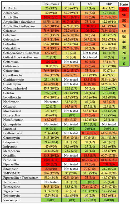
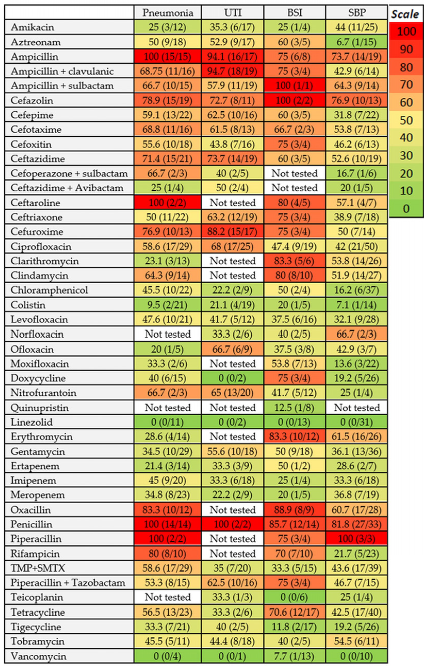
Antibiotic susceptibility patterns varied significantly across different anatomical sites of infection (Figure 2). For pneumonia, the lowest in vitro resistance rates were observed for linezolid (0%, predominantly Gram-positive strains), colistin (9.5%, Gram-negative strains only), ofloxacin (20%), clarithromycin (23.1%), and amikacin (25%). Moderate resistance rates (25–50%) were recorded for erythromycin, moxifloxacin, gentamicin, tigecycline, meropenem, chloramphenicol, doxycycline, levofloxacin, imipenem, and aztreonam. Conversely, resistance rates exceeded 50% for ampicillin (alone or combined with clavulanic acid/sulbactam), clindamycin, piperacillin–tazobactam, and most cephalosporins. Notably, endotracheal aspirates exhibited higher resistance rates than sputum samples for amikacin (33.3% vs. 16.7%) and ofloxacin (25% vs. 0.0%), whereas sputum samples demonstrated higher resistance to clarithromycin, chloramphenicol, aztreonam, ciprofloxacin, clindamycin, and moxifloxacin (Supplementary Figure S1).
Figure 2.
Antibiotic resistance according to the infection site. UTI = Urinary Tract Infection. BSI = Bloodstream Infection. SBP = Spontaneous Bacterial Peritonitis. TMP + SMTX = Trimethoprim + Sulfamethoxazole.
In urinary tract infections, the lowest resistance rates (20–25%) were restricted to colistin (Gram-negative strains), chloramphenicol, and meropenem. Resistance rates between 25% and 50% were noted for imipenem, ertapenem (mostly Gram-negative strains), norfloxacin, levofloxacin, amikacin, tetracycline, trimethoprim–sulfamethoxazole, tigecycline, and tobramycin. Within the cephalosporin class, only cefoxitin and cefoperazone–sulbactam maintained resistance rates below 50% (ranging between 40% and 50%). High resistance rates were observed for ciprofloxacin, gentamicin, and piperacillin–tazobactam.
For SBP, low resistance rates were observed for aztreonam, colistin (Gram-negative strains), linezolid, moxifloxacin (Gram-positive strains), and tigecycline. Carbapenem resistance ranged between 28% and 37%. In contrast, high resistance rates were found for ampicillin (with or without sulbactam), erythromycin, clindamycin, and cefazolin. Nosocomial SBP strains exhibited distinct resistance profiles compared to non-nosocomial strains, characterized by elevated carbapenem resistance among nosocomial Gram-negative bacilli (GNB), while Gram-positive bacteria (GPB) remained uniformly susceptible to linezolid and vancomycin.
In bloodstream infections, resistance rates remained below 25% for teicoplanin, vancomycin, tigecycline, quinupristin, linezolid (primarily in Gram-positive isolates), meropenem, and amikacin. In BSI, bacteriologic confirmation and antibiogram results may arrive late (sometimes after more than 7 days), with empirical therapy very important for prognosis. For these reasons, an accurate knowledge of the local particularities regarding possible strains and antibiotic susceptibility is of utmost importance for therapy.
3.10. Antibiotic Susceptibility in Cirrhotics with Ascites Stratified for Infection Type
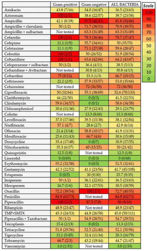
We analyzed the antibiotic susceptibility in relation to the strain type involved in bacterial infections in cirrhotics with ascites (Figure 3). In Gram-positive infections, low resistance rates were noted for linezolid, ertapenem, vancomycin, cefepime, cefotaxime, tigecycline, quinupristin, teicoplanin, meropenem, and ceftriaxone; high resistance rates were noted for ceftaroline, oxacillin, and penicillin. In Gram-negative infections, low resistance rates were recorded for doxycycline (no resistance) and colistin; high resistance rates were recorded for most cephalosporins, most quinolones, and for the combination of piperacillin + tazobactam, whereas moderate resistance was recorded for carbapenems. For Staphylococcus infections, no resistance was recorded to vancomycin, linezolid, quinupristin, and teicoplanin, and a low resistance rate was observed to tigecycline. No in vitro resistance was also recorded for linezolid, doxycycline, and ofloxacin in Enterococcus infections, but one VRE strain was recorded. Streptococcus infections have shown in vitro susceptibility to most antibiotics. In Gram-negative infections, low resistance rates were noted for ceftaroline (E. coli and Klebsiella), combinations of cefoperazone + sulbactam (especially for Klebsiella) and ceftazidime + avibactam (especially in E. coli infections), and colistin (all Gram-negative strain types). Low or moderate resistance rates were also recorded for tigecycline (25 to 36%) and carbapenems (19 to 45%). Acinetobacter infections were associated, however, with high resistance rates for most antibiotics, with low resistance rates only for colistin and doxycycline (no resistance), and tigecycline (25%). A detailed status for all isolated bacterial strains is found in Supplementary Figure S2. We want to emphasize that intrinsic resistance patterns were strictly accounted for in accordance with EUCAST guidelines and were excluded from cross-species susceptibility comparisons; Gram-positive isolates are intrinsically resistant to colistin and aztreonam, whereas Gram-negative isolates are intrinsically resistant to oxacillin, glycopeptides (vancomycin, teicoplanin), lincosamides (clindamycin), and macrolides (erythromycin). Averaged resistance metrics for ‘All Bacteria’ serve strictly as descriptive cohort summaries and must not be used to infer cross-class clinical efficacy or therapeutic equivalence.
Figure 3.
Heatmap of antibiotic resistance stratified by Gram status. TMP + SMTX = Trimethoprim + Sulfamethoxazole.
3.11. The Prevalence of MDR, XDR, and Specific Bacteria-Resistant Strains
We evaluated the presence of multidrug-resistant (MDR) and extensively drug-resistant (XDR) bacteria, ESBL (Enterobacteriaceae species with beta-lactamase), Staphylococcus aureus MRSA, and VRE. MDR strains were present in 104 of 148 bacteria with antibiograms (70.3%), and XDR strains in 13 cases (8.8%), including 4 of 8 Acinetobacter spp. XDR strains. MRSA was noted in 27 of 41 cases (65.9%). ESBL Gram-negative strains were found in 5 of 27 bacteria (18.5%); carbapenem resistance was present in 23 of 74 Gram-negative strains (31.1%). In strict accordance with CDC/ECDC consensus definitions for multidrug resistance, carbapenem resistance was systematically assigned if an isolate exhibited non-susceptibility to at least one tested carbapenem agent. Only one vancomycin-resistant Enterococcus (VRE) was found. In patients with pneumonia, regardless of the intubation status, the proportion of XDR and ESBL strains was higher (16.2 and 80%, respectively), whereas in bloodstream infection, the proportion of ESBL (50%) and MRSA (100%) was also higher.
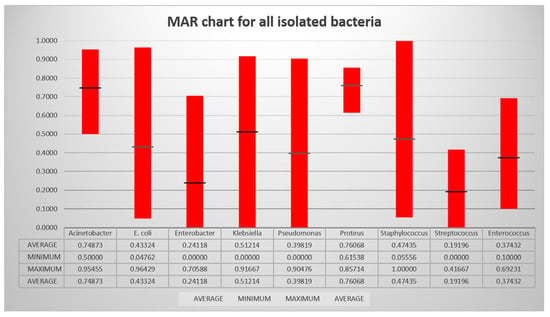
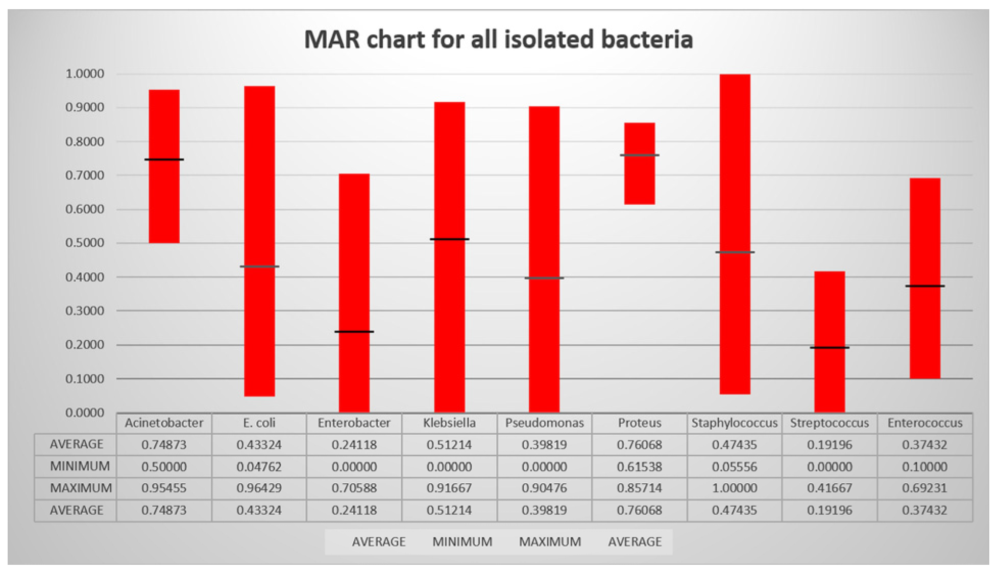
We evaluated the Mean Antibiotic Resistance (MAR) score for all isolated strains with available antibiogram results (148 of 151 strains). The MAR score was higher for Acinetobacter and Proteus, and lowest for Streptococcus and Enterobacter (Figure 4).
Figure 4.
The Mean Antibiotic Resistance for all isolated strains. Sample sizes contributing to each estimate are as follows: Staphylococcus (47), Streptococcus spp. (13), Enterococcus spp. (13), E. coli (26), Klebsiella spp. (27), Acinetobacter baumannii (8), Pseudomonas spp. (6), Enterobacter (5) and Proteus mirabilis (3).
3.12. Risk Factors for Mortality Related to the Presence and Type of Infection
To evaluate the independent risk factors for in-hospital mortality in patients with cirrhosis, ascites, and bacterial infection, we performed univariate and multivariate logistic regression analysis using a backward stepwise selection algorithm. In total, 778 patients with known Child–Pugh class (B or C), alcoholic versus non-alcoholic etiology, with or without acute kidney injury (AKI) or acute-on-chronic liver failure (ACLF), and with or without a documented infection were included in three multivariate models, with the MDR presence also included as a potential risk factor for mortality (Table 4).
Table 4.
Risk factors associated with mortality in patients with cirrhosis with ascites, with or without bacterial infection.
In the first multivariate model, which included baseline clinical characteristics and the presence of at least one infection, disease etiology was automatically eliminated due to a lack of statistical significance (p > 0.20). The finalized model demonstrated a robust global fit (Firth-penalized Likelihood Ratio chi2 = 94.61; p < 0.001), an appropriate explanatory capacity (Nagelkerke R2 = 0.284), and good clinical discrimination with an area under the receiver operating characteristic curve (AUC-ROC) of 0.765. The model exhibited robust calibration, as confirmed by the Hosmer–Lemeshow test (χ2 = 5.24, p = 0.732), indicating no significant deviation from perfect calibration. Multicollinearity tracking demonstrated excellent parameter stability, with all retained covariates displaying strict Variance Inflation Factors well below critical thresholds (Age VIF = 1.08, Child class VIF = 1.14, AKI VIF = 1.21, and ACLF VIF = 1.25). The overall classification accuracy was 78.66%, exhibiting a specificity of 81.85% for predicting survival and a sensitivity of 53.93% for detecting mortality. All four retained variables were directly and significantly associated with death. The presence of infection emerged as a strong independent factor associated with mortality, generating a nearly four-fold increase in the odds of mortality (adjusted Odds Ratio aOR = 3.93; 95% CI: 2.80–5.53; p < 0.001). This was followed closely by acute kidney injury (aOR = 3.72; 95% CI: 2.02–6.83; p < 0.001) and acute-on-chronic liver failure status (aOR = 3.66; 95% CI: 1.83–7.32; p = 0.002). Advanced liver disease severity, defined as Child–Pugh class C versus class B, also independently predicted death, increasing the odds of mortality more than two and a half times (aOR = 2.65; 95% CI: 1.81–3.87; p < 0.001).
A second multivariate model was constructed to assess the specific impact of single versus multiple infection sites. Mirroring the previous results, cirrhosis etiology was excluded from the equation (p > 0.20). This model demonstrated a highly significant global fit (Firth-penalized Likelihood Ratio chi2 = 112.29; p < 0.001), a Nagelkerke R2 = 0.327, and an AUC-ROC of 0.772, accurately classifying 78.53% of subjects with 81.71% specificity and 53.93% sensitivity. Notably, concurrent involvement at multiple distinct sites was associated with a higher death risk compared to a localized infection, showing increased adjusted odds of mortality (aOR = 51.82; 95% CI: 7.89–340.22; p < 0.001). The global calibration of the structural dissemination model was verified (χ2 = 4.88, Hosmer–Lemeshow p = 0.770). To assess the robustness of our clinical risk estimates against potential ascertainment artifacts, a sensitivity analysis was performed by restricting the multivariable framework strictly to the sub-cohort of culture-positive bacterial infections (N = 87). The predictive significance and effect directions of liver disease severity (Child C), acute kidney injury, and anatomical extension remained statistically stable, confirming that the model parameters are not driven by data sparsity or missing testing metrics. However, this high effect magnitude and its wide confidence interval must be interpreted with extreme caution. This numerical variation is driven by data sparsity and a small number of events within this specific sub-cohort cell, indicating that while multi-anatomical infectious dissemination carries a notable clinical risk, the exact mathematical weight remains sensitive to sample size constraints.
Finally, a third multivariate model analyzed mortality variations by specific infection types and multidrug resistance (MDR) profiles. While the presence of bacterial pneumonia, bloodstream infections, and SBP were significantly associated with the risk of in-hospital death, no independent mortality risk was associated with CDI or urinary tract infections. The presence of MDR strains carried additional risk of death in univariate analysis; however, this effect was completely blunted in multivariate analysis (aOR = 1.09) because multi-drug resistance collinearly clusters within severe clinical contexts, such as intensive care unit cases of pneumonia and bloodstream infections.
3.13. Importance of the Bacterial Type for Mortality Risk
We performed an analysis of mortality rates stratified by the bacterial type, including only SBP, BSI, and pneumonia cases. Mortality was higher in Gram-negative infections (85.7% in Pseudomonas aeruginosa, 77.8% in Acinetobacter infection, 72.7% in E. coli, and 52.9% in Klebsiella infections) compared to Gram-positive infections (36.4% in Enterococcus, 33.3% in Staphylococcus, and 30% in Streptococcus infections); however, MRSA infections were associated with a higher mortality than MSSA forms (43.5 versus 9.1%, OR = 7.69, 95%CI 0.8398 to 70.4597, p = 0.0710). To address the quasi-complete separation of data caused by the multiple site infection and to correct for the small-sample bias within the complete-case sub-cohort (N = 87), a multivariate Firth-penalized logistic regression model was performed (Table 5). The penalized model demonstrated excellent discriminative power with an Area Under the ROC Curve (AUROC) of 0.829 and robust calibration (Hosmer–Lemeshow p = 0.511). The microbiological classification of the pathogens (Gram status) emerged as a statistically significant independent factor associated with in-hospital mortality (Global Variable Wald chi2 = 6.85, p = 0.033, 2 degrees of freedom). Compared to the baseline reference group (Gram-positive), both mixed (Gram-positive and Gram-negative infection) and Gram-negative infections carried a significantly higher risk of death (adjusted OR = 4.83; 95% CI: 1.04–22.40; p = 0.044, and adjusted OR = 3.39; 95% CI: 1.17–9.85; p = 0.025, respectively).
Table 5.
Firth penalized logistic regression for mortality risk.
4. Discussion
Bacterial infections are a common complication in hospitalized patients with advanced cirrhosis, driven primarily by immunosuppression, gut microbiota alterations, and bacterial translocation [3,4,5,6,7,8,10,11,30]. In this study, spontaneous bacterial peritonitis (SBP) and Clostridioides difficile infection (CDI) were the most frequent complications, as other published studies also noted [1,3,11,12,30]. The high prevalence of CDI and also high MDR rates align with the heavy use of prophylactic and therapeutic antibiotics in this patient population, particularly for managing hepatic encephalopathy and gastrointestinal bleeding [24,27,37,38,39,40,41,42,43,44,45], although our study lacks precise data regarding previous antibiotic and PPI usage in our cohort. CDI represents a toxin-driven disease rather than a bacterial infection, with toxin A and B detection as the main diagnostic modality; however, a significant prevalence of CDI in patients with cirrhosis is frequently recorded, with a 2.7–2.8% CDI proportion in patients admitted for cirrhosis in two large studies, and 7.3, 6.8, and 11.8% prevalence in patients with hepatic encephalopathy, gastrointestinal bleeding, and previous SBP also noted [11,45]. Bacterial-confirmed pneumonia, urinary tract infections, and bloodstream infections were recorded in 3.3%, 3%, and 2.5%, whereas other bacterial types of infection were only encountered sporadically. Our findings were similar to other published data, with respiratory infections encountered in 6.4–21%, urinary tract infections were recorded in 2.9–22%, and bloodstream infections in 4–35.4% of patients with cirrhosis [3,4,5,6,7,8,9,10,12,15,28,29,46,47,48,49,50,51]. In some papers with higher prevalence rates, cases not confirmed by bacteriology were included [9], or data were reported as a percentage of the total number of tested patients [28]. Most bacterial infections occur in patients with more severe liver disease, with a higher percentage of ACLF, acute kidney injury, and Child C class. Spontaneous pleural empyema, central venous catheter infection, or wound infection were rarely encountered, similar to literature data [52,53,54].
Notably, we observed a distinct etiological split: Gram-negative bacteria predominated in pneumonia and urinary tract infections (UTIs), whereas Gram-positive species dominated in culture-positive SBP and bloodstream infections. This shift toward Gram-positive pathogens in SBP mirrors recent literature and is likely tied to increased instrumental maneuvers and healthcare-associated exposures [3,4,12,15,38,40,41]. Conversely, the high rate of Gram-negative pneumonia reflects the substantial proportion of critically ill patients in the ICU with nosocomial infections. These shifting etiological patterns have significant clinical implications, emphasizing the need to tailor empirical antimicrobial therapy in high-risk cirrhotic subgroups before susceptibility results are available.
Antimicrobial resistance poses a severe challenge to standard empirical regimens, such as third-generation cephalosporins [1,2]. Our cohort demonstrated high resistance rates to cephalosporins and ampicillin combinations, alongside 25–50% resistance rates to quinolones, aminoglycosides, piperacillin–tazobactam, and imipenem. Overall, multidrug-resistant (MDR) strains were found in 70.3% of cases, XDR strains in 8.8%, carbapenem-resistant Gram-negative strains were noted in 31.1%, and methicillin-resistant Staphylococcus aureus (MRSA) in 65.9%. The literature reports ESBL-producing strains in 11.7% to 35% of cases, often correlating with higher mortality rates [5,6,7,8,9,10,28,29,40,49,50,51,55]. A major methodological limitation is the reliance on phenotypic disk diffusion kits for ESBL and carbapenemase detection without molecular genotypic confirmation. This phenotypic approach carries inherent diagnostic drawbacks, as it may underdiagnose low-hydrolysis OXA-48-like enzymes or misclassify porin loss and hyperproduced AmpC beta-lactamases as true carbapenemase resistance. Rising MDR prevalence across numerous studies increasingly complicates infection management in liver cirrhosis [4,9,28,29,49,51,56,57]. In our cohort, the observed MRSA rate was 65.9%, thus exceeding the 11% to 40% range documented elsewhere. Previous studies report CRE rates of 5% to 18.2%; in our study, carbapenem resistance was noted in 31.1% of Gram-negative strains; these findings include, however, non-fermenters (Acinetobacter and Pseudomonas) traditionally associated with higher resistance rates to carbapenems. Lastly, VRE remains rare in cirrhotic patients, with an ambiguous effect on mortality [16,41,58].
Site-specific analyses indicated that linezolid, vancomycin, and colistin maintained low in vitro resistance rates for their respective target pathogens; low resistance rates were noted in SBP to Tigecycline, and also to Linezolid and Vancomycin (Gram-positive forms), and to Aztreonam and Colistin (Gram-negative forms). In patients with pneumonia, low in vitro resistance rates were recorded to linezolid (mostly Gram-positive strains tested), colistin (Gram-negative strains), ofloxacin, clarithromycin, and amikacin (with resistance rates between 20 and 25%). In urinary tract infections, only colistin, chloramphenicol, and meropenem had low resistance rates, and in bloodstream infections, resistance rates below 25% were recorded for teicoplanin, vancomycin, tigecycline, and linezolid (in Gram-positive cases), and also for meropenem and amikacin. Although in vitro resistance does not always equate to clinical failure, our findings support recent guidelines recommending extended-spectrum combinations (e.g., carbapenems plus glycopeptides) over cephalosporins for healthcare-associated infections, particularly in patients with documented high MRSA rates, prior MDR strain exposure, or severe cirrhosis presentations such as ACLF and high SOFA scores.
Several susceptibility patterns according to the bacterial strain were also identified. Staphylococcus strains had no in vitro resistance to vancomycin, linezolid, and teicoplanin, and Streptococcus strains were susceptible to most antibiotics; only one vancomycin-resistant Enterococcus strain was noted. Acinetobacter isolates showed extensive resistance to most antibiotics, remaining susceptible primarily to colistin and doxycycline. Overall, low resistance rates were recorded for colistin among all Gram-negative strains, for ceftaroline in E. coli and Klebsiella, and for the combinations of cefoperazone–sulbactam and ceftazidime–avibactam. These patterns must be, however, interpreted with prudence because in vitro resistance is not identical to clinical efficacy, and regional particularities and lack of uniform testing may restrict generalizability of our findings.
In-hospital mortality was high, particularly for patients with culture-confirmed bacterial pneumonia (63%) and bloodstream infections (55%). In SBP, mortality reached 28.2%, with no significant difference between culture-positive and culture-negative cases, similar to literature data [1,2,16,20,59,60]. Multivariate analysis identified the presence of any infection, multiple-site infections, pneumonia, bloodstream infections, SBP, and Gram-negative or mixed etiologies as significantly associated with the risk of death. Mortality risk in patients with pneumonia may be, however, augmented by the high proportion of intubated patients, suggesting that the severity of disease, with ICU-related nosocomial trauma and invasive airways as concomitant aggravating factors for mortality. Furthermore, acute kidney injury (AKI), acute-on-chronic liver failure (ACLF), and Child–Pugh class C were confirmed as strong, independent factors associated with mortality; however, these parameters should also be considered consequences of an ongoing infection rather than pure baseline covariates. Given the sparse data and very wide confidence interval for multiple-site infections (OR = 51.82), these findings should be interpreted as exploratory and limited by our single-center experience, rather than a definitive conclusion. Interestingly, the presence of MDR status did not independently predict mortality in our model, as other studies recorded [9,28,29,49], likely because of the high baseline prevalence of MDR strains (70.3%) and collinearity clusters within infection sites with traditionally high mortality rates (such as nosocomial ICU pneumonias and BSIs, which can obscure its statistical effect).
These outcomes broadly align with existing Romanian epidemiological data. A study of 1046 patients with liver cirrhosis has found a 4.9% infection rate, most frequent being SBP (2.5%), urinary (1.9%), and pneumonia (0.8%), with a 33.3% death rate in cases with documented infection [31]. In another study of 198 cases (83 with infections and 115 without infections, the mortality rate was 2-fold higher in patients with infection [32]. In a study of 155 patients with advanced liver disease on the waiting list for liver transplantation, the prevalence of active infections was 41.9%, the most frequent being SBP, urinary tract infection, and pneumonia [33]. A study of 367 cirrhotics with acute variceal bleeding found 25 cases of CDI (6.8%), with almost 2-fold higher mortality (44 versus 25.6%, p = 0.0446).
The primary strength of this study lies in its detailed characterization of localized antibiotic susceptibility and mortality risk factors within a region lacking extensive data, offering valuable insights for updating empirical treatment protocols. However, several limitations must be acknowledged. The single-center design limited the sample size, which may reduce the generalizability of the findings and restrict the statistical power of subgroup multivariate analyses; a large subset of patients with cirrhosis (2003 cases) had incomplete data and were excluded from the analysis. Our cohort lacks accurate data regarding previous antibiotic and PPI usage, ICU length of stay, empirical therapy, time-to-effective therapy, or healthcare-associated or community-acquired infections, which can also significantly influence mortality. For some infections (bloodstream and urinary tract infections), the presence of large uncollected cohorts (663 for urine, 761 for blood) creates a potential selection bias; in our patients, testing was performed strictly on indication (symptom-driven surveillance) to avoid treating asymptomatic bacteriuria, and our findings may represent a lower-bound diagnostic proportion of documented infections within a selected hospitalized cohort, rather than true population prevalence. A complete exclusion of contaminating flora remains an inherent clinical challenge. An internal audit has noted that our final cohort of 806 patients was inherently skewed toward more severe, decompensated liver disease, which serves as a necessary caveat when interpreting our reported infection and mortality metrics. The low prevalence of COVID-19 in our specific cohort (1.7%), due to institutional triaging protocols and patients’ reluctance regarding admission, makes the potential impact of SARS-CoV-2 on portal-hypertension-driven bacterial infections difficult to evaluate. Multivariate analysis may be inaccurate in small samples, especially in smaller subgroups such as Gram-negative versus Gram-positive infections, or infections stratified by bacterial strains. Future multi-center or long-term studies are needed to better track regional etiological shifts and optimize management strategies.
5. Conclusions
In our cohort of hospitalised patients with cirrhosis and ascites, 22.1% had at least one confirmed bacterial infection, with SBP (9.7%), CDI (9.2%), pneumonia (3.3%), urinary tract infection (3%), and bloodstream infection (2.5%) being the most frequent. Mortality rate was higher in culture-positive pneumonia (63%), bloodstream infection (55%), and SBP (28.2%); a trend to higher mortality in CDI was noted only in univariate analysis. Increased antibiotic resistance and MDR presence (70.3%) were noted; the isolated pathogens included MRSA (65.9%), ESBL-producing Gram-negative bacilli (18.5%), and carbapenem-resistant Gram-negative strains (31.1%); only one case of vancomycin-resistant Enterococcus (VRE) was recorded. The severity of liver disease; AKI and ACLF presence; the presence of pneumonia, BSI, and SBP; and Gram-negative and multiple infection sites were associated with higher mortality in multivariate analysis. Despite lacking clinical outcome correlation, our in vitro data demonstrate high regional resistance to standard third-generation cephalosporin regimens. Finally, our 70.3% baseline prevalence of MDR strains warrants further investigation into whether broader empirical regimens provide more appropriate coverage for critically ill patients pending susceptibility testing.
Supplementary Materials
The following supporting information can be downloaded at https://www.mdpi.com/article/10.3390/life16111873/s1: Supplementary File S1 (Supplementary Table S1: SBP in patients with cirrhosis and ascites; Supplementary Table S2: Clostridioides difficile infection in patients with cirrhosis and ascites; Supplementary Table S3: Characteristics of patients with cirrhosis and bacterial pneumonia; Supplementary Table S4: Bloodstream infection in patients with cirrhosis and ascites); Supplementary File S2 (Supplementary Figure S1: Antibiotic resistance in patients with pneumonia, stratified by endotracheal aspirates or sputum samples; Supplementary Figure S2: Antibiotic resistance in Gram-negative strains; Supplementary Figure S3: Antibiotic resistance in Gram-positive strains); Supplementary File S3 (Supplementary Table S5: Antibiotic susceptibility testing for all strains).
Author Contributions
Conceptualization, S.M.C., C.M.I., and D.N.F.; methodology, D.N.F., O.-M.Z., and I.R.; software, S.M.C., D.C.N., and V.C.; validation, C.M.I., D.N.F., and D.C.N.; formal analysis, S.M.C., C.M.I., and D.N.F.; investigation, V.C., C.M.I., A.G., and L.F.V.; resources, S.M.C., R.-C.S., D.C.N., A.G., V.C., and L.F.V.; data curation, C.M.I., D.C.N., A.G., and I.R.; writing—original draft preparation, S.M.C., C.M.I., I.R., and O.-M.Z.; writing—review and editing, S.M.C., D.C.N., R.-C.S., and I.R.; visualization, I.R., L.F.V., R.-C.S., and D.N.F.; supervision, S.M.C., D.N.F., and O.-M.Z.; project administration, S.M.C., D.C.N., C.M.I., and I.R. All authors have read and agreed to the published version of the manuscript.
Funding
This research received no external funding.
Institutional Review Board Statement
This study was approved on 19 April 2023 by the Ethics Committee at the Emergency Clinical Hospital of Craiova (Comisia de Etica a Spitalului Clinic Judetean de Urgentă Craiova) (approval No. 18690/19.04.2023).
Informed Consent Statement
Informed consent was obtained from all subjects involved in the study.
Data Availability Statement
The data presented in this study are available on request from the corresponding author.
Conflicts of Interest
The authors declare no conflicts of interest.
Abbreviations
| Clinical & Scores Abbreviations | |
| ACLF | Acute-on-Chronic Liver Failure |
| AKI | Acute Kidney Injury |
| ALT | Alanine Aminotransferase |
| AST | Aspartate Aminotransferase |
| CDI | Clostridioides difficile Infection |
| CTP | Child–Turcotte–Pugh score |
| HCC | Hepatocellular Carcinoma |
| ICU | Intensive Care Unit |
| INR | International Normalized Ratio |
| MELD | Model for End-Stage Liver Disease score |
| MELD-3 | Model for End-Stage Liver Disease version 3 score |
| MELD-Na | Model for End-Stage Liver Disease Sodium score |
| PMN | Polymorphonuclear Neutrophils |
| SBP | Spontaneous Bacterial Peritonitis |
| UTI | Urinary Tract Infection |
| Microbiological Abbreviations | |
| AST Card | Antimicrobial Susceptibility Testing Card |
| BSI | Bloodstream Infection |
| CFU/mL | Colony-Forming Units per Milliliter |
| CNS | Coagulase-Negative Staphylococci |
| CRE | Carbapenem-Resistant Enterobacteriaceae |
| ECDC | European Centre for Disease Prevention and Control |
| ESBL | Extended-Spectrum Beta-Lactamase |
| EUCAST | European Committee on Antimicrobial Susceptibility Testing |
| GDH | Glutamate Dehydrogenase |
| GNB | Gram-Negative Bacilli |
| GPB | Gram-Positive Bacteria |
| MAR index | Multiple Antibiotic Resistance index |
| MDR | Multidrug-Resistant |
| MRSA | Methicillin-Resistant Staphylococcus aureus |
| MSSA | Methicillin-Susceptible Staphylococcus aureus |
| VRE | Vancomycin-Resistant Enterococcus |
| XDR | Extensively Drug-Resistant |
| Statistical Abbreviations | |
| aOR | Adjusted Odds Ratio |
| AUC-ROC/AUROC | Area Under the Receiver Operating Characteristic curve |
| 95% CI | 95% Confidence Interval |
| IQR | Interquartile Range |
| OR | Odds Ratio |
| p-value | Probability value |
References
- European Association for the Study of the Liver. EASL Clinical Practice Guidelines for the management of patients with decompensated cirrhosis. J. Hepatol. 2018, 69, 406–460. [Google Scholar] [CrossRef] [Scilit] [PubMed]
- Biggins, S.W.; Angeli, P.; Garcia-Tsao, G.; Ginès, P.; Ling, S.C.; Nadim, M.K.; Wong, F.; Kim, W.R. Diagnosis, Evaluation, and Management of Ascites, Spontaneous Bacterial Peritonitis and Hepatorenal Syndrome: 2021 Practice Guidance by the American Association for the Study of Liver Diseases. Hepatology 2021, 74, 1014–1048. [Google Scholar] [CrossRef] [Scilit]
- Ascione, T.; Di Flumeri, G.; Boccia, G.; De Caro, F. Infections in patients affected by liver cirrhosis: An update. Infez. Med. 2017, 25, 91–97. [Google Scholar]
- Piano, S.; Singh, V.; Caraceni, P.; Maiwall, R.; Alessandria, C.; Fernandez, J.; Soares, E.C.; Kim, D.J.; Kim, S.E.; Marino, M.; et al. Epidemiology and Effects of Bacterial Infections in Patients With Cirrhosis Worldwide. Gastroenterology 2019, 156, 1368–1380.e10. [Google Scholar] [CrossRef] [Scilit] [PubMed]
- Bruns, T.; Zimmermann, H.W.; Stallmach, A. Risk factors and outcome of bacterial infections in cirrhosis. World J. Gastroenterol. 2014, 20, 2542–2554. [Google Scholar] [CrossRef] [Scilit]
- Strauss, E. The impact of bacterial infections on survival of patients with decompensated cirrhosis. Ann. Hepatol. 2013, 13, 7–19. [Google Scholar] [CrossRef] [Scilit] [PubMed]
- Jalan, R.; Fernandez, J.; Wiest, R.; Schnabl, B.; Moreau, R.; Angeli, P.; Stadlbauer, V.; Gustot, T.; Bernardi, M.; Canton, R.; et al. Bacterial infections in cirrhosis: A position statement based on the EASL Special Conference 2013. J. Hepatol. 2014, 60, 1310–1324. [Google Scholar] [CrossRef] [Scilit] [PubMed]
- Acevedo, J.; Fernández, J. New determinants of prognosis in bacterial infections in cirrhosis. World J. Gastroenterol. 2014, 20, 7252–7259. [Google Scholar] [CrossRef] [Scilit]
- Garg, P.; Verma, N.; Angrup, A.; Taneja, N.; Valsan, A.; Reddy, V.D.; Agarwal, J.; Chaudhary, R.; Kaur, P.; Rathi, S.; et al. Exploring the Prevalence, Predictors, and Impact of Bacterial Infections to Guide Empiric Antimicrobial Decisions in Cirrhosis (EPIC-AD). J. Clin. Exp. Hepatol. 2024, 14, 101352. [Google Scholar] [CrossRef] [Scilit]
- Mattos, A.A.; Wiltgen, D.; Jotz, R.F.; Dornelles, C.M.R.; Fernandes, M.V.; Mattos, Â.Z. Spontaneous bacterial peritonitis and extraperitoneal infections in patients with cirrhosis. Ann. Hepatol. 2020, 19, 451–457. [Google Scholar] [CrossRef] [Scilit]
- Voicu, M.N.; Popescu, F.; Florescu, D.N.; Rogoveanu, I.; Turcu-Stiolica, A.; Gheonea, D.I.; Iovanescu, V.F.; Iordache, S.; Cazacu, S.M.; Ungureanu, B.S. Clostridioides difficile Infection among Cirrhotic Patients with Variceal Bleeding. Antibiotics 2021, 10, 731. [Google Scholar] [CrossRef] [Scilit]
- Bunchorntavakul, C.; Chamroonkul, N.; Chavalitdhamrong, D. Bacterial infections in cirrhosis: A critical review and practical guidance. World J. Hepatol. 2016, 8, 307–321. [Google Scholar] [CrossRef] [Scilit] [PubMed]
- Aithal, G.P.; Palaniyappan, N.; China, L.; Härmälä, S.; Macken, L.; Ryan, J.M.; Wilkes, E.A.; Moore, K.; Leithead, J.A.; Hayes, P.C.; et al. Guidelines on the management of ascites in cirrhosis. Gut 2021, 70, 9–29. [Google Scholar] [CrossRef] [Scilit]
- Arzivian, A.; Duong, T. The Incidence of Spontaneous Bacterial Peritonitis in Patients With Cirrhosis-Related Ascites Undergoing Elective Outpatient Large-Volume Paracentesis. Cureus 2023, 15, e50191. [Google Scholar] [CrossRef] [Scilit]
- Ekpanyapong, S.; Reddy, K.R. Infections in Cirrhosis. Curr. Treat. Options Gastroenterol. 2019, 17, 254–270. [Google Scholar] [CrossRef] [Scilit]
- Tay, P.W.L.; Xiao, J.; Tan, D.J.H.; Ng, C.; Lye, Y.N.; Lim, W.H.; Teo, V.X.Y.; Heng, R.R.Y.; Yeow, M.W.X.; Lum, L.H.W.; et al. An Epidemiological Meta-Analysis on the Worldwide Prevalence, Resistance, and Outcomes of Spontaneous Bacterial Peritonitis in Cirrhosis. Front Med. 2021, 8, 693652. [Google Scholar] [CrossRef] [Scilit]
- Căruntu, F.A.; Benea, L. Spontaneous bacterial peritonitis: Pathogenesis, diagnosis, treatment. J. Gastrointest. Liver Dis. 2006, 15, 51–56. [Google Scholar]
- Long, B.; Gottlieb, M. Emergency medicine updates: Spontaneous bacterial peritonitis. Am. J. Emerg. Med. 2023, 70, 84–89. [Google Scholar] [CrossRef] [Scilit]
- Fiore, M.; Di Franco, S.; Alfieri, A.; Passavanti, M.B.; Pace, M.C.; Kelly, M.E.; Damiani, G.; Leone, S. Spontaneous bacterial peritonitis caused by Gram-negative bacteria: An update of epidemiology and antimicrobial treatments. Expert Rev. Gastroenterol. Hepatol. 2019, 13, 683–692. [Google Scholar] [CrossRef] [Scilit]
- Shizuma, T. Spontaneous bacterial and fungal peritonitis in patients with liver cirrhosis: A literature review. World J. Hepatol. 2018, 10, 254–266. [Google Scholar] [CrossRef] [Scilit]
- Fiore, M.; Maraolo, A.E.; Gentile, I.; Borgia, G.; Leone, S.; Sansone, P.; Passavanti, M.B.; Aurilio, C.; Pace, M.C. Current concepts and future strategies in the antimicrobial therapy of emerging Gram-positive spontaneous bacterial peritonitis. World J. Hepatol. 2017, 9, 1166–1175. [Google Scholar] [CrossRef] [Scilit]
- Nookala, V.K.; Sharzehi, K.; Lee, T.P. An Increase of Gram-Positive Bacteria in Spontaneous Bacterial Peritonitis. Gastroenterology 2012, 142, S951. [Google Scholar] [CrossRef] [Scilit]
- Cazacu, S.M.; Zlatian, O.M.; Alexandru, D.O.; Plesea, E.L.; Vacariu, I.A.; Cimpoeru, M.; Rogoveanu, I.; Bigea, C.C.; Marginean, C.M.; Iordache, S. Higher NLR Values Can Predict Gram-Negative Spontaneous Bacterial Peritonitis and a High In-Hospital Mortality Rate in Patients with Spontaneous Bacterial Peritonitis. Life 2025, 15, 1363. [Google Scholar] [CrossRef] [Scilit]
- Cazacu, S.M.; Zlatian, O.M.; Plesea, E.L.; Vacariu, A.I.; Cimpoeru, M.; Rogoveanu, I.; Bigea, C.C.; Iordache, S. Predominant Gram-Positive Etiology May Be Associated with a Lower Mortality Rate but with Higher Antibiotic Resistance in Spontaneous Bacterial Peritonitis: A 7-Year Study in a Tertiary Center in Romania. Life 2025, 15, 855. [Google Scholar] [CrossRef] [Scilit]
- Tariq, T.; Irfan, F.B.; Farishta, M.; Dykstra, B.; Sieloff, E.M.; Desai, A.P. Spontaneous fungal peritonitis: Micro-organisms, management and mortality in liver cirrhosis—A systematic review. World J. Hepatol. 2019, 11, 596–606. [Google Scholar] [CrossRef] [Scilit]
- MacIntosh, T. Emergency Management of Spontaneous Bacterial Peritonitis—A Clinical Review. Cureus 2018, 10, e2253. [Google Scholar] [CrossRef] [Scilit]
- Oliveira, J.C.; Carrera, E.; Petry, R.C.; Deutschendorf, C.; Mantovani, A.; Barcelos, S.T.A.; Cassales, S.; Schacher, F.C.; Lopes, A.B.; Alvares-da-Silva, M.R. High Prevalence of Multidrug Resistant Bacteria in Cirrhotic Patients with Spontaneous Bacterial Peritonitis: Is It Time to Change the Standard Antimicrobial Approach? Can. J. Gastroenterol. Hepatol. 2019, 2019, 6963910. [Google Scholar] [CrossRef] [Scilit]
- Varghese, G.; Jamwal, A.; Sarawat, D.; Singh, S.; Tejan, N.; Patel, S.S.; Sahu, C. Bacterial resistance profile and its association with poor outcome among cirrhosis patients attending a tertiary care referral center in northern India. Indian J. Gastroenterol. 2025, 44, 198–207. [Google Scholar] [CrossRef] [Scilit]
- Trad, N.; Mohamed, G.; Bizid, S.; Abdallah, H.B.; Bouali, R.; Abdelli, M.N. Clinical impact of multidrug-resistant bacterial infections in patients with cirrhosis. Future Sci. OA 2024, 10, FSO945. [Google Scholar] [CrossRef] [Scilit]
- Meyyur Aravamudan, V.; Khan, S.R.; Hussain, I. Clostridium difficile Infection in Liver Cirrhosis Carries a Higher Risk of Mortality: A Comprehensive Literature Review. Cureus 2019, 11, e5463. [Google Scholar] [CrossRef] [Scilit]
- Preda, C.M.; Ghita, R.; Ghita, C.; Mindru, C.; Vlaicu, L.; Andrei, A.; Andrei, S.; Diculescu, M. A retrospective study of bacterial infections in cirrhosis. Maedica 2011, 6, 185–192. [Google Scholar] [PubMed]
- Bacanu, I.; Ionescu, V.A.; Gheorghe, G.; Năstac, A.; Tiuca, C.L.; Diaconu, C.C. Infectious Complications in Patients with Liver Cirrhosis. Ann. Acad. Romanian Sci. Ser. Med. 2024, 5, 17–32. [Google Scholar] [CrossRef] [Scilit]
- Mihaela, G.; Ester, C.; Iacob, S.; Cerban, R.; Pietrareanu, C.; Popescu, I.; Ruta, S.; Grancea, C.; Gheorghe, C.; Gheorghe, L. Predictive Factors for Mortality and Infection Surveillance in Romanian Candidates for Liver Transplantation. Surg. Gastroenterol. Oncol. 2024, 29, 89. [Google Scholar] [CrossRef] [Scilit]
- The European Committee on Antimicrobial Susceptibility Testing. Breakpoint Tables for Interpretation of MICs and Zone Diameters. Version 14.0, 2024. Available online: http://www.eucast.org (accessed on 2 May 2026).
- Kidd, J.M.; Livermore, D.M.; Nicolau, D.P. The difficulties of identifying and treating Enterobacterales with OXA-48-like carbapenemases. Clin. Microbiol. Infect. 2020, 26, 401–403. [Google Scholar] [CrossRef] [Scilit]
- Magiorakos, A.P.; Srinivasan, A.; Carey, R.B.; Carmeli, Y.; Falagas, M.E.; Giske, C.G.; Harbarth, S.; Hindler, J.F.; Kahlmeter, G.; Olsson-Liljequist, B.; et al. Multidrug-resistant, extensively drug-resistant and pandrug-resistant bacteria: An international expert proposal for interim standard definitions for acquired resistance. Clin. Microbiol. Infect. 2012, 18, 268–281. [Google Scholar] [CrossRef] [Scilit]
- de Mattos, A.A.; Costabeber, A.M.; Lionço, L.C.; Tovo, C.V. Multidrug-resistant bacteria in spontaneous bacterial peritonitis: A new step in management? World J. Gastroenterol. 2014, 20, 14079–14086. [Google Scholar] [CrossRef] [Scilit]
- Piroth, L.; Pechinot, A.; Di Martino, V.; Hansmann, Y.; Putot, A.; Patry, I.; Hadou, T.; Jaulhac, B.; Chirouze, C.; Rabaud, C.; et al. Evolving epidemiology and antimicrobial resistance in spontaneous bacterial peritonitis: A two-year observational study. BMC Infect. Dis. 2014, 14, 287. [Google Scholar] [CrossRef] [Scilit]
- Pimentel, R.; Leitão, J.; Gregório, C.; Santos, L.; Carvalho, A.; Figueiredo, P. Spontaneous Bacterial Peritonitis in Cirrhotic Patients: A Shift in the Microbial Pattern? A Retrospective Analysis. GE Port. J. Gastroenterol. 2021, 29, 256–266. [Google Scholar] [CrossRef] [Scilit]
- Ratnasekera, I.U.; Johnson, A.; Powell, E.E.; Henderson, A.; Irvine, K.M.; Valery, P.C. Epidemiology of ascites fluid infections in patients with cirrhosis in Queensland, Australia from 2008 to 2017: A population-based study. Medicine 2022, 101, e29217. [Google Scholar] [CrossRef] [Scilit]
- Alexopoulou, A.; Vasilieva, L.; Agiasotelli, D.; Siranidi, K.; Pouriki, S.; Tsiriga, A.; Toutouza, M.; Dourakis, S.P. Extensively drug-resistant bacteria are an independent predictive factor of mortality in 130 patients with spontaneous bacterial peritonitis or spontaneous bacteremia. World J. Gastroenterol. 2016, 22, 4049–4056. [Google Scholar] [CrossRef] [Scilit]
- Khoury, A.; Rattanasuwan, T.; Ebied, A.M. Shifting microorganism incidence in cirrhotic patients with ascites: A 5-year retrospective cross-sectional analysis. J. Dig. Med. Res. 2020, 3, 45. [Google Scholar] [CrossRef] [Scilit]
- Friedrich, K.; Nüssle, S.; Rehlen, T.; Stremmel, W.; Mischnik, A.; Eisenbach, C. Microbiology and resistance in first episodes of spontaneous bacterial peritonitis: Implications for management and prognosis. J. Gastroenterol. Hepatol. 2016, 31, 1191–1195. [Google Scholar] [CrossRef] [Scilit]
- Li, Y.T.; Yu, C.B.; Huang, J.R.; Qin, Z.J.; Li, L.J. Pathogen profile and drug resistance analysis of spontaneous peritonitis in cirrhotic patients. World J. Gastroenterol. 2015, 21, 10409–10417. [Google Scholar] [CrossRef] [Scilit]
- Liu, Y.; Chen, M. Clostridioides difficile Infection in Liver Cirrhosis: A Concise Review. Can. J. Gastroenterol. Hepatol. 2022, 2022, 4209442. [Google Scholar] [CrossRef] [Scilit]
- Bartoletti, M.; Giannella, M.; Lewis, R.E.; Viale, P. Bloodstream infections in patients with liver cirrhosis. Virulence 2016, 7, 309–319. [Google Scholar] [CrossRef] [Scilit]
- Bartoletti, M.; Giannella, M.; Lewis, R.; Caraceni, P.; Tedeschi, S.; Paul, M.; Schramm, C.; Bruns, T.; Merli, M.; Cobos-Trigueros, N.; et al. A prospective multicentre study of the epidemiology and outcomes of bloodstream infection in cirrhotic patients. Clin. Microbiol. Infect. 2018, 24, 546.e1–546.e8. [Google Scholar] [CrossRef] [Scilit]
- Onorato, L.; Monari, C.; Capuano, S.; Grimaldi, P.; Coppola, N. Prevalence and Therapeutic Management of Infections by Multi-Drug-Resistant Organisms (MDROs) in Patients with Liver Cirrhosis: A Narrative Review. Antibiotics 2022, 11, 232. [Google Scholar] [CrossRef] [Scilit]
- Terra, C.; de Mattos, Â.Z.; Chagas, M.S.; Torres, A.; Wiltgen, D.; Souza, B.M.; Perez, R.M. Impact of multidrug resistance on the management of bacterial infections in cirrhosis. World J. Clin. Cases 2023, 11, 534–544. [Google Scholar] [CrossRef] [Scilit]
- Bartoletti, M.; Lewis, R.E.; Giannella, M.; Tedeschi, S.; Viale, P. The role of extended infusion β-lactams in the treatment of bloodstream infections in patients with liver cirrhosis. Expert Rev. Anti Infect. Ther. 2018, 16, 771–779. [Google Scholar] [CrossRef] [Scilit]
- Xie, X.Q.; Shao, J.W. Clinical impact and disparities in mortality and antimicrobial resistance between Gram-negative and Gram-positive bacteria in cirrhotic abdominal infections: A meta-analysis. Front Med. 2025, 12, 1714716. [Google Scholar] [CrossRef] [Scilit]
- Gupta, P.; Lee, T. Retrospective Outcomes of Patients With Spontaneous Bacterial Empyema Given Albumin. Chest 2025, 167, 694–696. [Google Scholar] [CrossRef] [Scilit]
- Chen, L.; Yuan, Z.; Yang, X.; Li, S.; Zhang, T.; Chu, W.; Chen, Z. Hepatic hydrothorax in the absence of ascites with mixed bacterial and fungal infection: A case report. BMC Infect. Dis. 2025, 25, 1278. [Google Scholar] [CrossRef] [Scilit]
- Soin, S.; Sher, N.; Saleem, N. Spontaneous bacterial empyema: An elusive diagnosis in a patient with cirrhosis. BMJ Case Rep. 2018, 2018, bcr2018224810. [Google Scholar] [CrossRef] [Scilit]
- Song, K.H.; Jeon, J.H.; Park, W.B.; Park, S.W.; Kim, H.B.; Oh, M.D.; Lee, H.S.; Kim, N.J.; Choe, K.W. Clinical outcomes of spontaneous bacterial peritonitis due to extended-spectrum beta-lactamase-producing Escherichia coli and Klebsiella species: A retrospective matched case-control study. BMC Infect. Dis. 2009, 9, 41. [Google Scholar] [CrossRef] [Scilit]
- Rostkowska, K.A.; Szymanek-Pasternak, A.; Simon, K.A. Spontaneous bacterial peritonitis—Therapeutic challenges in the era of increasing drug resistance of bacteria. Clin. Exp. Hepatol. 2018, 4, 224–231. [Google Scholar] [CrossRef] [Scilit]
- Ardolino, E.; Wang, S.S.; Patwardhan, V.R. Evidence of Significant Ceftriaxone and Quinolone Resistance in Cirrhotics with Spontaneous Bacterial Peritonitis. Dig. Dis. Sci. 2019, 64, 2359–2367. [Google Scholar] [CrossRef] [Scilit]
- Barger, M.; Blodget, E.; Pena, S.; Mack, W.; Fong, T.L. VRE in cirrhotic patients. BMC Infect. Dis. 2019, 19, 711. [Google Scholar] [CrossRef] [Scilit]
- Niu, B.; Kim, B.; Limketkai, B.N.; Sun, J.; Li, Z.; Woreta, T.; Chen, P.H. Mortality from Spontaneous Bacterial Peritonitis Among Hospitalized Patients in the USA. Dig. Dis. Sci. 2018, 63, 1327–1333. [Google Scholar] [CrossRef] [Scilit]
- Hung, T.H.; Wang, C.Y.; Tsai, C.C.; Lee, H.F. Short and long-term mortality of spontaneous bacterial peritonitis in cirrhotic patients. Medicine 2024, 103, e40851. [Google Scholar] [CrossRef] [Scilit]

Disclaimer/Publisher’s Note: The statements, opinions and data contained in all publications are solely those of the individual author(s) and contributor(s) and not of MDPI and/or the editor(s). MDPI and/or the editor(s) disclaim responsibility for any injury to people or property resulting from any ideas, methods, instructions or products referred to in the content. |
© 2026 by the authors. Licensee MDPI, Basel, Switzerland. This article is an open access article distributed under the terms and conditions of the Creative Commons Attribution (CC BY) license.