


Abstract
Background: Deoxycholic acid (DCA) causes adipocytolysis after subcutaneous injection and is approved for reducing submental fat. Use in the abdomen is off-label, and evidence for low-concentration, cannula-delivered protocols remains limited. Objective: Our objective is to describe clinical and ultrasound outcomes after a diluted DCA-based injection protocol for localized lower abdominal adiposity. Methods: Five healthy adult women aged 31–43 years underwent two lower-abdominal treatments 4 weeks apart. For each treatment, two 2 mL vials of DCA 10 mg/mL were diluted to a final concentration of 2 mg/mL using L-carnitine and a clinic-prepared tumescent solution, yielding 20 mL of total injectate. The formulation was delivered into the subcutaneous layer using a 23-gauge, 50 mm cannula. Body weight, abdominal circumference, caliper thickness, clinical photographs, and ultrasound-measured local tissue depth were reviewed at baseline and final follow-up; caliper thickness and circumference were also recorded at interim visits. Results: All five cases showed reductions in both caliper thickness and ultrasound-measured local tissue depth. Mean caliper thickness decreased from 41.14 ± 4.76 mm to 35.94 ± 5.15 mm, a mean reduction of 5.20 ± 1.20 mm. Mean ultrasound depth decreased from 3.22 ± 0.80 cm to 2.39 ± 0.81 cm, a mean reduction of 0.83 ± 0.15 cm; the mean individual relative reduction was 27.5%. Mean body weight changed by −0.08 kg and mean abdominal circumference by −0.64 cm, with substantial case-to-case variability. The available summary reported procedural pain scores of 0–1 on a 10-point visual analog scale; complete patient-level adverse-event records were unavailable, precluding systematic safety assessment. Conclusion: In this exploratory five-patient series, localized caliper and ultrasound tissue-depth measures decreased after two sessions of a 2 mg/mL diluted DCA-based protocol despite variable changes in body weight and circumference. The uncontrolled design, combined formulation, small sample, and incomplete safety documentation preclude attribution of effect to DCA alone. Controlled studies with standardized imaging and complete adverse-event capture are required.
1. Introduction
Deoxycholic acid (DCA) is a secondary bile acid that disrupts adipocyte cell membranes when injected into subcutaneous fat. Histologic studies have demonstrated early adipocytolysis and local inflammation followed by macrophage-mediated clearance, septal remodeling, and partial resolution of inflammation over subsequent weeks [1,2,3]. Clinical development has focused primarily on moderate-to-severe submental adiposity, for which standardized synthetic DCA formulations have established efficacy and a predominantly local adverse-event profile [2,4].
Interest in injection adipocytolysis has expanded to small, localized fat deposits of the trunk and extremities. A prospective pilot study of hypogastric fat and a larger real-world observational series have reported reductions after abdominal DCA treatment, although dosing, injection pattern, treatment volume, and safety findings varied substantially [5,6]. More recently, diluted DCA concentrations of 2.5 and 5 mg/mL were evaluated for upper-arm adiposity, with ultrasound reductions observed despite limited circumference change and less pain at the lower concentration [7]. The cited abdominal and upper-arm studies provide context but differ in treatment protocol and outcome assessment; their findings should not be considered directly interchangeable with those of the present five-case series.
The present series examines a still lower DCA concentration, 2 mg/mL, administered with a cannula as part of a diluted formulation containing L-carnitine and a tumescent solution. The purpose was to describe patient-level clinical, anthropometric, and ultrasound findings after two lower-abdominal treatments and to identify practical considerations for future controlled investigation. Because the protocol used a combined formulation and lacked a comparison group, the report is framed as an exploratory case series rather than an efficacy trial. The small sample and descriptive, uncontrolled design do not constitute a formal prospective pilot feasibility study. Recent relevant reports already cited include a hypogastric pilot study (2021), a real-world treatment series (2023), and a diluted DCA upper-arm series (2025) [5,6,7]. A wider literature review is warranted, but the number of Web of Science records and their eligibility have not been independently verified in this revision.
2. Materials and Methods
2.1. Study Design and Cases
This single-center, uncontrolled case series was prepared from the five cases contained in the V-OLET lower-abdominal clinical project dataset. The cases were healthy adult women aged 31–43 years with localized lower-abdominal adiposity who underwent the same two-session treatment schedule at the It’s Me Clinic, Sejong, Republic of Korea. Only de-identified case numbers were used for analysis and Figure preparation. The planned schedule consisted of treatment at baseline (week 0), a second treatment at week 4, an assessment 4 weeks after the second treatment (nominal week 8), and final follow-up 8 weeks after the second treatment (nominal week 12). Clinical photographs and ultrasound images used in the composite Figure represent baseline and the final available follow-up. Ethics approval: The study was reviewed and approved by the Public Institutional Review Board designated by the Ministry of Health and Welfare, Republic of Korea (Approval No. P01-202603-01-017; approval date: 24 March 2026).
2.2. Treatment Protocol
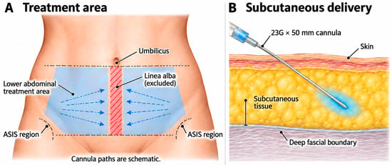
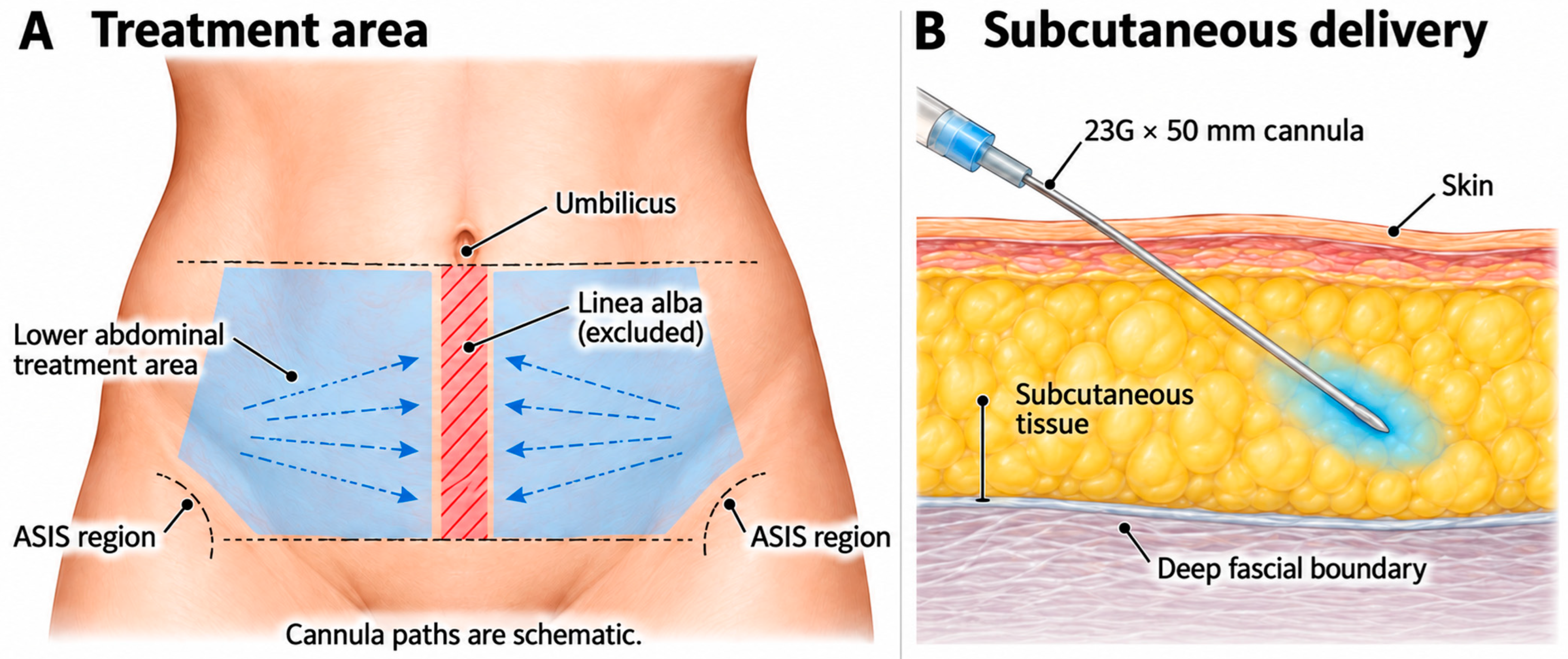
The source product was V-OLET injection (deoxycholic acid 20 mg/2 mL; Daewoong Pharmaceutical Co., Ltd., Seoul, Republic of Korea). Use in the lower abdomen was off-label relative to the product’s approved submental indication; the ethics approval information for this case series is reported in Section 2.1 and in the Institutional Review Board Statement. For each treatment, two 2 mL vials of DCA 10 mg/mL were used. Each vial was combined with 5 mL of L-carnitine solution and 3 mL of a clinic-prepared tumescent solution to produce 10 mL at a final DCA concentration of 2 mg/mL. Thus, each treatment delivered 40 mg of DCA in 20 mL of total injectate. The treatment zone was the lower abdomen below the umbilicus, extending bilaterally toward the anterior superior iliac spine region while excluding the linea alba. The diluted formulation was placed in the subcutaneous layer through bilateral cannula passes using a 23-gauge, 50 mm cannula. The source records did not provide a reproducible point-by-point dose map or calculated dose per square centimeter; therefore, these details are not reconstructed here.
The composition of the clinic-prepared tumescent solution is detailed in Table 1. The treatment area and subcutaneous cannula delivery are illustrated in Figure 1.
Table 1.
Composition of the clinic-prepared tumescent solution.
Figure 1.
Schematic of the lower-abdominal treatment area and subcutaneous cannula delivery. (A) Bilateral treatment areas below the umbilicus, with the linea alba excluded. Blue dashed arrows indicate schematic cannula paths and directions; the anterior superior iliac spine (ASIS) regions are shown for orientation. (B) Subcutaneous placement of a 23-gauge, 50 mm cannula above the deep fascial boundary. Cannula paths are schematic.
2.3. Outcome Assessments
Abdominal circumference was measured at the umbilical horizontal line, or at a consistently defined reference level, with the participant standing in the same position and after comfortable exhalation. Caliper thickness was measured at a consistent periumbilical site. The case-report form specifies millimeters for caliper values, and the present manuscript follows that unit. Ultrasound measurements were obtained at a consistent lower-abdominal location near the umbilicus. The recorded vertical ultrasound distance extended from the skin surface to the deep fascial boundary using on-screen calipers; this measurement may include skin thickness and cannot be assumed to represent adipose tissue alone. The available records do not establish the ultrasound device or transducer, examination position, precisely marked landmark, probe orientation or pressure, measurement repetition, operator identity or experience, or whether the same operator and location were used at baseline and follow-up. No intra-observer reliability assessment was documented. Baseline and final ultrasound images were selected to match the values in the study summary. Frontal clinical photographs were reviewed qualitatively. Image brightness and color were not altered; only cropping, layout assembly, and masking of identifiers were performed.
Pain was summarized using a 0–10 visual analog scale (VAS). The case-report form listed potential adverse events, including erythema, foreign-body sensation, nausea or vomiting, bruising, edema, pain, induration, nodules, fever, and nerve dysfunction. However, completed patient-level forms documenting event occurrence, severity, duration, management, and resolution by treatment session were not available for verification; infection and other systemic events likewise could not be systematically assessed.
2.4. Statistical Analysis
Because the sample comprised five cases and no control group, analyses were descriptive. Continuous variables are presented as individual values and mean ± standard deviation. Change was calculated as final follow-up minus baseline. Percentage reduction was calculated for caliper thickness and ultrasound fat depth for each case. No hypothesis testing was performed.
3. Results
3.1. Patient and Treatment Characteristics
The five participants had a mean age of 36.4 ± 4.8 years (range, 31–43 years). Each case underwent two sessions 4 weeks apart and had baseline and final measurements available. Baseline body weight ranged from 54.0 to 70.7 kg, caliper thickness from 35.4 to 46.3 mm, abdominal circumference from 79.5 to 99.0 cm, and ultrasound subcutaneous fat depth from 2.20 to 4.02 cm. Patient-level baseline and final outcomes are summarized in Table 2.
Table 2.
Patient-level baseline and final outcomes.
3.2. Objective Outcomes
Caliper thickness decreased in every case, with reductions ranging from 4.0 to 7.2 mm. The group means decreased from 41.14 ± 4.76 mm at baseline to 35.94 ± 5.15 mm at final follow-up. The mean absolute reduction was 5.20 ± 1.20 mm, and the mean individual relative reduction was 12.8% ± 3.5%. Longitudinal caliper measurements are shown in Table 3.
Table 3.
Longitudinal caliper thickness (mm).
Recorded ultrasound tissue depth also decreased in every case, with absolute reductions ranging from 0.61 to 0.98 cm. The group mean decreased from 3.22 ± 0.80 cm to 2.39 ± 0.81 cm. The mean absolute reduction was 0.83 ± 0.15 cm, and the mean individual relative reduction was 27.5% ± 10.1%.
By contrast, changes in body weight and abdominal circumference were inconsistent. Mean weight was 61.94 ± 6.34 kg at baseline and 61.86 ± 6.30 kg at final follow-up, a mean change of −0.08 ± 1.92 kg. Mean abdominal circumference was 86.70 ± 7.88 cm at baseline and 86.06 ± 4.99 cm at final follow-up, a mean change of −0.64 ± 3.08 cm. Two cases had increased final circumference despite reductions in both caliper and ultrasound thickness. Longitudinal circumference measurements are shown in Table 4.
Table 4.
Longitudinal abdominal circumference (cm).
3.3. Individual Case Summaries
Case 1. A 36-year-old woman maintained essentially stable weight (+0.1 kg). Caliper thickness decreased by 7.2 mm, abdominal circumference by 1.2 cm, and ultrasound depth by 0.91 cm (22.6%).
Case 2. A 33-year-old woman gained 2.4 kg and had a 1.5 cm increase in circumference. Despite these changes, caliper thickness decreased by 4.8 mm and ultrasound depth by 0.61 cm (18.9%).
Case 3. A 39-year-old woman lost 2.4 kg but had a 3.0 cm increase in circumference. Caliper thickness decreased by 4.8 mm and ultrasound depth by 0.98 cm (44.5%), the largest relative ultrasound reduction in the series.
Case 4. A 31-year-old woman gained 1.0 kg. Circumference decreased by 5.0 cm, caliper thickness by 5.2 mm, and ultrasound depth by 0.92 cm (23.1%).
Case 5. A 43-year-old woman lost 1.5 kg. Circumference decreased by 1.5 cm, caliper thickness by 4.0 mm, and ultrasound depth by 0.75 cm (28.1%).
3.4. Clinical Photographs and Ultrasound Findings
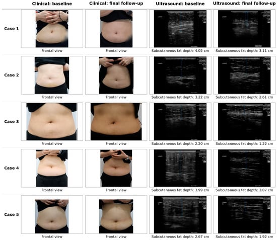
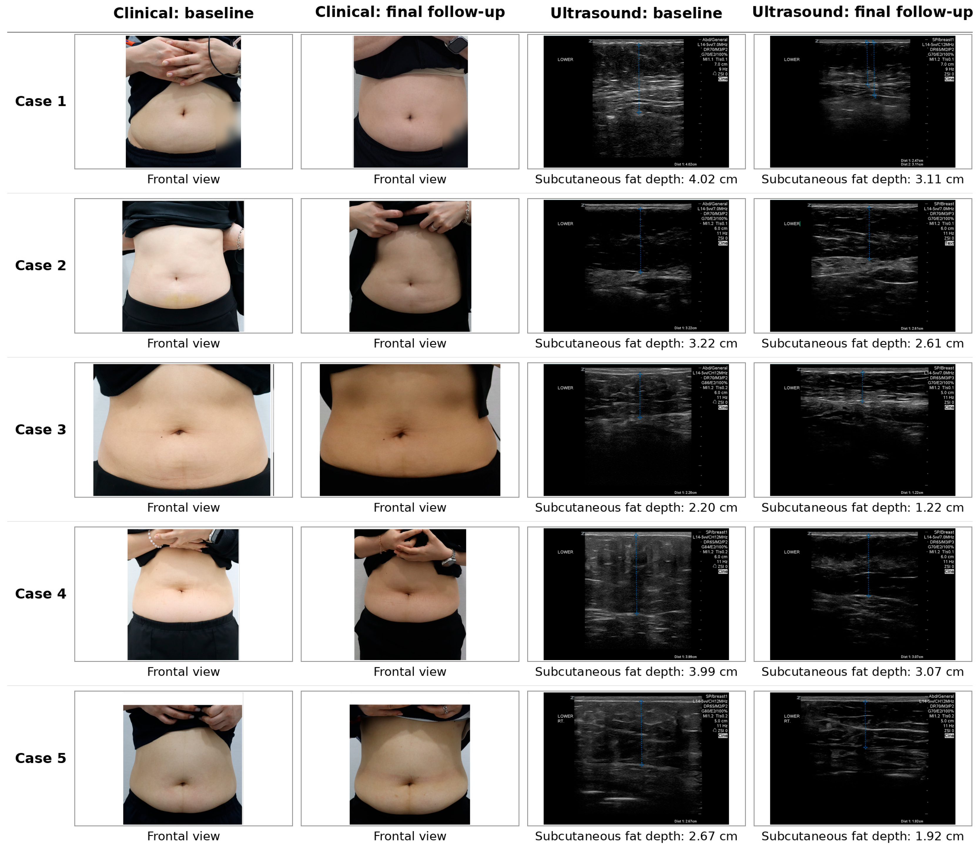
The descriptive composite presents the baseline and final frontal clinical photographs and the paired ultrasound images for all five cases (Figure 2). Differences in lighting, framing, magnification, posture, hand position, and possible abdominal muscle activation preclude reliable photographic comparisons of contour change. The paired ultrasound images illustrate the reported tissue-depth readings but do not establish measurement reproducibility.
Figure 2.
Clinical and ultrasound comparison before and after treatment in Cases 1–5. Cases are shown from top to bottom. The first two columns show frontal lower-abdominal photographs at baseline and final follow-up; the last two columns show baseline and final ultrasound images. Numeric labels reproduce the recorded skin-surface-to-deep-fascia distances in the study summary, which may include skin thickness. Ultrasound header identifiers and a potentially identifying skin mark were obscured. Photographs were cropped and assembled without color or brightness adjustment. Because lighting, framing, magnification, posture, hand position, and possible abdominal muscle activation were not standardized, photographs are descriptive only and do not constitute evidence of efficacy or quantitative contour improvement.
3.5. Available Safety Information and Documentation Limitations
The available study summary reported procedural pain scores of 0–1 on a 10-point visual analog scale. Complete patient-level adverse-event records were unavailable for verification. Therefore, the occurrence, severity, duration, management, and resolution of local or systemic adverse events could not be assessed systematically for each patient and treatment session. The absence of serious complications in the summary cannot be interpreted as confirmation that none occurred. These data do not establish the safety or tolerability of the off-label, combined injectable protocol.
4. Discussion
This five-patient series found consistent decreases in two localized thickness measures after a two-session, 2 mg/mL diluted DCA-based lower-abdominal protocol. Caliper thickness decreased by a mean of 5.2 mm, and recorded ultrasound tissue depth decreased by a mean of 0.83 cm. These findings occurred despite an essentially unchanged group mean body weight and variable circumference responses. Three cases had stable or increased body weight, yet all three demonstrated ultrasound reductions, supporting the interpretation that the recorded change was not fully explained by group-level weight change; however, nonstandardized acquisition limits causal interpretation.
The biologic plausibility of localized reduction is consistent with DCA-mediated adipocytolysis. Histologic work has shown that subcutaneous DCA produces focal membrane disruption, inflammatory cell recruitment, macrophage-mediated clearance, and subsequent septal remodeling [1,3]. However, abdominal application differs from the approved submental use in surface area, anatomy, total injectate volume, and potential exposure. Prior abdominal studies have used higher total volumes and have reported frequent but self-limited local edema, bruising, and erythema [5,6]. The reported pain scores cannot establish tolerability or safety in the absence of complete patient-level adverse-event documentation.
The present concentration of 2 mg/mL is lower than the 2.5 and 5 mg/mL concentrations reported in a four-patient upper-arm series [7]. That study also observed greater consistency in ultrasound thickness than in circumference, a pattern echoed here. Circumference is influenced by respiration, posture, intra-abdominal contents, visceral fat, and landmark placement, whereas caliper and ultrasound assessments more directly sample local subcutaneous tissue. The transient increases in caliper thickness at some interim visits may reflect measurement variability, post-treatment tissue response, or both.
A major interpretive limitation is that the injectate was not DCA alone. It included L-carnitine and a tumescent anesthetic solution. L-carnitine has shown lipolytic and fatty-acid-oxidation effects in cultured adipocytes, but this does not establish a clinically meaningful effect after local injection in humans [8]. The relative contributions of DCA, L-carnitine, tumescent fluid, cannula technique, and any unrecorded concomitant treatment therefore cannot be separated. For this reason, the title and conclusions describe a DCA-based protocol rather than attributing the outcome exclusively to DCA.
Additional limitations include the very small all-female sample, absence of a comparator, lack of blinded assessment, short follow-up, incomplete medical and treatment-history data, unverified ultrasound equipment and acquisition conditions, lack of repeated measures or intra-observer reliability data, possible inclusion of skin in the ultrasound distance, inconsistent photographic acquisition, and missing patient-level safety and satisfaction forms. The series also lacks a standardized treatment-area map and dose per square centimeter, limiting reproducibility. Finally, the abdominal use was off-label and requires careful ethical, regulatory, and consent documentation.
A staged research program is proposed. First, a prospectively designed feasibility study should define eligibility criteria, anatomical treatment boundaries, injection technique, formulation composition, dose per treatment area, and prespecified feasibility and safety endpoints. Appropriate ethics and regulatory review of off-label use should be completed before any prospective research procedures. Individual adverse events should be collected after every session with severity, onset, duration, treatment, and resolution documented. Ultrasound measurements should use a prespecified device and transducer, reproducible marked landmarks, consistent patient positioning, minimal and controlled probe compression, multiple repeated measurements, and blinded or reliability-assessed readings. Photographs should be acquired with fixed lighting, distance, posture, and framing. If feasibility and safety are adequately demonstrated, a larger controlled study with a suitable vehicle or comparator group, longer follow-up, and a design able to isolate the contribution of L-carnitine could evaluate treatment-related effects. These proposals concern future research and were not procedures performed in the present series.
5. Conclusions
Two sessions of a 2 mg/mL diluted DCA-based formulation were followed by reductions in caliper thickness and ultrasound-measured lower-abdominal tissue depth (skin surface to deep fascia) in all five cases. Body weight and circumference were less consistent, and the combined formulation and uncontrolled design prevent causal attribution to DCA alone [9]. These observations support further controlled evaluation but are not sufficient to establish efficacy or safety for routine lower-abdominal use.
Author Contributions
K.-T.B.: investigation, clinical case management, resources, data provision, and manuscript review. K.-H.Y.: manuscript strategy, data review and analysis, and writing—original draft. J.H.K.: journal selection, project administration, and conceptualization. J.H.K. and K.-H.Y. are the corresponding authors. All authors have read and agreed to the published version of the manuscript.
Funding
Daewoong Pharmaceutical Co., Ltd. provided the deoxycholic acid (DCA) vials used in this study and financial support for medical writing services for this manuscript.
Institutional Review Board Statement
The study was reviewed and approved by the Public Institutional Review Board designated by the Ministry of Health and Welfare, Republic of Korea (Approval No. P01-202603-01-017; approval date: 24 March 2026). The study was conducted in accordance with the Declaration of Helsinki.
Informed Consent Statement
Written informed consent for publication has been obtained from the participating patients.
Data Availability Statement
De-identified data may be made available from the corresponding author on reasonable request, subject to clinic approval, patient privacy protections, and the data-use agreement.
Conflicts of Interest
Kyung-Tae Bae is advisory board member of Daewoong Pharmaceutical.
Abbreviations
| DCA | Deoxycholic acid |
| VAS | Visual analog scale |
References
- Deeks, E.D. Deoxycholic acid: A review in submental fat contouring. Am. J. Clin. Dermatol. 2016, 17, 701–707. [Google Scholar] [CrossRef] [Scilit] [PubMed]
- Ascher, B.; Fellmann, J.; Monheit, G. ATX-101 (deoxycholic acid injection) for reduction of submental fat. Expert Rev. Clin. Pharmacol. 2016, 9, 1131–1143. [Google Scholar] [CrossRef] [Scilit] [PubMed]
- Walker, P.S.; Lee, D.R.; Toth, B.A.; Bowen, B. Histological analysis of the effect of ATX-101 (deoxycholic acid injection) on subcutaneous fat: Results from a phase 1 open-label study. Dermatol. Surg. 2020, 46, 70–77. [Google Scholar] [CrossRef] [Scilit] [PubMed]
- Ascher, B.; Hoffmann, K.; Walker, P.; Lippert, S.; Wollina, U.; Havlickova, B. Efficacy, patient-reported outcomes and safety profile of ATX-101 (deoxycholic acid), an injectable drug for the reduction of unwanted submental fat: Results from a phase III, randomized, placebo-controlled study. J. Eur. Acad. Dermatol. Venereol. 2014, 28, 1707–1715. [Google Scholar] [CrossRef] [Scilit] [PubMed]
- Salvador, L.; Benito-Ruiz, J. Safety and efficacy of deoxycholic acid injection for hypogastric fat reduction: A pilot study. Dermatol. Surg. 2021, 47, e132–e137. [Google Scholar] [CrossRef] [Scilit] [PubMed]
- Shridharani, S.M.; Tisch, G.M.; Kennedy, M.L. Injection adipocytolysis for body and jawline contouring: Real-world experience and treatment considerations. Aesthetic Surg. J. 2023, 43, 470–483. [Google Scholar] [CrossRef] [Scilit] [PubMed]
- Hong, J.Y.; Lee, Y.H.; Yeoum, S.H.; Youn, C.S.; Park, K.Y. The effect of diluted deoxycholic acid on arm fat reduction: Evaluation of its potential in minimally invasive fat loss treatment. J. Cosmet. Dermatol. 2025, 24, e70088. [Google Scholar] [CrossRef] [Scilit] [PubMed]
- Lee, M.S.; Lee, H.J.; Lee, H.S.; Kim, Y. L-carnitine stimulates lipolysis via induction of the lipolytic gene expression and suppression of the adipogenic gene expression in 3T3-L1 adipocytes. J. Med. Food 2006, 9, 468–473. [Google Scholar] [CrossRef] [Scilit] [PubMed]
- Daewoong Pharmaceutical Co., Ltd. V-OLET® Injection (Deoxycholic Acid 20 mg/2 mL): Prescribing Information; Daewoong Pharmaceutical Co., Ltd.: Seoul, Republic of Korea, 2021. [Google Scholar]

Disclaimer/Publisher’s Note: The statements, opinions and data contained in all publications are solely those of the individual author(s) and contributor(s) and not of MDPI and/or the editor(s). MDPI and/or the editor(s) disclaim responsibility for any injury to people or property resulting from any ideas, methods, instructions or products referred to in the content. |
© 2026 by the authors. Licensee MDPI, Basel, Switzerland. This article is an open access article distributed under the terms and conditions of the Creative Commons Attribution (CC BY) license.