


Abstract
Histone H2A family member X phosphorylated at serine 139 (γH2AX) is a marker of the cellular response to DNA double-strand breaks. To our knowledge, this study reports the detection of γH2AX by flow cytometry in equine spermatozoa and provides preliminary evidence through its association with DNA status n and apoptotic-like changes. A total of 64 semen samples from 12 stallions were analyzed under routine field conditions. Sperm viability, mitochondrial membrane potential, Annexin V positivity, and γH2AX levels were assessed using multiparametric flow cytometry. γH2AX showed a wide distribution among samples and was correlated with Annexin V positivity and PI positivity. Linear mixed-effects models, including stallion as a random effect, identified significant associations between γH2AX levels and both Annexin V positivity and PI positivity. In contrast, γH2AX was not significantly related to sperm viability or mitochondrial membrane potential. These findings indicate that γH2AX-associated fluorescence can be detected in equine spermatozoa and is associated with indicators of apoptotic-like changes and DNA instability, which may be related to nuclear DNA damage rather than general sperm functional status. The observed associations suggest that γH2AX may have potential as an additional parameter in multiparametric flow cytometry approaches for the assessment of sperm nuclear status in stallions. However, further validation against established DNA damage assays and appropriate specificity controls is required to determine whether γH2AX fluorescence provides a specific measure of sperm nuclear DNA damage.
1. Introduction
Flow cytometry has become an essential tool in reproductive biology, enabling rapid, objective, and multiparametric evaluation of sperm quality. Unlike conventional semen analysis, this technique allows the simultaneous assessment of thousands of spermatozoa at the single-cell level [1,2], providing detailed information on functional and structural heterogeneity within sperm populations [3]. In equine reproduction, where artificial insemination with fresh or chilled semen is widely used, accurate assessment of sperm quality is critical for optimizing fertility outcomes. However, traditional parameters such as motility and morphology do not fully reflect the functional status or fertilizing potential of spermatozoa [4,5].
The application of flow cytometry in equine andrology has expanded considerably, allowing the evaluation of key cellular features including membrane integrity, mitochondrial activity, DNA structure, and apoptotic-like changes [3,6]. Fluorescent probes such as SYBR-14, propidium iodide, PNA-FITC, and JC-1 enable the simultaneous analysis of viability, acrosomal integrity, and mitochondrial function [7]. Multiparametric approaches further allow the simultaneous evaluation of functional parameters such as mitochondrial activity, membrane integrity, apoptosis, and DNA damage [3,8]. These approaches provide a more comprehensive understanding of sperm physiology and reveal sublethal alterations that are not detectable by conventional methods. Nevertheless, the assessment of sperm DNA integrity in equine species remains limited, and conventional semen evaluation does not adequately reflect nuclear status. The incorporation of novel biomarkers could therefore improve the characterization of sperm functional competence [3,4,5].
DNA damage in spermatozoa has been widely associated with impaired fertility, defective embryo development, increased pregnancy loss, and long-term effects on offspring health [5,9,10,11]. Sperm DNA fragmentation, defined as the presence of single- or double-strand breaks, represents a key parameter for evaluating male fertility potential and has been extensively assessed using assays such as the Sperm Chromatin Structure Assay (SCSA), Terminal deoxynucleotidil Transferase dUTP Nick End Labeling (TUNEL), and single-cell gel electrophoresis (COMET) [12]. In mammalian sperm, DNA damage is closely linked to oxidative stress, defective DNA remodeling, and apoptosis-like processes [13,14]. In addition, cytometric approaches have been widely used to characterize apoptotic pathways, including DNA fragmentation, mitochondrial dysfunction, and membrane alterations, highlighting their value for studying sperm cell fate at the single-cell level [1]. These findings underscore the importance of evaluating nuclear integrity alongside conventional functional parameters.
Phosphorylation of histone H2AX (γH2AX) is a well-established marker of DNA double-strand breaks and is widely used in cell biology to detect DNA damage at the single-cell level. The formation of γH2AX foci occurs rapidly in response to DNA double-strand breaks, making it a sensitive indicator of genomic instability [15]. In human spermatozoa, γH2AX has been successfully detected by flow cytometry and has been proposed as a biomarker of DNA damage, showing significant associations with semen quality parameters and DNA fragmentation levels [16]. Moreover, Coban et al., who assessed γH2AX immunofluorescence in human spermatozoa, concluded that γH2AX levels were associated with live-birth outcomes following IVF but were not correlated with conventional semen parameters or male age [17]. These findings suggest that γH2AX may provide valuable information on sperm nuclear integrity beyond traditional assessments.
Recently, γH2AX immunolabelling has also been applied to assess DNA damage in paternal pronuclei derived from frozen–thawed equine spermatozoa following xenogeneic ICSI, demonstrating increased DNA damage particularly in pronuclei originating from immotile spermatozoa [18]. However, to our knowledge, γH2AX has not previously been evaluated in equine spermatozoa by flow cytometry. Therefore, the aim of the present study was to report the detection of γH2AX by flow cytometry in equine spermatozoa and to evaluate its biological relevance through its association with established sperm functional and DNA parameters [18].
2. Materials and Methods
2.1. Semen Collection and Processing
Ejaculates were collected from twelve stallions aged between 5 and 19 years, including Pura Raza Española, Arabian, Anglo-Arabian, Hispano-Arabian, and Spanish Sport Horse (CDE) breeds, during the 2022 and 2024 breeding seasons. A total of 64 semen samples were obtained through repeated collections, reflecting typical field conditions. All stallions were maintained at the CENSYRA (Centro de Selección y Reproducción Animal, Badajoz, Spain). The interval between successive semen collections from the same stallion ranged from 48 to 72 h, according to the routine breeding activities at CENSYRA.
Semen was collected using a pre-warmed (45 °C) artificial vagina (INRA model; IMV, L’Aigle, France) equipped with a filter to remove the gel fraction. Ejaculates were maintained at 37 °C, and sperm concentration was determined using a hemocytometer (Makler® chamber, Trumbull, CT, USA).
Ejaculate volume ranged approximately from 60 to 120 mL, depending on the stallion. Before dilution, sperm concentration was measured using a hemocytometer and ranged from 73 to 518 × 106 spermatozoa/mL. Motility data were available for a subset of samples from four stallions. At the initial assessment (0 h), mean total and progressive motility were 84.0 ± 7.7% and 53.7 ± 11.7%, respectively (mean ± SD).
Each ejaculate was divided into aliquots and extended to a final concentration of 25 × 106 spermatozoa/mL using commercial extenders (INRA 96® [IMV, L’Aigle, France], Equiplus® [Minitube, Tiefenbach, Germany], Beyond® [Minitube], or INRA 96® after centrifugation at 600× g for 10 min). Two aliquots of each extended semen sample were transported to the Research Support Services (STAB) of the University of Extremadura on the day of collection in insulated expanded polystyrene boxes containing cold packs. One aliquot was processed upon arrival, with the initial evaluation performed within 3 h of semen collection; this assessment was designated as 0 h. The second aliquot was stored in a refrigerator at 5 °C at STAB and evaluated approximately 24 h after the initial assessment.
2.2. Flow Cytometry Analysis
Flow cytometry analyses were performed at STAB. The following probes were used: Hoechst 33258 (5 µM, 10 min, room temperature [Thermo Fisher Scientific/eBioscience, Waltham, MA, USA]) to identify non-viable spermatozoa, Tetra Methyl Rhodamine Methylesther (TMRM) (100 nM, 20 min, 37 °C [Thermo Fisher Scientific/eBioscience]) to assess mitochondrial membrane potential, Annexin V-FITC ([Beckman Coulter, Brea, CA, USA] 5 µL per sample, 15 min, room temperature) to detect phosphatidylserine externalization, and propidium iodide (PI) ([Thermo Fisher Scientific/eBioscience] 5–6 µM, added immediately before acquisition) to assess membrane integrity. An aliquot of each sample was fixed and permeabilized (Fix & Perm kit, Beckman Coulter) according to the manufacturer’s instructions. Intracellular staining was performed using a monoclonal antibody directed against phospho-H2AX (Ser139; clone CR55T33; Cat. No. 14-9865-80; Thermo Fisher Scientific/eBioscience). Following incubation with the primary antibody, fluorescence was detected using a goat anti-mouse IgG (H+L) secondary antibody conjugated to DyLight 549 (Thermo Fisher Scientific; Cat. No. 35507). The specificity of the detected antibody-associated fluorescence signal was not independently validated in equine spermatozoa.
Data acquisition was performed using a MACSQuant VYB flow cytometer (Miltenyi Biotec, Bensberg, Germany), recording at least 10,000 events per sample, and data were analyzed using FlowLogic™ software Version 8.6 (Inivai Technologies, Mentone, Australia). Spermatozoa were identified based on forward and side scatter properties, and doublets were excluded using FSC-A/FSC-H parameters. Staining controls were used to establish positivity thresholds and compensation, and a secondary-antibody-only control was included for the phospho-H2AX assay.
Instrument settings were applied throughout the study, following established methodological guidelines [19].
All parameters were expressed as percentage of the total sperm population.
2.3. Statistical Analysis
Statistical analyses were performed using Jamovi software (version 2.6.44). Data are presented as mean ± standard deviation and median, and normality was assessed using visual inspection and statistical tests. Pearson correlation coefficients were calculated to evaluate the relationships between γH2AX levels and sperm parameters, including viability (Live Total), mitochondrial membrane potential measured with TMRM, Annexin V positivity, and PI-based fluorescence associated with DNA decondensation.
To account for repeated measures, linear mixed-effects models were constructed with γH2AX as the dependent variable. Annexin V positivity and PI-positivity were included as fixed effects, and stallion was included as a random effect. Additional models were performed to evaluate the association between γH2AX and individual sperm functional parameters, including viability and mitochondrial membrane potential. Model assumptions were verified by visual inspection of residuals, and model fit was assessed using marginal and conditional R2 values. Statistical significance was set at p < 0.05.
Samples obtained under different extender conditions and at different time points were included to reflect field conditions and increase biological variability. These observations were treated as repeated measures, and non-independence was accounted for by including stallion as a random effect.
Ethical Statement
All procedures were carried out under standard management practices and in accordance with national animal welfare regulations.
3. Results
3.1. Descriptive Analysis
A total of 64 samples were analyzed for sperm functional and nuclear parameters. Sperm viability (Live Total) showed a mean value of 52.9 ± 23.2%, while mitochondrial membrane potential (TMRM) averaged 40.0 ± 23.9%. Annexin V positivity presented a mean of 21.3 ± 24.7%, indicating a wide variability in apoptotic-like changes across samples.
γH2AX levels displayed a broad range (2.74–85.9%), with a mean of 22.7 ± 20.9% and a moderately skewed distribution (median: 15.8%), indicating variability among semen samples in the proportion of spermatozoa classified as positive for the anti-phospho-H2AX antibody. Similarly, the percentage of uncondensed nuclei showed considerable variability (20.5 ± 13.1%; median: 18.5%), supporting heterogeneity in DNA status within the analyzed samples (Table 1).
Table 1.
Descriptive statistics of sperm functional and nuclear parameters. Data are presented as mean ± standard deviation (SD) and median. γH2AX and DNA decondensation were evaluated in all samples.
3.2. Correlation Analysis
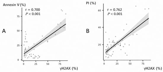
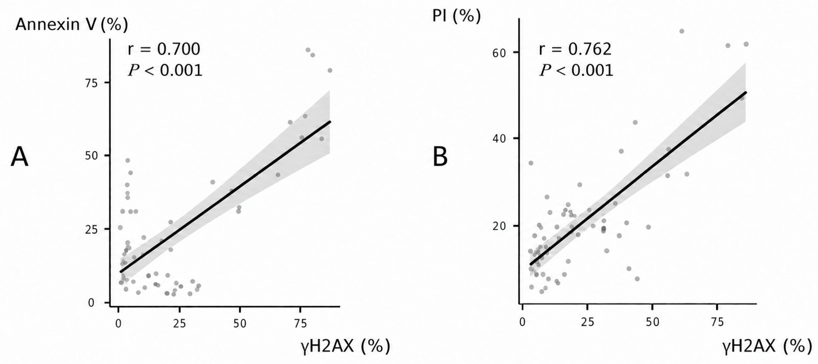
At the semen-sample level, the proportion of spermatozoa classified as positive for the anti-phospho-H2AX antibody was positively correlated with Annexin V positivity (r = 0.700, p < 0.001), as well as with PI-positivity (r = 0.762, p < 0.001), supporting a possible relationship between DNA integrity and apoptotic-like changes. In the linear mixed-effects model including stallion as a random effect, both Annexin V positivity (β = 0.315 ± 0.094, p = 0.002) and PI positivity (β = 0.806 ± 0.173, p < 0.001) were associated with the antibody-derived signal. In contrast, weaker inverse relationships were observed between γH2AX and sperm viability (Live Total) and mitochondrial membrane potential (TMRM); however, these associations were not statistically significant. These associations concern measurements obtained from semen samples and do not establish marker co-occurrence within individual spermatozoa.
3.3. Linear Mixed Model Analysis
To account for the non-independence of observations, a linear mixed model was performed including stallion as a random effect. The model demonstrated a high explanatory power (marginal R2 = 0.852; conditional R2 = 0.876; p < 0.001), indicating that the included variables explained a substantial proportion of the variability in γH2AX levels.
Both Annexin V positivity and PI positivity were significantly associated with γH2AX after adjusting for stallion variability. Annexin V showed a positive effect (β = 0.315 ± 0.094, p = 0.002), while PI showed a stronger association (β = 0.806 ± 0.173, p < 0.001). The variance attributable to stallion was moderate (ICC = 0.165), suggesting that most of the variation in γH2AX was explained by the measured cellular parameters rather than individual differences (Table 2).
Table 2.
Results of the linear mixed-effects model evaluating the association between γH2AX levels and sperm parameters. Annexin V positivity and PI positivity were included as fixed effects, and stallion was included as a random effect. β: regression coefficient; SE: standard error; CI: confidence interval.
3.4. Additional Mixed Model Analyses
Additional linear mixed models showed that γH2AX was not significantly associated with sperm viability (Live Total; p = 0.969) or mitochondrial membrane potential (TMRM; p = 0.211), providing no evidence of associations between the antibody-derived fluorescence signal and these two functional parameters in the models fitted.
3.5. Graphical Analysis
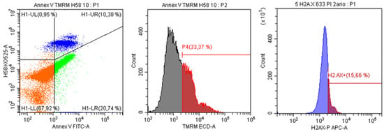
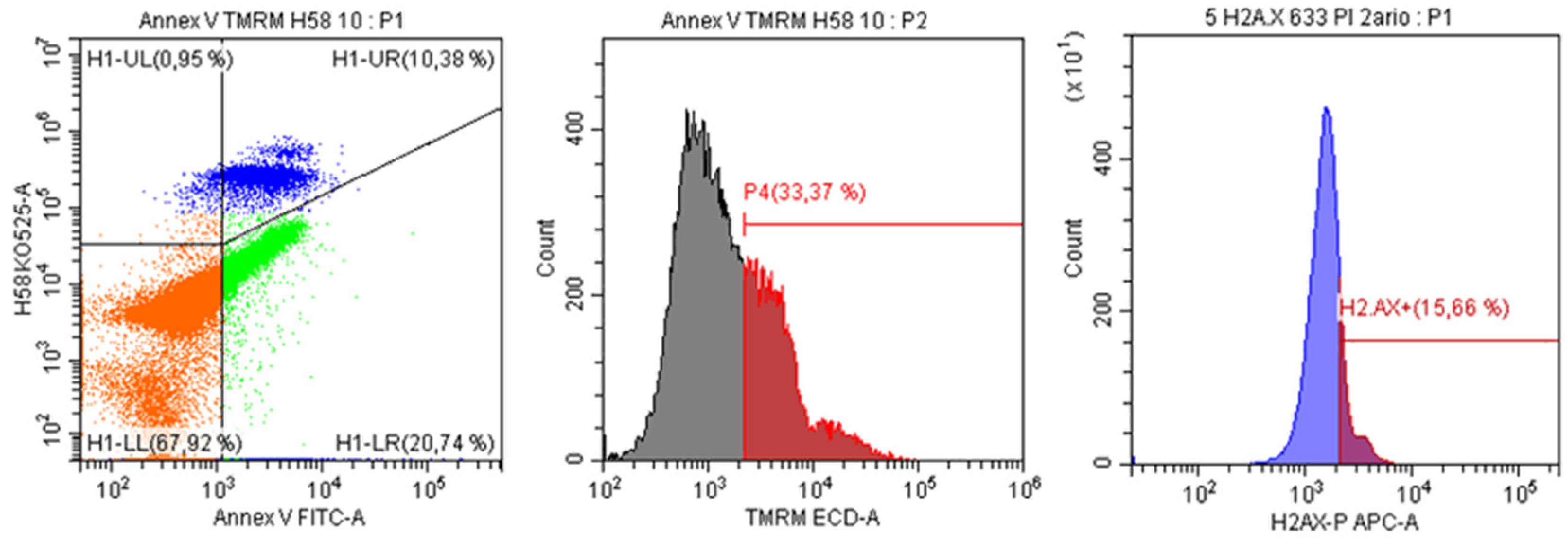
Scatter plot analysis confirmed the statistical associations observed. A positive relationship was identified between γH2AX levels and Annexin V positivity, as well as between γH2AX and PI positivity. In contrast, no clear relationship was observed between γH2AX and sperm viability or mitochondrial membrane potential. These graphical patterns illustrate the sample-level associations between antibody positivity, Annexin V positivity, and PI positivity and possibly the proportion of uncondensed nuclei (Figure 1). Representative flow cytometry plots illustrating the gating strategy and identification of γH2AX-positive spermatozoa are presented in Figure 2.
Figure 1.
Scatter plots showing the relationships between γH2AX and markers of sperm quality in equine spermatozoa. (A) Relationship between γH2AX and Annexin V positivity. (B) Relationship between γH2AX and PI positivity. The horizontal axis represents γH2AX-positive spermatozoa (%) in both panels. The vertical axis represents Annexin V-positive spermatozoa (%) in panel A and PI-positive spermatozoa (%) in panel B. Pearson correlation coefficients were r = 0.700 for panel A and r = 0.762 for panel B (both p < 0.001).
Figure 2.
Representative flow cytometry analysis of equine spermatozoa. The dot plot illustrates Annexin V-FITC and TMRM staining for the identification of viable (non-stained H1-LL orange), early apoptotic (Annexin V positive/Hoechst 33258 negative H1-LR green), debris (Hoechst 33258 positive/Annexin V negative H1-UL blue) and late apoptotic/dead (Hoechst 33258 positive/Annexin V positive H1-UR blue) sperm populations. Histograms show TMRM fluorescence, reflecting mitochondrial membrane potential, and anti-phospho-H2AX antibody-associated fluorescence percentages indicate the proportion of positive cells within each gated population.
4. Discussion
To our knowledge, this study provides the first report of anti-phospho-H2AX antibody-associated fluorescence detected by flow cytometry in equine spermatozoa, with sample-level probable associations between antibody positivity, Annexin V positivity, PI positivity and therefore the proportion of uncondensed nuclei. The ability to detect γH2AX in equine sperm and its integration within a multiparametric flow cytometry approach may represent a novel methodological and biological contribution to equine andrology.
Flow cytometry has been widely used in equine reproduction to assess sperm functional integrity, including membrane status, mitochondrial activity, DNA structure, and apoptotic-like changes [3,6]. However, the incorporation of markers directly related to DNA damage remains limited. In this context, anti-phospho-H2AX staining may provide an additional parameter for investigating sperm nuclear status, although its interpretation as a specific indicator of DNA double-strand breaks in equine spermatozoa requires further validation. This approach complements existing functional assessments and provides a more comprehensive characterization of sperm quality.
Higher γH2AX levels were observed in association with Annexin V positivity, indicating a possible relationship between DNA damage and apoptotic-like changes in equine spermatozoa. This finding agrees with previous reports in mammalian sperm linking DNA fragmentation to apoptosis-related processes and membrane alterations [13,14]. In particular, early apoptotic spermatozoa identified by Annexin V binding have been associated with increased DNA fragmentation and reduced functional competence [20]. The sample-level association with Annexin V positivity suggests a possible relationship with apoptotic-like changes, but does not establish co-occurrence within individual spermatozoa. This finding suggests that DNA double-strand breaks, as detected by γH2AX, may arise as a downstream consequence of oxidative stress and defective DNA remodeling in spermatozoa. Oxidative stress and altered DNA remodeling are possible explanations that require investigation; the present data do not establish the mechanisms underlying the observed associations.
Interestingly, Rychtarova et al. [18] recently demonstrated that frozen–thawed immotile equine spermatozoa frequently produced paternal pronuclei with extensive γH2AX labelling following xenogeneic ICSI, whereas pronuclei derived from motile spermatozoa showed little or no detectable DNA damage. This xenogeneic ICSI approach had previously been proposed as a method for assessing sperm DNA damage through γH2AX evaluation in sperm-derived paternal pronuclei [21]. Although their experimental approach differs substantially from the direct flow-cytometric assessment used here, these findings further demonstrate that γH2AX can reveal biologically relevant nuclear damage in equine sperm and highlight the importance of assessing nuclear integrity independently of conventional sperm quality parameters [18]. While previous studies in other species have reported associations between sperm DNA damage and reduced viability or mitochondrial function [9,13,22], the present findings indicate that γH2AX is more closely linked to nuclear instability in equine spermatozoa.
The linear mixed model analysis further supports the robustness of these associations, demonstrating that Annexin V, PI and possibly DNA decondensation can consist on significant explanations for the variability in γH2AX levels independently of stallion variability. The associations remained statistically significant after including stallion as a random effect; however, their consistency across ages, breeds, and management conditions requires further investigation.
Importantly, the study included samples obtained under different conditions, including multiple extenders and two time points (0 h and 24 h), which may have contributed to the variability observed in sperm quality parameters. However, the objective of the present study was not to compare extenders or storage conditions, but rather to determine whether γH2AX could be reliably detected across a heterogeneous population of equine sperm samples displaying a wide range of functional and DNA characteristics. From this perspective, the inclusion of samples processed under different field conditions increased biological variability and allowed the evaluation of γH2AX under scenarios commonly encountered in routine equine reproduction practice. Rather than representing a limitation, this heterogeneity may strengthen the findings, as it suggests that the associations between γH2AX and sperm parameters are maintained across a range of physiological and processing conditions. These findings highlight the value of incorporating DNA damage markers into multiparametric flow cytometry approaches for a more comprehensive evaluation of sperm quality.
The use of γH2AX as a biomarker of DNA damage has been well established in somatic cells; it has been explored in human spermatozoa, where it has been associated with DNA fragmentation and semen quality [16]; as well as live-birth outcomes following IVF procedures [17]. The present study extends these findings to equine sperm, supporting the applicability of this marker in veterinary reproductive research. However, further studies are needed to determine its relationship with fertility outcomes and to compare it with established DNA fragmentation assays such as TUNEL or SCSA.
Some limitations of this study should be acknowledged. Although the number of stallions included (n = 12) is within the range commonly reported in equine reproductive studies, γH2AX detection was not directly validated against established sperm DNA damage assays such as TUNEL or SCSA. Therefore, while associations were observed between PI positivity and Annexin V positivity, these findings should be interpreted cautiously, as the specificity of CR55T33 binding in equine spermatozoa was not independently established. Although the antibody is characterized by the manufacturer as recognizing phospho-H2AX (Ser139), this characterization alone cannot confirm the identity of the fluorescence signal detected in the present specimens. Moreover, DNA decondensation may increase epitope accessibility and contribute to the measured signal. Thus, the association between antibody-derived fluorescence and PI fluorescence associated with sperm DNA status cannot distinguish increased H2AX phosphorylation from increased antibody accessibility, or a contribution from both. Because the endpoints were assessed in separate aliquots, their sample-level associations do not demonstrate co-occurrence within individual spermatozoa or establish a shared cellular mechanism.
The extent to which γH2AX reflects the same biological dimension assessed by conventional DNA fragmentation tests remains to be determined. In addition, no direct fertility outcomes were available for statistical analysis, and thus the predictive value of γH2AX for reproductive performance could not be assessed. Nevertheless, all stallions included in the study were routinely used in commercial artificial insemination programs at a specialized equine reproduction center, reflecting a population of reproductively active and clinically fertile animals. Samples were obtained under routine field conditions using different extenders and storage times, which may have contributed to variability in sperm quality parameters. However, this heterogeneity also enhances the practical relevance of the findings, as the observed associations were maintained across a range of conditions commonly encountered in equine reproduction practice. Future studies should validate the specificity of the anti-phospho-H2AX antibody signal in equine spermatozoa using appropriate controls and compare it with established DNA damage assays, such as TUNEL and the Comet assay. The influence of DNA decondensation on antibody accessibility should also be investigated. Combined single-cell analyses would help determine whether the fluorescence signal co-occurs with apoptotic-like changes within individual spermatozoa. Once analytical validity has been established, studies should assess its potential associations with fertilization, embryo development, and pregnancy outcomes. The biological relevance of γH2AX-detectable damage in equine sperm is further supported by recent xenogeneic ICSI experiments, in which increasing paternal γH2AX labelling was accompanied by a higher incidence of micronuclei and, in severely affected samples, impaired cleavage to the two-cell stage [18].
In conclusion, a flow cytometric fluorescence signal was obtained in equine spermatozoa using an antibody directed to γH2AX (Ser139). Across semen samples, the proportion of antibody-positive spermatozoa was associated with the proportions positive for Annexin V and PI, whereas no significant association was found with viability or mitochondrial membrane potential. These exploratory findings require validation of signal specificity in equine sperm and combined single-cell assessment before the biological basis of the associations or the potential value of this assay as a DNA-damage marker can be established. Further studies comparing γH2AX with established DNA fragmentation assays and fertility outcomes are warranted to determine its potential value as a complementary biomarker in stallion fertility assessment.
Author Contributions
Conceptualization: C.M.B.d.S., E.M.-P. Methodology: E.M.-P., C.M.B.d.S. Investigation: E.M.-P., C.M.B.d.S., V.G.-A., A.Á.-B. Writing—Original Draft: C.M.B.d.S., E.M.-P., A.Á.-B. Writing—Review & Editing: C.M.B.d.S., E.M.-P., M.M.-C. Supervision: E.M.-P. All authors have read and agreed to the published version of the manuscript.
Funding
This research was funded by Fundação para a Ciência e a Tecnologia (grant UID/05064/2025—https://doi.org/10.54499/UID/05064/2025).
Institutional Review Board Statement
Ethical review and approval as an animal experimentation project were not required because the study was limited to the laboratory analysis of semen samples obtained during routine, non-experimental reproductive activities. No procedures were performed on animals specifically for research purposes. This activity falls outside the scope of Royal Decree 53/2013 of 1 February, particularly Article 2(5)(b), (d), and (f).
Informed Consent Statement
Prior written authorization for the scientific use of the semen samples and associated anonymized data was obtained from the relevant associations. No information allowing the identification of individual owners or holdings is reported.
Data Availability Statement
The data supporting the findings of this study are available from the corresponding author upon reasonable request.
Conflicts of Interest
The authors declare no conflict of interest.
References
- Wlodkowic, D.; Telford, W.; Skommer, J.; Darzynkiewicz, Z. Apoptosis and beyond: Cytometry in studies of programmed cell death. Methods Cell Biol. 2011, 103, 55–98. [Google Scholar] [CrossRef] [Scilit] [PubMed]
- Petrunkina, A.M.; Harrison, R.A. Cytometric solutions in veterinary andrology. Cytom. Part A 2011, 79, 338–348. [Google Scholar] [CrossRef] [Scilit] [PubMed]
- Peña, F.J.; Ortega-Ferrusola, C.; Martín-Muñoz, P. New flow cytometry approaches in equine andrology. Theriogenology 2016, 86, 366–372. [Google Scholar] [CrossRef] [Scilit] [PubMed]
- Love, C.C. Relationship between sperm motility, morphology, and the fertility of stallions. Theriogenology 2011, 76, 547–557. [Google Scholar] [CrossRef] [Scilit] [PubMed]
- Evenson, D.P.; Wixon, R. Clinical aspects of sperm DNA fragmentation detection and male infertility. Theriogenology 2006, 65, 979–991. [Google Scholar] [CrossRef] [Scilit] [PubMed]
- Ortega-Ferrusola, C.; García, B.M.; Gallardo-Bolaños, J.M.; González-Fernández, L.; Rodríguez-Martínez, H.; Tapia, J.A.; Peña, F.J. Apoptotic markers can be used to forecast the freezeability of stallion spermatozoa. Anim. Reprod. Sci. 2009, 114, 393–403. [Google Scholar] [CrossRef] [Scilit] [PubMed]
- Robles, V.; Martínez-Pastor, F. Flow cytometric methods for sperm assessment. In Spermatogenesis: Methods and Protocols; Carrell, D.T., Aston, K.I., Eds.; Springer: Berlin/Heidelberg, Germany, 2013; pp. 175–186. [Google Scholar] [CrossRef] [Scilit] [PubMed]
- da Silva, C.M.B.; Cano, F.E.M.; Gaitskell-Phillips, G.; Vega, F.J.P. Multiparametric flow cytometry for determination of viability, caspase activity, and lipid peroxidation in equine spermatozoa. Curr. Protoc. 2023, 3, e885. [Google Scholar] [CrossRef] [Scilit] [PubMed]
- Sedó, C.A.; Bilinski, M.; Lorenzi, D.; Uriondo, H.; Noblía, F.; Longobucco, V.; Chemes, H. Effect of sperm DNA fragmentation on embryo development: Clinical and biological implications. Fertil. Steril. 2017, 107, 847–854. [Google Scholar] [CrossRef] [Scilit] [PubMed]
- Alahmar, A.T. Sperm DNA fragmentation: A review. Reprod. Biol. Endocrinol. 2022, 20, 122. [Google Scholar] [CrossRef] [Scilit] [PubMed]
- Esteves, S.C.; Zini, A.; Coward, R.M.; Evenson, D.P.; Gosálvez, J.; Lewis, S.E.M.; Sharma, R.; Agarwal, A. The assessment of sperm DNA integrity: A clinical practice guideline. World J. Men’s Health 2024, 42, 92–132. [Google Scholar] [CrossRef] [Scilit] [PubMed]
- Evenson, D.P. The sperm chromatin structure assay (SCSA®) and other sperm DNA fragmentation tests for evaluation of sperm nuclear DNA integrity as related to fertility. Anim. Reprod. Sci. 2016, 169, 56–75. [Google Scholar] [CrossRef] [Scilit] [PubMed]
- Agarwal, A.; Saleh, R.A.; Bedaiwy, M.A. Role of reactive oxygen species in the pathophysiology of human reproduction. Reprod. Biol. Endocrinol. 2003, 1, 107. [Google Scholar] [CrossRef] [Scilit] [PubMed]
- Mateo-Otero, Y.; Romar, R.; Coy, P.; Gadea, J. Sperm DNA damage compromises embryo development: A review. Biol. Res. 2022, 55, 15. [Google Scholar] [CrossRef] [Scilit] [PubMed]
- Gorczyca, W.; Traganos, F.; Jesionowska, H.; Darzynkiewicz, Z. Presence of DNA strand breaks and increased sensitivity of DNA in situ to denaturation in abnormal human sperm cells: Analogy to apoptosis of somatic cells. Exp. Cell Res. 1993, 207, 202–205. [Google Scholar] [CrossRef] [Scilit]
- Zhong, H.; Huang, Y.; Yang, X.; Zhang, L.; Liu, J.; Li, L. Evaluating γH2AX in spermatozoa from male infertility patients. Andrologia 2015, 47, 790–795. [Google Scholar] [CrossRef] [Scilit] [PubMed]
- Coban, O.; Serdarogullari, M.; Yarkiner, Z.; Serakinci, N. Investigating the level of DNA double-strand break in human spermatozoa and its relation to semen characteristics and IVF outcome using phospho-histone H2AX antibody as a biomarker. Andrologia 2020, 8, 421–426. [Google Scholar] [CrossRef] [Scilit] [PubMed]
- Rychtarova, J.; Fulka, H.; Loi, P.; Fulka, J. Assessing the sperm head DNA damage in frozen/thawed horse spermatozoa via xenogeneic ICSI. Reprod. Domest. Anim. 2026, 61, e70172. [Google Scholar] [CrossRef] [Scilit] [PubMed]
- Barranco, I.; Padilla, L.; Parrilla, I.; Álvarez-Barrientos, A.; Martínez, E.A.; Rodríguez-Martínez, H. Immunophenotype profile by flow cytometry reveals different subtypes of extracellular vesicles in porcine seminal plasma. Cell Commun. Signal. 2024, 22, 63. [Google Scholar] [CrossRef] [Scilit] [PubMed]
- Barroso, G.; Morshedi, M.; Oehninger, S. Analysis of DNA fragmentation, plasma membrane translocation of phosphatidylserine, and oxidative stress in human spermatozoa. Hum. Reprod. 2000, 15, 1338–1344. [Google Scholar] [CrossRef] [Scilit] [PubMed]
- Rychtarova, J.; Langerova, A.; Fulka, H.; Loi, P.; Benc, M.; Fulka, J. Interspecific ICSI for the assessment of sperm DNA damage: Technology report. Animals 2021, 11, 1250. [Google Scholar] [CrossRef] [Scilit] [PubMed]
- Hernández-Avilés, C.; Ramírez-Agámez, L.; Varner, D.D.; Raudsepp, T.; Love, C.C. Spermatozoal acrosome dysfunction and its role in stallion subfertility. J. Equine Vet. Sci. 2024, 143, 105213. [Google Scholar] [CrossRef] [Scilit] [PubMed]

Disclaimer/Publisher’s Note: The statements, opinions and data contained in all publications are solely those of the individual author(s) and contributor(s) and not of MDPI and/or the editor(s). MDPI and/or the editor(s) disclaim responsibility for any injury to people or property resulting from any ideas, methods, instructions or products referred to in the content. |
© 2026 by the authors. Licensee MDPI, Basel, Switzerland. This article is an open access article distributed under the terms and conditions of the Creative Commons Attribution (CC BY) license.