


Abstract
Parkinson’s disease (PD) is traditionally defined by dopaminergic loss in the substantia nigra, yet its heterogeneous phenotypes and prodromal trajectories challenge a linear, dopamine-centered model. The α-synuclein origin and connectome (SOC) model proposes two major trajectories: a brain-first pathway, with the pathology initiating in limbic and brainstem structures and spreading ipsilaterally to the nigrostriatal system, and a body-first pathway, with the pathology originating in enteric and peripheral autonomic nerves before ascending to the brain. In this review, we integrate the SOC model into a broader framework, reconceptualizing PD as a progressive disorder of spatial–temporal symmetry. Spatial symmetry encompasses left–right and cranio-caudal balance of neural and musculoskeletal systems, whereas temporal symmetry denotes the coherence of biological rhythms from circadian and autonomic cycles, coupled with metabolic health and mitochondrial function, to sub-second timing governed by dopaminergic and basal ganglia–cortical network dynamics. We outline how systemic insulin resistance and mitochondrial stress erode temporal symmetry, while cranio-cervical malalignment and temporomandibular disorders perturb spatial symmetry. We discuss the neurobiological implementation of these symmetry axes via large-scale networks and dopaminergic modulation of spatial–temporal sensorimotor dynamics, framing PD as a multiscale symmetry-breaking process, and explore the implications for symmetry-oriented biomarkers, subtyping, and future interventions.
1. Introduction
Parkinson’s disease (PD) is conventionally defined as a dopaminergic neurodegenerative disorder characterized by the progressive loss of substantia nigra pars compacta (SNpc) neurons and resulting basal ganglia circuit dysfunction [1]. Pathologically, the spread and deposition of Lewy bodies, mainly composed of α-synuclein, in vulnerable neural systems, including the SNpc, is classically viewed as a hallmark of PD [2]. While this conventional view adequately explains the motor symptoms of PD (i.e., bradykinesia, resting tremor, rigidity, and posture instability), it fails to fully encompass the extensive non-motor symptoms and marked clinical heterogeneity of PD patients [3].
Recent evidence suggests that PD originates through two distinct pathways: a brain-first route, in which α-synuclein pathology begins in the central nervous system, and a body-first (also known as gut-first) route, characterized by initial involvement of the enteric and autonomic nervous systems [4]. These distinct trajectories suggest that PD may not be merely a focal degenerative process, but rather a systemic disorder affecting the whole body.
Building on this, the α-synuclein origin and connectome (SOC) model suggests that the brain-first subtype initiates unilaterally in limbic or brainstem nuclei (e.g., amygdala, locus coeruleus) and then propagates to the ipsilateral substantia nigra via strong monosynaptic connections. Meanwhile, the body-first subtype begins in the enteric nervous system (ENS). From there, it propagates along peripheral autonomic pathways, such as the dorsal motor nucleus of the vagus nerve, before ascending to brainstem structures and the SNpc [5].
Concurrently, PD is increasingly recognized as a systems-level disorder that involves not only central motor circuits but also enteric, autonomic, circadian, and postural systems. Notably, asymmetry is not restricted to motor symptoms—it is present across neural degeneration, coordination, balance, gait, and broader disease presentation [6]. Accordingly, symmetry may serve as a fundamental concept, providing an integrating framework across different subtypes of PD. Rather than treating the SOC model as the sole determinant, we view it as one important instantiation within a broader spatiotemporal symmetry framework.
While the SOC model provides critical insights into the anatomical pathways of α-synucleinopathy, our framework shifts the focus toward the dynamic phenotype of systemic order disruption. We hypothesize that PD is not merely a linear spread of pathology, but a progressive process of global symmetry breaking across both the spatial and temporal axes. Unlike existing pathogen-centric models, our framework offers an integrative organizing principle for clinical features that are often treated as disparate phenomena. Under this framework, we can re-interpret the clinical observation of reduced lateral asymmetry in advanced PD not as a recovery of symmetry, but as a collapse of the system’s adaptive capacity. As the initial left–right imbalance appears to diminish, it may be replaced by a more profound breakdown of anterior–posterior postural instability and a blunting of circadian and ultradian rhythms.
By integrating these multi-scale disruptions into a single spatial–temporal symmetry framework, this model predicts specific intermediate phenotypes—such as spatial–temporal mismatch in internal timing signals—and identifies self-reinforcing feedback loops between musculoskeletal alignment and metabolic–circadian health. These systemic interactions are not explicitly captured by brain-first/body-first models, yet they provide essential clues for precision subtyping and symmetry-oriented interventions.
In this review, we (1) define spatial and temporal symmetry in biological systems; (2) summarize the SOC model and map brain-first and body-first trajectories onto a dual-axis symmetry space; (3) discuss clinical and experimental evidence for spatial symmetry disruption (including hemispheric nigrostriatal asymmetry, postural and cranio-cervical changes, temporomandibular disorders, and bruxism) and temporal symmetry disruption (including circadian and autonomic dysregulation and interval timing abnormalities); and (4) explore the implications of this framework for PD subtyping, biomarker development, and potential symmetry-oriented interventions.
2. Symmetry in Biological Systems
In this manuscript, symmetry is defined not merely as a static geometric mirror-image, but as a dynamic regulatory equilibrium that is essential for maintaining systemic homeostasis. Biological symmetry represents the capacity of a complex system to coordinate multiple scales—from sub-second neural oscillations to 24 h circadian cycles, and from orofacial mechanics to axial postural control—into a coherent and adaptable state.
The mechanistic rationale for integrating these seemingly disparate domains into a unified symmetry framework lies in the dopaminergic modulation of sensorimotor and homeostatic dynamics. Dopamine in the basal ganglia regulates both the spatial amplitude of movement and the temporal precision of biological clocks [7]. Consequently, PD pathology induces a multiscale symmetry-breaking process. The depletion of dopamine not only causes slowness of movement but also affects the internal calibration of the system.
2.1. Spatial Symmetry
In biological systems, spatial symmetry is generally defined as the balanced arrangement and coordinated function of structures across different axes. For the human body, the most intuitive form is left–right (bilateral) symmetry, but anterior–posterior and cranio-caudal balance are equally critical for maintaining efficient posture, locomotion, and sensorimotor integration. In this review, we operationalize spatial symmetry as quantifiable left–right or axis-specific imbalance indices derived from imaging, kinematics, posture metrics, and orofacial measures.
In the context of PD, critical domains of spatial symmetry encompass the following:
- In PD, hemispheric symmetry of the nigrostriatal pathway, basal ganglia circuitry, and cortical motor networks is reflected by lateralized abnormalities in sensorimotor network connectivity in cortico-basal ganglia loops, closely tracking motor laterality and postural imbalance.
- Cranio-cervical and axial alignment, specifically involving the head, mandible, cervical spine, thoracic spine, pelvis, and lower limbs.
- A multiscale biomechanical organization of myofascial and connective-tissue networks that distributes mechanical loads and coordinates force transmission across left–right and anterior–posterior axes, spanning from cellular structural elements to whole-body musculoskeletal function.
In a healthy state, these systems maintain integrity and approximate functional symmetry—not as perfect mirror images, but as functionally balanced biomechanical configurations in which myofascial and connective-tissue networks distribute loads efficiently, keeping local asymmetries small enough not to disturb the overall posture, gait, or orofacial function [8]. In parallel, complex-systems accounts of multiscale biomechanics extend this structural concept to behavior dynamics, proposing that posture and action emerge from nonlinear, cross-scale, cascade-like interactions between the connective-tissue matrix and the contractile apparatus, resulting in multifractal fluctuations in postural sway, gait, and language [9].
Within this framework, spatial symmetry is maintained when the multiscale load-distributing and mechanical equilibrium remains even and coherent across axes and scales. When PD disrupts spatial symmetry, the system may exhibit directionally biased loading, asymmetric mechanical stiffness, and structural misalignment, which can manifest clinically as motor laterality, scoliosis, camptocormia, Pisa syndrome, or cranio-cervical imbalance. It may also be reflected in altered orofacial and temporomandibular mechanics.
2.2. Temporal Symmetry
From a system perspective, temporal symmetry refers to the degree to which biological rhythms across multiple scales remain coherent, stable, and appropriately entrained, preserving relatively consistent phase relationships and amplitudes over time. This includes:
- Circadian and ultradian rhythms, including sleep–wake cycles, melatonin and cortisol secretion, core body temperature, 24 h blood pressure profiles, and rest–activity patterns.
- Autonomic oscillations such as heart rate variability (HRV), cardiorespiratory coupling, and short-range fluctuations in blood pressure are influenced by the balance between the sympathetic and parasympathetic nervous systems.
- Millisecond-to-second timing processes supporting sensorimotor integration and interval timing.
In practice, temporal symmetry can be operationalized using rhythmicity and amplitude metrics, phase stability or coherence, and fragmentation- or entropy-based indices, depending on the time scale and modality. In a healthy state, circadian and autonomic oscillations maintain stable phase relationships and amplitudes relative to each other and to environmental zeitgebers (e.g., light-dark cycles, feeding schedules, social routines), and they adapt coherently to changing physiological demands [10]. Temporal symmetry loss arises when these oscillations show phase drift, reduced amplitude, increased irregularity or fragmentation, or lose their ability to remain entrained to external zeitgebers [11].
Circadian and ultradian rhythms are deeply linked to energy homeostasis and neuroendocrine regulation [12]. The suprachiasmatic nucleus (SCN), serving as the central circadian pacemaker, aligns hypothalamic–pituitary–adrenal (HPA) axis activity, autonomic outflow, and peripheral metabolic clocks with environmental cycles [13], while hypothalamic nuclei such as the arcuate and paraventricular nuclei integrate signals from leptin, insulin, glucose, and other metabolites to coordinate feeding behavior, thermoregulation, and stress responses [14,15]. When this SCN–hypothalamus–periphery axis undergoes chronic misalignment, insulin resistance, metabolic dysregulation, mood disturbance, and sleep–wake fragmentation can emerge, sometimes years before an overt neurodegenerative disease such as PD is diagnosed [16,17].
In line with this, longitudinal actigraphy studies in community-dwelling older adults have shown that reduced circadian rhythmicity is associated with a substantially increased risk of incident PD. Individuals in the lowest quartile of these circadian parameters have been reported to have roughly a threefold higher risk of subsequently developing PD than those in the highest quartile, after adjustment for age, comorbidities, and nighttime sleep disturbances. This finding supports the view that attenuated and blunted circadian rhythms may represent an important prodromal feature of PD [18].
Temporal organization of biological rhythms also has a network-level correlate. In healthy conditions, circadian and sleep–wake processes modulate large-scale brain network dynamics, including interactions among default-mode, frontoparietal executive, and salience networks, with salience-network-mediated switching implicated in flexible state transitions [19,20,21]. This day–night structuring of network states supports stable patterns of cognition, arousal, and autonomic tone [22,23]. In PD, particularly in the body-first trajectory characterized by prodromal dysautonomia and rapid eye movement (REM) sleep behavior disorder (RBD), evidence suggests reduced DMN integrity and impaired salience-network-mediated transitions, potentially reflecting a temporal symmetry loss in which circadian and autonomic rhythms fail to impose a coherent 24 h structure on brain dynamics [24,25,26].
Finally, metabolic health and mitochondrial function are tightly intertwined with these temporal dynamics. Systemic insulin resistance and chronic metabolic stress have been linked to blunted circadian metabolic amplitude and reduced physiological flexibility, plausibly accelerating cellular stress and vulnerability in neurodegenerative contexts [27,28]. Mechanistically, circadian disruption may also distort the precision of interoceptive processing [29] and degrade the reliability of allostatic signaling across the 24 h cycle [22,30]. In PD, the collapse of circadian and ultradian rhythms, combined with impaired allostatic anticipation of temporal demands, may serve as a common mechanistic bridge linking fragmented sleep–wake cycles, autonomic dysregulation, and deficits in interoceptive awareness [29,31]. Within our symmetry framework, these converging pathways illustrate how temporal organization can progressively degrade across scales.
3. The α-Synuclein Origin and Connectome (SOC) Model
The SOC model proposes that PD is a set of related trajectories defined by the initiation site and propagation of pathogenic α-synuclein [5]. Within this framework, two principal trajectories are commonly delineated: brain-first and body-first PD.
3.1. Brain-First PD
In the brain-first trajectory, pathogenic α-synuclein is hypothesized to be seeded unilaterally within the central nervous system, most often in limbic structures such as the amygdala, or in closely connected regions. Other possible sites of entry include the olfactory bulb or upper brainstem nuclei such as the substantia nigra or locus coeruleus [5]. From these sites, the pathology is proposed to propagate predominantly in a top-down, ipsilateral pattern through strongly connected cortico-basal ganglia and nigrostriatal circuits, producing a rostro-caudal gradient of involvement. As a result, the substantia nigra is primarily affected, the neighboring pons shows modest pathology, and the medulla and peripheral autonomic nervous systems are relatively spared at the time of motor onset [4,32].
Consequently, early brain-first PD is typically associated with asymmetric dopaminergic deficit on nigrostriatal imaging, with pronounced reduction in putaminal [18F] dihydroxyphenylalanine (FDOPA) uptake but largely preserved cardiac sympathetic [123I] metaiodobenzylguanidine (MIBG) binding and colonic cholinergic innervation compared with body-first cases at a similar motor stage. Clinically, this pattern corresponds to unilateral or strongly lateralized motor symptoms, relative preservation of autonomic function, and a lower prevalence of premotor RBD. When RBD occurs in the brain-first trajectory, it tends to emerge later in the disease course as descending pathology reaches pontine REM sleep circuits [4,32].
3.2. Body-First PD
α-Synuclein pathology is hypothesized to initiate in peripheral autonomic structures—most prominently the enteric nervous system (ENS) and cardiac sympathetic nerves—and subsequently ascend to the CNS. Environmental, metabolic, or inflammatory triggers at the level of the gut–brain axis or peripheral autonomic system are thought to initiate misfolding, after which pathology spreads via parasympathetic (e.g., vagal) and sympathetic connectomes to the dorsal motor nucleus of the vagus, intermediolateral cell column, and other lower brainstem nuclei, and only later to the substantia nigra. A commonly proposed explanation for this pattern is that autonomic pathways provide more bilateral routes of propagation, which may contribute to relatively more symmetrical dopaminergic involvement in body-first PD [4,5,32].
Patients with premotor RBD, as well as de novo PD with premotor RBD, often show pronounced cardiac sympathetic denervation (reduced [123I] MIBG heart-to-mediastinum ratios) and reduced colonic [11C] donepezil uptake, often accompanied by enlarged colon volume and delayed colonic transit, already at or before the time when putaminal [18F] FDOPA uptake has declined to the threshold for motor Parkinsonism. By contrast, many de novo RBD-negative PD patients retain relatively preserved autonomic imaging markers at a comparable level of nigrostriatal dopaminergic loss. Clinically, early body-first PD thus presents as a diffuse autonomic-predominant phenotype with several non-motor symptoms—constipation, orthostatic hypotension, and RBD are common. In body-first PD, relatively symmetric nigrostriatal degeneration and typical motor symptoms occur in the later stage [4,5,32].
This trajectory is consistent with the classic Braak staging of sporadic PD, in which Lewy pathology first appears in the dorsal motor nucleus of the glossopharyngeal and vagal nerves, and then follows an overall ascending pattern through lower brainstem nuclei before substantial involvement of the substantia nigra, basal forebrain, limbic mesocortex, and finally widespread neocortical territories [33]. Within the SOC framework, Braak’s brainstem-predominant sequence can be interpreted as capturing a central segment of the body-first pathway—one in which early involvement of the dorsal IX/X motor nucleus and related autonomic nuclei forms the central hub linking peripheral autonomic initiation in the gut and heart to later nigrostriatal and cortical spread—while also acknowledging that not all PD cases strictly conform to this stereotyped progression [5].
Recent large-scale postmortem studies combined with data-driven disease progression modeling further refine the body-first construct. Using the SuStaIn machine learning algorithm, Andersen et al. suggested that body-first PD may include at least two partially distinct sub-trajectories: a parasympathetic-dominant pattern and a sympathetic-dominant pattern. The parasympathetic-dominant body-first trajectory showed early and severe involvement of the dorsal motor nucleus of the vagus and the locus coeruleus, with subsequent spread to the sympathetic chain, basal forebrain, substantia nigra, limbic regions, and, eventually, the neocortex. In contrast, the sympathetic-dominant body-first trajectory showed very early, often isolated Lewy body pathology in the sympathetic trunk and cardiac nerves, often with adrenal and other peripheral autonomic ganglia, before converging in brainstem autonomic nuclei, substantia nigra, and cortical regions. This suggests that body-first PD is not a single homogeneous entity but rather encompasses at least two partially distinct peripheral autonomic trajectories that converge on brainstem, nigrostriatal, and cortical networks within the SOC framework [34].
In the remainder of this article, we map and integrate these SOC trajectories onto our spatial–temporal symmetry framework, treating SOC not as the etiological foundation of symmetry disruption but as one important instantiation of how symmetry can be broken in large-scale neural and body networks.
4. Spatial Symmetry Disruption in Brain-First Parkinson’s Disease
4.1. Hemispheric Nigrostriatal Asymmetry
In brain-first PD, unilateral initiation of α-synuclein pathology in limbic or brainstem nuclei is proposed to be associated with hemispheric asymmetry within the nigrostriatal system. Dopamine transporter imaging often shows a marked reduction in tracer uptake in the putamen and caudate on one side compared with the contralateral side, and post-mortem studies reveal greater neuronal loss and α-synuclein accumulation in the substantia nigra corresponding to the clinically more affected body side [35,36]. Electrophysiological recordings further demonstrate asymmetric β-band oscillatory activity within basal ganglia circuits, particularly in the subthalamic nucleus, where β-band power, the proportion of β-oscillatory neurons, and β-γ phase-amplitude coupling are greater in the hemisphere contralateral to the more affected body side, consistent with a lateralized disturbance of network dynamics [37,38,39].
4.2. Motor Laterality as a Macroscopic Projection of Spatial Asymmetry
Clinically, brain-first PD frequently presents with unilateral tremor, rigidity, or bradykinesia. Even when symptoms eventually become bilateral, the initial side usually remains more severely impaired [5,40,41]. This motor laterality is not confined to distal limbs; it extends to axial control and subtle postural biases [42,43,44]. From a symmetry standpoint, motor laterality can be interpreted as a macroscopic projection of hemispheric nigrostriatal asymmetry onto the musculoskeletal system [36,40]. Over time, we hypothesize that this biased pattern of motor output may alter biomechanical interaction with gravity and promote unilateral overuse, compensatory movement strategies, and secondary musculoskeletal pain [45,46,47,48].
5. Temporal Symmetry Disruption in Body-First Parkinson’s Disease
5.1. Enteric and Autonomic Origins
In body-first PD, α-synuclein pathology is hypothesized to initiate in the enteric nervous system (ENS), with a subsequent ascent to brainstem nuclei via parasympathetic and sympathetic pathways [5]. Clinically, this trajectory is typically associated with a long prodromal phase characterized by chronic constipation and other gastrointestinal symptoms, orthostatic hypotension and other manifestations of autonomic failure, RBD, sleep fragmentation, severe hyposmia, and other non-motor symptoms [33,49]. Such cases frequently demonstrate relatively symmetrical nigrostriatal involvement in imaging at the time of PD diagnosis, reflecting the predominantly bilateral nature of autonomic innervation [4,5,32,49].
Within a multidimensional framework, this body-first pattern can be positioned at one pole of an origin-site axis. However, the origin-site alone does not exhaust the relevant heterogeneity. Systemic insulin resistance, mitochondrial dysfunction, and metabolic syndrome have been identified as both risk factors and disease modifiers of PD: large-scale epidemiological studies have shown that insulin resistance and type 2 diabetes are associated with an increased PD incidence and faster progression, while mechanistic work implicates impaired insulin signaling and mitochondrial stress in α-synuclein aggregation, oxidative damage, and neuroinflammation [50,51,52,53]. Since these metabolic dysfunctions are tightly coupled to circadian and sleep–wake regulation, these phenomena provide a plausible pathway through which body-first PD may exhibit pronounced disruption of temporal symmetry—i.e., the coordination of biological rhythms across the 24 h cycle and across multiple timescales. At present, it remains unclear whether insulin resistance and metabolic syndrome are systematically more prevalent in body-first than in brain-first trajectories. Resolving this question will require future studies that stratify prodromal and early PD cohorts by origin-site subtype while simultaneously assessing their metabolic status.
5.2. Circadian and Infradian Rhythms
Circadian rhythms act as an internal timekeeping system that tunes physiology toward an alert state during the day and a restorative state at night. In PD—particularly in body-first and RBD-positive phenotypes—circadian abnormalities include reduced melatonin amplitude and phase delays, attenuated or misaligned rhythms of core body temperature and blood pressure, and fragmented sleep–wake cycles characterized by reduced sleep efficiency, increased daytime napping and nocturnal awakenings [54,55,56]. Collectively, these changes indicate a breakdown of temporal organization, in which the 24 h organization of physiological processes becomes unstable and desynchronized from environmental zeitgebers [27,57,58].
Recent studies suggest that this circadian breakdown is closely intertwined with enteric, metabolic, and mitochondrial disturbances, features that are often emphasized in the body-first type of PD. In idiopathic RBD, colonic biopsy shows at most a non-significant trend toward reduced complex I activity without clear increases in oxidative damage [59], indicating that substantial mitochondrial failure in the intestinal mucosa is not yet demonstrable at this stage. More pronounced mitochondrial respiratory deficits have instead been detected in patient-derived fibroblasts from iRBD cohorts, where they are being investigated as candidate biomarkers of phenoconversion to PD [60]. This pattern suggests that metabolic and mitochondrial vulnerability may be systemic. One plausible pathway involves gut dysbiosis and increased intestinal permeability, allowing bacterial toxins such as lipopolysaccharides (LPS) to trigger systemic inflammation Toll-like receptor 4 (TLR4), which directly impairs peripheral mitochondrial respiration. In this context, skin fibroblasts may serve as a sensitive indicator of mitochondrial instability and subclinical bioenergetic failure [61].
This systemic bioenergetic vulnerability is inherently coupled with insulin resistance, forming a self-reinforcing pathological loop. Systemic insulin resistance has been identified as an important risk factor as well as a modifier of PD [50]. Epidemiological studies suggest that type 2 diabetes increases PD risk [62,63] and may accelerate disease progression [63]. Because insulin signaling and mitochondrial dynamics are under circadian regulation [64,65], their mutual degradation can blunt the metabolic oscillations necessary to support daytime energy utilization and nighttime restorative processes [66,67]. This blunting of the circadian rhythm may compromise the maintenance processes such as mitophagy and protein-aggregate clearance [68].
In this context, we hypothesize that such metabolic instability may act in synergy with the initial α-synuclein pathology emerging from the enteric nervous system, potentially eroding the temporal scaffold specifically within body-first trajectories, transforming a rhythmic internal milieu toward chronic low-grade inflammation and oxidative stress that can no longer effectively constrain the spread of neurodegeneration [69]. However, it must be noted that current evidence linking insulin resistance, mitochondrial dysfunction, and circadian disruption to PD symmetry loss remains primarily correlative. It also remains to be established whether this metabolic–temporal cascade operates specifically within the body-first subtype or as a general modifier across all PD trajectories, as the current epidemiological data lack origin-site stratification.
5.3. Ultradian/Physiological Oscillations
While circadian rhythms organize daily temporal symmetry, shorter timescale homeostatic stability depends on the dynamic oscillation of the autonomic nervous system. In body-first PD, this rhythmic coordination of the autonomic nervous system is often compromised during the prodromal phase [70]. A commonly reported marker is reduced heart rate variability (HRV), particularly in high-frequency components reflecting vagal and parasympathetic modulation [71,72]. This decline suggests reduced temporal flexibility, in which cardiac dynamics become more rigid and less adaptive to internal and external demands.
This autonomic rigidity extends to the short-term blood pressure regulation, where impaired baroreflex sensitivity leads to unstable oscillatory control and subsequent orthostatic symptoms [73]. Furthermore, the cardiopulmonary synchronization may also be blunted, indicating reduced coherence between cardiac and respiratory rhythms [74,75]. From a symmetry perspective, these abnormalities reflect a loss of coherent alternation and coupling between sympathetic and parasympathetic states. In body-first phenotypes, this autonomic dysfunction serves as an early functional signature of disease progression and may interact bidirectionally with circadian disruption to further destabilize systemic resilience [73].
6. Dual-Axis Spatial–Temporal Symmetry Framework
6.1. Defining the Two Axes
We represent PD phenotypes in a two-dimensional conceptual space defined by two conceptually separable, yet biologically coupled, axes. This framework is intended as a phenotype-organizing map, rather than a strict causal model.
- Spatial symmetry axis (horizontal, x-axis): Left = relatively symmetric organization; right = marked asymmetry (e.g., hemispheric nigrostriatal imbalance, motor laterality, asymmetric posture).
- Temporal symmetry axis (vertical, y-axis): Bottom = coherent and entrained rhythms; top = disrupted circadian, autonomic, micro-temporal organization (e.g., circadian misalignment, dysautonomia, sleep fragmentation, timing instability).
6.2. Mapping Brain-First and Body-First PD Trajectories
Within this conceptual space, brain-first and body-first PD can be viewed as different trajectories that often follow partially distinct progression vectors (Figure 1).
Figure 1.
The dual-axis spatial–temporal symmetry framework for Parkinson’s disease.
- Brain-first PD (asymmetry-first): Early in the disease course, temporal organization is relatively preserved, while spatial asymmetry is toward the lower-right region on the map. As sleep–wake regulation, autonomic control, and perceptual timing become increasingly affected, trajectories tend to shift upward, approaching combined spatial–temporal symmetry loss.
- Body-first PD (rhythm-first): Early in the disease course, temporal organization is often disrupted (e.g., dysautonomia, circadian disturbance and RBD), while spatial symmetry is comparatively preserved (e.g., more bilateral nigrostriatal involvement on imaging appears later in the disease course). De novo body-first PD cases can thus be placed toward the upper-left region. As nigrostriatal degeneration progresses and postural control deteriorates, trajectories may tend to shift rightward, approaching combined spatial–temporal symmetry loss.
Thus, brain-first and body-first PD can be conceptualized as heterogeneous entry points and routes through a shared symmetry landscape, with partial convergence over time toward a global loss of coordinated spatial–temporal organization. This mapping generates testable expectations: early brain-first cases should show stronger laterality-linked biomarkers, whereas early body-first cases should show stronger rhythm and autonomic biomarkers.
6.3. Cross-Axis Interfaces
Certain domains sit at the interface of the two axes and can serve as cross-axis readouts.
6.3.1. Sub-Second/Interval Timing
Beyond circadian and autonomic rhythms, PD significantly impairs time processing within the millisecond-to-second range. While some sub-second timing (e.g., <500 ms) largely governed by automatic cerebellar networks may show relative preservation compared to longer intervals, PD patients still exhibit increased variability and overestimation, even in the 100–700 ms range [76,77]. Marked impairments are most evident in supra-second interval timing (>1 s) mediated by basal ganglia–cortical loops, characterized by increased difference thresholds and altered Weber fractions [76].
Within the symmetry framework, PD produces a scale-selective temporal distortion characterized by a “migration effect”: shorter durations are over-reproduced while longer durations are under-reproduced, consistent with an inherent time scale tied to short-term memory [77]. This distortion represents a critical junction where two distinct PD phenotypes converge through unique neuroanatomical pathways [78].
Evidence from neuroimaging studies demonstrates that brain-first trajectories are characterized by early, highly unilateral nigrostriatal denervation [78]. Within this framework, we hypothesize that this profound hemispheric asymmetry may produce a corresponding asymmetry in internal timing signals, creating what we term a “spatial–temporal mismatch”. Specifically, the “internal clock” in the more affected hemisphere may generate timing signals that lack coherence with those of the less affected hemisphere, potentially resulting in fragmented temporal processing and reduced inter-regional coordination [79].
While the current literature addresses general timing deficits, it remains to be established whether unilateral dopamine depletion directly manifests as lateralized neural clock asymmetry. This hypothesis generates a testable prediction: brain-first PD patients with strong motor laterality should exhibit greater inter-hemispheric timing discordance, which is measurable via lateralized EEG oscillations or asymmetric performance on unilateral timing tasks, compared to body-first patients at a similar motor stage. Future studies are needed to determine whether such lateralized timing signals can serve as a neurophysiological biomarker for the brain-first trajectory.
Body-first trajectories are characterized by a more symmetric bilateral progression of dopaminergic loss. In these cases, temporal distortion may be consistent with a later involvement of the basal ganglia–cortical loops. Unlike the early motor laterality of brain-first cases, body-first cases may follow a bottom-up progression where temporal organization gradually breaks down from circadian to perceptual scales [78,79].
These deficits are often partially refractory to standard treatment; while dopaminergic medication can normalize the internal clock speed (improving overproduction), it often fails to restore temporal precision or correct memory-based distortions, such as the migration effect [76,80]. Thus, the inherent time scale in PD remains distorted, requiring personalized interventions such as rhythmic auditory cueing to stabilize the dysfunctional network [77,79]. Taken together, interval timing can be interpreted as a cross-axis marker whose impairment may reflect both temporal distortion and reduced inter-hemispheric coherence, with potentially different early signatures across brain-first and body-first trajectories. This timing vulnerability also has translational relevance, as it suggests that cue-based intervention, both visual and auditory, may act as external temporal scaffolds to stabilize sensorimotor timing and reduce output variability [6,79].
6.3.2. Olfactory–Limbic Asymmetry and Unilateral Hyposmia
The olfactory system is also involved in PD pathology. While traditional staging schemes emphasized early olfactory bulb (OB) involvement [33,81,82], the SOC model suggests that in many brain-first cases, the amygdala and other limbic structures may actually be the sites closest to the origin, with OB involvement emerging later via centrifugal spread [5]. Clinically, hyposmia is common in PD, but its severity and symmetry vary [83]. Case reports and small series have described hemihyposmia in patients with asymmetric motor PD, wherein the smell function is more severely compromised on the side of the more affected hemisphere. Such cases support the concept that olfactory–limbic structures can participate in the same lateralized gradient as the nigrostriatal system [5,84,85].
Conversely, severe, often bilateral hyposmia is more typical in body-first and RBD-positive PD, reflecting the widespread involvement of olfactory and limbic structures within a more global pattern of temporal and autonomic dysregulation [86,87]. Olfactory findings thus sit at the intersection of spatial and temporal axes: in brain-first PD, they extend the lateralized spatial asymmetry from motor to limbic–sensory networks, whereas in body-first PD, they form part of a diffuse early breakdown of temporal and autonomic symmetry. Accordingly, olfaction may distinguish whether early dysfunction behaves more like a lateralized spatial extension (brain-first PD-like) or a diffuse temporal or autonomic phenotype (body-first PD-like).
6.3.3. Cranio-Cervical Alignment, Temporomandibular Disorders, Bruxism, and Spatial Symmetry
Spatial symmetry disruption in PD extends beyond the limbs to axial postural deformities such as antecollis, camptocormia, thoracic hyperkyphosis (i.e., dropped head, bent spine syndrome), and scoliosis are common, especially in more advanced stages, and can affect roughly a fifth of patients when camptocormia and Pisa are considered together [45,88,89]. These postural imbalances may reflect asymmetric loading from the cranio-cervical junction to the pelvis. Within this chain, the upper cervical complex (C0–C2) provides the main axes for head motion and is anatomically and functionally coupled to the mandible and stomatognathic system via shared musculature and ligamentous connections. Thus, altered head–neck posture can modify temporomandibular joint (TMJ) loading, mandibular position, and masticatory muscle activity [90,91].
Epidemiologic and clinical data suggest that temporomandibular joint disorder (TMD) and orofacial dysfunction are more frequent in people with PD than in matched controls. Cohort studies indicate that PD patients had increased odds of TMD and vice versa, even after adjustment for vascular and lifestyle factors [92,93]. Hospital-based studies show TMD prevalence of around one-third in PD, with clinically relevant impacts on oral health, masticatory function, psychological disability, and daily activities [94,95,96].
Beyond PD-specific cohorts, a large TMD study links TMD to neck–shoulder pain, reduced neck muscle endurance, altered cranio-cervical posture, and changes in cervical range of motion [97]. Systematic reviews document that TMD patients are more likely to report cervical pain, headache, and back pain than controls, despite substantial heterogeneity and moderate effect sizes [97,98]. A meta-analysis based on the diagnostic criteria for temporomandibular disorders (DC/TMD) indicates a measurable correlation between whole-body posture and TMD and suggests that nerve pathways may regulate both body and mandibular posture [99]. A controlled study on young adults shows that individuals with TMD often exhibit reduced cervical extension and subtle asymmetries in their cervical range of motion compared with healthy subjects, supporting a bidirectional coupling between mandibular function and upper cervical mechanics [91]. Systematic reviews and meta-analyses of manual therapy report that mobilization or manipulation directed at the upper cervical segments (C0–C3) can reduce TMJ pain and improve maximal mouth opening in people with TMD [99,100].
Besides TMD, bruxism is another common symptom of PD. Questionnaire-based and pilot clinical studies suggest that both awake and sleep bruxism are reported more frequently in PD than in age-matched controls, often co-occurring with TMD pain, non-specific orofacial pain, and tooth wear. A recent scoping review estimates bruxism prevalence in PD cohorts between approximately 2–57%, with TMD pain in 0–33%, which is higher than general-population estimates but with clinical heterogeneity [101]. In addition, sleep bruxism may reduce sleep quality and disrupt autonomic balance. Polysomnographic and autonomic studies in subjects with sleep bruxism show that sleep bruxism episodes cluster around micro-arousals and are preceded by a shift in sympatho-vagal balance toward increased sympathetic activity [102].
Within our framework, TMD and bruxism occupy an interface between the spatial and temporal axes. Repeated clenching and grinding impose asymmetric loading on the TMJ, masticatory muscles, and cranio-cervical myofascial chains, potentially amplifying pre-existing head–neck–shoulder asymmetry within the PD-related axial deformity spectrum, while their association with micro-arousals and autonomic surges reflects a fragmented temporal symmetry in sleep–wake and autonomic control [101,102]. However, most available studies are cross-sectional or retrospective, many rely on administrative diagnostic codes or self-reporting rather than standardized DC/TMD or polysomnographic criteria, and effect sizes are generally moderate [92,93,101]. Although data stratifying TMD or bruxism by brain-first versus body-first trajectories are currently lacking, they serve clinically relevant but non-specific cross-axis indicators of cranio-cervical spatial imbalance and sleep-autonomic fragility in PD.
6.4. Bidirectional Coupling and Self-Reinforcing Loops
The following bidirectional coupling mechanisms are proposed based on physiological plausibility and indirect evidence. A direct empirical demonstration of this integrated feedback system in the PD population is currently lacking. Within this framework, we hypothesize that spatial and temporal symmetry loss are likely coupled through bidirectional, self-reinforcing dynamics, rather than unfolding independently. This is consistent with the view that persistent postural and axial asymmetry may reduce mobility and spontaneous physical activity, potentially contributing to deconditioning, reduced ventilatory efficiency, and downstream metabolic slowing. This functional downshift can be interpreted as a factor that aggravates circadian misalignment, sleep fragmentation, and autonomic instability, amplifying temporal symmetry loss.
Conversely, we propose that chronic sleep–wake disruption and dysautonomia may impair alertness, motor learning, and energy availability. Such deficits may worsen gait variability and postural control; as a result, this accelerates axial deformity and musculoskeletal imbalance.
Importantly, this feedback is not mediated by a single organ system. Instead, multiple domains can act as coupling points between spatial and temporal organization. Future studies are needed to determine whether progressive changes in gastrointestinal motility and feeding-activity timing interact with autonomic and circadian regulation in a way that promotes global spatial–temporal disorganization. Whether these cross-domain couplings represent a unified pathological feedback loop remains an empirical question that requires multi-modal, longitudinal investigation.
7. Genetic Influences: LRRK2 and GBA1 Variants
PD is best understood as the product of complex interactions between genetic susceptibility and environmental exposures, rather than a purely monogenic or purely environmental disorder. Idiopathic PD shows only modest heritability, and multiple environmental factors, including pesticides, solvents, air pollution, head trauma, and lifestyle-related exposures, appear to modify risk, often in a gene–environment-dependent manner [103,104]. Within this framework, genetic risk factors span a continuum from rare, highly penetrant Mendelian mutations to common, low-effect-risk alleles identified by genome-wide association studies (GWAS), with several loci occupying an intermediate-risk space [105,106,107].
Pathogenic LRRK2 and GBA1 variants are prototypical examples of such intermediate-penetrance, medium-effect risk factors [107]. Across large cohorts, age-dependent lifetime penetrance for the common LRRK2 Gly2019Ser mutation has been estimated at roughly 15–40% (often reported by late life, depending on ancestry and study design), clearly above background risk but far below that of fully penetrant Mendelian forms [108,109,110,111]. Likewise, heterozygous GBA1 variants confer odds ratios of about 3–5 for mild variants and 10–30 for severe or complex variants, yet many carriers never develop PD [112,113,114]. In population terms, pathogenic GBA1 and LRRK2 variants together account for a non-trivial minority of PD cases, but are neither extremely rare nor ubiquitous in the general population [106,107,111]. Thus, both loci are best conceptualized as medium-risk, intermediate-penetrance modifiers whose clinical impact is strongly shaped by their genetic background and environmental context [107].
Within our symmetry framework, genetic risk factors can be viewed as modulators of where and how trajectories enter and move through the spatial–temporal symmetry space, rather than as deterministic brain-first or body-first labels. Two of the best-studied loci in this respect are LRRK2 and GBA1 [115,116]. Evidence from clinical genetics demonstrates that LRRK2 carriers often show milder or more variable non-motor prodromal symptoms than GBA1 carriers, with relatively preserved or only modestly impaired olfaction in many cohorts and motor features that can resemble idiopathic PD [116,117]. LRRK2 encodes a large, multifunctional kinase expressed not only in neurons but also in multiple immune cell types and in the enteric nervous system, and pathogenic variants have been implicated in inflammatory bowel disease and other immune-mediated disorders [118,119,120]. Rather than functioning as a pure brain-first gene, LRRK2 is therefore better understood as a systems-level susceptibility factor whose phenotypic expression may depend on where within the gut–brain–immune axis pathology first gains a foothold. Within this framework, we hypothesize that LRRK2’s role in TLR4 and inflammatory signaling may modulate systemic vulnerability without an inherent temporal bias, leading to the more variable clinical presentations observed.
By contrast, GBA1 variants are consistently associated with a more aggressive non-motor phenotype. Across cohorts, GBA1-PD is characterized by more severe hyposmia, higher burdens of RBD and dysautonomia, and an increased risk of earlier and faster cognitive decline relative to non-carriers, with effect sizes that scale with variant severity [113,121,122]. This profile resembles a body-first-like trajectory with an early, diffuse α-synuclein spread and the involvement of olfactory, autonomic, and sleep–wake systems. We propose that GBA1-mediated lysosomal dysfunction may accelerate mitochondrial stress and circadian dysregulation, plausibly biasing affected individuals toward greater temporal symmetry loss even before significant nigrostriatal involvement.
From a symmetry perspective, the effects of LRRK2 and GBA1 are therefore best integrated into a multidimensional risk landscape, in which intermediate-penetrance genetic variants, environmental exposures, and network-level resilience jointly shape how and where symmetry loss emerges and manifests as PD [103,106,115]. This integration generates a testable prediction: GBA1 carriers should show a stronger early burden of temporal symmetry biomarkers (e.g., HRV reduction, circadian fragmentation) relative to spatial laterality indices, compared to sporadic LRRK2-PD. Whether these variants specifically shift a patient’s position on the dual-axis map remains an empirical question that future studies must resolve.
8. Clinical Implications and Translational Potential
8.1. Symmetry-Oriented Subtyping
A symmetry-oriented lens complements the existing subtyping schemes by providing a functional profile that prioritizes the dominant mode of spatial–temporal disorganization. Here, symmetry refers not to equal bilateral deficits, but to the integrity of coordinated organization across spatial axes and temporal scales. As a result, our symmetry-oriented subtyping framework asks whether the clinical presentation is dominated by spatial miscoordination, temporal desynchronization, or their coupling.
- Spatial-dominant profiles: Pronounced motor laterality and hemispheric nigrostriatal asymmetry on imaging in early stages [35,36,37,38,39,40,41,42,43,44], often followed later in the disease course by clinically meaningful axial and postural deviations (e.g., camptocormia, Pisa syndrome) [45,88,89]. Cranio-cervical–oromandibular involvement may also fall within this profile [90,91,92,93]. Many brain-first PD trajectories may initially align with this profile.
- Temporal-dominant profiles: Early and prominent dysautonomia, RBD, circadian disruption, and diffuse non-motor burden, often accompanied by relatively symmetric nigrostriatal involvement at the time of diagnosis [33,49,50,51,52,53,54,55,56,60,61,62,63,70,71,72,73,74,75]. Many body-first trajectories—particularly those with RBD—may align with this profile.
- Mixed profiles: Concurrent and mutually reinforcing spatial and temporal abnormalities. These mixed patterns may become more prevalent with disease progression and may reflect convergence toward global spatial–temporal disorganization.
Symmetry profiles could be further refined using multimodal data (e.g., genetic risk variants such as GBA1 and LRRK2, oromandibular findings, and time-perception measures).
8.2. Spatial and Temporal Symmetry Proxies
To transform the conceptual dual-axis map into a testable clinical framework, we operationalize spatial and temporal symmetry using specific quantitative proxies. The absolute absence of variance does not define symmetry, but rather the maintenance of dynamic regulatory equilibrium within the established physiological thresholds.
8.2.1. Spatial Symmetry Proxies
Spatial symmetry can be quantified through indices of hemispheric and axial alignment:
- Hemispheric asymmetry index (AI): Calculated using DaTSCAN or SPECT as .
- Axial deviation magnitude: Quantified by the degrees of deviation in the coronal or sagittal planes using 3D motion capture or standardized radiography.
- Orofacial asymmetry ratio: Can be measured by surface EMG of the masseter muscles during maximum voluntary clenching.
8.2.2. Temporal Symmetry Proxies
- Circadian robustness: Can be derived from actigraphy, where interdaily stability measures the strength of entrainment and intradaily variability measures the rhythm fragmentation. Decreased interdaily stability and increased intradaily variability may serve as proxies for temporal symmetry loss. A blunted circadian rhythm represents a loss of robust 24 h oscillations.
- Autonomic phase coherence: Quantified by HRV parameters, specifically SDNN, RMSSD, and power spectral analysis. A reduction and flattening of diurnal rhythms in these measures below age-matched norms reflect a loss of parasympathetic–sympathetic oscillatory balance.
- Scalar timing precision: Operationalized by the coefficient of variation in interval timing tasks, where PD patients require a larger difference in duration defined by the Weber fraction, for example, to perceive two intervals as distinct.
8.3. Symmetry-Based Biomarkers
The framework motivates a measurable biomarker set that quantifies spatial and temporal organization (Table 1). Composite indices integrating multiple spatial and temporal dimensions may yield more clinically relevant characterization than any single marker, particularly when standardized for medication state, sleep disorders, and comorbidities.
Table 1.
Potential symmetry-based biomarkers.
8.4. Oromandibular Phenomena as Clinical Clues
TMD and bruxism should be treated as clinically informative manifestations of spatial–temporal vulnerability, not as defining signatures of specific SOC subtypes. Nevertheless, for clinicians, recognizing TMD and bruxism as possible features of PD has several direct and practical implications:
- Quality of life and management: They contribute to pain, eating difficulties, oral health decline, and disability—supporting routine screening and targeted management [92,93,94,95,96].
- Spatial-organization clue: When co-occurring with forward head posture, neck and shoulder symptoms, or axial imbalance, they may indicate broader cranio-cervical–orofacial involvement within a spatial disruption profile [97,98,99,132].
- Temporal-organization clue: Sleep bruxism, given its association with micro-arousals and autonomic surges, can serve as a pragmatic indicator of sleep-autonomic fragility [101,102].
8.5. Symmetry-Oriented Therapeutic Strategies
Although few therapies were explicitly designed to restore symmetry, many interventions can be interpreted and optimized with this goal, particularly when paired with symmetry-relevant outcome measures.
8.5.1. Spatial-Organization Strategies
- Motor rehabilitation: Physiotherapy and individualized exercise programs targeting left–right and anterior–posterior coordination (candidate outcomes: gait asymmetry, stride-time variability, arm swing asymmetry, postural sway metrics). Advanced technologies such as robotics can be designed as precision assessment platforms for spatial symmetry. These systems can provide real-time, objective metrics of axial and limb imbalance, allowing for personalized feedback loops that dynamically adjust to restore the patient’s biomechanical equilibrium [133,134,135].
- Axial and cranio-cervical strategies: Postural re-education, targeted axial training, and selected manual and rehabilitation approaches [136]; in research contexts, structured oromandibular care may be explored as adjuncts [137] (candidate outcomes: axial alignment, pain and function scales, swallowing and oral function).
8.5.2. Temporal-Organization Strategies
- Medication timing: Optimization of dosing schedules to reduce fluctuations and better align with daily functional demands (candidate outcomes: wearing-off patterns, sleep fragmentation, daytime alertness) [138,139,140].
- Circadian stabilization: Light-based interventions and phase-targeted behavioral programs (candidate outcomes: actigraphy outcomes, depression questionnaire, sleep efficiency) [141,142].
- Autonomic modulation: HRV-guided paced breathing, exercise prescriptions, and exploratory neuromodulation approaches (candidate outcomes: HRV metrics, orthostatic symptoms, fatigue). Emerging functional electrical stimulation techniques, such as transcutaneous auricular vagus nerve stimulation (taVNS), could be strategically applied to improve autonomic dysfunction. By targeting the auricular branch of the vagus nerve, taVNS may enhance HRV and physiological coherence [143,144].
- Metabolic and feeding-timing optimization: Dietary restriction and time-structured eating aimed at improving metabolic flexibility and stabilizing feeding–activity rhythms [145]; microbiome-targeted strategies (e.g., prebiotic and probiotic supplementation [146]) may be explored as adjuncts where evidence and safety are appropriate (candidate outcomes: fasting glucose, insulin or HOMA-IR, lipid profile, inflammatory markers, constipation severity, and circadian robustness on actigraphy).
8.5.3. Multimodal Symmetry Programs
- Integrated interventions: Interventions explicitly targeting spatial and temporal domains simultaneously, using pre-defined composite symmetry indices as outcomes.
- Cue-integrated movement programs (visual and/or auditory entrainment): Programs that combine posture–gait retraining (spatial axis) with externally provided rhythmic structure (temporal axis), such as rhythmic auditory stimulation (metronome beats or music) and visual floor cues. These interventions can be framed as symmetry scaffolding, aiming to reduce gait variability and freezing while improving step-time consistency, trunk control, and functional mobility (candidate outcomes: gait parameters) [147,148,149,150].
While evidence that these approaches modify long-term neurodegenerative trajectories remains limited, they are likely to improve functional organization and quality of life and may strengthen the residual compensatory capacity—particularly when delivered early and tracked with standardized spatial–temporal metrics.
9. Discussion and Future Directions
The spatial–temporal symmetry framework proposed in this review reconceptualizes PD not merely as a localized dopaminergic deficiency, but as a progressive disintegration of systemic spatial–temporal organization. While the SOC model and the brain-first and body-first hypotheses provide important mechanistic accounts of PD heterogeneity—particularly regarding where pathology may originate and how it may propagate—the clinical reliance on imaging-based asymmetry indices warrants cautious re-evaluation. As the disease advances, asymmetry indices derived from DaTSCAN or SPECT (single-photon emission computed tomography) often diminish as nigrostriatal degeneration progresses and bilateral involvement increases. Interpreting this decline as a “recovery of symmetry” is an interpretive pitfall: biological symmetry is better understood as a flexible, dynamically coordinated equilibrium—i.e., the capacity for multi-scale co-adjustment—rather than the numerical equality of bilateral deficits.
The intensification of axial features such as Pisa syndrome and camptocormia, together with severe postural instability in later stages, points to a fundamental collapse of spatial-axis organization (including gravity-referenced postural control), rather than a simple “loss of laterality.” This spatial breakdown is paralleled by deterioration in temporal organization. In body-first trajectories, early sleep–wake and autonomic disruption can extend across timescales, with increasing gait variability and loss of motor rhythm complexity.
While much of the evidence linking metabolic dysregulation, mitochondrial stress, and circadian disruption to symmetry-breaking is correlative, we propose a mechanistic integration based on the concept of allostatic load. Systemic metabolic failure, quantified by indices such as HOMA-IR or mitochondrial respiratory deficits, erodes the system’s bioenergetic reserve and impairs the brain’s capacity for dynamic coordination, making the system more vulnerable for both spatial axis failure and rhythm disruption. Accordingly, PD progression may be more usefully characterized as a multidimensional symmetry-breaking process, with different subtypes entering this landscape at distinct coordinates determined by genetics and environmental context.
Integrating environmental toxicants into this risk landscape generates a testable hypothesis: brain-first patients with prominent exposure to inhaled toxicants may represent a unique subgroup that is exceptionally vulnerable to temporal symmetry loss. This is plausible because inhaled toxicants may initiate brain-first pathology through the olfactory system, while toxicant-based models, such as rotenone, directly perturb circadian clock gene expression, body temperature rhythms, and sleep–wake amplitude. We acknowledge, however, that current clinical data stratifying brain-first PD by specific toxicant exposure are limited, and other factors such as MPTP model inconsistency and exposure heterogeneity warrant cautious interpretation of these links.
These perspectives suggest concrete priorities for future research and clinical application. First, integrated quantitative metrics are needed that extend beyond hemispheric brain imaging to capture whole-body spatial organization (postural and axial alignment, including the cranio-cervical–mandibular complex) and temporal organization (the robustness and complexity of biological rhythms, e.g., actigraphy-based circadian stability, non-linear HRV indices, and gait variability/complexity measures). Second, therapeutic strategies should evolve beyond dopamine-centered replacement toward multimodal approaches that aim to recalibrate spatial function (posture, respiration, and orofacial mechanics) while resynchronizing temporal function (sleep, autonomic, and metabolic rhythms). Ultimately, PD is not simply a collection of symptoms but a progressive loss of spatial–temporal order; therefore, the long-term goal of treatment may shift from restoring dopamine levels alone to restoring systemic spatial–temporal coordination.
10. Conclusions
Parkinson’s disease is fundamentally more than a dopamine deficiency or a focal basal ganglia disorder. When viewed through the conceptual lens of spatial–temporal symmetry, PD emerges as a complex systems-level disease in which the location and nature of the initial symmetry breakdown—whether in hemispheric motor circuits, enteric and autonomic rhythms, sleep architecture, or cranio-cervical and/or oromandibular alignment—critically shapes individual phenotypic trajectories.
The SOC model, with its brain-first and body-first trajectories, fits naturally into this broader symmetry framework: (1) brain-first PD is characterized by an initial spatial symmetry breakdown and (2) body-first PD is characterized by an initial temporal symmetry breakdown. Over time, both trajectories inexorably converge toward a state of global spatial–temporal symmetry loss, impacting the motor, autonomic, sleep, cognitive, and orofacial domains.
Recognizing PD in this manner does not negate the existing pathological or etiological models; rather, it integrates them into a unified systems perspective that emphasizes patterns of asymmetry, cross-scale interactions, and positive feedback loops. This perspective suggests powerful new directions for subtyping, biomarker development, and therapeutic innovation—directions in which cranio-cervical posture, TMD, and bruxism are not peripheral curiosities, but integral, measurable components of the PD symmetry story.
Author Contributions
Conceptualization, M.S.P. and H.Y.; methodology, M.S.P. and H.Y.; writing—original draft preparation, M.S.P.; writing—review and editing, M.S.P. and H.Y.; visualization, M.S.P. and H.Y.; funding acquisition, H.Y. All authors have read and agreed to the published version of the manuscript.
Funding
This work has been supported by the National Research Foundation of Korea (NRF) grant funded by the Korea government (MSIT) (RS-2022-NR070207).
Institutional Review Board Statement
Ethical review and approval were waived for this study due to its nature as a review of the existing literature.
Informed Consent Statement
Informed consent for participation was waived as no new human participants were involved in this study.
Data Availability Statement
No new data were created or analyzed in this study. Data sharing is not applicable to this article.
Conflicts of Interest
The authors declare no conflicts of interest. The funders had no role in the design of the study; in the collection, analyses, or interpretation of data; in the writing of the manuscript; or in the decision to publish the results.
Abbreviations
The following abbreviations are used in this manuscript:
| ANS | Autonomic nervous system |
| CNS | Central nervous system |
| DaTSCAN | Ioflupane 123I-FP-CIT dopamine transporter (DaT) SPECT |
| DC/TMD | Diagnostic criteria for temporomandibular disorders |
| DMN | Default-mode network |
| ENS | Enteric nervous system |
| FDOPA | Fluorodopa |
| GBA1 | Glucosylceramidase beta 1 |
| HOMA-IR | Homeostatic Model Assessment of Insulin Resistance |
| HRV | Heart rate variability |
| LRRK2 | Leucine-rich repeat kinase 2 |
| MDS-UPDRS | Movement disorder society-sponsored revision of the unified Parkinson’s disease rating scale |
| MIBG | Meta-iodo-benzyl-guanidine |
| PD | Parkinson’s disease |
| RBD | REM sleep behavior disorder |
| REM | Rapid eye movement |
| SCN | Suprachiasmatic nucleus |
| SPECT | Single-photon emission computed tomography |
| SNpc | Substantia nigra pars compacta |
| SOC | Alpha-synuclein origin and connectome |
| TMD | Temporomandibular disorder |
| TMJ | Temporomandibular joint |
References
- Bloem, B.R.; Okun, M.S.; Klein, C. Parkinson’s disease. Lancet 2021, 397, 2284–2303. [Google Scholar] [CrossRef] [Scilit] [PubMed]
- Koeglsperger, T.; Rumpf, S.L.; Schließer, P.; Struebing, F.L.; Brendel, M.; Levin, J.; Trenkwalder, C.; Höglinger, G.U.; Herms, J. Neuropathology of incidental Lewy body & prodromal Parkinson’s disease. Mol. Neurodegener. 2023, 18, 32. [Google Scholar] [CrossRef] [Scilit]
- Kalia, L.V.; Lang, A.E. Parkinson’s disease. Lancet 2015, 386, 896–912. [Google Scholar] [CrossRef] [Scilit]
- Borghammer, P.; Van Den Berge, N. Brain-first versus gut-first Parkinson’s disease: A hypothesis. J. Park. Dis. 2019, 9, S281–S295. [Google Scholar] [CrossRef] [Scilit]
- Borghammer, P. The α-synuclein origin and connectome model (SOC Model) of Parkinson’s disease: Explaining motor asymmetry, non-motor phenotypes, and cognitive decline. J. Park. Dis. 2021, 11, 455–474. [Google Scholar] [CrossRef] [Scilit]
- Park, M.S.; Park, S.; Hur, W.; Yoo, H. Asymmetry of motor symptoms and neuronal degeneration in Parkinson’s disease: A brief scoping review. Kinesiology 2022, 7, 59–70. [Google Scholar] [CrossRef] [Scilit]
- Jones, C.R.; Jahanshahi, M. Dopamine modulates striato-frontal functioning during temporal processing. Front. Integr. Neurosci. 2011, 5, 70. [Google Scholar] [CrossRef] [Scilit]
- Ingber, D.E.; Wang, N.; Stamenović, D. Tensegrity, cellular biophysics, and the mechanics of living systems. Rep. Prog. Phys. 2014, 77, 046603. [Google Scholar] [CrossRef] [Scilit] [PubMed]
- Mangalam, M.; Seleznov, I.; Kolosova, E.; Popov, A.; Kelty-Stephen, D.C.; Kiyono, K. Postural control in gymnasts: Anisotropic fractal scaling reveals proprioceptive reintegration in vestibular perturbation. Front. Netw. Physiol. 2024, 4, 1393171. [Google Scholar] [CrossRef] [Scilit] [PubMed]
- Roenneberg, T.; Merrow, M. The circadian clock and human health. Curr. Biol. 2016, 26, R432–R443. [Google Scholar] [CrossRef] [Scilit]
- Hood, S.; Amir, S. The aging clock: Circadian rhythms and later life. J. Clin. Investig. 2017, 127, 437–446. [Google Scholar] [CrossRef] [Scilit]
- McGinnis, G.R.; Young, M.E. Circadian regulation of metabolic homeostasis: Causes and consequences. Nat. Sci. Sleep 2016, 8, 163–180. [Google Scholar] [CrossRef] [Scilit]
- Kalsbeek, A.; Van der Spek, R.; Lei, J.; Endert, E.; Buijs, R.M.; Fliers, E. Circadian rhythms in the hypothalamo–pituitary–adrenal (HPA) axis. Mol. Cell. Endocrinol. 2012, 349, 20–29. [Google Scholar] [CrossRef] [Scilit]
- Morita-Takemura, S.; Wanaka, A. Blood-to-brain communication in the hypothalamus for energy intake regulation. Neurochem. Int. 2019, 128, 135–142. [Google Scholar] [CrossRef] [Scilit]
- Bruinstroop, E.; Fliers, E.; Kalsbeek, A. Hypothalamic control of hepatic lipid metabolism via the autonomic nervous system. Best Pract. Res. Clin. Endocrinol. Metab. 2014, 28, 673–684. [Google Scholar] [CrossRef] [Scilit]
- Park, M.S. Subhealth as a consequence of circadian rhythm disruption and insulin resistance. Korean J. Subhealth Med. 2025, 6, 11–20. [Google Scholar] [CrossRef] [Scilit]
- Vallée, A.; Lecarpentier, Y.; Vallée, J.N. Circadian rhythms and energy metabolism reprogramming in Parkinson’s disease. Curr. Issues Mol. Biol. 2019, 31, 21–44. [Google Scholar] [CrossRef] [Scilit]
- Leng, Y.; Blackwell, T.; Cawthon, P.M.; Ancoli-Israel, S.; Stone, K.L.; Yaffe, K. Association of circadian abnormalities in older adults with an increased risk of developing Parkinson disease. JAMA Neurol. 2020, 77, 1270–1278. [Google Scholar] [CrossRef] [Scilit]
- Hodkinson, D.J.; O’daly, O.; Zunszain, P.A.; Pariante, C.M.; Lazurenko, V.; Zelaya, F.O.; Howard, M.A.; Williams, S.C. Circadian and homeostatic modulation of functional connectivity and regional cerebral blood flow in humans under normal en-trained conditions. J. Cereb. Blood Flow Metab. 2014, 34, 1493–1499. [Google Scholar] [CrossRef] [Scilit]
- Facer-Childs, E.R.; Campos, B.M.; Middleton, B.; Skene, D.J.; Bagshaw, A.P. Circadian phenotype impacts the brain’s resting-state functional connectivity, attentional performance, and sleepiness. Sleep 2019, 42, zsz033. [Google Scholar] [CrossRef] [Scilit]
- Menon, V. Large-scale brain networks and psychopathology: A unifying triple network model. Trends Cogn. Sci. 2011, 15, 483–506. [Google Scholar] [CrossRef] [Scilit]
- Wright, K.P.; Lowry, C.A.; LeBourgeois, M.K. Circadian and wakefulness-sleep modulation of cognition in humans. Front. Mol. Neurosci. 2012, 5, 50. [Google Scholar] [CrossRef] [Scilit]
- Riganello, F.; Prada, V.; Soddu, A.; Di Perri, C.; Sannita, W.G. Circadian rhythms and measures of CNS/autonomic interaction. Int. J. Environ. Res. Public Health 2019, 16, 2336. [Google Scholar] [CrossRef] [Scilit]
- De Micco, R.; Piramide, N.; Di Nardo, F.; Siciliano, M.; Cirillo, M.; Russo, A.; Silvestro, M.; Tedeschi, G.; Esposito, F.; Tessitore, A. Resting-state network connectivity changes in drug-naive Parkinson’s disease patients with probable REM sleep behavior disorder. J. Neural Transm. 2023, 130, 43–51. [Google Scholar] [CrossRef] [Scilit]
- Chung, S.J.; Bae, Y.J.; Jun, S.; Yoo, H.S.; Kim, S.W.; Lee, Y.H.; Sohn, Y.H.; Lee, S.K.; Seong, J.K.; Lee, P.H. Dysautonomia is associated with structural and functional alterations in Parkinson disease. Neurology 2019, 92, e1456–e1467. [Google Scholar] [CrossRef] [Scilit]
- Fifel, K.; Videnovic, A. Circadian and sleep dysfunctions in neurodegenerative disorders—An update. Front. Neurosci. 2021, 14, 627330. [Google Scholar] [CrossRef] [Scilit]
- Bass, J.; Takahashi, J.S. Circadian integration of metabolism and energetics. Science 2010, 330, 1349–1354. [Google Scholar] [CrossRef] [Scilit]
- Rathor, P.; Ch, R. Metabolic basis of circadian dysfunction in Parkinson’s disease. Biology 2023, 12, 1294. [Google Scholar] [CrossRef] [Scilit]
- Arora, T.; Barbato, M.; Al Hemeiri, S.; Omar, O.M.; AlJassmi, M.A. A mysterious sensation about sleep and health: The role of interoception. BMC Public Health 2021, 21, 1584. [Google Scholar] [CrossRef] [Scilit]
- Hastings, M.H.; Maywood, E.S.; Brancaccio, M. The mammalian circadian timing system and the suprachiasmatic nucleus as its pacemaker. Biology 2019, 8, 13. [Google Scholar] [CrossRef] [Scilit]
- Videnovic, A.; Golombek, D. Circadian and sleep disorders in Parkinson’s disease. Exp. Neurol. 2013, 243, 45–56. [Google Scholar] [CrossRef] [Scilit]
- Horsager, J.; Andersen, K.B.; Knudsen, K.; Skjærbæk, C.; Fedorova, T.D.; Okkels, N.; Schaeffer, E.; Bonkat, S.K.; Geday, J.; Otto, M.; et al. Brain-first versus body-first Parkinson’s disease: A multimodal imaging case-control study. Brain 2020, 143, 3077–3088. [Google Scholar] [CrossRef] [Scilit]
- Braak, H.; Del Tredici, K.; Rüb, U.; De Vos, R.A.; Steur, E.N.; Braak, E. Staging of brain pathology related to sporadic Parkinson’s disease. Neurobiol. Aging 2003, 24, 197–211. [Google Scholar] [CrossRef] [Scilit]
- Andersen, K.B.; Krishnamurthy, A.; Just, M.K.; Van Den Berge, N.; Skjærbæk, C.; Horsager, J.; Knudsen, K.; Vogel, J.W.; Toledo, J.B.; Attems, J.; et al. Sympathetic and parasympathetic subtypes of body-first Lewy body disease observed in postmortem tissue from prediagnostic individuals. Nat. Neurosci. 2025, 28, 925–936. [Google Scholar] [CrossRef] [Scilit]
- Borghammer, P. The brain-first vs. body–first model of Parkinson’s disease with comparison to alternative models. J. Neural Transm. 2023, 130, 737–753. [Google Scholar] [CrossRef] [Scilit]
- Riederer, P.; Jellinger, K.A.; Kolber, P.; Hipp, G.; Sian–Hülsmann, J.; Krüger, R. Lateralisation in Parkinson disease. Cell Tissue Res. 2018, 373, 297–312. [Google Scholar] [CrossRef] [Scilit]
- Shreve, L.A.; Velisar, A.; Malekmohammadi, M.; Koop, M.M.; Trager, M.; Quinn, E.J.; Hill, B.C.; Blumenfeld, Z.; Kilbane, C.; Mantovani, A.; et al. Subthalamic oscillations and phase amplitude coupling are greater in the more affected hemisphere in Parkinson’s disease. Clin. Neurophysiol. 2017, 128, 128–137. [Google Scholar] [CrossRef] [Scilit]
- Zhao, X.; Zhuang, P.; Hallett, M.; Zhang, Y.; Li, J.; Wen, Y.; Li, J.; Wang, Y.; Hu, Y.; Li, Y. Differences in subthalamic oscillatory activity in the two hemispheres associated with severity of Parkinson’s disease. Front. Aging Neurosci. 2023, 15, 1185348. [Google Scholar] [CrossRef] [Scilit]
- Bocci, T.; Ferrara, R.; Albizzati, T.; Averna, A.; Guidetti, M.; Marceglia, S.; Priori, A. Asymmetries of the subthalamic activity in Parkinson’s disease: Phase-amplitude coupling among local field potentials. Brain Commun. 2024, 6, fcae201. [Google Scholar] [CrossRef] [Scilit]
- Djaldetti, R.; Ziv, I.; Melamed, E. The mystery of motor asymmetry in Parkinson’s disease. Lancet Neurol. 2006, 5, 796–802. [Google Scholar] [CrossRef] [Scilit]
- Miller-Patterson, C.; Buesa, R.; McLaughlin, N.; Jones, R.; Akbar, U.; Friedman, J.H. Motor asymmetry over time in Parkinson’s disease. J. Neurol. Sci. 2018, 393, 14–17. [Google Scholar] [CrossRef] [Scilit]
- Wright, W.G.; Gurfinkel, V.V.; King, L.A.; Nutt, J.G.; Cordo, P.J.; Horak, F. Axial kinesthesia is impaired in Parkinson’s disease: Effects of levodopa. Exp. Neurol. 2010, 225, 202–209. [Google Scholar] [CrossRef] [Scilit]
- Seuthe, J.; Hermanns, H.; Hulzinga, F.; D’Cruz, N.; Deuschl, G.; Ginis, P.; Nieuwboer, A.; Schlenstedt, C. Gait asymmetry and symptom laterality in Parkinson’s disease: Two of a kind? J. Neurol. 2024, 271, 4373–4382. [Google Scholar] [CrossRef] [Scilit]
- Barone, P.; Santangelo, G.; Amboni, M.; Pellecchia, M.T.; Vitale, C. Pisa syndrome in Parkinson’s disease and parkinsonism: Clinical features, pathophysiology, and treatment. Lancet Neurol. 2016, 15, 1063–1074. [Google Scholar] [CrossRef] [Scilit]
- Doherty, K.M.; Van De Warrenburg, B.P.; Peralta, M.C.; Silveira-Moriyama, L.; Azulay, J.P.; Gershanik, O.S.; Bloem, B.R. Postural deformities in Parkinson’s disease. Lancet Neurol. 2011, 10, 538–549. [Google Scholar] [CrossRef] [Scilit]
- Yoshii, F.; Moriya, Y.; Ohnuki, T.; Ryo, M.; Takahashi, W. Postural deformities in Parkinson’s disease–Mutual relationships among neck flexion, fore-bent, knee-bent and lateral-bent angles and correlations with clinical predictors. J. Clin. Mov. Disord. 2016, 3, 1. [Google Scholar] [CrossRef] [Scilit]
- Ha, A.D.; Jankovic, J. Pain in Parkinson’s disease. Mov. Disord. 2012, 27, 485–491. [Google Scholar] [CrossRef] [Scilit]
- Li, J.; Zhu, B.F.; Gu, Z.Q.; Zhang, H.; Mei, S.S.; Ji, S.Z.; Liu, S.Y.; Han, C.; Chen, H.Z.; Chan, P. Musculoskeletal pain in Parkinson’s disease. Front. Neurol. 2022, 12, 756538. [Google Scholar] [CrossRef] [Scilit]
- Horsager, J.; Knudsen, K.; Sommerauer, M. Clinical and imaging evidence of brain-first and body-first Parkinson’s disease. Neurobiol. Dis. 2022, 164, 105626. [Google Scholar] [CrossRef] [Scilit]
- Zagare, A.; Hemedan, A.; Almeida, C.; Frangenberg, D.; Gomez-Giro, G.; Antony, P.; Halder, R.; Krüger, R.; Glaab, E.; Ostaszewski, M.; et al. Insulin resistance is a modifying factor for Parkinson’s disease. Mov. Disord. 2025, 40, 67–76. [Google Scholar] [CrossRef] [Scilit]
- Hong, C.T.; Chen, K.Y.; Wang, W.; Chiu, J.Y.; Wu, D.; Chao, T.Y.; Hu, C.J.; Chau, K.Y.; Bamodu, O.A. Insulin resistance promotes Parkinson’s disease through aberrant expression of α-synuclein, mitochondrial dysfunction, and deregulation of the polo-like kinase 2 signaling. Cells 2020, 9, 740. [Google Scholar] [CrossRef] [Scilit]
- Athauda, D.; Evans, J.; Wernick, A.; Virdi, G.; Choi, M.L.; Lawton, M.; Vijiaratnam, N.; Girges, C.; Ben-Shlomo, Y.; Ismail, K.; et al. The impact of type 2 diabetes in Parkinson’s disease. Mov. Disord. 2022, 37, 1612–1623. [Google Scholar] [CrossRef] [Scilit] [PubMed]
- Ebadpour, N.; Mahmoudi, M.; Kheder, R.K.; Abavisani, M.; Baridjavadi, Z.; Abdollahi, N.; Esmaeili, S.A. From mitochondrial dysfunction to neuroinflammation in Parkinson’s disease: Pathogenesis and mitochondrial therapeutic approaches. Int. Immunopharmacol. 2024, 142, 113015. [Google Scholar] [CrossRef] [Scilit] [PubMed]
- Hunt, J.; Coulson, E.J.; Rajnarayanan, R.; Oster, H.; Videnovic, A.; Rawashdeh, O. Sleep and circadian rhythms in Parkinson’s disease and preclinical models. Mol. Neurodegener. 2022, 17, 2. [Google Scholar] [CrossRef] [Scilit] [PubMed]
- Yalçin, M.; Grande, V.; Outeiro, T.F.; Relógio, A. Circadian clock dysfunction in Parkinson’s disease: Mechanisms, consequences, and therapeutic strategy. npj Park. Dis. 2025, 11, 213. [Google Scholar] [CrossRef] [Scilit]
- Asadpoordezaki, Z.; Coogan, A.N.; Henley, B.M. Chronobiology of Parkinson’s disease: Past, present and future. Eur. J. Neurosci. 2023, 57, 178–200. [Google Scholar] [CrossRef] [Scilit]
- Vallée, A.; Lecarpentier, Y.; Guillevin, R.; Vallée, J.N. Circadian rhythms, neuroinflammation and oxidative stress in the story of Parkinson’s disease. Cells 2020, 9, 314. [Google Scholar] [CrossRef] [Scilit]
- Duret, L.C.; Nagoshi, E. The intertwined relationship between circadian dysfunction and Parkinson’s disease. Trends Neurosci. 2025, 48, 62–76. [Google Scholar] [CrossRef] [Scilit]
- Morén, C.; González-Casacuberta, Í.; Navarro-Otano, J.; Juárez-Flores, D.; Vilas, D.; Garrabou, G.; Milisenda, J.C.; Pont-Sunyer, C.; Catalán-García, M.; Guitart-Mampel, M.; et al. Colonic oxidative and mitochondrial function in Parkinson’s disease and idiopathic REM sleep behavior disorder. Park. Dis. 2017, 2017, 9816095. [Google Scholar] [CrossRef] [Scilit]
- Ongari, G.; Ghezzi, C.; Di Martino, D.; Pisani, A.; Terzaghi, M.; Avenali, M.; Valente, E.M.; Cerri, S.; Blandini, F. Impaired mitochondrial respiration in REM-sleep behavior disorder: A biomarker of Parkinson’s disease? Mov. Disord. 2024, 39, 294–304. [Google Scholar] [CrossRef] [Scilit]
- Jakubczyk-Słabicka, A.; Kasprzak, J.; Skonieczna-Żydecka, K.; Sławek, J.; Górska-Ponikowska, M. Gut and skin microbiome profiles as promising biomarkers in Parkinson’s disease—Preliminary results. Neurol. Neurochir. Pol. 2026, 60, 114–116. [Google Scholar] [CrossRef] [Scilit]
- Hu, G.; Jousilahti, P.; Bidel, S.; Antikainen, R.; Tuomilehto, J. Type 2 diabetes and the risk of Parkinson’s disease. Diabetes Care 2007, 30, 842–847. [Google Scholar] [CrossRef] [Scilit]
- Chohan, H.; Senkevich, K.; Patel, R.K.; Bestwick, J.P.; Jacobs, B.M.; Bandres Ciga, S.; Gan–Or, Z.; Noyce, A.J. Type 2 diabetes as a determinant of Parkinson’s disease risk and progression. Mov. Disord. 2021, 36, 1420–1429. [Google Scholar] [CrossRef] [Scilit]
- Stenvers, D.J.; Scheer, F.A.; Schrauwen, P.; la Fleur, S.E.; Kalsbeek, A. Circadian clocks and insulin resistance. Nat. Rev. Endocrinol. 2019, 15, 75–89. [Google Scholar] [CrossRef] [Scilit] [PubMed]
- Gabriel, B.M.; Altıntaş, A.; Smith, J.A.; Sardon-Puig, L.; Zhang, X.; Basse, A.L.; Laker, R.C.; Gao, H.; Liu, Z.; Dollet, L.; et al. Disrupted circadian oscillations in type 2 diabetes are linked to altered rhythmic mitochondrial metabolism in skeletal muscle. Sci. Adv. 2021, 7, eabi9654. [Google Scholar] [CrossRef] [Scilit] [PubMed]
- Catalano, F.; De Vito, F.; Cassano, V.; Fiorentino, T.V.; Sciacqua, A.; Hribal, M.L. Circadian clock desynchronization and insulin resistance. Int. J. Environ. Res. Public Health 2022, 20, 29. [Google Scholar] [CrossRef] [Scilit]
- Amorim, J.A.; Coppotelli, G.; Rolo, A.P.; Palmeira, C.M.; Ross, J.M.; Sinclair, D.A. Mitochondrial and metabolic dysfunction in ageing and age-related diseases. Nat. Rev. Endocrinol. 2022, 18, 243–258. [Google Scholar] [CrossRef] [Scilit]
- Xu, K.; Zhang, Y.; Shi, Y.; Zhang, Y.; Zhang, C.; Wang, T.; Lv, P.; Bai, Y.; Wang, S. Circadian rhythm disruption: A potential trigger in Parkinson’s disease pathogenesis. Front. Cell. Neurosci. 2024, 18, 1464595. [Google Scholar] [CrossRef] [Scilit]
- Liang, Y.; Cui, L.; Gao, J.; Zhu, M.; Zhang, Y.; Zhang, H.L. Gut microbial metabolites in Parkinson’s disease: Implications of mitochondrial dysfunction in the pathogenesis and treatment. Mol. Neurobiol. 2021, 58, 3745–3758. [Google Scholar] [CrossRef] [Scilit]
- Horsager, J.; Borghammer, P. Brain-first vs. body-first Parkinson’s disease: An update on recent evidence. Park. Relat. Disord. 2024, 122, 106101. [Google Scholar] [CrossRef] [Scilit] [PubMed]
- Heimrich, K.G.; Lehmann, T.; Schlattmann, P.; Prell, T. Heart rate variability analyses in Parkinson’s disease: A systematic review and meta-analysis. Brain Sci. 2021, 11, 959. [Google Scholar] [CrossRef] [Scilit]
- Cernanova Krohova, J.; Oleksakova, J.; Turianikova, Z.; Czippelova, B.; Grofik, M.; Kurca, E.; Javorka, M. Early parasympathetic dysfunction in Parkinson’s disease: Insights from information-theoretic analysis. Front. Netw. Physiol. 2025, 5, 1680069. [Google Scholar] [CrossRef] [Scilit]
- Chen, Z.; Li, G.; Liu, J. Autonomic dysfunction in Parkinson’s disease: Implications for pathophysiology, diagnosis, and treatment. Neurobiol. Dis. 2020, 134, 104700. [Google Scholar] [CrossRef] [Scilit] [PubMed]
- Bouhaddi, M.; Vuillier, F.; Fortrat, J.O.; Cappelle, S.; Henriet, M.T.; Rumbach, L.; Regnard, J. Impaired cardiovascular autonomic control in newly and long-term-treated patients with Parkinson’s disease: Involvement of L-dopa therapy. Auton. Neurosci. 2004, 116, 30–38. [Google Scholar] [CrossRef] [Scilit]
- Trachani, E.; Constantoyannis, C.; Sakellaropoulos, G.C.; Stavrinou, M.L.; Nikiforidis, G.; Chroni, E. Heart rate variability in Parkinson’s disease unaffected by deep brain stimulation. Acta Neurol. Scand. 2012, 126, 56–61. [Google Scholar] [CrossRef] [Scilit] [PubMed]
- Smith, J.G.; Harper, D.N.; Gittings, D.; Abernethy, D. The effect of Parkinson’s disease on time estimation as a function of stimulus duration range and modality. Brain Cogn. 2007, 64, 130–143. [Google Scholar] [CrossRef] [Scilit]
- Terao, Y.; Honma, M.; Asahara, Y.; Tokushige, S.I.; Furubayashi, T.; Miyazaki, T.; Inomata-Terada, S.; Uchibori, A.; Miyagawa, S.; Ichikawa, Y.; et al. Time distortion in Parkinsonism. Front. Neurosci. 2021, 15, 648814. [Google Scholar] [CrossRef] [Scilit] [PubMed]
- Knudsen, K.; Fedorova, T.D.; Horsager, J.; Andersen, K.B.; Skjærbæk, C.; Berg, D.; Schaeffer, E.; Brooks, D.J.; Pavese, N.; Van Den Berge, N.; et al. Asymmetric dopaminergic dysfunction in brain-first versus body-first Parkinson’s disease subtypes. J. Park. Dis. 2021, 11, 1677–1687. [Google Scholar] [CrossRef] [Scilit]
- Schwartze, M.; Kotz, S.A. Regional interplay for temporal processing in Parkinson’s disease: Possibilities and challenges. Front. Neurol. 2016, 6, 270. [Google Scholar] [CrossRef] [Scilit]
- Jones, C.R.; Malone, T.J.; Dirnberger, G.; Edwards, M.; Jahanshahi, M. Basal ganglia, dopamine and temporal processing: Performance on three timing tasks on and off medication in Parkinson’s disease. Brain Cogn. 2008, 68, 30–41. [Google Scholar] [CrossRef] [Scilit]
- Wattendorf, E.; Welge-Lüssen, A.; Fiedler, K.; Bilecen, D.; Wolfensberger, M.; Fuhr, P.; Hummel, T.; Westermann, B. Olfactory impairment predicts brain atrophy in Parkinson’s disease. J. Neurosci. 2009, 29, 15410–15413. [Google Scholar] [CrossRef] [Scilit]
- Burke, R.E.; Dauer, W.T.; Vonsattel, J.P. A critical evaluation of the Braak staging scheme for Parkinson’s disease. Ann. Neurol. 2008, 64, 485–491. [Google Scholar] [CrossRef] [Scilit]
- Haehner, A.; Hummel, T.; Reichmann, H. Olfactory loss in Parkinson’s disease. Park. Dis. 2011, 2011, 450939. [Google Scholar] [CrossRef] [Scilit]
- Heckmann, J.G.; Höcherl, C.; Platsch, G.; Hummel, T. Hemihyposmia in a case of hemiparkinsonism. J. Neurol. Neurosurg. Psychiatry 2003, 74, 1165–1166. [Google Scholar] [CrossRef] [Scilit]
- Hansen, F.O.; Knudsen, K.; Damholdt, M.F.; Bek, T.; Borghammer, P.; Okkels, N. Non-motor asymmetry and dopamine degeneration in Parkinson’s disease. Brain Commun. 2025, 7, fcaf002. [Google Scholar] [CrossRef] [Scilit] [PubMed]
- Janzen, A.; Vadasz, D.; Booij, J.; Luster, M.; Librizzi, D.; Henrich, M.T.; Timmermann, L.; Habibi, M.; Sitig, E.; Mayer, G.; et al. Progressive olfactory impairment and cardiac sympathetic denervation in REM sleep behavior disorder. J. Park. Dis. 2022, 12, 1921–1935. [Google Scholar] [CrossRef] [Scilit] [PubMed]
- Velucci, V.; Gigante, A.F.; Iliceto, G.; Pellicciari, R.; Vitucci, B.; Idrissi, S.; Mascia, M.M.; Muroni, M.; Ercoli, T.; Defazio, P.S.; et al. The natural history of body-first versus brain-first Parkinson’s disease subtypes. J. Neurol. 2025, 272, 325. [Google Scholar] [CrossRef] [Scilit]
- Rosenbohm, A.; Nguyen-Younossi, N.; Kassubek, J. Parkinson’s disease-associated camptocormia: Biopsy-confirmed focal myositis and treatment options. J. Neurol. 2025, 272, 724. [Google Scholar] [CrossRef] [Scilit]
- Gdynia, H.J.; Sperfeld, A.D.; Unrath, A.; Ludolph, A.C.; Sabolek, M.; Storch, A.; Kassubek, J. Histopathological analysis of skeletal muscle in patients with Parkinson’s disease and ‘dropped head’/‘bent spine’ syndrome. Park. Relat. Disord. 2009, 15, 633–639. [Google Scholar] [CrossRef] [Scilit] [PubMed]
- Minervini, G.; Franco, R.; Marrapodi, M.M.; Crimi, S.; Badnjević, A.; Cervino, G.; Bianchi, A.; Cicciù, M. Correlation between temporomandibular disorders (TMD) and posture evaluated trough the diagnostic criteria for temporomandibular disorders (DC/TMD): A systematic review with meta-analysis. J. Clin. Med. 2023, 12, 2652. [Google Scholar] [CrossRef] [Scilit]
- Nota, A.; Pittari, L.; Lannes, A.C.; Vaghi, C.; Calugi Benvenuti, C.; Tecco, S. Analysis of cervical range of motion in subjects affected by temporomandibular disorders: A controlled study. Medicina 2023, 60, 37. [Google Scholar] [CrossRef] [Scilit]
- Chen, Y.Y.; Fan, H.C.; Tung, M.C.; Chang, Y.K. The association between Parkinson’s disease and temporomandibular disorder. PLoS ONE 2019, 14, e0217763. [Google Scholar] [CrossRef] [Scilit] [PubMed]
- Choi, H.G.; Yoon, J.H.; Chung, T.H.; Min, C.; Yoo, D.M.; Wee, J.H.; Kang, S.Y.; Choi, Y.; Hong, S.J.; Byun, S.H. Association between temporomandibular joint disorder and Parkinson’s disease. Brain Sci. 2021, 11, 747. [Google Scholar] [CrossRef] [Scilit] [PubMed]
- Silva, R.M.; Santos, V.L.; Silva, T.V.; Lins, C.C. Prevalence of temporomandibular joint disorder in people with Parkinson’s disease in a public university hospital. Rev. CEFAC 2019, 21, e17618. [Google Scholar] [CrossRef] [Scilit]
- da Costa Silva, P.F.; Biasotto-Gonzalez, D.A.; Motta, L.J.; Silva, S.M.; Ferrari, R.A.; Fernandes, K.P.; Bussadori, S.K. Impact in oral health and the prevalence of temporomandibular disorder in individuals with Parkinson’s disease. J. Phys. Ther. Sci. 2015, 27, 887–891. [Google Scholar] [CrossRef] [Scilit] [PubMed]
- Baram, S.; Thomsen, C.E.; Øzhayat, E.B.; Karlsborg, M.; Bakke, M. Orofacial function and temporomandibular disorders in Parkinson’s Disease: A case-controlled study. BMC Oral Health 2023, 23, 381. [Google Scholar] [CrossRef] [Scilit]
- de Oliveira-Souza, A.I.; Ferro, J.K.; Barros, M.M.; de Oliveira, D.A. Cervical musculoskeletal disorders in patients with temporomandibular dysfunction: A systematic review and meta-analysis. J. Bodyw. Mov. Ther. 2020, 24, 84–101. [Google Scholar] [CrossRef] [Scilit]
- Chisari, E.; Buccheri, E.; Cubisino, G.; D’Arma, F.; Catania, E.; Di Paolo, C.; Cascone, P. Temporomandibular disorders and cervical spine: A systematic review. Ann. Stomatol. 2018, 9, 95–105. [Google Scholar]
- Lam, A.C.; Liddle, L.J.; MacLellan, C.L. The effect of upper cervical mobilization/manipulation on temporomandibular joint pain, maximal mouth opening, and pressure pain thresholds: A systematic review and meta-analysis. Arch. Rehabil. Res. Clin. Transl. 2023, 5, 100242. [Google Scholar] [CrossRef] [Scilit]
- De Castro, M.E.; Da Silva, R.M.; Basilio, F.B. Effects of manual therapy in the treatment of temporomandibular dysfunction—A review of the literature. Man. Ther. Posturol. Rehabil. J. 2017, 1–7. [Google Scholar] [CrossRef] [Scilit]
- Verhoeff, M.C.; Koutris, M.; Tambach, S.; Eikenboom, D.; de Vries, R.; Berendse, H.W.; van Dijk, K.D.; Lobbezoo, F. Orofacial pain and dysfunction in patients with Parkinson’s disease: A scoping review. Eur. J. Pain 2022, 26, 2036–2059. [Google Scholar] [CrossRef] [Scilit] [PubMed]
- Huynh, N.; Kato, T.; Rompré, P.H.; Okura, K.; Saber, M.; Lanfranchi, P.A.; Montplaisir, J.Y.; Lavigne, G.J. Sleep bruxism is associated to micro-arousals and an increase in cardiac sympathetic activity. J. Sleep Res. 2006, 15, 339–346. [Google Scholar] [CrossRef] [Scilit]
- Ball, N.; Teo, W.P.; Chandra, S.; Chapman, J. Parkinson’s disease and the environment. Front. Neurol. 2019, 10, 421551. [Google Scholar] [CrossRef] [Scilit] [PubMed]
- Cannon, J.R.; Greenamyre, J.T. Gene–environment interactions in Parkinson’s disease: Specific evidence in humans and mammalian models. Neurobiol. Dis. 2013, 57, 38–46. [Google Scholar] [CrossRef] [Scilit] [PubMed]
- Billingsley, K.J.; Bandres-Ciga, S.; Saez-Atienzar, S.; Singleton, A.B. Genetic risk factors in Parkinson’s disease. Cell Tissue Res. 2018, 373, 9–20. [Google Scholar] [CrossRef] [Scilit]
- Blauwendraat, C.; Nalls, M.A.; Singleton, A.B. The genetic architecture of Parkinson’s disease. Lancet Neurol. 2020, 19, 170–178. [Google Scholar] [CrossRef] [Scilit]
- Salles, P.; Tirapegui, J.M.; Chaná-Cuevas, P. Genetics of Parkinson’s disease: Dominant forms and GBA. Neurol. Perspect. 2024, 4, 100153. [Google Scholar] [CrossRef] [Scilit]
- Bonifati, V. LRRK2 low-penetrance mutations (Gly2019Ser) and risk alleles (Gly2385Arg)—Linking familial and sporadic Parkinson’s disease. Neurochem. Res. 2007, 32, 1700–1708. [Google Scholar] [CrossRef] [Scilit]
- Goldwurm, S.; Zini, M.; Mariani, L.; Tesei, S.; Miceli, R.; Sironi, F.; Clementi, M.; Bonifati, V.; Pezzoli, G. Evaluation of LRRK2 G2019S penetrance: Relevance for genetic counseling in Parkinson disease. Neurology 2007, 68, 1141–1143. [Google Scholar] [CrossRef] [Scilit]
- Iwaki, H.; Blauwendraat, C.; Makarious, M.B.; Bandrés-Ciga, S.; Leonard, H.L.; Gibbs, J.R.; Hernandez, D.G.; Scholz, S.W.; Faghri, F.; IPDGC; et al. Penetrance of Parkinson’s disease in LRRK2 p. G2019S carriers is modified by a polygenic risk score. Mov. Disord. 2020, 35, 774–780. [Google Scholar] [CrossRef] [Scilit]
- Pitz, V.; Makarious, M.B.; Bandres-Ciga, S.; Iwaki, H.; Singleton, A.B.; Nalls, M.; Heilbron, K.; Blauwendraat, C. Analysis of rare Parkinson’s disease variants in millions of people. npj Park. Dis. 2024, 10, 11. [Google Scholar] [CrossRef] [Scilit] [PubMed]
- Avenali, M.; Blandini, F.; Cerri, S. Glucocerebrosidase defects as a major risk factor for Parkinson’s disease. Front. Aging Neurosci. 2020, 12, 97. [Google Scholar] [CrossRef] [Scilit]
- Menozzi, E.; Toffoli, M.; Schapira, A.H. Targeting the GBA1 pathway to slow Parkinson disease: Insights into clinical aspects, pathogenic mechanisms and new therapeutic avenues. Pharmacol. Ther. 2023, 246, 108419. [Google Scholar] [CrossRef] [Scilit] [PubMed]
- Vieira, S.R.; Mezabrovschi, R.; Toffoli, M.; Del Pozo, S.L.; Menozzi, E.; Mullin, S.; Yalkic, S.; Limbachiya, N.; Koletsi, S.; Loefflad, N.; et al. Consensus guidance for genetic counseling in GBA1 variants: A focus on Parkinson’s disease. Mov. Disord. 2024, 39, 2144–2154. [Google Scholar] [CrossRef] [Scilit]
- von Linstow, C.U.; Gan-Or, Z.; Brundin, P. Precision medicine in Parkinson’s disease patients with LRRK2 and GBA risk variants—Let’s get even more personal. Transl. Neurodegener. 2020, 9, 39. [Google Scholar] [CrossRef] [Scilit] [PubMed]
- Smith, L.J.; Lee, C.Y.; Menozzi, E.; Schapira, A.H. Genetic variations in GBA1 and LRRK2 genes: Biochemical and clinical consequences in Parkinson disease. Front. Neurol. 2022, 13, 971252. [Google Scholar] [CrossRef] [Scilit]
- Saunders-Pullman, R.; Mirelman, A.; Wang, C.; Alcalay, R.N.; San Luciano, M.; Ortega, R.; Raymond, D.; Mejia-Santana, H.; Ozelius, L.; Clark, L.; et al. Olfactory identification in LRRK 2 G2019S mutation carriers: A relevant marker? Ann. Clin. Transl. Neurol. 2014, 1, 670–678. [Google Scholar] [CrossRef] [Scilit]
- Wallings, R.L.; Herrick, M.K.; Tansey, M.G. LRRK2 at the interface between peripheral and central immune function in Parkinson’s. Front. Neurosci. 2020, 14, 443. [Google Scholar] [CrossRef] [Scilit]
- Herrick, M.K.; Tansey, M.G. Is LRRK2 the missing link between inflammatory bowel disease and Parkinson’s disease? npj Park. Dis. 2021, 7, 26. [Google Scholar] [CrossRef] [Scilit]
- de Guilhem de Lataillade, A.; Pellegrini, C.; Neunlist, M.; Rolli-Derkinderen, M.; Derkinderen, P. Are LRRK2 mysteries lurking in the gut? Am. J. Physiol. Gastrointest. Liver Physiol. 2023, 325, G429–G435. [Google Scholar] [CrossRef] [Scilit]
- Menozzi, E.; Schapira, A.H. Exploring the genotype–phenotype correlation in GBA-Parkinson disease: Clinical aspects, biomarkers, and potential modifiers. Front. Neurol. 2021, 12, 694764. [Google Scholar] [CrossRef] [Scilit]
- Devigili, G.; Straccia, G.; Cereda, E.; Garavaglia, B.; Fedeli, A.; Elia, A.E.; Piacentini, S.H.; Prioni, S.; Amami, P.; Invernizzi, F.; et al. Unraveling autonomic dysfunction in GBA-related Parkinson’s disease. Mov. Disord. Clin. Pract. 2023, 10, 1620–1638. [Google Scholar] [CrossRef] [Scilit]
- Voruz, P.; Péron, J. Motor asymmetry in Parkinson’s disease: Diagnostic thresholds based on clinical scores and DaTSCAN imaging. Clin. Park. Relat. Disord. 2025, 12, 100350. [Google Scholar] [CrossRef] [Scilit]
- Lakhani, D.A.; Zhou, X.; Tao, S.; Patel, V.; Wen, S.; Okromelidze, L.; Greco, E.; Lin, C.; Westerhold, E.M.; Straub, S.; et al. Diagnostic utility of 7T neuromelanin imaging of the substantia nigra in Parkinson’s disease. npj Park. Dis. 2024, 10, 13. [Google Scholar] [CrossRef] [Scilit] [PubMed]
- Zhong, Z.; Merkitch, D.; Karaman, M.M.; Zhang, J.; Sui, Y.; Goldman, J.G.; Zhou, X.J. High-spatial-resolution diffusion MRI in Parkinson disease: Lateral asymmetry of the substantia nigra. Radiology 2019, 291, 149–157. [Google Scholar] [CrossRef] [Scilit]
- Navarro-Lopez, V.; Fernandez-Vazquez, D.; Molina-Rueda, F.; Cuesta-Gomez, A.; Garcia-Prados, P.; del-Valle-Gratacos, M.; Carratala-Tejada, M. Arm-swing kinematics in Parkinson’s disease: A systematic review and meta-analysis. Gait Posture 2022, 98, 85–95. [Google Scholar] [CrossRef] [Scilit] [PubMed]
- Fling, B.W.; Curtze, C.; Horak, F.B. Gait asymmetry in people with Parkinson’s disease is linked to reduced integrity of callosal sensorimotor regions. Front. Neurol. 2018, 9, 215. [Google Scholar] [CrossRef] [Scilit]
- Goetz, C.G.; Tilley, B.C.; Shaftman, S.R.; Stebbins, G.T.; Fahn, S.; Martinez-Martin, P.; Poewe, W.; Sampaio, C.; Stern, B.M.; Dodel, R.; et al. Movement Disorder Society-sponsored revision of the Unified Parkinson’s Disease Rating Scale (MDS-UPDRS): Scale presentation and clinimetric testing results. Mov. Disord. 2008, 23, 2129–2170. [Google Scholar] [CrossRef] [Scilit]
- Raschellà, F.; Scafa, S.; Puiatti, A.; Martin Moraud, E.; Ratti, P.L. Actigraphy enables home screening of rapid eye movement behavior disorder in Parkinson’s disease. Ann. Neurol. 2023, 93, 317–329. [Google Scholar] [CrossRef] [Scilit] [PubMed]
- Marano, M.; Rosati, J.; Magliozzi, A.; Casamassa, A.; Rappa, A.; Sergi, G.; Iannizzotto, M.; Yekutieli, Z.; Vescovi, A.L.; Lazzaro, V.D. Circadian profile, daytime activity, and the Parkinson’s phenotype: A motion sensor pilot study with neurobiological underpinnings. Neurobiol. Sleep Circadian Rhythm. 2023, 14, 100094. [Google Scholar] [CrossRef] [Scilit]
- Iranzo, A.; De Cock, V.C.; Fantini, M.L.; Pérez-Carbonell, L.; Trotti, L.M. Sleep and sleep disorders in people with Parkinson’s disease. Lancet Neurol. 2024, 23, 925–937. [Google Scholar] [CrossRef] [Scilit] [PubMed]
- Xiao, C.Q.; Wan, Y.D.; Li, Y.Q.; Yan, Z.B.; Cheng, Q.Y.; Fan, P.D.; Huang, Y.; Wang, X.Y.; Xiong, X. Do temporomandibular disorder patients with joint pain exhibit forward head posture? A cephalometric study. Pain Res. Manag. 2023, 2023, 7363412. [Google Scholar] [CrossRef] [Scilit]
- Jellish, J.; Abbas, J.J.; Ingalls, T.M.; Mahant, P.; Samanta, J.; Ospina, M.C.; Krishnamurthi, N. A system for real-time feedback to improve gait and posture in Parkinson’s disease. IEEE J. Biomed. Health Inform. 2015, 19, 1809–1819. [Google Scholar] [CrossRef] [Scilit]
- Mayo, N.E.; Mate, K.K.; Fellows, L.K.; Morais, J.A.; Sharp, M.; Lafontaine, A.L.; Hill, E.T.; Dawes, H.; Sharkh, A.A. Real-time auditory feedback for improving gait and walking in people with Parkinson’s disease: A pilot and feasibility trial. Pilot Feasibility Stud. 2024, 10, 115. [Google Scholar] [CrossRef] [Scilit]
- Mazumdar, H.; Khondakar, K.R.; Das, S.; Kaushik, A. Soft Robotics for Parkinson’s Disease Supported by Functional Materials and Artificial Intelligence. BME Front. 2025, 6, 0143. [Google Scholar] [CrossRef] [Scilit] [PubMed]
- Geroin, C.; Artusi, C.A.; Nonnekes, J.; Aquino, C.; Garg, D.; Dale, M.L.; Schlosser, D.; Lai, Y.; Al-Wardat, M.; Salari, M.; et al. Axial postural abnormalities in parkinsonism: Gaps in predictors, pathophysiology, and management. Mov. Disord. 2023, 38, 732–739. [Google Scholar] [CrossRef] [Scilit]
- Nomoto, S.; Nakamura, M.; Sato, T.; Hisanaga, R. Occlusal treatment with bite splint improves dyskinesia in Parkinson’s disease patient: A case report. Bull. Tokyo Dent. Coll. 2013, 54, 157–161. [Google Scholar] [CrossRef] [Scilit]
- Titova, N.; Chaudhuri, K.R. Personalized medicine in Parkinson’s disease: Time to be precise. Mov. Disord. 2017, 32, 1147. [Google Scholar] [CrossRef] [Scilit] [PubMed]
- Grosset, K.A.; Grosset, D.G. Effect of educational intervention on medication timing in Parkinson’s disease: A randomized controlled trial. BMC Neurol. 2007, 7, 20. [Google Scholar] [CrossRef] [Scilit] [PubMed]
- Urwin, R.; Wabe, N.; Neupane, S.; Timothy, A.; Raban, M.Z.; Westbrook, J.I. Parkinson’s Disease Medication Administration Timing Deviations and Contraindicated Medicine Use in Residential Aged Care: A Retrospective Cohort Study. J. Am. Med. Dir. Assoc. 2025, 26, 105889. [Google Scholar] [CrossRef] [Scilit]
- Videnovic, A.; Klerman, E.B.; Wang, W.; Marconi, A.; Kuhta, T.; Zee, P.C. Timed light therapy for sleep and daytime sleepiness associated with Parkinson disease: A randomized clinical trial. JAMA Neurol. 2017, 74, 411–418. [Google Scholar] [CrossRef] [Scilit]
- Huang, H.T.; Huang, T.W.; Hong, C.T. Bright light therapy for Parkinson disease: A literature review and meta-analysis of randomized controlled trials. Biology 2021, 10, 1205. [Google Scholar] [CrossRef] [Scilit]
- Matsuoka, M.; Yamaguchi, T.; Fujiwara, T. Transcutaneous auricular vagus nerve stimulation in healthy individuals, stroke, and Parkinson’s disease: A narrative review of safety, parameters, and efficacy. Front. Physiol. 2025, 16, 1693907. [Google Scholar] [CrossRef] [Scilit]
- Zhang, H.; Cao, X.Y.; Wang, L.N.; Tong, Q.; Sun, H.M.; Gan, C.T.; Shan, A.D.; Yuan, Y.S.; Zhang, K.Z. Transcutaneous auricular vagus nerve stimulation improves gait and cortical activity in Parkinson’s disease: A pilot randomized study. CNS Neurosci. Ther. 2023, 29, 3889–3900. [Google Scholar] [CrossRef] [Scilit]
- Wang, Z.; Cui, Y.; Wen, L.; Yu, H.; Feng, J.; Yuan, W.; He, X. Dietary restriction against Parkinson’s disease: What we know so far. Nutrients 2022, 14, 4108. [Google Scholar] [CrossRef] [Scilit]
- Hong, C.T.; Chen, J.H.; Huang, T.W. Probiotics treatment for Parkinson disease: A systematic review and meta-analysis of clinical trials. Aging 2022, 14, 7014. [Google Scholar] [CrossRef] [Scilit] [PubMed]
- Ghai, S.; Ghai, I.; Schmitz, G.; Effenberg, A.O. Effect of rhythmic auditory cueing on parkinsonian gait: A systematic review and meta-analysis. Sci. Rep. 2018, 8, 506. [Google Scholar] [CrossRef] [Scilit]
- Spaulding, S.J.; Barber, B.; Colby, M.; Cormack, B.; Mick, T.; Jenkins, M.E. Cueing and gait improvement among people with Parkinson’s disease: A meta-analysis. Arch. Phys. Med. Rehabil. 2013, 94, 562–570. [Google Scholar] [CrossRef] [Scilit] [PubMed]
- Rocha, P.A.; Porfírio, G.M.; Ferraz, H.B.; Trevisani, V.F. Effects of external cues on gait parameters of Parkinson’s disease patients: A systematic review. Clin. Neurol. Neurosurg. 2014, 124, 127–134. [Google Scholar] [CrossRef] [Scilit]
- Lee, S.J.; Yoo, J.Y.; Ryu, J.S.; Park, H.K.; Chung, S.J. The effects of visual and auditory cues on freezing of gait in patients with Parkinson disease. Am. J. Phys. Med. Rehabil. 2012, 91, 2–11. [Google Scholar] [CrossRef] [Scilit] [PubMed]
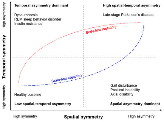
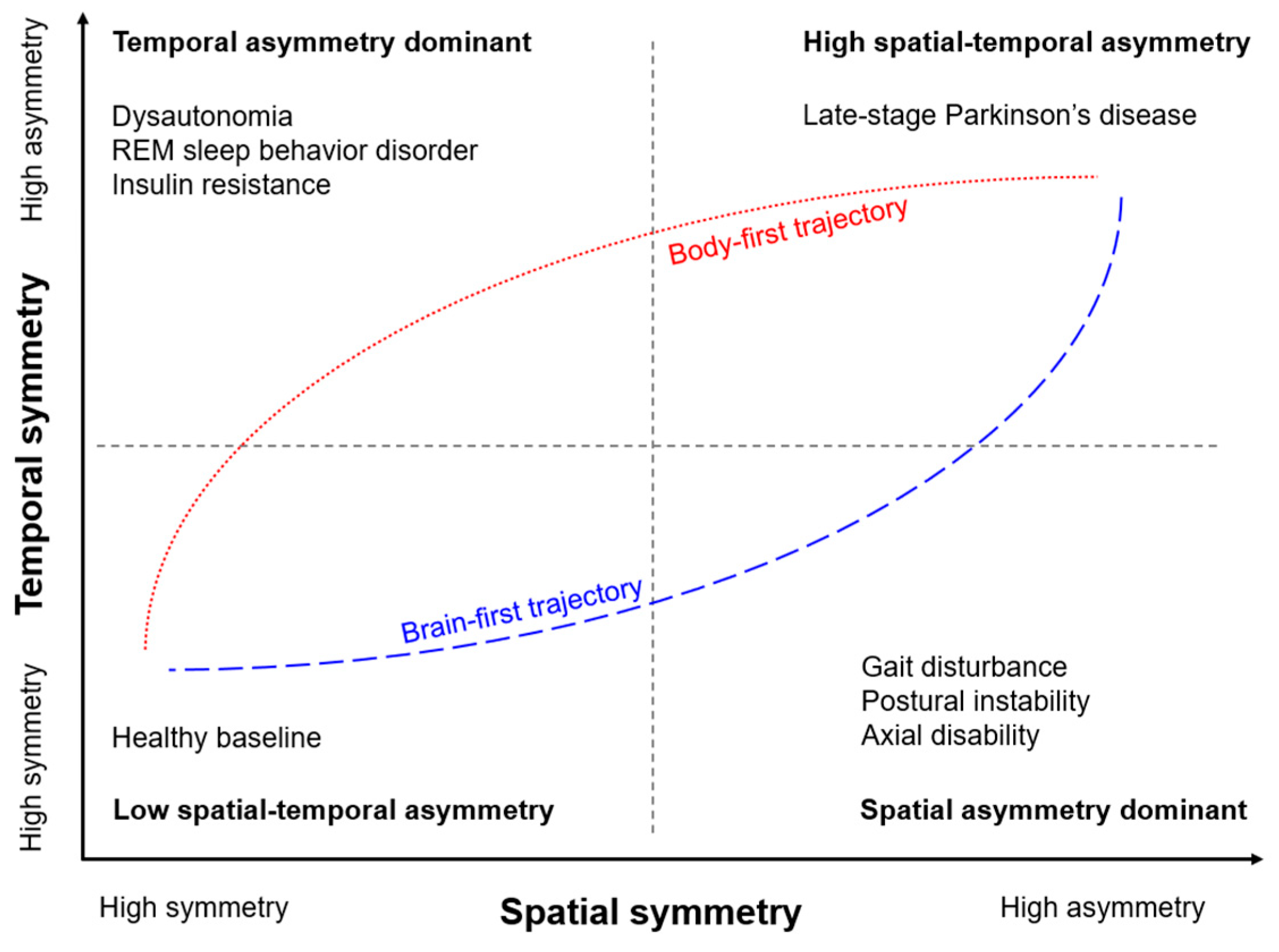

Disclaimer/Publisher’s Note: The statements, opinions and data contained in all publications are solely those of the individual author(s) and contributor(s) and not of MDPI and/or the editor(s). MDPI and/or the editor(s) disclaim responsibility for any injury to people or property resulting from any ideas, methods, instructions or products referred to in the content. |
© 2026 by the authors. Licensee MDPI, Basel, Switzerland. This article is an open access article distributed under the terms and conditions of the Creative Commons Attribution (CC BY) license.