


Abstract
Poor indoor air quality is linked to respiratory symptoms and disease, but interactions between indoor pollutants and microbial communities remain unclear. Evidence suggests microorganisms can interact with polycyclic aromatic hydrocarbons (PAHs) through degradation or attachment, potentially shaping indoor microbiomes. In the COPSACsevere cohort, we analyzed bed dust from 84 children (58 asthma, 26 controls). PAHs were measured by HPLC-FLD and microbiota profiled by 16S rRNA and ITS2 sequencing. Questionnaires captured sociodemographic and lifestyle factors. Associations were tested using PERMANOVA, Spearman correlations, and MaAsLin2. Bacterial community composition varied with the host factors age, sex, and parental education, but showed no association with PAH concentrations. Fungal diversity and composition were significantly associated with PAHs, especially pyrene and fluoranthene, which explained 6.1–6.3% variance (p < 0.05) and were positively correlated with Shannon diversity and evenness. Several fungal taxa including Lecanoraceae, Helotiales, and Circinaria correlated positively with PAHs. Asthma status was not associated with bacterial or fungal alpha and beta diversity metrics. PAHs selectively influenced fungal but not bacterial communities in bed dust. Although no asthma associations were detected and no mechanisms can be inferred, these findings highlight potential chemical–fungal interactions in indoor environments and their relevance for future respiratory health research. These results suggest that chemical exposures may shape specific components of indoor microbiomes independent of host disease status, emphasizing the need to integrate chemical–biological interactions in indoor exposure assessments in future environmental health studies and interventions.
1. Introduction
It has been estimated that Europeans spend as much as 85–90% of their time indoors [1,2]. Poor indoor air quality (IAQ) has been linked to various health outcomes, including allergies, eczema, skin and mucous membrane issues (eyes, nose, and throat), as well as behavioral symptoms including hyperactivity, inattention, and sleep difficulties [3,4,5,6,7,8]. Numerous pollutants have been extensively studied in relation to health, such as house dust mites, mold, pet dander, volatile organic compounds (VOCs), and particulate matter, which often contain harmful compounds like polycyclic aromatic hydrocarbons (PAHs) [9,10,11,12]. In addition to studying primary sources of such compounds, there is growing interest in household dust as a potential reservoir or secondary source of pollution, not only in terms of chemical contamination but also biological pollution, such as fungal and bacterial components, collectively referred to as the house dust microbiome or more generally as the indoor microbiome [13,14,15,16].
The significance of indoor microbial exposure for human health has gained increasing recognition. Research across homes, schools, and public spaces consistently shows that household dust microbiome composition reflects local environmental influences and serves as a source of microbes that colonize the respiratory tract of young children, directly affecting immune development [17,18,19,20,21,22,23,24,25,26].
Among indoor chemical pollutants, PAHs are of particular concern. They originate primarily from combustion of fossil fuels, are transported by particulate matter, and readily enter indoor environments, where they accumulate in settled dust at concentrations relevant to human health [10,12,27]. PAHs display a well-characterized genotoxic profile [28], and their capacity to alter microbial community structure and to be biotransformed by resident microbes represents a mechanistically plausible but poorly studied interaction in residential settings. Jarma et al. [29] demonstrated that semi-volatile organic compounds (SVOCs), including PAHs, and bacterial and fungal communities can be analyzed from the same house dust samples, confirming that settled dust is a good medium for parallel chemical–biological investigation in indoor environments.
This study, conducted within the EDIAQI project—an EU Horizon-funded initiative integrating chemical, biological, and health data to develop evidence-based strategies for improving indoor air quality and reducing associated health risks [10], addresses the following primary research question: do PAH concentrations in bed dust co-vary with the composition of the bacterial and fungal microbiome in the same samples? We hypothesize that higher PAH levels will be associated with shifts in microbial community structure, reflecting shared contamination sources, chemical pressure on microbial communities, or both. The investigation was conducted within the Danish COPSACsevere cohort, which was established to examine phenotypic subtypes of severe asthma. Although the cohort does not include indoor air quality as a primary covariate, we utilized the presence of both children with and without asthma to evaluate potential health effects. Therefore, this study is an ancillary investigation that features novel measurements of the microbial and chemical composition of bed dust from participants’ mattresses. As secondary objectives, we (i) characterize the PAH and microbiome profiles of bed dust in this cohort and (ii) explore whether either pollutant class is associated with respiratory health status.
2. Materials and Methods
2.1. Study Population
The Copenhagen Prospective Studies on Asthma in Childhood COPSACsevere cohort is a Danish national case–control study comprising children aged 6–18 years with severe or exacerbation-prone asthma (n = 111) and their healthy peers (n = 73). The study was conducted between 2020 and 2024. Children with asthma were recruited from sites across Denmark, either from hospital departments or through the Danish National Patient Registry, which prospectively registers all hospitalizations nationwide. Inclusion criteria for children with severe asthma were based on Global Initiative for Asthma (GINA) guidelines: a confirmed diagnosis of asthma and treatment with (1) high-dose inhaled corticosteroids (ICS) (>400 μg of budesonide or equivalent ICS) daily plus a second controller medication over the past year to prevent uncontrolled asthma, or (2) treatment with systemic oral corticosteroids (ORS) for ≥50% of the year to prevent uncontrolled asthma, or (3) current biological treatment due to previously diagnosed severe asthma. Inclusion criteria for exacerbation-prone asthma included a confirmed diagnosis of asthma and (1) at least one acute hospitalization due to an asthma exacerbation in the past two years and at least three hospitalizations due to asthma exacerbations after age 2, or (2) six or more hospitalizations for asthma exacerbations over a lifetime, with at least two of those after age 2. All hospitalizations should be separated by at least 3 months. Exclusion criteria for children with severe or exacerbation-prone asthma included (1) known immunodeficiencies, (2) previous major lung surgery, (3) other chronic lung diseases, or (4) severe neurological disorders. Healthy children were recruited through invitation letters at local institutions or via a personal network of children with asthma already included. The exclusion criteria for healthy children were (1) current infection, (2) daily medication use except for allergy medications, (3) chronic diseases except common atopic comorbidities unrelated to asthmatic symptoms (e.g., food allergies and allergic rhinoconjunctivitis), or (4) being a first-degree relative of one of the children with asthma included in the study.
All children attended a baseline visit. Children with asthma had a similar follow-up visit approximately one year later, while healthy controls completed a telephone interview after one year to confirm they had not developed asthma within that period. Throughout the follow-up year, children with asthma were monitored weekly with spirometry and seen during acute visits if their symptoms worsened (defined as three consecutive days of impaired spirometry or an exacerbation requiring hospitalization or treatment with high-dose ICS or OCS). Participants with an available dust sample were included in this study (n = 84). The sampled subgroup did not differ from the remainder of the cohort with respect to demographic and clinical characteristics.
2.2. Metadata
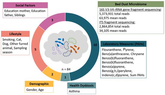
Gender, age, health (asthma status), parental education, siblings, pets, and maternal smoking behavior were assessed using questionnaires (Table 1, Figure 1). The highest education level of the parents was classified into five categories: (1) high school, (2) vocational education, (3) college, (4) bachelor’s, and (5) master’s degree. Age and sampling season of dust samples were categorized into four groups, while maternal smoking during pregnancy was categorized as sometimes, yes, or no. Ownership of cats, dogs, and other furred pets, as well as asthma and siblings, was classified as yes or no. To summarize baseline characteristics, we used the tableone package in R, specifying asthma status as the stratification variable. Sociodemographic and household variables were coded as factors, and descriptive statistics were reported for all levels of each categorical variable. Group differences in the baseline characteristics were assessed using chi-squared or Fisher’s exact tests for categorical variables and t-tests or analysis of variance (ANOVA) for continuous variables. Missing entries in the questionnaires were coded as NA and handled by pairwise exclusion in the respective analyses. To investigate the relationship between exposure level to PAHs and the bed dust microbiome, the PAHs were discretized into five categories (very low, low, medium, high, very high) according to the distribution of the variable to obtain balanced groups containing a similar number of individuals and with a distinct level of concentrations of PAHs between groups.
Table 1.
Summary of sociodemographic and household variables stratified by asthma status.
Figure 1.
Overview of the study design and the included variables. Bed dust microbiome samples were obtained, along with 21 variables from five categories, from 84 individuals.
2.3. Collection and Preprocessing of the Dust Samples
Participants were asked to collect dust samples from their beds by vacuuming their mattresses with an external filter kit (DUSTREAM® Collector, Indoor Biotechnologies, Charlottesville, VA, USA, or Dust Collecting Device from ALK-Abello, Hørsholm, Denmark) attached to the family’s vacuum cleaner with instructions to vacuum the mattresses, sheets and pillow for 5 min The samples were initially stored at −20 °C before transfer to the central storage facility, where they were weighed, aliquoted into 2 mL cryotubes, and stored at −80 °C. Next, the samples were thoroughly homogenized, divided into two portions, and distributed to separate laboratories in Zagreb, Croatia: one half was sent to the Institute for Anthropological Research for microbiome sequencing and preprocessing, while the other half was sent to the Institute for Medical Research and Occupational Health for PAH analysis and preprocessing. The laboratory personnel performing the downstream procedures were blinded to the asthma status of the participants. The PAH analytical pipeline is described in the following sections, while the microbiome laboratory procedures have been described in detail previously [15,16].
2.3.1. Polycyclic Aromatic Hydrocarbon (PAH) Measurements
The extraction method for PAHs from dust samples has been described previously [27]. Briefly, approximately 0.1 g of dust was extracted by accelerated solvent extraction (ASE) with two static extraction cycles at 125 °C. The extraction solvent was a 30:70 v/v mixture of cyclohexane and toluene. The extracts were dried using Rocket GeneVac evaporation (SP Industries, Warminster, PA, USA). Following evaporation, the samples were dissolved in 500 µL of acetonitrile, centrifuged for 10 min at 3000 rpm, and passed through a polytetrafluoroethylene (PTFE) syringe filter into the vial. The mass concentration of PAHs was measured using high-performance liquid chromatography with a fluorescence detector (HPLC-FCD). The following PAHs were determined: fluoranthene (Flu), pyrene (Pyr), chrysene (Chry), benzo(a)anthracene (BaA), benzo(j)fluoranthene (BjF), benzo(b)fluoranthene (BbF), benzo(a)pyrene (BaP), benzo(k)fluoranthene (BkF), benzo(ghi)perylene (BghiP), dibenzo(a,h)anthracene (DahA), and indeno(1,2,3-c,d)pyrene (IP). DahA and BjF were removed from the analysis due to a high proportion of samples below the detection limit.
2.3.2. DNA Extraction, Library Preparation, and Sequencing
Microbial DNA was extracted, amplified, and sequenced according to protocols previously described [15,16]. Briefly, fungal communities were profiled by targeting the Internal Transcribed Spacer 2 region (ITS2), and bacterial communities by targeting the V3–V4 regions of the 16S rRNA (ribosomal ribonucleic acid) gene. Quality controls included both positive and negative standards. Sequencing was performed on the Illumina MiSeq platform using the MiSeq Reagent Kit v3at the Laboratory for Molecular Anthropology, Centre for Applied Bioanthropology, Institute for Anthropological Research, Zagreb, Croatia.
2.3.3. Bioinformatics and Statistical Analysis
The sequencing data were subjected to quality filtering, denoising, and removal of chimeric sequences using the DADA2 algorithm [30] within the QIIME2 environment [31]. The resulting representative sequences (amplicon sequence variants (ASVs)) were taxonomically classified using the SILVA v132 database for bacteria [32] and the UNITE V7.0 database for fungi [33] with the vsearch algorithm [34]. Microbial community analysis was performed using the package Phyloseq [35] in R (version 4.4.2) [36] within R Studio (2024.12.0) (Posit team, 2025) [37]. Visualizations were prepared using the package ggplot2 [37]. Before further statistical analysis, the datasets were rarefied to 13,412 reads for bacteria and 1494 reads for fungi. Rarefaction to a common depth was applied to standardise the substantially differing sequencing efforts across samples (spanning roughly two orders of magnitude for fungi) before alpha- and beta-diversity analyses. One sample had to be removed from the bacterial dataset due to low read counts. Alpha diversity was calculated as ASV richness and Shannon H’ index. To determine evenness, the Shannon H’ index was divided by the natural logarithm of species richness [38]. Spearman’s rank correlations were calculated for PAHs and alpha diversity indices. Correlations were considered statistically significant if the FDR-adjusted p-value (q-value) was <0.1. For beta-diversity analysis, rarefied datasets were used to calculate Bray–Curtis dissimilarity matrices and compared with covariates and PAH using adonis and permanova from the package VEGAN [39] and visualized by principal coordinates analysis (PCoA). Prior to interpreting PERMANOVA results, homogeneity of multivariate dispersion (PERMDISP) among groups was assessed using the betadisper function from the package VEGAN on Bray–Curtis dissimilarity, followed by permutation tests of dispersion. To assess correlations between the relative abundance of bacterial and fungal genera and PAH levels, as well as other sociodemographic factors, MaAsLin2 [40] was used. Associations were considered statistically significant if the FDR-adjusted p-value (q-value) was < 0.1. Functional analysis of selected fungal ASVs was conducted using the FUNGuild online tool [41].
Generative AI was not utilized for data analysis, figure creation, or the development of substantive manuscript content.
3. Results
Parental education varied across case and control groups (mothers in both groups most frequently held a bachelor’s or master’s degree, while fathers’ education ranged from high school to master’s degree level). Most participants had siblings (n = 76, 90.5%), and most mothers reported no smoking during pregnancy (n = 59, 70.2%). Exposure to pets was common, particularly dogs, while ownership of cats and other furred animals was less frequent. Sampling occurred across all seasons, with spring and summer being the most common periods of data collection. Some variables contained missing (NA) values due to the online, self-reported nature of the questionnaire.
3.1. Cohort Characteristics and Sequencing Results
A total of 84 mattress dust samples from the baseline visit were analyzed since dusting was not mandatory. This includes 58 from children diagnosed with severe or exacerbation-prone asthma and 26 from children without asthma. In addition to these variables, additional metadata were collected to facilitate the integration of microbial data with environmental and host factors. Overall, 21 variables were evaluated across five categories: demographic, lifestyle, social, health status, and laboratory measures (PAHs) (Figure 1).
Table 1 presents the sociodemographic and household characteristics of the study population stratified by asthma status. There were no statistically significant differences between groups in gender distribution, age at sampling, maternal education, presence of siblings, maternal smoking during pregnancy, pet ownership (cats, dogs, or other furred pets), or sampling season (all p > 0.05). Paternal education differed significantly between groups (p = 0.01), with a higher proportion of fathers of asthmatic children having high school education and a lower proportion having a master’s degree compared with the non-asthma group.
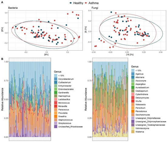
After quality filtering and removal of non-target sequences, a total of 5,373,931 bacterial reads (average: 63,975) and 2,864,854 fungal reads (average: 34,105) were retained. Bacterial reads were classified into 39 phyla and 23,401 ASVs, while fungal reads were assigned to 11 phyla and 2575 ASVs (Figure 1). The microbial community mainly consisted of Firmicutes (43%), followed by Actinobacteria (24.7%) and Proteobacteria (21.4%). The fungal community was mainly composed of Ascomycota (68.4%) and Basidiomycota (29.9%). Dominant bacterial genera included Staphylococcus (20.5%) and Corynebacterium (9.9%), while the dominant fungal genera were Cladosporium (26.5%) and Malassezia (12.3%) (Figure 2B).
Figure 2.
(A) Visualization of bacterial and fungal community composition using PCoA-plots based on Bray–Curtis dissimilarity. Each point corresponds to a sample and is colored in respect to the variable “health dysbiosis”. (B) Taxonomic composition of bacterial and fungal communities in bed dust microbiomes at the genus level. Each sample is represented by a stacked bar chart. The “< 10%” group was created from ASVs with relative abundance < 10%.
3.2. Variables Associated with Microbial Composition and Diversity
Beta diversity analysis based on Bray–Curtis dissimilarity showed that bacterial and fungal communities varied among individuals but did not form clear groups by asthma status. PERMANOVA analysis confirmed that asthma contributed only a small part to the differences in bacterial and fungal community composition, indicating no significant separation at the community level (Figure 2A).
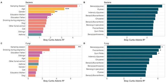
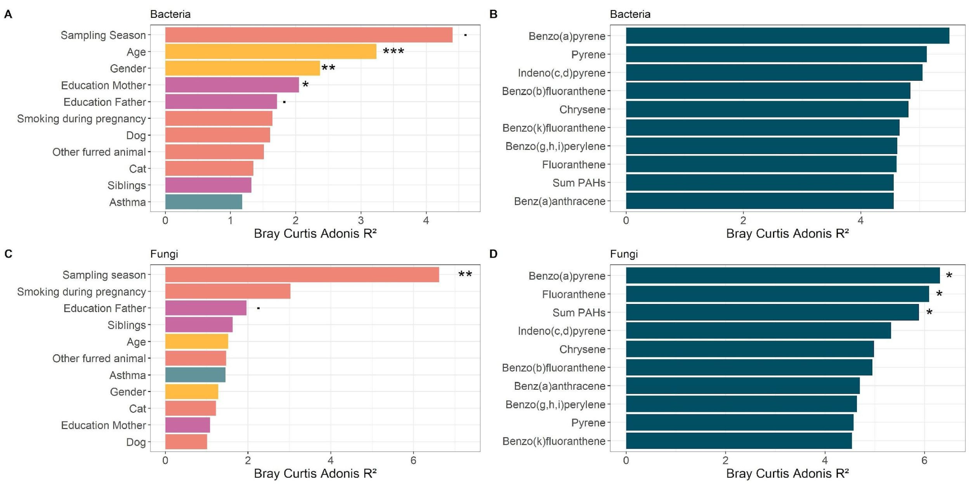
The influence of sociodemographic and lifestyle factors, as well as PAH levels, on the bed dust microbiome was studied on a compositional level (Figure 3A–D). PERMANOVA analysis showed that the bacterial community was mainly affected by demographic factors including age (R2: 3.2%, p < 0.001) and gender (R2: 2.4%, p = 0.003), as well as social variables including parental education (Education Mother: R2: 2.1%, p = 0.008; Education Father: R2: 1.7%, p = 0.06) (Figure 3A). Neither lifestyle factors nor PAH levels in dust significantly influenced the bacterial community (Figure 3B). In contrast, fungal community composition was not affected by sociodemographic or lifestyle factors, except for the sampling season (R2: 7.6%, p = 0.003) (Figure 3C). Additionally, the amounts of specific PAHs explain a significant portion of the variation in fungal community composition (Figure 3D). Specifically, levels of BaP (R2: 6.3%, p = 0.01), Flu (R2: 6.1%, p = 0.018), and the overall PAH burden (R2: 5.9%, p = 0.034) significantly affected the fungal community structure.
Figure 3.
Relationships between microbiome community composition and evaluated variables. The bar graphs show the proportion of variation explained by each tested variable. The left panels display the association between (A) bacterial and (C) fungal community composition and sociodemographic and lifestyle factors. The right panels present the association between (B) bacterial and (D) fungal community composition and PAH levels. The significance thresholds are indicated as follows: p ≤ 0.1 (.); p ≤ 0.05 (*); p ≤ 0.01 (**); p ≤ 0.001 (***).
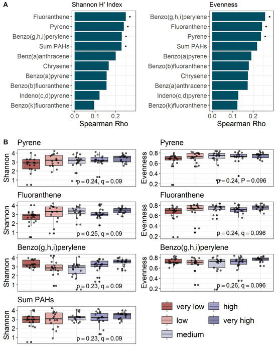
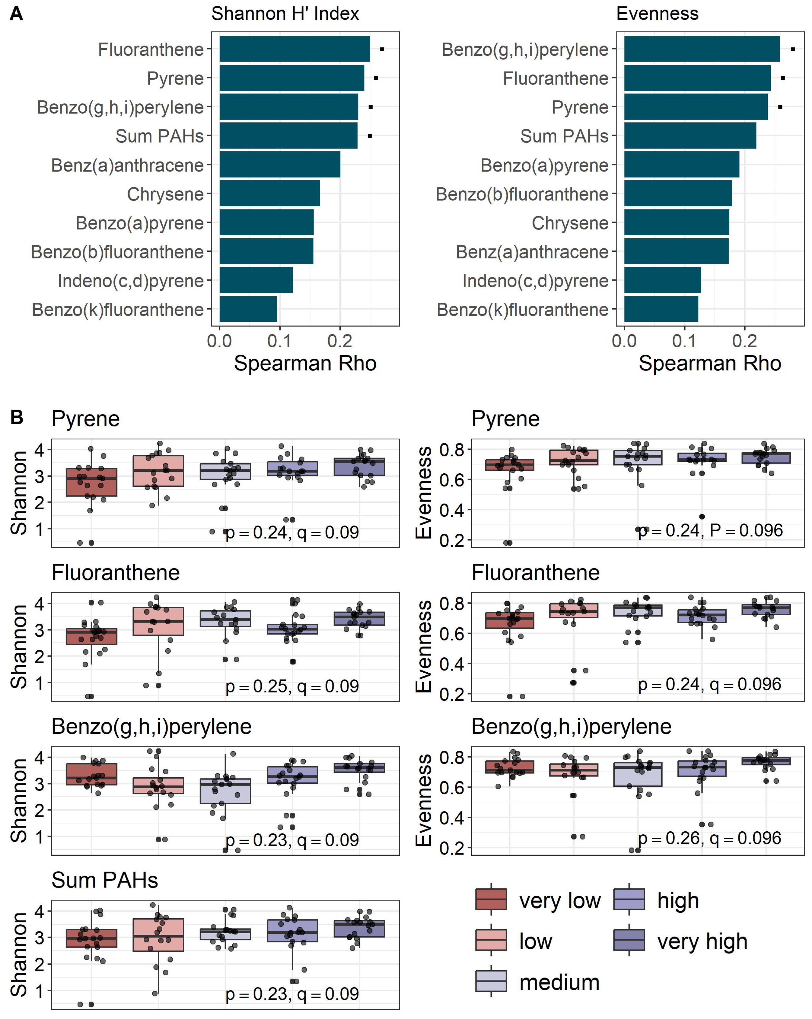
Asthma status was not associated with either bacterial or fungal composition (see Figure 3A,C). Sociodemographic and lifestyle factors also showed no significant link with bacterial and fungal diversity, richness, and evenness. Additionally, bacterial alpha diversity was unaffected by PAH levels. However, fungal diversity and evenness showed significant correlations with PAH concentrations, whereas fungal richness did not exhibit a significant relationship after FDR correction (Figure 4A, Table S1). Specifically, fungal diversity was positively correlated with several PAHs, including Flu (Spearman’s p = 0.25, q = 0.09), Pyr (Spearman’s p = 0.24, q = 0.09), BghiP (Spearman’s p = 0.23, q = 0.09), and the total PAH levels (Spearman’s p = 0.23, q = 0.09) (Figure 4B). Moreover, fungal evenness showed positive correlations with Pyr (Spearman’s p = 0.24, q = 0.096), Flu (Spearman’s p = 0.24, q = 0.096), and BghiP (Spearman’s p = 0.26, p = 0.096) (Figure 4B).
Figure 4.
Spearman correlation analysis examining the relationship between PAH levels and fungal alpha diversity metrics. (A). Bar plots show Spearman’s Rho (p) values representing correlations between PAH concentrations and alpha diversity indices, such as Shannon H’ index, and evenness. The significance levels after FDR-correction (q) are indicated as follows: q ≤ 0.1 (.); q ≤ 0.05 (*); q ≤ 0.01 (**); q ≤ 0.001 (***) (B) Boxplots further illustrate significant correlations.
3.3. Identification of Microbial Taxa Associated with Specific Variables
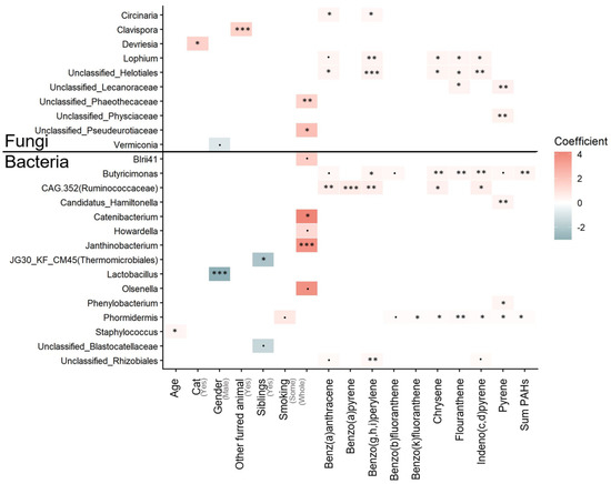
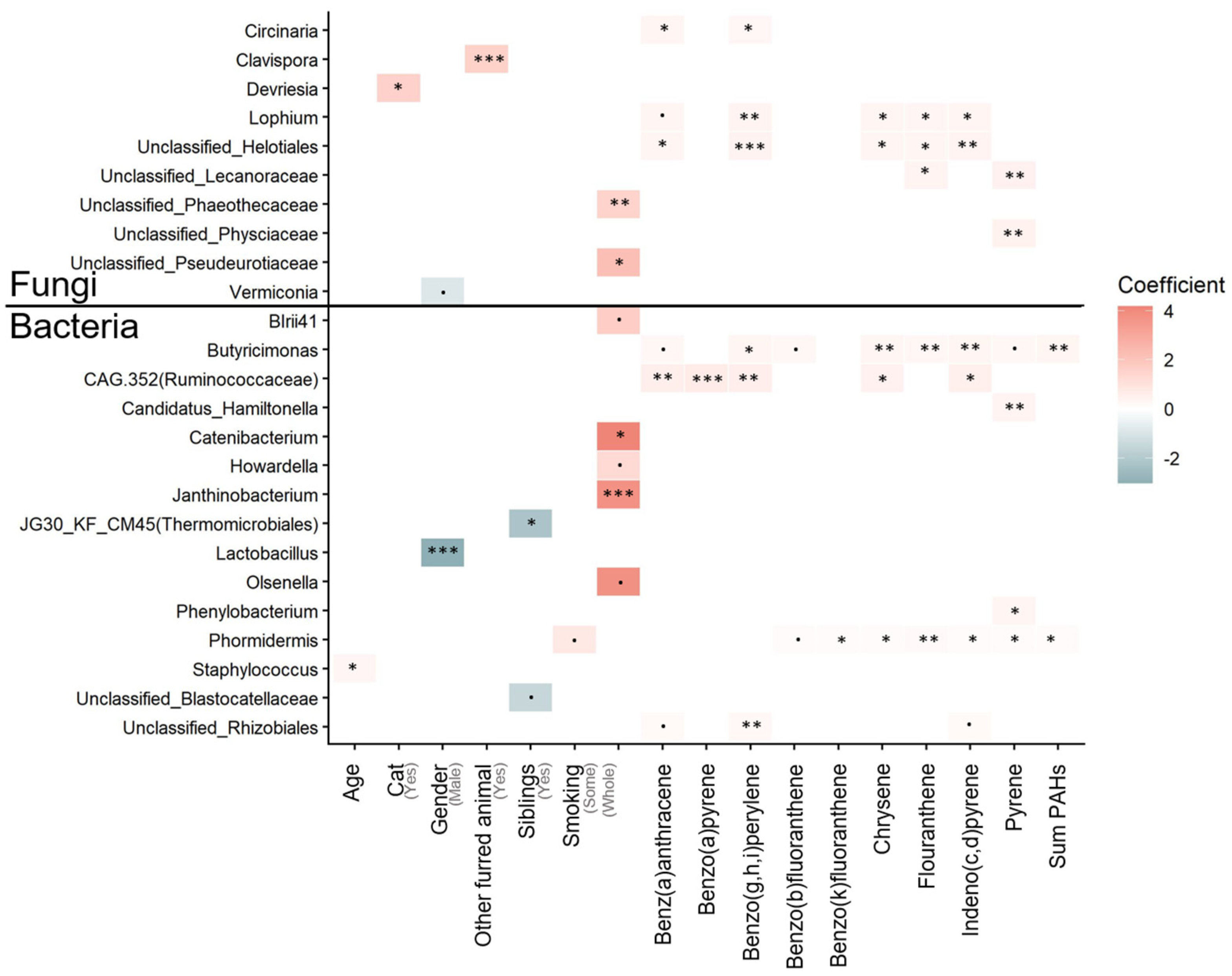
Microbiome multivariate association with linear models (MaAsLin2) was used to identify bacterial and fungal genera linked to the collected variables. Asthma, sampling season, dog ownership, and parental education level did not show significant associations with any microbial genera. However, 15 bacterial and 10 fungal genera were significantly associated with at least one variable (Figure 5). Notably, Rhizobiales, Phormidermis, CAG.352 (Ruminococcaceae), and Butyricimonas were identified as bacterial taxa positively associated with three or more PAHs. Interestingly, Lactobacillus showed a strong association with gender, with higher relative abundance in female participants. The relative abundance of Staphylococcus was positively associated with age, while bacterial genera including Janthinobacterium, Olsenella, Howardella, Catenibacterium, and BIrii41 demonstrated positive correlations with smoking. Several fungal taxa, including Lecanoraceae, Helotiales, Lophium, and Circinaria, were observed to be positively linked to at least two different PAHs. Additionally, pet ownership showed a positive correlation with certain fungi, with cats influencing the relative abundance of Devriesia, and other furred animals associated with higher relative abundance of Clavispora. Notably, Clavispora and Devriesia are fungal genera that have been shown to potentially possess animal pathogenic characteristics.
Figure 5.
Bacterial and fungal genera that were found to be significantly associated with different variables using multivariate association with linear models (MaAsLin). The significance thresholds after FDR-correction (q) are indicated as follows: q ≤ 0.1 (.); q ≤ 0.05 (*); q ≤ 0.01 (**); q ≤ 0.001 (***).
4. Discussion
In this study, we hypothesized that PAHs are believed to originate primarily from outdoor combustion sources [10] and could influence the bed dust microbiome, which might be linked to severe asthma. Our findings show that PAHs are not inert pollutants but can act as ecological factors, selectively shaping fungal communities in bed dust. In contrast, bacterial communities appear more influenced by demographic and household factors. Our main findings suggest that PAH concentrations were significantly associated with fungal diversity in bed dust, while bacterial communities are not affected. Although no clear associations with asthma were found, this differential sensitivity highlights the importance of considering chemical–fungal interactions when evaluating potential downstream health effects. The differential sensitivity aligns with recent observations in other environmental matrices, in which fungal communities were found to be more influenced by air quality than bacterial communities [42]. A plausible reason for this differential response is that many fungi possess versatile extracellular enzyme systems able to transform aromatic compounds and occupy ecological niches distinct from those of bacteria, which may render fungal communities more responsive to PAH exposure [43]. Our analyses demonstrated that indoor fungal diversity and community composition consistently relate to PAH levels, particularly BaP and Flu. These PAHs are structural isomers, and their consistent associations across diversity measures may indicate a selective association with specific indoor fungi. PERMANOVA analyses confirmed that BaP (R2 = 6.3%, p = 0.01), Flu (R2 = 6.1%, p = 0.018), and overall PAH burden (R2 = 5.9%, p = 0.034) each explained a significant portion of variation in fungal community composition. At the taxa level members of Helotiales, Lecanoraceae, and Circinaria, exhibited positive correlations with PAH levels. Interestingly, the latter two groups are known for their roles in lichen symbioses, as they absorb nutrients from the air and can indicate air pollution levels. Notably, Clavispora and Devriesia are fungal genera that have been shown to potentially possess animal pathogenic characteristics. Overall, these findings are consistent with the initial hypothesis: fungi can directly interact with PAHs, potentially degrading them or using them as substrates, thereby influencing the fungal ecology of household dust. However, enzymatic degradation is only one possible explanation; the observed correlations may equally reflect PAH tolerance of certain taxa, or shared sources and household characteristics (e.g., wood or biomass combustion), rather than a direct causal relationship [43,44] PAHs may be associated with the presence of fungal taxa possessing specific metabolic capabilities or tolerance mechanisms. Although direct mechanistic investigations on fungi in household dust are limited, numerous studies have found significant links between PAH levels and microbial community composition. Al Hunaiti et al. [45] found that PAHs in floor dust in Jordanian households influenced fungal and bacterial population abundances. These shifts shape the indoor fungal environment and could have downstream indirect effects on health. Analogous interactions of fungi and PAHs have been observed in previous studies [43,44] as various fungi can metabolize PAHs. Similarly, metagenomic analyses of the cutaneous microbiota have identified specific microbial genes associated with aromatic compound degradation in high-PAH environments, supporting the view that chronic exposure can drive functional adaptations in microbial communities [46]. Our findings suggest that assessing indoor PAH levels alongside fungal community profiles may inform indoor exposure assessment and environmental monitoring, although any respiratory or immunological relevance remains to be demonstrated. Preventive strategies could include reducing indoor PAH sources (e.g., minimizing combustion-related emissions and improving ventilation) and implementing targeted indoor air quality interventions to limit both chemical burden and secondary ecological shifts in the indoor mycobiome.
In contrast, bacterial communities were not significantly impacted by PAH levels, but instead reflected host- and household-related factors such as age (R2 = 3.2%, p < 0.001), gender (R2 = 2.4%, p = 0.003), and parental education (R2 = 1.7%, p = 0.06). This limited impact in dust contrasts with studies on human-associated matrices, such as meconium, where prenatal PAH exposure has been directly linked to altered bacterial alpha diversity and colonization patterns [47], suggesting that matrix-specific properties of bed dust may mitigate chemical pressure on bacteria. However, some bacterial taxa, including Rhizobiales and Phormidermis (both soil- and plant-associated), as well as Ruminococcaceae and Butyricimonas (both gut-associated), were positively associated with multiple PAHs. These associations indicate that while PAHs may not significantly alter overall bacterial community structure in bed dust, they may affect the transport and deposition of specific taxa. By demonstrating that PAHs influence fungal communities in bed dust or vice versa, this study highlights a pathway through which chemical exposures may indirectly affect health, even if clinical associations to severe asthma were not identified here.
4.1. Limitations and Outlook
Our study results are influenced by several limiting factors, including a small sample size (n = 84) and an uneven distribution of healthy controls (n = 26) versus asthma cases (n = 58). This limited and unbalanced sample reduces statistical power and increases the possibility of a Type II error, which may partly account for the absence of asthma associations; accordingly, the modest effect sizes observed should be interpreted accordingly. Furthermore, asthma, especially its severe form, is a complex disease that is difficult to predict from either individual or complex factors [48,49]. Given the transmission rate of PAHs from ambient into indoor air [10], a location-based bias could propagate into this analysis. PAH concentrations in Danish ambient air are lower than in some European countries [50], and a broader research setting would reveal distinct mechanisms. Denmark also has low soil biodiversity [51], which may affect the dust microbiome, as inferred from other relationships, such as soil–gut associations [52]. Together, these characteristics limit the range of environmental exposures and may reduce the likelihood of detecting concentration-dependent effects. In addition, the cohort was not designed around indoor air-quality or building assessments, so potential key covariates (e.g., ventilation, humidity, mold, building age, building material) were not available. Unmeasured confounding by these factors may influence PAH levels and fungal communities. Future work should include expanded metadata to better account for these variables. Furthermore, it remains unclear whether the associated fungal taxa are directly related or belong to separate clades. Further research, such as isolating fungi or reconstructing their genomes from metagenomic data, will be necessary.
4.2. Implications for Future Research
Overall, these findings support the main premise of this study that indoor environmental health cannot be understood by examining chemical and biological exposures separately. Instead, PAHs and microbiomes may interact in ways that could impact immune development in future findings. This integrative approach underscores the importance of future research that continues to link chemical and microbial factors when evaluating indoor exposures on health.
5. Conclusions
This study of Danish children from the COPSACsevere cohort highlights a distinct divergence in how indoor microbes respond to chemical pollution. PAHs were strongly associated with fungal community composition and diversity in bed dust, whereas demographic factors predominantly shaped bacterial communities. These findings align with recent environmental research suggesting that fungal communities are often more sensitive to air quality indices than bacteria. Although no direct connections were observed between asthma status and either microbiome structure or PAH exposure, likely due to the limited sample size and the relatively low PAH levels in Denmark, an association between PAH exposure and fungal community structure is evident. Our results suggest that PAHs may help shape indoor fungal communities in bed dust; whether such chemical-microbial interactions influence respiratory outcomes will require larger, longitudinal studies. Additionally, given evidence that prenatal PAH exposure can impede early microbial colonization in other tissues, future studies should investigate whether these chemical-microbial interactions exert health effects across different developmental windows, extending beyond the indoor dust environment.
Supplementary Materials
The following supporting information can be downloaded at: https://www.mdpi.com/article/10.3390/atmos17080755/s1, Table S1: Spearman correlations between PAH concentrations in bed dust and fungal alpha diversity metrics. For each PAH-metric pair the table reports Spearman’s rho, p-value, FDR-adjusted q value, and the 95% confidence interval for rho. Alpha diversity metrics were computed as Shannon H’, observed ASV richness, and evenness.
Author Contributions
Conceptualization, K.B.; methodology, J.Š., D.H.A. and K.B.; software, K.M. and M.F.; formal analysis, K.M. and M.F.; investigation, K.M., M.F., I.Š., J.Š., D.H.A., I.J., G.P., S.R., S.H. and K.A.; resources, J.Š. and D.H.A.; data curation, K.M., M.F. and K.A.; writing—original draft preparation, K.M., M.F., I.Š., S.K.J., T.C. and M.L.; writing—review and editing, K.M., M.F., I.Š., J.Š., D.H.A., I.J., G.P., M.A.R., S.K.J., K.B., S.R., S.H., K.A., T.C. and M.L.; visualization, K.M.; supervision, K.B., T.C. and M.L.; project administration, J.Š., S.R., S.H., K.A. and K.B.; funding acquisition, K.B., T.C. and M.L.; data collection, S.K.J. All authors have read and agreed to the published version of the manuscript.
Funding
This research was funded by the European Union’s Horizon research and innovation program under grant number 101057497 (Evidence-driven Indoor Air Quality Improvement—EDIAQI).
Institutional Review Board Statement
Ethical approval was given by the National Committee on Health Research Ethics (H-20006157).
Informed Consent Statement
The study, approved by the National Committee on Health research Ethics (H-20006157), was conducted in accordance with the guiding principles of the Declaration of Helsinki. Both parents gave oral and written informed consent before participation in the study. An independent oral and written informed consent was obtained from study participants above the age of 18 years.
Data Availability Statement
Raw sequences for each sample used in this study were deposited at the European Nucleotide Archive (ENA) in the FASTQ format and are available under the Bioproject number PRJEB105098.
Acknowledgments
We express our deepest gratitude to the children and families of the COPSACsevere cohort for all their support and commitment. We acknowledge and appreciate the unique efforts of the COPSAC research team and all our national collaborators. We would like to acknowledge the huge work of the late Hans Bisgaard, who was the founder of COPSAC and was head of the COPSAC clinical research center for more than 25 years.
Conflicts of Interest
All authors declare no potential, perceived, or real conflict of interest regarding the content of this manuscript. The funding agencies did not have any role in the design and conduct of the study; collection, management, and interpretation of the data; or preparation, review, or approval of the manuscript. No pharmaceutical company was involved in the study.
References
- Schweizer, C.; Edwards, R.D.; Bayer-Oglesby, L.; Gauderman, W.J.; Ilacqua, V.; Juhani Jantunen, M.; Lai, H.K.; Nieuwenhuijsen, M.; Künzli, N. Indoor Time–Microenvironment–Activity Patterns in Seven Regions of Europe. J. Expo. Sci. Environ. Epidemiol. 2007, 17, 170–181. [Google Scholar] [CrossRef] [Scilit] [PubMed]
- WHO. Combined or Multiple Exposure to Health Stressors in Indoor Built Environments: An Evidence-Based Review; WHO Regional Office for Europe: Bonn, Germany, 2014. [Google Scholar]
- Belo, J.; Meira e Cruz, M.; Viegas, C.; Lage, J.; Almeida, S.M.; Verde, S.C.; Alves, C.; Canha, N. Sleep and Indoor Air Quality: An Exploratory Polysomnographic Evaluation of Potential Associations. Atmos. Pollut. Res. 2025, 16, 102557. [Google Scholar] [CrossRef] [Scilit]
- Breysse, P.N.; Diette, G.B.; Matsui, E.C.; Butz, A.M.; Hansel, N.N.; McCormack, M.C. Indoor Air Pollution and Asthma in Children. Proc. Am. Thorac. Soc. 2010, 7, 102–106. [Google Scholar] [CrossRef] [Scilit] [PubMed]
- Chen, Y.-C.; Gui, Z.-H.; Bao, W.-W.; Liang, J.-H.; Zhang, S.-X.; Zhao, Y.; Jiang, N.; Chen, Y.-J. Chronic Exposure to Indoor Air Pollutants in Association with Attention-Deficit/Hyperactivity Disorder Symptoms in Chinese Schoolchildren: A Cross-Sectional Study. Neurotoxicology 2023, 94, 182–190. [Google Scholar] [CrossRef] [Scilit] [PubMed]
- Jones, A.P. Indoor Air Quality and Health. Atmos. Environ. 1999, 33, 4535–4564. [Google Scholar] [CrossRef] [Scilit]
- Raaschou-Nielsen, O.; Hermansen, M.N.; Loland, L.; Buchvald, F.; Pipper, C.B.; Sørensen, M.; Loft, S.; Bisgaard, H. Long-Term Exposure to Indoor Air Pollution and Wheezing Symptoms in Infants. Indoor Air 2010, 20, 159–167. [Google Scholar] [CrossRef] [Scilit] [PubMed]
- Tran, V.V.; Park, D.; Lee, Y.-C. Indoor Air Pollution, Related Human Diseases, and Recent Trends in the Control and Improvement of Indoor Air Quality. Int. J. Environ. Res. Public Health 2020, 17, 2927. [Google Scholar] [CrossRef] [Scilit] [PubMed]
- Lovrić, M.; Gajski, G.; Fernández-Agüera, J.; Pöhlker, M.; Gursch, H.; EDIAQI Consortium; Borg, A.; Switters, J.; Mureddu, F. Evidence Driven Indoor Air Quality Improvement: An Innovative and Interdisciplinary Approach to Improving Indoor Air Quality. Biofactors 2024, 51, e2126. [Google Scholar] [CrossRef] [Scilit] [PubMed]
- Lovrić, M.; Račić, N.; Pehnec, G.; Horvat, T.; Lovrić Štefiček, M.J.; Jakovljević, I. Indoor Polycyclic Aromatic Hydrocarbons—Relationship to Ambient Air, Risk Estimation, and Source Apportionment Based on Household Measurements. Atmosphere 2024, 15, 1525. [Google Scholar] [CrossRef] [Scilit]
- Martins, C.; Teófilo, V.; Clemente, M.; Corda, M.; Fermoso, J.; Aguado, A.; Rodriguez, S.; Moshammer, H.; Kristian, A.; Ferri, M.; et al. Sources, Levels, and Determinants of Indoor Air Pollutants in Europe: A Systematic Review. Sci. Total Environ. 2025, 964, 178574. [Google Scholar] [CrossRef] [Scilit] [PubMed]
- Račić, N.; Terzić, I.; Karlović, N.; Bošnjaković, A.; Terzić, T.; Jakovljević, I.; Pehnec, G.; Horvat, T.; Gajski, G.; Gerić, M.; et al. Volatile Organic Compounds (VOCs) and Polycyclic Aromatic Hydrocarbons (PAHs) in Indoor Environments: A Review and Analysis of Measured Concentrations in Europe. Indoor Air 2025, 2025, 5945455. [Google Scholar] [CrossRef] [Scilit]
- Lehtimäki, J.; Gupta, S.; Hjelmsø, M.; Shah, S.; Thorsen, J.; Rasmussen, M.A.; Soverini, M.; Li, X.; Russel, J.; Trivedi, U.; et al. Fungi and Bacteria in the Beds of Rural and Urban Infants Correlate with Later Risk of Atopic Diseases. Clin. Exp. Allergy 2023, 53, 1268–1278. [Google Scholar] [CrossRef] [Scilit] [PubMed]
- National Academies of Sciences, Engineering, and Medicine; Division on Earth and Life Studies; Health and Medicine Division; Board on Infrastructure and the Constructed Environment; Board on Environmental Studies and Toxicology; Committee on Microbiomes of the Built Environment: From Research to Application. Microorganisms in Built Environments: Impacts on Human Health. In Microbiomes of the Built Environment: A Research Agenda for Indoor Microbiology, Human Health, and Buildings; National Academies Press: Washington, DC, USA, 2017. [Google Scholar]
- Šarac, J.; Havaš Auguštin, D.; Šunić, I.; Michl, K.; Berg, G.; Cernava, T.; Marjanović, D.; Jakobsen, R.R.; Lovrić, M. Linking the Bed Dust Microbiome with Environmental Factors and Child Respiratory Health. Ann. Hum. Biol. 2025, 52, 2509606. [Google Scholar] [CrossRef] [Scilit] [PubMed]
- Šunić, I.; Havaš Auguštin, D.; Šarac, J.; Michl, K.; Cernava, T.; Jakobsen, R.R.; Mešić, A.; Novokmet, N.; Lovrić, M. Associations Between Indoor Fungal Community Structures and Environmental Factors: Insights from the Evidence-Driven Indoor Air-Quality Improvement Study. J. Fungi 2025, 11, 261. [Google Scholar] [CrossRef] [Scilit] [PubMed]
- Cox, J.; Stone, T.; Ryan, P.; Burkle, J.; Jandarov, R.; Mendell, M.J.; Niemeier-Walsh, C.; Reponen, T. Residential Bacteria and Fungi Identified by High-Throughput Sequencing and Childhood Respiratory Health. Environ. Res. 2022, 204, 112377. [Google Scholar] [CrossRef] [Scilit] [PubMed]
- Ege, M.J.; Mayer, M.; Normand, A.-C.; Genuneit, J.; Cookson, W.O.C.M.; Braun-Fahrländer, C.; Heederik, D.; Piarroux, R.; von Mutius, E.; GABRIELA Transregio 22 Study Group. Exposure to Environmental Microorganisms and Childhood Asthma. N. Engl. J. Med. 2011, 364, 701–709. [Google Scholar] [CrossRef] [Scilit] [PubMed]
- Guo, J.; Xiong, Y.; Kang, T.; Xiang, Z.; Qin, C. Bacterial Community Analysis of Floor Dust and HEPA Filters in Air Purifiers Used in Office Rooms in ILAS, Beijing. Sci. Rep. 2020, 10, 6417. [Google Scholar] [CrossRef] [Scilit] [PubMed]
- Kirjavainen, P.V.; Karvonen, A.M.; Adams, R.I.; Täubel, M.; Roponen, M.; Tuoresmäki, P.; Loss, G.; Jayaprakash, B.; Depner, M.; Ege, M.J.; et al. Farm-like Indoor Microbiota in Non-Farm Homes Protects Children from Asthma Development. Nat. Med. 2019, 25, 1089–1095. [Google Scholar] [CrossRef] [Scilit] [PubMed]
- Shan, Y.; Wu, W.; Fan, W.; Haahtela, T.; Zhang, G. House Dust Microbiome and Human Health Risks. Int. Microbiol. 2019, 22, 297–304. [Google Scholar] [CrossRef] [Scilit] [PubMed]
- Tischer, C.; Weikl, F.; Probst, A.J.; Standl, M.; Heinrich, J.; Pritsch, K. Urban Dust Microbiome: Impact on Later Atopy and Wheezing. Environ. Health Perspect. 2016, 124, 1919–1923. [Google Scholar] [CrossRef] [Scilit] [PubMed]
- Vandenborght, L.-E.; Enaud, R.; Urien, C.; Coron, N.; Girodet, P.-O.; Ferreira, S.; Berger, P.; Delhaes, L. Type 2–High Asthma Is Associated with a Specific Indoor Mycobiome and Microbiome. J. Allergy Clin. Immunol. 2021, 147, 1296–1305.e6. [Google Scholar] [CrossRef] [Scilit] [PubMed]
- Gupta, S.; Hjelmsø, M.H.; Lehtimäki, J.; Li, X.; Mortensen, M.S.; Russel, J.; Trivedi, U.; Rasmussen, M.A.; Stokholm, J.; Bisgaard, H.; et al. Environmental Shaping of the Bacterial and Fungal Community in Infant Bed Dust and Correlations with the Airway Microbiota. Microbiome 2020, 8, 115. [Google Scholar] [CrossRef] [Scilit] [PubMed]
- Man, W.H.; de Steenhuijsen Piters, W.A.A.; Bogaert, D. The Microbiota of the Respiratory Tract: Gatekeeper to Respiratory Health. Nat. Rev. Microbiol. 2017, 15, 259–270. [Google Scholar] [CrossRef] [Scilit] [PubMed]
- Mortensen, M.S.; Brejnrod, A.D.; Roggenbuck, M.; Abu Al-Soud, W.; Balle, C.; Krogfelt, K.A.; Stokholm, J.; Thorsen, J.; Waage, J.; Rasmussen, M.A.; et al. The Developing Hypopharyngeal Microbiota in Early Life. Microbiome 2016, 4, 70. [Google Scholar] [CrossRef] [Scilit] [PubMed]
- Jakovljević, I.; Dvoršćak, M.; Jagić, K.; Klinčić, D. Polycyclic Aromatic Hydrocarbons in Indoor Dust in Croatia: Levels, Sources, and Human Health Risks. Int. J. Environ. Res. Public Health 2022, 19, 11848. [Google Scholar] [CrossRef] [Scilit] [PubMed]
- Štampar, M.; Sedighi Frandsen, H.; Rogowska-Wrzesinska, A.; Wrzesinski, K.; Filipič, M.; Žegura, B. Hepatocellular Carcinoma (HepG2/C3A) Cell-Based 3D Model for Genotoxicity Testing of Chemicals. Sci. Total Environ. 2021, 755, 143255. [Google Scholar] [CrossRef] [Scilit] [PubMed]
- Jarma, D.; Maestre, J.P.; Sanchez, J.; Brodfuehrer, S.; Katz, L.E.; Horner, S.; Kinney, K.A. Participant-Collected Household Dust for Assessing Microorganisms and Semi-Volatile Organic Compounds in Urban Homes. Sci. Total Environ. 2024, 908, 168230. [Google Scholar] [CrossRef] [Scilit] [PubMed]
- Callahan, B.J.; McMurdie, P.J.; Rosen, M.J.; Han, A.W.; Johnson, A.J.A.; Holmes, S.P. DADA2: High-Resolution Sample Inference from Illumina Amplicon Data. Nat. Methods 2016, 13, 581–583. [Google Scholar] [CrossRef] [Scilit] [PubMed]
- Bolyen, E.; Rideout, J.R.; Dillon, M.R.; Bokulich, N.A.; Abnet, C.C.; Al-Ghalith, G.A.; Alexander, H.; Alm, E.J.; Arumugam, M.; Asnicar, F.; et al. Reproducible, Interactive, Scalable and Extensible Microbiome Data Science Using QIIME 2. Nat. Biotechnol. 2019, 37, 852–857. [Google Scholar] [CrossRef] [Scilit] [PubMed]
- Pruesse, E.; Quast, C.; Knittel, K.; Fuchs, B.M.; Ludwig, W.; Peplies, J.; Glöckner, F.O. SILVA: A Comprehensive Online Resource for Quality Checked and Aligned Ribosomal RNA Sequence Data Compatible with ARB. Nucleic Acids Res. 2007, 35, 7188–7196. [Google Scholar] [CrossRef] [Scilit] [PubMed]
- Abarenkov, K.; Nilsson, R.H.; Larsson, K.-H.; Taylor, A.F.S.; May, T.W.; Frøslev, T.G.; Pawlowska, J.; Lindahl, B.; Põldmaa, K.; Truong, C.; et al. The UNITE Database for Molecular Identification and Taxonomic Communication of Fungi and Other Eukaryotes: Sequences, Taxa and Classifications Reconsidered. Nucleic Acids Res. 2024, 52, D791–D797. [Google Scholar] [CrossRef] [Scilit] [PubMed]
- Rognes, T.; Flouri, T.; Nichols, B.; Quince, C.; Mahé, F. VSEARCH: A Versatile Open Source Tool for Metagenomics. PeerJ 2016, 4, e2584. [Google Scholar] [CrossRef] [Scilit] [PubMed]
- McMurdie, P.J.; Holmes, S. Phyloseq: An R Package for Reproducible Interactive Analysis and Graphics of Microbiome Census Data. PLoS ONE 2013, 8, e61217. [Google Scholar] [CrossRef] [Scilit] [PubMed]
- R Core Team. R: A Language and Environment for Statistical Computing; R Core Team: Vienna, Austria, 2022. [Google Scholar]
- Wickham, H. Ggplot2: Elegant Graphics for Data Analysis; Use R! Springer International Publishing: Cham, Switzerland, 2016. [Google Scholar]
- Hill, T.C.J.; Walsh, K.A.; Harris, J.A.; Moffett, B.F. Using Ecological Diversity Measures with Bacterial Communities. FEMS Microbiol. Ecol. 2003, 43, 1–11. [Google Scholar] [CrossRef] [PubMed]
- Oksanen, J.; Blanchet, F.G.; Kindt, R.; Legendre, P.; Minchin, P.; O’Hara, B.; Simpson, G.; Solymos, P.; Stevens, H.; Wagner, H. Vegan: Community Ecology Package. R Package Version 2.2-1 2015, 2, 1–2. [Google Scholar]
- Mallick, H.; Rahnavard, A.; McIver, L.J.; Ma, S.; Zhang, Y.; Nguyen, L.H.; Tickle, T.L.; Weingart, G.; Ren, B.; Schwager, E.H.; et al. Multivariable Association Discovery in Population-Scale Meta-Omics Studies. PLoS Comput. Biol. 2021, 17, e1009442. [Google Scholar] [CrossRef] [Scilit] [PubMed]
- Nguyen, N.; Song, Z.; Bates, S.; Branco, S.; Tedersoo, L.; Menke, J.; Schilling, J.; Kennedy, P. FUNGuild: An Open Annotation Tool for Parsing Fungal Community Datasets by Ecological Guild. Fungal Ecol. 2015, 20, 241–248. [Google Scholar] [CrossRef] [Scilit]
- Schweitzer, M.; Kögl, I.; Wassermann, B.; Abdelfattah, A.; Wicaksono, W.A.; Berg, G. Urban Air Quality Affects the Apple Microbiome Assembly. Environ. Res. 2024, 262, 119858. [Google Scholar] [CrossRef] [Scilit] [PubMed]
- Sack, U.; Günther, T. Metabolism of PAH by Fungi and Correlation with Extracellular Enzymatic Activities. J. Basic. Microbiol. 1993, 33, 269–277. [Google Scholar] [CrossRef] [Scilit] [PubMed]
- Zain ul Arifeen, M.; Ma, Y.; Wu, T.; Chu, C.; Liu, X.; Jiang, J.; Li, D.; Xue, Y.-R.; Liu, C.-H. Anaerobic Biodegradation of Polycyclic Aromatic Hydrocarbons (PAHs) by Fungi Isolated from Anaerobic Coal-Associated Sediments at 2.5 Km below the Seafloor. Chemosphere 2022, 303, 135062. [Google Scholar] [CrossRef] [Scilit] [PubMed]
- Al-Hunaiti, A.; Arar, S.; Täubel, M.; Wraith, D.; Maragkidou, A.; Hyvärinen, A.; Hussein, T. Floor Dust Bacteria and Fungi and Their Coexistence with PAHs in Jordanian Indoor Environments. Sci. Total Environ. 2017, 601–602, 940–945. [Google Scholar] [CrossRef] [Scilit] [PubMed]
- Leung, M.H.Y.; Tong, X.; Bastien, P.; Guinot, F.; Tenenhaus, A.; Appenzeller, B.M.R.; Betts, R.J.; Mezzache, S.; Li, J.; Bourokba, N.; et al. Changes of the Human Skin Microbiota upon Chronic Exposure to Polycyclic Aromatic Hydrocarbon Pollutants. Microbiome 2020, 8, 100. [Google Scholar] [CrossRef] [Scilit] [PubMed]
- Keerthy, D.; Spratlen, M.J.; Wen, L.; Seeram, D.; Park, H.; Calero, L.; Uhlemann, A.-C.; Herbstman, J.B. An Evaluation of in Utero Polycyclic Aromatic Hydrocarbon Exposure on the Neonatal Meconium Microbiome. Environ. Res. 2024, 263, 120053. [Google Scholar] [CrossRef] [Scilit] [PubMed]
- Banić, I.; Lovrić, M.; Cuder, G.; Kern, R.; Rijavec, M.; Korošec, P.; Turkalj, M. Treatment Outcome Clustering Patterns Correspond to Discrete Asthma Phenotypes in Children. Asthma Res. Pract. 2021, 7, 11. [Google Scholar] [CrossRef] [Scilit] [PubMed]
- Lovrić, M.; Banić, I.; Lacić, E.; Pavlović, K.; Kern, R.; Turkalj, M. Predicting Treatment Outcomes Using Explainable Machine Learning in Children with Asthma. Children 2021, 8, 376. [Google Scholar] [CrossRef] [Scilit] [PubMed]
- Guerreiro, C.B.B.; Horálek, J.; de Leeuw, F.; Couvidat, F. Benzo(a)Pyrene in Europe: Ambient Air Concentrations, Population Exposure and Health Effects. Environ. Pollut. 2016, 214, 657–667. [Google Scholar] [CrossRef] [Scilit] [PubMed]
- Aksoy, E.; Louwagie, G.; Gardi, C.; Gregor, M.; Schröder, C.; Löhnertz, M. Assessing Soil Biodiversity Potentials in Europe. Sci. Total Environ. 2017, 589, 236–249. [Google Scholar] [CrossRef] [Scilit] [PubMed]
- Roslund, M.; Laitinen, O.; Sinkkonen, A. Scoping Review on Soil Microbiome and Gut Health—Are Soil Microorganisms Missing from the Planetary Health Plate? People Nat. 2024, 6, 1078–1095. [Google Scholar] [CrossRef] [Scilit]

Disclaimer/Publisher’s Note: The statements, opinions and data contained in all publications are solely those of the individual author(s) and contributor(s) and not of MDPI and/or the editor(s). MDPI and/or the editor(s) disclaim responsibility for any injury to people or property resulting from any ideas, methods, instructions or products referred to in the content. |
© 2026 by the authors. Licensee MDPI, Basel, Switzerland. This article is an open access article distributed under the terms and conditions of the Creative Commons Attribution (CC BY) license.