


Abstract
Background: Ovarian granulosa cells (GCs) play a pivotal role in folliculogenesis, and their dysfunction is central to disorders such as polycystic ovary syndrome (PCOS) and premature ovarian failure (POF). MicroRNAs (miRNAs) have emerged as crucial post-transcriptional regulators of GC homeostasis. Method: This review synthesizes current evidence by systematically analyzing relevant studies, integrating data from in vitro GC models, animal experiments, human cell lines, and clinical samples to elucidate the specific mechanisms by which miRNAs regulate GCs. Results: miRNAs precisely modulate GC proliferation, apoptosis, steroidogenesis, and oxidative stress responses by targeting key signaling pathways (e.g., PI3K/AKT/mTOR, TGF-β/SMAD) and functional genes (e.g., TP53, CYP19A1). Exosomal miRNAs serve as vital mediators of communication within the follicular microenvironment. To date, nearly 200 miRNAs have been associated with PCOS. Conclusions: miRNAs constitute a decisive regulatory network governing GC fate, offering promising therapeutic targets for PCOS and POF. However, significant challenges remain, primarily miRNA pleiotropy and the lack of follicle-specific delivery systems. Future clinical translation requires rigorous validation in human-relevant models.
1. Introduction
Ovarian granulosa cells (GCs) are the essential somatic microenvironment for oocytes, governing folliculogenesis through metabolic support, steroid hormone synthesis, and precise regulation of proliferation and apoptosis. Dysregulation of GC homeostasis is a hallmark of polycystic ovary syndrome (PCOS) and premature ovarian failure (POF), which are disorders affecting millions of women worldwide [1]. The functional integrity of GCs determines follicular fate, with external stressors further compromising their function and highlighting their central importance in female reproductive health.
MicroRNAs (miRNAs), approximately 22-nucleotide non-coding RNAs, have emerged as dominant post-transcriptional regulators across biological systems. Recent genomic studies have expanded their functional repertoires dramatically: systematic re-analysis of 14,035 small-RNA-seq libraries uncovered >6000 newly annotated candidate loci, approximately doubling the searchable repertoire for granulosa-cell studies and revealing ovary-enriched clusters that had escaped canonical annotations [2]. Given this pivotal role in gene regulation and intricate link to reproductive pathologies, miRNAs have justifiably become a major research focus [3].
In the ovarian context, miRNAs serve dual functions: they regulate gene expression cell autonomously while also mediating intercellular communication via extracellular vesicles, particularly exosomes. These vesicles enable trafficking of functional miRNAs between cells within the follicular niche, with exosomal miRNAs derived from sources such as stem cells directly modulating GC gene expression and steroidogenic function [4]. This convergence of GC biology and miRNA mechanisms underscores a vital research frontier.
The clinical urgency is underscored by demographic trends: ovarian function declines with age, a process significantly accelerated by modern lifestyle factors such as circadian disruption and hypercaloric intake, which correlate with altered follicular miRNA profiles in young women. Accumulating evidence indicates that clinical control of follicular atresia, initiated by GC apoptosis, is crucial to alleviate diseases such as PCOS and POF. During gonadotropin-induced follicular development, GC mitochondria generate ATP via oxidative phosphorylation, while reactive oxygen species (ROS) production increases dramatically. Excessive ROS accumulation induces oxidative damage, mitochondrial DNA mutations, and lipid peroxidation, ultimately compromising GC-oocyte communication and oocyte quality [5].
This review systematically synthesizes current evidence on miRNA regulation of GC proliferation, apoptosis, steroidogenesis, and oxidative stress responses. By integrating data from in vitro GC cultures, animal models (mouse, rat, goat, sheep, pig, chicken), human granulosa-like tumor cell lines, and clinical samples—as exemplified by studies that map miRNA-mRNA interactomes in specific models [6]—we elucidate specific mechanisms by which miRNAs target key signaling pathways and functional genes. We emphasize that nearly 200 miRNAs are now associated with PCOS, yet the field faces significant translational challenges, primarily miRNA pleiotropy and the absence of follicle-specific delivery systems [3]. Most evidence derives from in vitro cultures or animal models, highlighting the critical need for validation in human-relevant systems such as organoids before miRNA-based strategies can be considered viable clinical tools for managing ovarian dysfunction. Deciphering the specific miRNA networks that control GC proliferation, apoptosis, and differentiation is therefore a research frontier of both fundamental and translational importance.
2. miRNAs Regulating Proliferation and Apoptosis of Ovarian Granulosa Cells
Post-transcriptional control by miRNAs constitutes a decisive layer governing granulosa-cell fate. The latest release of miRNA databases (e.g., mirDB 4.0) incorporates > 6000 newly annotated human miRNA loci—many enriched in ovarian clusters—providing an expanded repertoire for granulosa-cell-specific target prediction and functional annotation. Recent omics-level datasets now offer a systems-level view of these interactions. By integrating 12 shared miRNAs with matched mRNA profiles across two GC sub-populations, Yousuf et al. constructed a high-confidence miRNA–mRNA interactome that positions miR-129, miR-30a-5p, and miR-146a-5p as master regulators of proliferation vs. apoptosis hubs in human granulosa cells [6]. This extensive miRNA network directly targets key genes controlling cell survival, with major implications for follicular atresia and PCOS development [1,7].
2.1. miRNAs Targeting Core Proliferation and Apoptosis Genes
MiRNAs directly target key genes controlling cell survival, with major implications for follicular atresia and PCOS development. The tumor suppressor TP53 is a critical regulator of GC apoptosis, and several miRNAs modulate its expression. It has been demonstrated that miR-644-5p, delivered via bone mesenchymal stem cell (BMSC)-derived exosomes, regulates p53 to inhibit ovarian granulosa cell apoptosis, showing therapeutic potential for POF [8]. In contrast, Xie et al. found that miR-4110 increases Sp1 expression by suppressing Smad2, leading to p53 upregulation, increased Bax levels, and ultimately GC apoptosis [9]. Interestingly, p53 can also regulate miRNA expression: Liang et al. reported that p53 binds to the proximal promoter region of the GABAA receptor ε subunit of the miR-224 host gene, regulating miR-224 and SMAD4 expression, consequently affecting GC proliferation and estradiol release [10]. Additionally, Tao et al. showed that p53 inhibits miR-27a expression by targeting its promoter, resulting in upregulation of NFAT5 and promotion of mouse granulosa cell proliferation [11].
Potassium voltage-gated channel subfamily A member 5 (KCNA5) represents another important target. In PCOS, miR-3188 has been identified as a key miRNA that inhibits apoptosis and promotes GC proliferation through targeting KCNA5, with downstream upregulation of Bcl-2 and downregulation of Bax and Caspase-3 [12]. Similarly, miR-3940-5p was shown to target KCNA5, thereby promoting GC proliferation in PCOS [13].
Mitochondrial fusion proteins Mfn1 and Mfn2 are essential for female fertility. Hou et al. showed that Mfn1 deficiency leads to follicular development arrest and mitochondrial dysfunction in oocytes [14]. Wang et al. linked low Mfn2 expression in GCs with age and worse ART outcomes by impairing mitochondrial function and promoting apoptosis [15]. Shi et al. reported that miR-214-3p directly regulates Mfn2, inhibiting GC proliferation and estradiol synthesis [16]. These findings highlight mitochondrial fusion proteins as critical, though understudied, miRNA targets in GC biology.
Additional key functional genes are regulated by miRNAs. miR-107 suppresses porcine GC proliferation and estradiol synthesis while promoting apoptosis by targeting PTGS2 [17]. Wang et al. demonstrated that miR-17-5p transcriptionally activates KPNA2 expression via a non-canonical RNA activation mechanism, counteracting high-glucose-induced oxidative stress and apoptosis in sheep granulosa cells [18]. The insulin-like growth factor pathway is also heavily targeted: The IGF-1 pathway is heavily targeted by miRNAs in various contexts. For instance, exposure to bisphenol A can dysregulate the IGF-1/miR-27b-3p axis [19], while in PCOS, mechanisms such as lncRNA HCP5 competitively binding miR-27a-3p [20] or miR-130a directly reducing IGF-1 expression have been reported [21]; Geng et al. demonstrated that miR-99a regulates proliferation and apoptosis of human GCs by targeting IGF1R [22]; Wu et al. found that miR-323-3p regulates steroidogenesis and apoptosis in KGN cells by targeting IGF-1 [23]; Zhong et al. showed that inhibition of miR-19b promotes GC proliferation by targeting IGF-1 in PCOS [24]; and Yang et al. revealed that miR-451a targets the ATF2 signaling pathway, modulating GC proliferation and apoptosis in a rat PCOS model [25]. Key miRNAs and their target genes discussed above are summarized in Table 1.
Table 1.
Key miRNAs regulating core proliferation and apoptosis genes in granulosa cells.
2.2. miRNAs Regulating Signaling Pathway Activity
MiRNAs precisely modulate core signaling cascades, with pathway-level integration determining GC fate decisions.
2.2.1. PI3K/AKT/mTOR Signaling Pathway
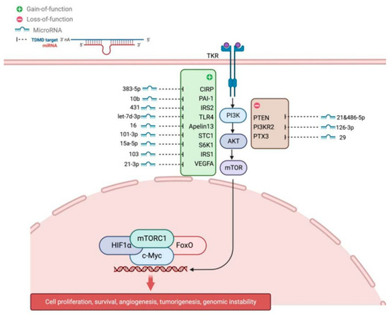
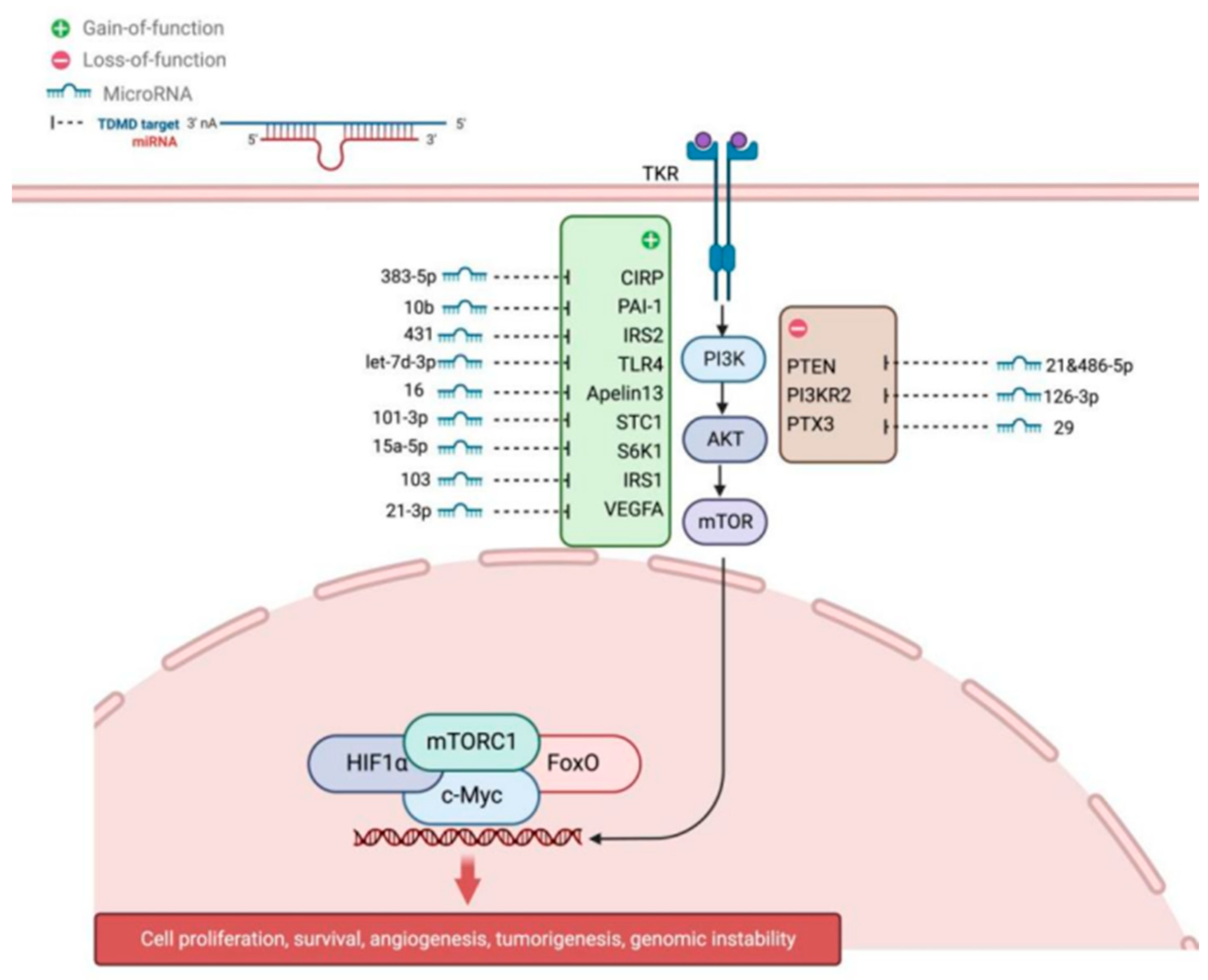
The PI3K/AKT/mTOR axis is central to GC proliferation and follicle dominance. In PCOS, miR-let-7d-3p inhibits the PI3K/Akt pathway and GC proliferation by targeting TLR4 [23]. Guo et al. demonstrated that miR-10b promotes apoptosis by targeting PAI-1 to regulate the PI3K/AKT pathway [24]. An et al. showed that miR-101-3p inhibits PI3K/AKT/mTOR by targeting STC1 in goat ovarian development [26]. High levels of miR-15a-5p correlate with poor ovarian response, likely via inhibiting granulosa cell proliferation through this pathway [27]. Yang et al. reported that miR-431 inhibits GC proliferation by targeting IRS2 and suppressing PI3K/AKT signaling [28]. Wang et al. found that miR-29 regulates goat GC function by targeting PTX3, activating both PI3K/AKT/mTOR and Erk1/2 pathways [29]. Han et al. demonstrated that miR-21 in oocyte secretory factors inhibits PI3K/Akt signaling and promotes GC apoptosis [30]. Recent work in sheep showed that miR-134-3p suppresses proliferation and induces apoptosis by targeting INHBA and dampening the TGF-β/PI3K/AKT cascade [31]. Clinically, Han et al. discovered that PD-L1 upregulation in PCOS suppresses GC apoptosis by activating PI3K/AKT, highlighting a potential immune-endocrine axis [32]. The miRNA-mediated regulation of PI3K/AKT/mTOR signaling is depicted in Figure 1.
Figure 1.
miRNAs regulating the PI3K/AKT/mTOR signaling pathway. This schematic illustrates complex post-transcriptional regulation of the central PI3K/AKT/mTOR cascade by multiple miRNAs. Inhibitory miRNAs (red lines) suppress pathway components: let-7d-3p targets TLR4; miR-431 targets IRS2; miR-10b targets PAI-1; miR-101-3p targets STC1; and PTEN acts as a natural brake by converting PIP3 to PIP2. Modulatory miRNAs (black arrows) target regulatory nodes: miR-126-3p targets PIK3R2; miR-29 targets PTX3; miR-21-3p targets VEGFA; miR-103 modulates mTOR; miR-15a-5p targets IRS1; miR-383-5p targets CIRP; and miR-486-5p targets APPL1. Upon activation, PI3K phosphory lates PIP2 to PIP3, leading to AKT phosphorylation and mTORC1 activation. Downstream effectors regulate proliferation, survival, and metabolism.
Beyond the well-characterized miRNA regulators, emerging evidence from high-throughput screening has identified additional miRNA-target interactions that modulate PI3K/AKT/mTOR signaling, though their functional roles in granulosa cells remain to be fully validated. These include miR-126-3p targeting PIK3R2 (the regulatory subunit of PI3K), miR-103 directly regulating mTOR expression, and miR-21-3p controlling VEGFA-mediated angiogenic signals. A study in endometrial cancer cells demonstrated that miR-126-3p directly binds the 3′UTR of PIK3R2, reducing PI3K activity and downstream AKT phosphorylation [33]. Similarly, miR-103 has been shown to suppress mTOR expression in hepatocellular carcinoma, influencing cell metabolism and proliferation [34]. While these specific interactions have not yet been confirmed in ovarian granulosa cells, the conserved nature of miRNA targeting suggests they may represent novel regulatory nodes in follicular development. Additionally, the cold-inducible RNA-binding protein (CIRP) has been identified as a target of miR-383-5p in stress responses [35], and the tumor suppressor PTEN is co-targeted by miR-21 and miR-486-5p within the PI3K/AKT/mTOR network [36,37], offering potential new avenues for investigating granulosa cell metabolism and survival.
2.2.2. JAK/STAT and Hippo Signaling Pathways
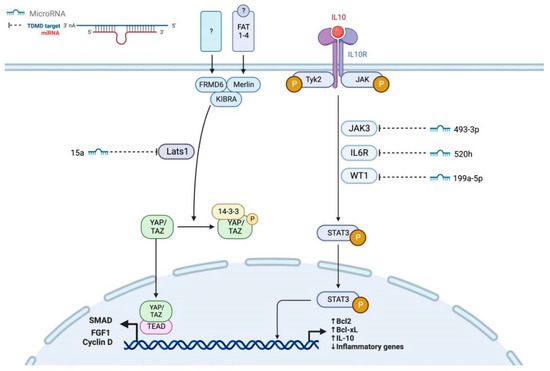
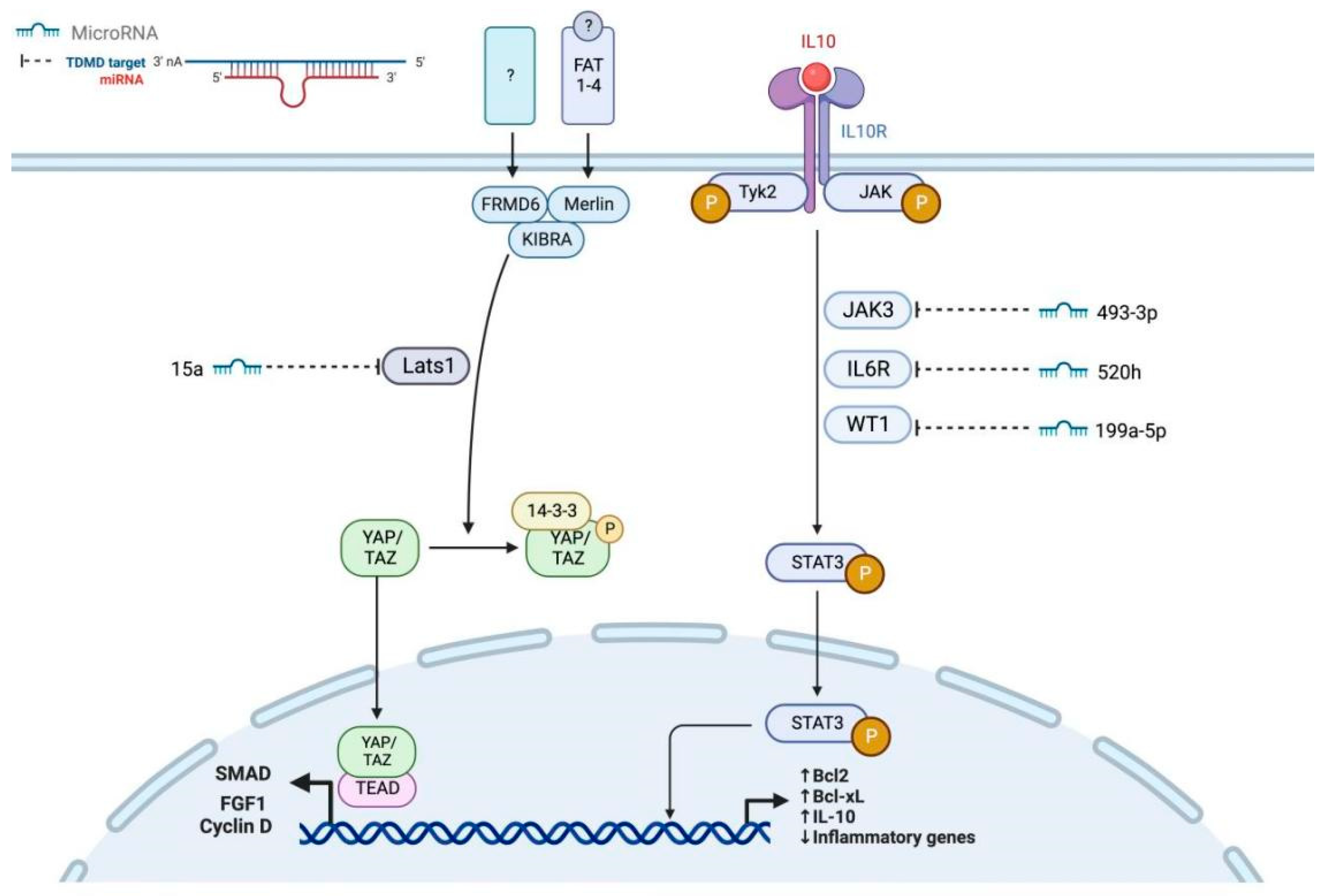
The JAK/STAT pathway is essential for cytokine signaling in GCs. Frost et al. reported conserved roles in human follicle development [38]. Hall et al. confirmed PI3K/AKT and JAK/STAT signaling as conserved biomarkers for ovarian development [39]. Yang et al. showed that IL-6 promotes FSH-induced VEGF expression through JAK/STAT3 in bovine GCs [40]. Ndiaye et al. identified novel JAK3-interacting proteins in GCs, including LEPROTL1, INHBA, and CDKN1B [41]. The highly expressed miR-199a-5p in PCOS patients inhibits WT1-mediated JAK/STAT3 activation, and its own inhibition has been shown to improve PCOS pathology [42].
The Hippo pathway is emerging as a critical regulator. Ai et al. determined that miR-15a targets Lats1 to inhibit GC proliferation and induce senescence. Recent studies expand Hippo crosstalk: Wang et al. demonstrated that RSPO2 coordinates with GDF9:BMP15 heterodimers to promote GC development via Wnt/β-catenin signaling, which extensively crosstalks with Hippo [43]. Shan et al. identified miR-184 as a transcriptional activator that acts as a transcriptional enhancer that indirectly induces SMAD3 expression by targeting an inhibitory factor, potentially interfacing with Hippo through TGF-β crosstalk [44]. The cytokine sensitivity of granulosa cells is further fine-tuned by miRNAs targeting receptor components of the JAK/STAT pathway. Recent transcriptomic analyses have identified miR-520h as a post-transcriptional regulator of IL6R expression, modulating granulosa cell responsiveness to IL-6-mediated signals [45]. Similarly, miR-493-3p has been implicated in controlling MIF (macrophage migration inhibitory factor) expression, thereby influencing macrophage infiltration and inflammatory responses [46]. While these specific receptor-targeting mechanisms have been characterized primarily in immune cells, their presence in ovarian granulosa cell transcriptome datasets suggests they may play important roles in regulating the follicular immune microenvironment during folliculogenesis and atresia. Validation of these interactions in granulosa cell-specific models represents an important area for future investigation. Taken together, these recent studies highlight that the Hippo pathway does not act in isolation; rather, through extensive crosstalk with other key pathways such as Wnt/β-catenin and TGF-β, it forms an integral part of the complex network determining granulosa cell fate. The miRNAs regulating JAK/STAT3 and Hippo signaling pathways are illustrated in Figure 2.
Figure 2.
miRNAs regulating JAK/STAT3 and Hippo signaling pathways. This schematic depicts the crosstalk between two critical signaling axes. Hippo pathway (Left): Upstream proteins Merlin, KIBRA, FAT, and FRMD6Y phosphorylate and activate Lats1 kinase. Lats1 subsequently phosphorylates YAP/TAZ, sequestering them in the cytoplasm via 14-3-3 binding and preventing TEAD-mediated transcription of downstream genes, including Bcl2, Bcl-xL, CyclinD, and FGF1. The miRNA miR-15a directly targets Lats1 (red inhibition line), derepressing YAP/TAZ nuclear translocation. JAK/STAT3 Pathway (Right): Ligands bind to cytokine receptors IL10R and IL6R, activating Tyk2 and JAK3 kinases. Phosphorylated STAT3 dimerizes, translocates to the nucleus, and drives transcription of target genes, including Bcl2, Bcl-xL, CyclinD, FGF1, and inflammatory mediators. The miRNAs miR-199a-5p, miR-520h, and miR-493-3p interface with pathway components by targeting WT1, IL6R, and IL10R, respectively (note: connection directionality is not specified in the diagram). Pathway Crosstalk: Both pathways converge on the regulation of proliferation and apoptosis-related genes (Bcl2, Bcl-xL, CyclinD, FGF1). The Hippo pathway primarily controls developmental signals, while JAK/STAT3 mediates inflammatory and survival responses.
2.2.3. MAPK Signaling Pathway
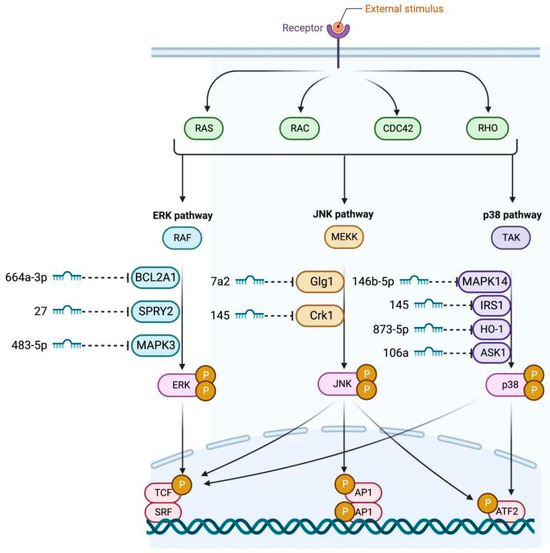
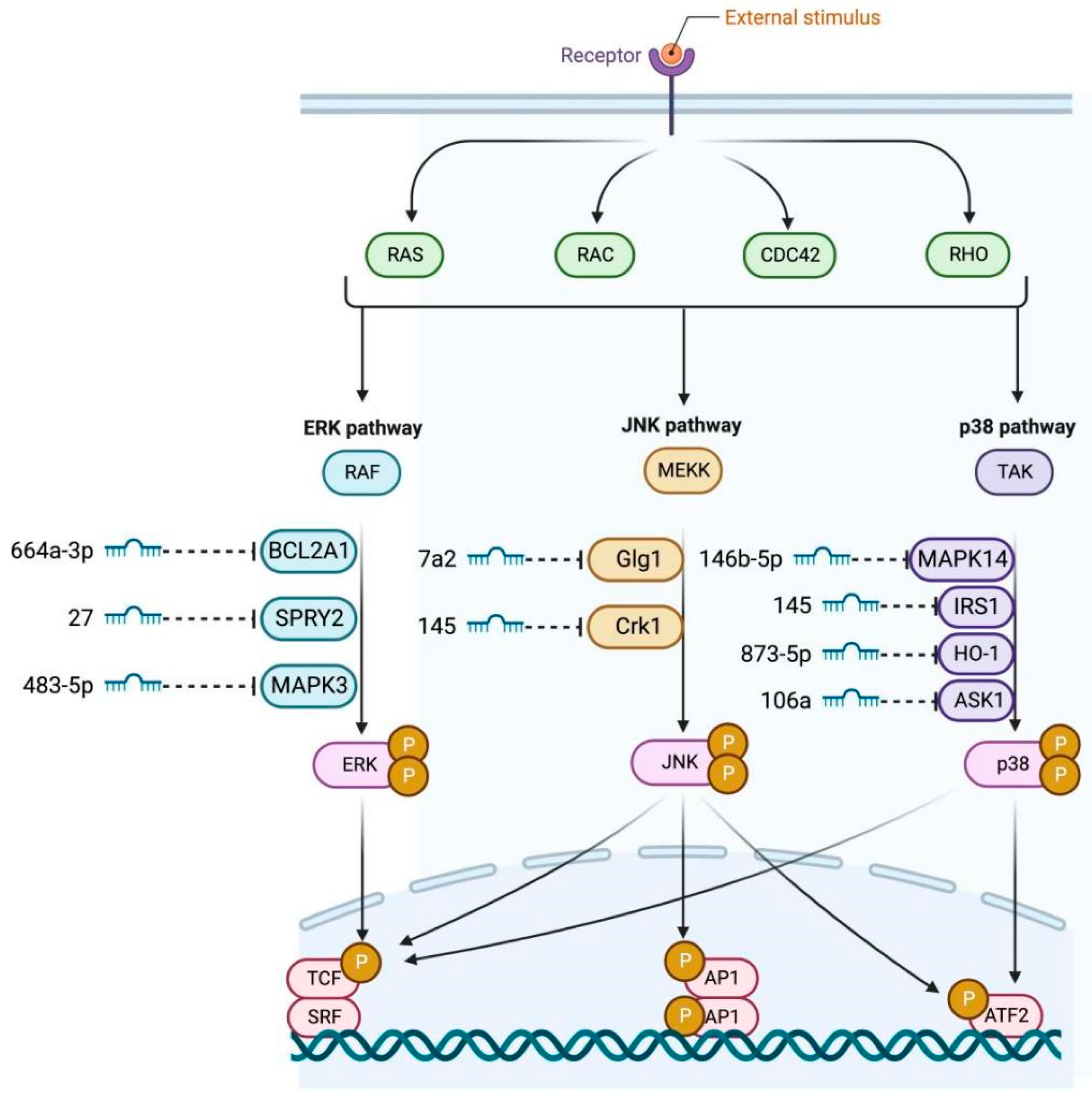
The MAPK pathway is a major signal transducer in GCs. miR-664a-3p inhibits KGN cell proliferation and promotes apoptosis by targeting BCL2A1 and consequently blocking MAPK/ERK signaling [47]. Liu et al. found that miR-146b-5p overexpression attenuates premature ovarian failure in mice by inhibiting the Dab2ip/Ask1/p38MAPK pathway [7]. Hong et al. showed that miR-106a increases GC viability by inhibiting ASK1 mRNA and p38MAPK phosphorylation [48]. Cai et al. proposed that miR-145 inhibits MAPK/ERK by targeting IRS1 in PCOS [49]. Wang et al. found that miR-145 targets Crkl to promote GC proliferation, differentiation, and steroidogenesis via JNK/p38MAPK [50]. The MAPK pathway also interfaces with less-characterized miRNA-target pairs that may have granulosa cell-specific functions. The diagram depicts miR-146b-5p targeting Golgi glycoprotein 1 (Glg1), a protein involved in vesicular trafficking and secretion [51]. Additionally, miR-873-5p is shown to regulate heme oxygenase-1 (HO-1), an enzyme critical for cellular stress response and antioxidant defense [52]. While these interactions have been documented in cancer and endothelial cell lines, their roles in granulosa cell oxidative stress responses during follicular development remain unexplored. The presence of miR-483-5p in the pathway network without an explicit target suggests additional regulatory complexity that warrants further investigation. These emerging miRNA-target pairs highlight knowledge gaps that must be addressed to fully understand MAPK-mediated control of granulosa cell fate. The miRNAs regulating the MAPK signaling pathway are illustrated in Figure 3.
Figure 3.
miRNAs regulating the MAPK signaling pathway. This schematic depicts an integrated network where external stimuli activate receptors to stimulate small GTPases (RAS, RAC, CDC42, RHO), initiating three convergent MAPK cascades. The ERK branch signals through RAF, the JNK branch through MEKK, and the p38 branch through TAK/ASK1, ultimately activating ERK, JNK, and p38 kinases, respectively. Multiple miRNAs exert inhibitory control: miR-664a-3p targets BCL2A1; miR-7a-2-3p targets Glg1; miR-146b-5p targets MAPK14; miR-27a targets SPRY2; miR-145 targets IRS1 and Crk1; miR-873-5p targets HO-1; miR-483-5p targets MAPK3; and miR-106a targets ASK1. These cascades coalesce on common transcription factors (TCF, AP1, SRF, ATF2) that orchestrate cell proliferation, differentiation, and stress responses.
2.2.4. TGF-β/SMAD Signaling Pathway
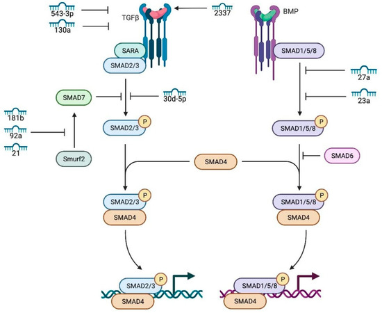
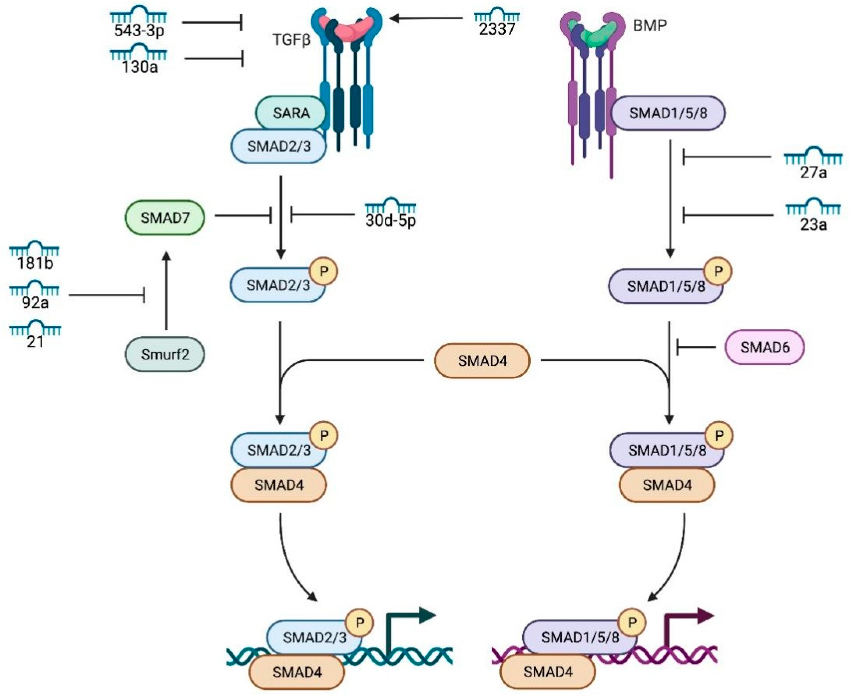
TGF-β/SMAD signaling is fundamental to female fertility. miR-543-3p targets decorin (DCN), leading to reduced expression of TGF-β pathway-related genes [53]. Du et al. reported that miR-130a promotes GC apoptosis by inhibiting TGF-β1 expression [54]. Wang et al. identified miR-2337 as a small activating RNA that enhances TGF-β1 promoter activity [55]. Several miRNAs target SMAD proteins: Nie et al. showed miR-23a and miR-27a promote apoptosis by targeting SMAD5 [56]; Yu et al. found miR-30d-5p targets SMAD2, inhibiting proliferation and promoting apoptosis [57]; Yao et al. showed miR-181b-induced SMAD7 downregulation controls GC apoptosis [58]; Liu et al. reported miR-92a inhibits apoptosis by targeting SMAD7 [59]; Yin et al. demonstrated that miR-224 targets SMAD4, regulating GC proliferation and estradiol production [60].
TGF-β1 also trans-regulates miRNA expression: Du et al. found that TGF-β1 induces miR-764-3p expression, which enhances GC proliferation via SMAD4 while inhibiting estradiol synthesis via SF-1 [61]. Yin et al. showed miR-383 transactivates miR-320, inhibiting estradiol synthesis and proliferation by targeting E2F1 and SF-1 [62]. Importantly, SMAD4 activates Wnt signaling to inhibit GC apoptosis [61] and represses multiple miRNAs that negatively regulate TGF-β signaling [63]. Recent evidence identifies the TGF-β1–SMAD4–BMF module as a conserved pro-survival axis in ruminant and human GCs [64,65,66]. The TGF-β/SMAD network also includes miRNAs with less-defined roles in granulosa cells. Figure 4 places miR-33 within the pathway without specifying a direct molecular target, suggesting it may function as a fine-tuner of overall pathway activity rather than a single-gene regulator. Studies in ovarian granulosa cells indicate that miR-33 can modulate TGF-β signaling by targeting key components of the pathway. For instance, in the context of polycystic ovary syndrome, miR-33b is upregulated and directly targets the TGF-β receptor TGFBR1, thereby influencing the signaling cascade and cellular outcomes [67]. In the ovarian context, miR-33 expression has been detected in follicular fluid exosomes, though its specific granulosa cell targets remain uncharacterized. Elucidating the mechanism by which miR-33 interfaces with the TGF-β/SMAD cascade could reveal novel regulatory nodes controlling follicular selection and atresia.
Figure 4.
miRNAs regulating the TGF-β1 signaling pathway. This schematic depicts miRNA regulatory nodes within the canonical TGF-β/SMAD cascade. Ligand binding to the TGF-β/BMP receptor complex recruits the SARA adaptor protein, facilitating phosphorylation of SMAD1/5/8 (BMP pathway) and SMAD2/3 (TGF-β pathway). Phosphorylated R-SMADs form heteromeric complexes with SMAD4, translocating to the nucleus to regulate transcription. The inhibitory arm comprises SMAD6, SMAD7, and the E3 ubiquitin ligase Smurf2. Multiple miRNAs interface with this network: miR-130a targets SARA; miR-30d-5p and miR-23a target SMAD2/3; miR-27a, miR-181b, miR-92a, and miR-21 target SMAD7; 543-3p and 33 are shown as pathway-associated miRNAs without explicit target connections. The diagram uses numerical miRNA identifiers, phosphorylation is denoted by “P”, and connection directionality is not specified. Key takeaway: This network demonstrates multi-level miRNA control targeting adaptor proteins, signal-transducing SMADs, and inhibitory SMADs, thereby regulating signal flow through the TGF-β pathway.
2.3. MiRNAs Regulating Steroidogenesis and Oxidative Stress Homeostasis
2.3.1. Steroid Hormone Production
MiRNAs critically regulate estrogen synthesis by targeting CYP19A1 (aromatase) and CYP11A1. Several miRNAs directly target CYP19A1: miR-210, miR-32, miR-146b, miR-10b, and miR-378 reduce estradiol secretion and promote apoptosis [68,69,70]. Others target transcriptional regulators: miR-29a impairs CREB1 to inhibit CYP19A1 [71]; Ding et al. showed miR-202-5p transactivated by SF-1 targets TGFβR2 [72]; Zhang et al. found miR-17-5p targets E2F1 [73]; Liu et al. reported miR-1275 impairs LRH-1/CYP19A1 axis [74]; Wang et al. showed miR-27a-3p targets CREB1 [75]; and Du et al. found miR-764-3p targets SF-1 [76]. Circular RNAs also participate: Zhang et al. identified hsa_circ_0043532 upregulating CYP19A1 by acting as a ceRNA for miR-1270 [77], while Tao et al. revealed the KRTAP5-AS1/miR-199b-5p/CYP19A1 axis [78]. Conversely, some miRNAs promote steroidogenesis: Zhang et al. showed miR-320a targets RUNX2 to enhance steroidogenesis [79]; Wu et al. found miR-132 targets Nurr1 [80]; and Dai et al. demonstrated miR-133b targets Foxl2 [81]. A 2024 study in chickens showed miR-7 promotes apoptosis and autophagy by targeting KLF4 via JAK/STAT3 [82].
2.3.2. Reactive Oxygen Species (ROS) Balance
MiRNAs regulate ROS metabolism, critical for GC survival. Liu et al. reported that miR-27 increases ROS stress and apoptosis by targeting SPRY2 and downregulating the p-ERK/Nrf2 pathway [83]. Ding et al. found hAMSC exosome-derived miR-320a targeting SIRT4 reduces ROS production [84], while hUMMSC exosome-derived miR-17-5p targeting SIRT7 exerts similar effects [85]. FOXO1 is a key mediator: it induces TXNIP expression, activates NLRP3 inflammasomes, causing oxidative stress and apoptosis [86]. Several miRNAs target FOXO1: Yan et al. found miR-1224-5p reduces PCOS by inhibiting NLRP3 activation via FOXO1 [87]; Wei et al. showed miR-221-3p reduces ovarian reserve by targeting FOXO1 in elderly women [88]; and Zhang et al. found miR-181a increases FOXO1 acetylation and promotes apoptosis through SIRT1 downregulation [77]. A 2025 study demonstrated that miR-361-5p alleviates GC dysfunction in diminished ovarian reserve by targeting SLC25A24 to maintain mitochondrial function [89].
3. Conclusions and Future Perspectives
This review establishes miRNAs as a decisive regulatory network governing GC proliferation, apoptosis, steroidogenesis, and oxidative stress responses. Integrated analyses identify miR-129, miR-30a-5p, and miR-146a-5p as master regulators in human GCs [6], while the TGF-β1–SMAD4–BMF module represents a conserved pro-survival axis across species [64,65,66]. MiRNAs target core components of PI3K/AKT/mTOR, TGF-β/SMAD, JAK/STAT, MAPK, and Hippo pathways, as well as key functional genes including TP53, BCL2 family members, KCNA5, Mfn1/2, and steroidogenic enzymes CYP19A1/CYP11A1.
However, significant translational challenges remain. First, miRNA pleiotropy poses off-target risks—miR-21 alone has four validated targets (PTEN, SMAD7) in PCOS [90,91]. Second, the vast majority of evidence derives from in vitro cultures or animal models, creating a substantial translational gap [1]. Third, the absence of follicle-specific delivery systems limits therapeutic applicability. Recent advances in tissue-specific miRNA delivery using cationic polymer nanocomplexes and peptide-modified AAV variants offer promise, but require further optimization for ovarian applications [3].
Future research must prioritize validation in human-relevant models. The development of human “ovaroid” organoids from induced pluripotent stem cells without exogenous transcription factors represents a crucial step toward bridging this gap [92]. Single-cell and spatial transcriptomics will further refine our understanding of cell type-specific miRNA functions within the follicular niche [2,6]. Addressing these challenges is paramount before miRNA-based strategies can be translated into viable clinical tools for managing PCOS, POF, and other ovarian disorders.
Author Contributions
Writing—original draft, Investigation: S.C. and J.L.; Writing—review and editing: Y.S., L.C., Y.Z., L.N., Y.W., X.Z., L.S., Y.T., M.G., and L.Z.; Supervision, Methodology, Investigation: M.G. and L.Z. Conceptualization: L.Z. All authors have read and agreed to the published version of the manuscript.
Funding
This research was funded by National The Natural Science Foundation of China (32402752), Sichuan Science and Technology Program (2021ZDZX0008, SCSZTD-2026-08); Postdoctoral Fellowship Program of CPSF (GZC20231860), China Postdoctoral Science Foundation (2023M732514), China Agriculture Research System (CARS-35), Independent Research Fund of State Key Laboratory of Swine and Poultry Breeding Industry (20252QQZ-G51).
Institutional Review Board Statement
Not applicable.
Informed Consent Statement
Not applicable.
Data Availability Statement
No new data were created or analyzed in this study.
Conflicts of Interest
The authors declare no conflicts of interest.
Abbreviations
The following abbreviations are used in this manuscript:
| miRNAs | MicroRNAs |
| GCs | Granulosa Cells |
| CGCs | Cumulus Granulosa Cells |
| mGCs | Mural Granulosa Cells |
| POF | Premature Ovarian Failure |
| PCOS | Polycystic Ovary Syndrome |
| POI | Premature Ovarian Insufficiency |
| FSH | Follicle-Stimulating Hormone |
| LH | Luteinizing Hormone |
| GnRH | Gonadotropin-Releasing Hormone |
| E2 | Estradiol |
| P4 | Progesterone |
| IGF-1 | Insulin-like Growth Factor 1 |
| IGF-1R | Insulin-like Growth Factor 1 Receptor |
| TP53 | Tumor Protein P53 |
| BCL2 | B-cell Lymphoma 2 |
| BAX | BCL2-Associated X Protein |
| KCNA5 | Potassium Voltage-Gated Channel Subfamily A Member 5 |
| Mfn1/2 | Mitofusin 1/2 |
| CYP19A1 | Cytochrome P450 Family 19 Subfamily A Member 1 (Aromatase) |
| CYP11A1 | Cytochrome P450 Family 11 Subfamily A Member 1 |
| SF-1 | Steroidogenic Factor-1 (NR5A1) |
| FOXO1 | Forkhead Box O1 |
| SIRTs | Sirtuins |
| Nrf2 | Nuclear Factor Erythroid 2–Related Factor 2 |
| PI3K/AKT/mTOR | Phosphoinositide 3-Kinase / Protein Kinase B / Mammalian Target of Rapamycin |
| TGF-β/SMAD | Transforming Growth Factor Beta / Mothers Against Decapentaplegic Homolog |
| JAK/STAT | Janus Kinase / Signal Transducer and Activator of Transcription |
| MAPK | Mitogen-Activated Protein Kinase |
| ERK | Extracellular Signal-Regulated Kinase |
| Abbreviation | Full Term |
| ROS | Reactive Oxygen Species |
| mt-OXPHOS | Mitochondrial Oxidative Phosphorylation |
| ATP | Adenosine Triphosphate |
| cAMP | Cyclic Adenosine Monophosphate |
| ECM | Extracellular Matrix |
| HPO axis | Hypothalamic-Pituitary-Ovarian axis |
| KGN cells | Human Granulosa-like Tumor Cell Line |
| BMSC | Bone Mesenchymal Stem Cell |
| ART | Assisted Reproductive Technology |
| scRNA-seq | Single-Cell RNA Sequencing |
References
- Yi, Y.; Zhong, Y.; Liang, R.N.; Liu, C.Y.; Zhang, Y.F.; Zhang, Y.X. Polycystic ovary syndrome: The role of granulosa cell proliferation and apoptosis in disease development. J. Steroid Biochem. Mol. Biol. 2025, 254, 106838. [Google Scholar] [CrossRef] [Scilit]
- Gao, F.; Wang, F.; Chen, Y.; Deng, B.; Yang, F.; Cao, H.; Chen, J.; Chen, H.; Qi, F.; Kapranov, P. The human genome encodes a multitude of novel miRNAs. Nucleic Acids Res. 2025, 53, Gkaf070. [Google Scholar] [CrossRef] [Scilit]
- Liu, L.; Fang, Y. The Role of Ovarian Granulosa Cells Related-ncRNAs in Ovarian Dysfunctions: Mechanism Research and Clinical Exploration. Reprod. Sci. 2025, 32, 2098–2120. [Google Scholar] [CrossRef] [Scilit]
- Cai, J.H.; Sun, Y.T.; Bao, S. HucMSCs-exosomes containing miR-21 promoted estrogen production in ovarian granulosa cells via LATS1-mediated phosphorylation of LOXL2 and YAP. Gen. Comp. Endocrinol. 2022, 321–322, 114015. [Google Scholar] [CrossRef] [Scilit]
- Lin, L.; Gao, W.; Chen, Y.; Li, T.; Sha, C.; Chen, L.; Yang, M.; Wei, H.; Chen, Y.; Zhu, X. Reactive oxygen species-induced SIAH1 promotes granulosa cells’ senescence in premature ovarian failure. J. Cell. Mol. Med. 2022, 26, 2417–2427. [Google Scholar] [CrossRef] [Scilit]
- Yousuf, S.; Malik, W.A.; Feng, H.; Liu, T.; Xie, L.; Miao, X. Integrated analysis of microRNA and mRNA interactions regulating fecundity in the ovaries of two distinct sheep breeds. BMC Genom. 2025, 26, 707. [Google Scholar] [CrossRef] [Scilit]
- Liu, T.; Lin, J.; Chen, C.; Nie, X.; Dou, F.; Chen, J.; Wang, Z.; Gong, Z. MicroRNA-146b-5p overexpression attenuates premature ovarian failure in mice by inhibiting the Dab2ip/Ask1/p38-Mapk pathway and γH2A.X phosphorylation. Cell Prolif. 2021, 54, E12954. [Google Scholar] [CrossRef] [Scilit] [PubMed]
- Sun, B.; Ma, Y.; Wang, F.; Hu, L.; Sun, Y. miR-644-5p carried by bone mesenchymal stem cell-derived exosomes targets regulation of p53 to inhibit ovarian granulosa cell apoptosis. Stem Cell Res. Ther. 2019, 10, 360. [Google Scholar] [CrossRef] [Scilit]
- Xie, H.L.; Zhu, S.; Zhang, J.; Wen, J.; Yuan, H.J.; Pan, L.Z.; Luo, M.J.; Tan, J.H. Glucose metabolism during in vitro maturation of mouse oocytes: An study using RNA interference. J. Cell. Physiol. 2018, 233, 6952–6964. [Google Scholar] [CrossRef] [Scilit] [PubMed]
- Liang, M.; Yao, G.; Yin, M.; Lü, M.; Tian, H.; Liu, L.; Lian, J.; Huang, X.; Sun, F. Transcriptional cooperation between p53 and NF-κB p65 regulates microRNA-224 transcription in mouse ovarian granulosa cells. Mol. Cell. Endocrinol. 2013, 370, 119–129. [Google Scholar] [CrossRef] [Scilit] [PubMed]
- Tao, H.; Xiong, Q.; Ji, Z.; Zhang, F.; Liu, Y.; Chen, M. NFAT5 is Regulated by p53/miR-27a Signal Axis and Promotes Mouse Ovarian Granulosa Cells Proliferation. Int. J. Biol. Sci. 2019, 15, 287–297. [Google Scholar] [CrossRef] [Scilit]
- Zhou, S.; Xia, L.; Chen, Y.; Guo, W.; Hu, J. miR-3188 Regulates proliferation and apoptosis of granulosa cells by targeting KCNA5 in the polycystic ovary syndrome. Acta Biochim. Pol. 2021, 68, 83–89. [Google Scholar] [CrossRef] [Scilit] [PubMed]
- Gao, L.; Wu, D.; Wu, Y.; Yang, Z.; Sheng, J.; Lin, X.; Huang, H. MiR-3940-5p promotes granulosa cell proliferation through targeting KCNA5 in polycystic ovarian syndrome. Biochem. Biophys. Res. Commun. 2020, 524, 791–797. [Google Scholar] [CrossRef] [Scilit] [PubMed]
- Hou, X.; Zhu, S.; Zhang, H.; Li, C.; Qiu, D.; Ge, J.; Guo, X.; Wang, Q. Mitofusin1 in oocyte is essential for female fertility. Redox Biol. 2019, 21, 101110. [Google Scholar] [CrossRef] [Scilit]
- Wang, L.; Song, S.; Liu, X.; Zhang, M.; Xiang, W. Low MFN2 expression related to ageing in granulosa cells is associated with assisted reproductive technology outcome. Reprod. Biomed. Online 2019, 38, 152–158. [Google Scholar] [CrossRef] [Scilit]
- Shi, S.; Zhou, X.; Li, J.; Zhang, L.; Hu, Y.; Li, Y.; Yang, G.; Chu, G. MiR-214-3p promotes proliferation and inhibits estradiol synthesis in porcine granulosa cells. J. Anim. Sci. Biotechnol. 2020, 11, 94. [Google Scholar] [CrossRef] [Scilit]
- Liu, S.; Chen, J.; Liu, M.; Zhang, C.; Chao, X.; Yang, H.; Wang, T.; Bi, H.; Ding, Y.; Wang, Z.; et al. miR-107 suppresses porcine granulosa cell proliferation and estradiol synthesis while promoting apoptosis via targeting PTGS2. Theriogenology 2025, 238, 117367. [Google Scholar] [CrossRef] [Scilit]
- Wang, Y.; Tian, F.; Yue, S.; Li, J.; Li, A.; Liu, Y.; Liang, J.; Gao, Y.; Xue, S. miR-17-5p-Mediated RNA Activation Upregulates KPNA2 Expression and Inhibits High-Glucose-Induced Apoptosis of Sheep Granulosa Cells. Int. J. Mol. Sci. 2025, 26, 943. [Google Scholar] [CrossRef] [Scilit] [PubMed]
- Rodosthenous, R.S.; Baccarelli, A.A.; Mansour, A.; Adir, M.; Israel, A.; Racowsky, C.; Hauser, R.; Bollati, V.; Machtinger, R. Supraphysiological Concentrations of Bisphenol A Alter the Expression of Extracellular Vesicle-Enriched miRNAs From Human Primary Granulosa Cells. Toxicol. Sci. 2019, 169, 5–13. [Google Scholar] [CrossRef] [Scilit]
- Chen, Y.; Zhang, X.; An, Y.; Liu, B.; Lu, M. LncRNA HCP5 promotes cell proliferation and inhibits apoptosis via miR-27a-3p/IGF-1 axis in human granulosa-like tumor cell line KGN. Mol. Cell. Endocrinol. 2020, 503, 110697. [Google Scholar] [CrossRef] [Scilit]
- Jiang, B.; Xue, M.; Xu, D.; Song, J.; Zhu, S. Down-regulated lncRNA HOTAIR alleviates polycystic ovaries syndrome in rats by reducing expression of insulin-like growth factor 1 via microRNA-130a. J. Cell. Mol. Med. 2020, 24, 451–464. [Google Scholar] [CrossRef] [Scilit]
- Geng, Y.; Sui, C.; Xun, Y.; Lai, Q.; Jin, L. MiRNA-99a can regulate proliferation and apoptosis of human granulosa cells via targeting IGF-1R in polycystic ovary syndrome. J. Assist. Reprod. Genet. 2019, 36, 211–221. [Google Scholar] [CrossRef] [Scilit] [PubMed]
- Wu, W.; Duan, C.; Lv, H.; Song, J.; Cai, W.; Fu, K.; Xu, J. MiR-let-7d-3p inhibits granulosa cell proliferation by targeting TLR4 in polycystic ovary syndrome. Reprod. Toxicol. 2021, 106, 61–68. [Google Scholar] [CrossRef] [Scilit] [PubMed]
- Guo, L.; Huang, Q.; Zhao, J.; Liu, H.; Lu, W.; Wang, J. microRNA-10b promotes the apoptosis of bovine ovarian granulosa cells by targeting plasminogen activator inhibitor-1. Theriogenology 2021, 176, 206–216. [Google Scholar] [CrossRef] [Scilit] [PubMed]
- Yang, T.; Wang, L.; Zhang, Y.; Zheng, J.; Liu, L. MicroRNA-451a plays a role in polycystic ovary syndrome by regulating ovarian granulosa cell proliferation and apoptosis. Exp. Ther. Med. 2021, 21, 583. [Google Scholar] [CrossRef] [Scilit]
- An, X.; Ma, H.; Liu, Y.; Li, F.; Song, Y.; Li, G.; Bai, Y.; Cao, B. Effects of miR-101-3p on goat granulosa cells in vitro and ovarian development in vivo via STC1. J. Anim. Sci. Biotechnol. 2020, 11, 102. [Google Scholar] [CrossRef] [Scilit]
- Zhang, K.; Zhong, W.; Li, W.P.; Chen, Z.J.; Zhang, C. miR-15a-5p levels correlate with poor ovarian response in human follicular fluid. Reproduction 2017, 154, 483–496. [Google Scholar] [CrossRef] [Scilit]
- Yang, L.; Lv, Q.; Liu, J.; Qi, S.; Fu, D. miR-431 regulates granulosa cell function through the IRS2/PI3K/AKT signaling pathway. J. Reprod. Dev. 2020, 66, 231–239. [Google Scholar] [CrossRef] [Scilit]
- Wang, P.; Liu, S.; Zhu, C.; Duan, Q.; Jiang, Y.; Gao, K.; Bu, Q.; Cao, B.; An, X. MiR-29 regulates the function of goat granulosa cell by targeting PTX3 via the PI3K/AKT/mTOR and Erk1/2 signaling pathways. J. Steroid Biochem. Mol. Biol. 2020, 202, 105722. [Google Scholar] [CrossRef] [Scilit]
- Han, X.; Xue, R.; Yuan, H.J.; Wang, T.Y.; Lin, J.; Zhang, J.; Liang, B.; Tan, J.H. MicroRNA-21 plays a pivotal role in the oocyte-secreted factor-induced suppression of cumulus cell apoptosis†. Biol. Reprod. 2017, 96, 1167–1180. [Google Scholar] [CrossRef] [Scilit]
- Huang, X.; Bao, Y.; Yang, F.; Li, X.; Wang, F.; Zhang, C. miR-134-3p Regulates Cell Proliferation and Apoptosis by Targeting INHBA via Inhibiting the TGF-β/PI3K/AKT Pathway in Sheep Granulosa Cells. Biology 2024, 14, 24. [Google Scholar] [CrossRef] [Scilit]
- Han, R.; Zhu, Y.; Zhang, X.; Liu, X.; Xia, Y.; Ye, E.; La, X.; Ding, J. Upregulation of PD-L1 contributes to improving the apoptosis of granulosa cells via the PI3K/AKT pathway in PCOS. J. Assist. Reprod. Genet. 2025, 42, 611–625. [Google Scholar] [CrossRef] [Scilit]
- Zheng, X.; Liu, M.; Song, Y.; Feng, C. Long Noncoding RNA-ATB Impairs the Function of Tumor Suppressor miR-126-Mediated Signals in Endometrial Cancer for Tumor Growth and Metastasis. Cancer Biother. Radiopharm. 2019, 34, 47–55. [Google Scholar] [CrossRef] [Scilit]
- Xia, W.; Ni, J.; Zhuang, J.; Qian, L.; Wang, P.; Wang, J. MiR-103 regulates hepatocellular carcinoma growth by targeting AKAP12. Int. J. Biochem. Cell Biol. 2016, 71, 1–11. [Google Scholar] [CrossRef] [Scilit]
- Li, Y.; Wu, X.; Miao, S.; Cao, Q. MiR-383-5p promotes apoptosis of ovarian granulosa cells by targeting CIRP through the PI3K/AKT signaling pathway. Arch. Gynecol. Obstet. 2022, 306, 501–512. [Google Scholar] [CrossRef] [Scilit] [PubMed]
- Chawra, H.S.; Agarwal, M.; Mishra, A.; Chandel, S.S.; Singh, R.P.; Dubey, G.; Kukreti, N.; Singh, M. MicroRNA-21′s role in PTEN suppression and PI3K/AKT activation: Implications for cancer biology. Pathol.-Res. Pract. 2024, 254, 155091. [Google Scholar] [CrossRef] [Scilit] [PubMed]
- Wang, W.; Liu, B.; Sun, S.; Lan, L.; Chen, Y.; Han, S.; Li, X.; Li, Z. Downregulation of miR-486-5p Enhances the Anti-Tumor Effect of 5-Fluorouracil on Pancreatic Cancer Cells. Onco Targets Ther. 2020, 13, 1649–1659, Erratum in Onco Targets Ther. 2020, 13, 4507–4508. [Google Scholar] [CrossRef] [Scilit] [PubMed]
- Frost, E.; Ford, E.; Peters, A.; Reed, N.; McLaughlin, E.; Baker, M.; Lovell-Badge, R.; Sutherland, J. STAT1 and STAT3 are expressed in the human ovary and have JAK1-independent functions in the COV434 human granulosa cell line. Reprod. Fertil. Dev. 2020, 32, 1027–1039. [Google Scholar] [CrossRef] [Scilit]
- Hall, S.E.; Upton, R.M.O.; McLaughlin, E.A.; Sutherland, J.M. Phosphoinositide 3-kinase/protein kinase B (PI3K/AKT) and Janus kinase/signal transducer and activator of transcription (JAK/STAT) follicular signalling is conserved in the mare ovary. Reprod. Fertil. Dev. 2018, 30, 624–633. [Google Scholar] [CrossRef] [Scilit]
- Yang, M.; Wang, L.; Wang, X.; Wang, X.; Yang, Z.; Li, J. IL-6 Promotes FSH-Induced VEGF Expression Through JAK/STAT3 Signaling Pathway in Bovine Granulosa Cells. Cell. Physiol. Biochem. 2017, 44, 293–302. [Google Scholar] [CrossRef] [Scilit]
- Ndiaye, K.; Castonguay, A.; Benoit, G.; Silversides, D.W.; Lussier, J.G. Differential regulation of Janus kinase 3 (JAK3) in bovine preovulatory follicles and identification of JAK3 interacting proteins in granulosa cells. J. Ovarian Res. 2016, 9, 71. [Google Scholar] [CrossRef] [Scilit]
- Shao, S.; Wang, H.; Shao, W.; Liu, N. miR-199a-5p stimulates ovarian granulosa cell apoptosis in polycystic ovary syndrome. J. Mol. Endocrinol. 2020, 65, 187–201. [Google Scholar] [CrossRef] [Scilit]
- Wang, Y.; Li, H.; You, L.; Wang, S.; Bie, J.; Su, Z.; Shi, L.; Su, Y. RSPO2 Coordinates with GDF9:BMP15 Heterodimers to Promote Granulosa Cell and Oocyte Development in Mice. Adv. Sci. 2025, 12, E01973. [Google Scholar] [CrossRef] [Scilit]
- Shan, B.; Huo, Y.; Guo, Z.; Li, Q.; Pan, Z.; Li, Q.; Du, X. miR-184, a downregulated ovary-elevated miRNA transcriptionally activated by SREBF2, exerts anti-apoptotic properties in ovarian granulosa cells through inducing SMAD3 expression. Cell Death Dis. 2024, 15, 892. [Google Scholar] [CrossRef] [Scilit]
- Huang, Y.H.; Dong, L.P.; Cui, Y.G.; Lu, H.Y. MiR-520h inhibits viability and facilitates apoptosis of KGN cells through modulating IL6R and the JAK/STAT pathway. Reprod. Biol. 2022, 22, 100607. [Google Scholar] [CrossRef] [Scilit] [PubMed]
- Zhu, W.; Wu, C.; Zhou, Z.; Zhang, G.; Luo, L.; Liu, Y.; Huang, Z.; Ai, G.; Zhao, Z.; Zhong, W.; et al. Acetate attenuates hyperoxaluria-induced kidney injury by inhibiting macrophage infiltration via the miR-493-3p/MIF axis. Commun. Biol. 2023, 6, 270. [Google Scholar] [CrossRef] [Scilit] [PubMed]
- He, M.; Mao, G.; Xiang, Y.; Li, P.; Wu, Y.; Zhao, D.; Li, T. MicroRNA-664a-3p inhibits the proliferation of ovarian granulosa cells in polycystic ovary syndrome and promotes apoptosis by targeting BCL2A1. Ann. Transl. Med. 2021, 9, 852. [Google Scholar] [CrossRef] [Scilit] [PubMed]
- Hong, L.; Peng, S.; Li, Y.; Fang, Y.; Wang, Q.; Klausen, C.; Yin, C.; Wang, S.; Leung, P.C.K.; Yang, X. miR-106a Increases Granulosa Cell Viability and Is Downregulated in Women With Diminished Ovarian Reserve. J. Clin. Endocrinol. Metab. 2018, 103, 2157–2166. [Google Scholar] [CrossRef] [Scilit]
- Cai, G.; Ma, X.; Chen, B.; Huang, Y.; Liu, S.; Yang, H.; Zou, W. MicroRNA-145 Negatively Regulates Cell Proliferation Through Targeting IRS1 in Isolated Ovarian Granulosa Cells From Patients With Polycystic Ovary Syndrome. Reprod. Sci. 2017, 24, 902–910. [Google Scholar] [CrossRef] [Scilit]
- Wang, S.; Tang, W.; Ma, L.; Yang, J.; Huang, K.; Du, X.; Luo, A.; Shen, W.; Ding, T.; Ye, S.; et al. MiR-145 regulates steroidogenesis in mouse primary granulosa cells through targeting Crkl. Life Sci. 2021, 282, 119820. [Google Scholar] [CrossRef] [Scilit]
- Li, L.; Zhang, J.; Lu, C.; Wang, B.; Guo, J.; Zhang, H.; Cui, S. MicroRNA-7a2 Contributes to Estrogen Synthesis and Is Modulated by FSH via the JNK Signaling Pathway in Ovarian Granulosa Cells. Int. J. Mol. Sci. 2022, 23, 8565. [Google Scholar] [CrossRef] [Scilit]
- Zhang, H.; Gao, Z.; Zhang, Y.; Wang, H.; Li, Y. MiR-873-5p regulated LPS-induced oxidative stress via targeting heme oxygenase-1 (HO-1) in KGN cells. J. Ovarian Res. 2021, 14, 85. [Google Scholar] [CrossRef] [Scilit]
- Yao, X.; Gao, X.; Bao, Y.; El-Samahy, M.A.; Yang, J.; Wang, Z.; Li, X.; Zhang, G.; Zhang, Y.; Liu, W.; et al. lncRNA FDNCR promotes apoptosis of granulosa cells by targeting the miR-543-3p/DCN/TGF-β signaling pathway in Hu sheep. Mol. Ther.-Nucleic Acids 2021, 24, 223–240. [Google Scholar] [CrossRef] [Scilit] [PubMed]
- Du, X.; Wang, L.; Li, Q.; Wu, W.; Shang, P.; Chamba, Y.; Pan, Z.; Li, Q. miR-130a/TGF-β1 axis is involved in sow fertility by controlling granulosa cell apoptosis. Theriogenology 2020, 157, 407–417. [Google Scholar] [CrossRef] [Scilit] [PubMed]
- Wang, L.; Du, X.; Li, Q.; Wu, W.; Pan, Z.; Li, Q. miR-2337 induces TGF-β1 production in granulosa cells by acting as an endogenous small activating RNA. Cell Death Discov. 2021, 7, 253. [Google Scholar] [CrossRef] [Scilit]
- Nie, M.; Yu, S.; Peng, S.; Fang, Y.; Wang, H.; Yang, X. miR-23a and miR-27a Promote Human Granulosa Cell Apoptosis by Targeting SMAD5. Biol. Reprod. 2015, 93, 98. [Google Scholar] [CrossRef] [Scilit] [PubMed]
- Yu, M.; Liu, J. MicroRNA-30d-5p promotes ovarian granulosa cell apoptosis by targeting Smad2. Exp. Ther. Med. 2020, 19, 53–60. [Google Scholar] [CrossRef] [Scilit]
- Yao, W.; Pan, Z.; Du, X.; Zhang, J.; Li, Q. miR-181b-induced SMAD7 downregulation controls granulosa cell apoptosis through TGF-β signaling by interacting with the TGFBR1 promoter. J. Cell. Physiol. 2018, 233, 6807–6821. [Google Scholar] [CrossRef] [Scilit]
- Liu, J.; Yao, W.; Yao, Y.; Du, X.; Zhou, J.; Ma, B.; Liu, H.; Li, Q.; Pan, Z. MiR-92a inhibits porcine ovarian granulosa cell apoptosis by targeting Smad7 gene. FEBS Lett. 2014, 588, 4497–4503. [Google Scholar] [CrossRef] [Scilit]
- Yao, G.; Yin, M.; Lian, J.; Tian, H.; Liu, L.; Li, X.; Sun, F. MicroRNA-224 Is Involved in Transforming Growth Factor-β-Mediated Mouse Granulosa Cell Proliferation and Granulosa Cell Function by Targeting Smad4. Mol. Endocrinol. 2010, 24, 540–551. [Google Scholar] [CrossRef] [Scilit]
- Du, X.; Li, Q.; Yang, L.; Liu, L.; Cao, Q.; Li, Q. SMAD4 activates Wnt signaling pathway to inhibit granulosa cell apoptosis. Cell Death Dis. 2020, 11, 373. [Google Scholar] [CrossRef] [Scilit]
- Yin, M.; Wang, X.; Yao, G.; Lü, M.; Liang, M.; Sun, Y.; Sun, F. Transactivation of MicroRNA-320 by MicroRNA-383 Regulates Granulosa Cell Functions by Targeting E2F1 and SF-1 Proteins. J. Biol. Chem. 2014, 289, 18239–18257. [Google Scholar] [CrossRef] [Scilit]
- Liu, J.; Tu, F.; Yao, W.; Li, X.; Xie, Z.; Liu, H.; Li, Q.; Pan, Z. Conserved miR-26b enhances ovarian granulosa cell apoptosis through HAS2-HA-CD44-Caspase-3 pathway by targeting HAS2. Sci. Rep. 2016, 6, 21197. [Google Scholar] [CrossRef] [Scilit] [PubMed]
- Li, M.; Liang, W.; Luo, Y.; Wang, J.; Liu, X.; Li, S.; Hao, Z. Transforming growth factor-β1 mediates the SMAD4/BMF pathway to regulate ovarian granulosa cell apoptosis in small tail Han sheep. Theriogenology 2024, 214, 360–369. [Google Scholar] [CrossRef] [Scilit] [PubMed]
- Ren, S.; Liu, Y.; Guo, Y.; Zhao, Z.; Cui, J.; Li, M.; Wang, J. TGF-β1 Mediates Novel-m0297-5p Targeting WNT5A to Participate in the Proliferation of Ovarian Granulosa Cells in Small-Tailed Han Sheep. Int. J. Mol. Sci. 2025, 26, 1961. [Google Scholar] [CrossRef] [Scilit]
- Li, M.; Wang, L.; Luo, Y.; Wang, J.; Liu, X.; Li, S.; Hao, Z. RNA-seq analysis of the biological process and regulatory signal of TGF-β1-mediated changes in ovarian granulosa cells in small-tail Han sheep. Theriogenology 2025, 234, 9–18. [Google Scholar] [CrossRef] [Scilit] [PubMed]
- Li, Y.; Xiang, Y.; Song, Y.; Wan, L.; Yu, G.; Tan, L. Dysregulated miR-142, -33b and -423 in granulosa cells target TGFBR1 and SMAD7: A possible role in polycystic ovary syndrome. Mol. Hum. Reprod. 2019, 25, 638–646. [Google Scholar] [CrossRef] [Scilit]
- Li, Q.; Du, X.; Liu, L.; Liu, H.; Pan, Z.; Li, Q. Upregulation of miR-146b promotes porcine ovarian granulosa cell apoptosis by attenuating CYP19A1. Domest. Anim. Endocrinol. 2021, 74, 106509. [Google Scholar] [CrossRef] [Scilit]
- Li, Q.; Du, X.; Pan, Z.; Zhang, L.; Li, Q. The transcription factor SMAD4 and miR-10b contribute to E2 release and cell apoptosis in ovarian granulosa cells by targeting CYP19A1. Mol. Cell. Endocrinol. 2018, 476, 84–95. [Google Scholar] [CrossRef] [Scilit]
- Toms, D.; Xu, S.; Pan, B.; Wu, D.; Li, J. Progesterone receptor expression in granulosa cells is suppressed by microRNA-378-3p. Mol. Cell. Endocrinol. 2015, 399, 95–102. [Google Scholar] [CrossRef] [Scilit]
- Li, Y.; Liu, Y.D.; Zhou, X.Y.; Chen, S.L.; Chen, X.; Zhe, J.; Zhang, J.; Zhang, Q.Y.; Chen, Y.X. MiR-29a regulates the proliferation, aromatase expression, and estradiol biosynthesis of human granulosa cells in polycystic ovary syndrome. Mol. Cell. Endocrinol. 2019, 498, 110540. [Google Scholar] [CrossRef] [Scilit] [PubMed]
- Ding, Q.; Jin, M.; Wang, Y.; Liu, J.; Kalds, P.; Wang, Y.; Yang, Y.; Wang, X.; Chen, Y. Transactivation of miR-202-5p by Steroidogenic Factor 1 (SF1) Induces Apoptosis in Goat Granulosa Cells by Targeting TGF?R2. Cells 2020, 9, 445. [Google Scholar] [CrossRef] [Scilit]
- Zhang, S.; Wang, L.; Wang, L.; Chen, Y.; Li, F. miR-17-5p affects porcine granulosa cell growth and oestradiol synthesis by targeting E2F1 gene. Reprod. Domest. Anim. 2019, 54, 1459–1469. [Google Scholar] [CrossRef] [Scilit]
- Liu, J.; Li, X.; Yao, Y.; Li, Q.; Pan, Z.; Li, Q. miR-1275 controls granulosa cell apoptosis and estradiol synthesis by impairing LRH-1/CYP19A1 axis. Biochim. Biophys. Acta BBA-Gene Regul. Mech. 2018, 1861, 246–257. [Google Scholar] [CrossRef] [Scilit] [PubMed]
- Wang, M.; Liu, M.; Sun, J.; Jia, L.; Ma, S.; Gao, J.; Xu, Y.; Zhang, H.; Tsang, S.Y.; Li, X. MicroRNA-27a-3p affects estradiol and androgen imbalance by targeting Creb1 in the granulosa cells in mouse polycytic ovary syndrome model. Reprod. Biol. 2017, 17, 295–304. [Google Scholar] [CrossRef] [Scilit] [PubMed]
- Wang, L.; Li, C.; Li, R.; Deng, Y.; Tan, Y.; Tong, C.; Qi, H. MicroRNA-764-3p regulates 17β-estradiol synthesis of mouse ovarian granulosa cells by targeting steroidogenic factor-1. In Vitro Cell. Dev. Biol.-Anim. 2016, 52, 365–373. [Google Scholar] [CrossRef] [Scilit]
- Zhang, H.; Fang, J.; Liu, Y.; Zhu, W.; Xu, Y.; Zhang, Y.; Shen, W.; Li, D.; Hao, C. Hsa_circ_0043532 contributes to PCOS through upregulation of CYP19A1 by acting as a ceRNA for hsa-miR-1270. J. Ovarian Res. 2024, 17, 151. [Google Scholar] [CrossRef] [Scilit]
- Tao, P.; Yan, X.; Wang, Z. Mechanistic role of the KRTAP5-AS1/miR-199b-5p/CYP19A1 axis in polycystic ovary syndrome pathogenesis. J. Ovarian Res. 2025, 18, 176. [Google Scholar] [CrossRef] [Scilit]
- Zhang, C.; Wang, H.; Yan, C.; Gao, X.; Ling, X. Deregulation of RUNX2 by miR-320a deficiency impairs steroidogenesis in cumulus granulosa cells from polycystic ovary syndrome (PCOS) patients. Biochem. Biophys. Res. Commun. 2017, 482, 1469–1476. [Google Scholar] [CrossRef] [Scilit]
- Wu, S.; Sun, H.; Zhang, Q.; Jiang, Y.; Fang, T.; Cui, I.; Yan, G.; Hu, Y. MicroRNA-132 promotes estradiol synthesis in ovarian granulosa cells via translational repression of Nurr1. Reprod. Biol. Endocrinol. 2015, 13, 94. [Google Scholar] [CrossRef] [Scilit]
- Dai, A.; Sun, H.; Fang, T.; Zhang, Q.; Wu, S.; Jiang, Y.; Ding, L.; Yan, G.; Hu, Y. MicroRNA-133b stimulates ovarian estradiol synthesis by targeting Foxl2. FEBS Lett. 2013, 587, 2474–2482. [Google Scholar] [CrossRef] [Scilit]
- Wei, Y.; Zhao, X.; Zhang, Y.; Cui, C.; Han, S.; Yang, C.; Yin, H. miR-7 promotes apoptosis and autophagy of granulosa cells by targeting KLF4 via JAK/STAT3 signaling pathway in chickens. Theriogenology 2024, 230, 322–329. [Google Scholar] [CrossRef] [Scilit]
- Liu, K.; Yang, W.; Hu, M.; Xie, W.; Huang, J.; Cui, M.; He, X.; Nie, X. Exosomal miR-27 negatively regulates ROS production and promotes granulosa cells apoptosis by targeting SPRY2 in OHSS. J. Cell. Mol. Med. 2021, 25, 3976–3990. [Google Scholar] [CrossRef] [Scilit]
- Ding, C.; Qian, C.; Hou, S.; Lu, J.; Zou, Q.; Li, H.; Huang, B. Exosomal miRNA-320a Is Released from hAMSCs and Regulates SIRT4 to Prevent Reactive Oxygen Species Generation in POI. Mol. Ther.-Nucleic Acids 2020, 21, 37–50. [Google Scholar] [CrossRef] [Scilit]
- Ding, C.; Zhu, L.; Shen, H.; Lu, J.; Zou, Q.; Huang, C.; Li, H.; Huang, B. Exosomal miRNA-17-5p derived from human umbilical cord mesenchymal stem cells improves ovarian function in premature ovarian insufficiency by regulating SIRT7. Stem Cells 2020, 38, 1137–1148. [Google Scholar] [CrossRef] [Scilit]
- Nyandwi, J.B.; Ko, Y.S.; Jin, H.; Yun, S.P.; Park, S.W.; Kim, H.J. Rosmarinic acid inhibits oxLDL-induced inflammasome activation under high-glucose conditions through downregulating the p38-FOXO1-TXNIP pathway. Biochem. Pharmacol. 2020, 182, 114246. [Google Scholar] [CrossRef] [Scilit] [PubMed]
- Li, Y.; Yao, N.; Gao, Y.; Wang, Y.; Bai, L.; Xu, J.; Wang, H. MiR-1224-5p attenuates polycystic ovary syndrome through inhibiting NOD-like receptor protein 3 inflammasome activation via targeting Forkhead box O 1. Bioengineered 2021, 12, 8555–8569. [Google Scholar] [CrossRef] [Scilit] [PubMed]
- Wei, C.; Xiang, S.; Yu, Y.; Song, J.; Zheng, M.; Lian, F. miR-221-3p regulates apoptosis of ovarian granulosa cells via targeting FOXO1 in older women with diminished ovarian reserve (DOR). Mol. Reprod. Dev. 2021, 88, 251–260. [Google Scholar] [CrossRef] [Scilit]
- Xu, J.; Jia, Y. miR-361-5p regulates SLC25A24 to maintain mitochondrial function and alleviate granulosa cell dysfunction in diminished ovarian reserve. J. Assist. Reprod. Genet. 2025, 42, 923–936. [Google Scholar] [CrossRef] [Scilit]
- Zhang, X.; Chen, Y.; Yang, M.; Shang, J.; Xu, Y.; Zhang, L.; Wu, X.; Ding, Y.; Liu, Y.; Chu, M.; et al. MiR-21-5p actions at the Smad7 gene during pig ovarian granulosa cell apoptosis. Anim. Reprod. Sci. 2020, 223, 106645. [Google Scholar] [CrossRef] [Scilit] [PubMed]
- Li, Q.; Bai, Y.; Cavender, S.M.; Miao, Y.; Nguele Meke, F.; Lasse-Opsahl, E.L.; Zhu, P.; Doody, G.M.; Tao, W.A.; Zhang, Z.Y. The PRL2 phosphatase up-regulates miR-21 through activation of the JAK2/STAT3 pathway to down-regulate the PTEN tumor suppressor. Biochem. J. 2025, 482, BCJ20240626. [Google Scholar] [CrossRef] [Scilit] [PubMed]
- Mu, C.; Li, X.; Yang, J.; Tian, G.G.; Bai, H.; Lin, W.; Wang, L.; Wu, J. Spatial Transcriptome and Single Nucleus Transcriptome Sequencing Reveals Tetrahydroxy Stilbene Glucoside Promotes Ovarian Organoids Development Through the Vegfa-Ephb2 Pair. Adv. Sci. 2024, 12, 2410098. [Google Scholar] [CrossRef] [Scilit] [PubMed]
Disclaimer/Publisher’s Note: The statements, opinions and data contained in all publications are solely those of the individual author(s) and contributor(s) and not of MDPI and/or the editor(s). MDPI and/or the editor(s) disclaim responsibility for any injury to people or property resulting from any ideas, methods, instructions or products referred to in the content. |
© 2026 by the authors. Licensee MDPI, Basel, Switzerland. This article is an open access article distributed under the terms and conditions of the Creative Commons Attribution (CC BY) license.