


Abstract
Background: Late-Onset Retinal Degeneration (L-ORD) is an inherited retinal dystrophy (IRD) that typically manifests with delayed dark adaptation by the fifth decade of life and progresses to initial central visual impairment after age 60 years. Here, we describe the clinical and molecular characterisation of two C1QTNF5 missense variants identified in individuals with IRD that do not share the characteristic retinal phenotype of L-ORD and exhibit variable age of onset and disease severity. Methods: Probands from two unrelated families underwent genetic testing and comprehensive ophthalmic phenotyping, including multimodal retinal imaging and electrophysiological assessment. This allowed comparison to the previously reported ophthalmic phenotype in two additional families for which the genetic cause of IRD had been previously reported. Genetic analysis was performed using next-generation sequencing, with variants classified according to ACMG/AMP guidelines. In silico analyses assessed evolutionary conservation, predicted pathogenicity, and structural consequences. To experimentally assess variant effects, wild-type and mutant C1QTNF5 constructs were expressed in hTERT-RPE1 cells, and protein folding, stability, and secretion were evaluated. Results: A de novo heterozygous variant, c.548G>A p.(G183E) in C1QTNF5 in a proband, was classified as likely pathogenic. The genetic cause of IRD in the other unrelated probands was C1QTNF5 c.538C>G, p.(Q180E), a known pathogenic variant. The missense variants affect highly conserved residues and are absent from population databases. Structural modelling of both variants predicted destabilisation of C1QTNF5 for both variants, although significantly less severe than for previously identified pathogenic L-ORD variants. In vitro analyses revealed reduced stability of both mutant proteins expressed alone, but when co-expressed with wild-type (WT) C1QTNF5, differences between these variants were observed. In contrast to L-ORD variants, C1QTNF5 containing p.G183E or p.Q180E, could multimerise, generating the expected ratio of trimer and high molecular weight C1QTNF5 multimers. C1QTNF5 p.Q180E/WT co-assembly destabilised the protein and decreased the amount of total C1QTNF5 secreted, but p.G183E was secreted at an equivalent level to WT C1QTNF5. Conclusions: These findings expand the variant and mechanistic spectrum of C1QTNF5-associated retinal disease and highlight C1QTNF5 protein misfolding as a key pathogenic mechanism. Integrating clinical phenotyping with structural and functional analyses is critical for accurate variant interpretation and molecular diagnosis in C1QTNF5-associated retinal disease.
1. Introduction
Late-Onset Retinal Degeneration (L-ORD; OMIM#605670) is a rare, fully penetrant, autosomal dominant inherited retinal dystrophy (IRD) and is associated with extensive sub-retinal pigment epithelium (RPE) deposits, RPE atrophy, choroidal neovascularisation (CNV), and photoreceptor death. For many years, a single heterozygous missense variant in C1QTNF5 (C1q-And Tumour Necrosis Factor-Related Protein 5), c.489C>G p.(S163R), was known to cause L-ORD [1]. While this remains the most common cause of L-ORD, we [2,3] and others have since identified additional pathogenic missense variants in C1QTNF5 that cause L-ORD [4] and an autosomal dominant gyrate atrophy-like choroidal dystrophy (adGALCD) [5]. Alongside allelic heterogeneity, a picture of phenotypic variability is emerging: deposit formation, CNV occurrence, and disease time-course may vary [1,2,5,6].
The C1QTNF5 gene is located on chromosome 11q23, comprises 3 exons, and encodes a 24 kDa secreted and membrane-associated protein. C1QTNF5 has proposed roles in energy metabolism [7,8,9] and cell adhesion [10] but its function remains to be fully elucidated. It is highly expressed by RPE cells, a critical cell layer in maintaining retinal health and integrity. RPE is the primary site of dysfunction in L-ORD. The C1QTNF5 protein contains a signal peptide, an N-terminal collagen domain with 23 G-X-Y repeats, and a globular complement 1q/tumour necrosis factor-like (gC1q) domain. Like other members of the C1q/TNF family, C1QTNF5 forms trimers and high molecular weight (HMW) multimers (assembled from six trimers) [7,10]. HMW multimers may have different functions to trimeric isoforms, as seen for other family members including adiponectin [11]. We previously identified and characterised pathogenic C1QTNF5 variants in L-ORD [2,3] and showed that the heterozygous p.S163R, p.P188T, p.P186S/p.S190W, and p.G216C missense variants compromise stability, folding, and assembly of wild-type (WT) C1QTNF5 [2,3]. The findings implicated a dominant-negative mechanism, with loss of HMW C1QTNF5 multimers, a key determinant in the pathogenesis of L-ORD [2].
Next-generation sequencing has become a cornerstone in the genetic diagnosis of IRDs [4,12,13], improving diagnostic yield and identifying variants of uncertain significance (VUS). These lack sufficient functional, segregation, or population data to allow definitive pathogenic or benign classification according to the stringent evidence-based criteria applied using the American College of Medical Genetics and Genomics and Association for Molecular Pathology (ACMG/AMP) guidelines [14]. Even when novel variants are assigned a likely pathogenic or pathogenic status, gaps in understanding can contribute to diagnostic uncertainty and prognostic confusion. In rare IRDs, this challenge is amplified by genetic heterogeneity, limited cohort sizes, and incomplete genotype–phenotype correlations. These gaps can be filled by ongoing data sharing, functional studies, and variant reclassification. This study aimed to investigate the molecular consequences of two different C1QTNF5 variants, one of which is novel, for which clinical presentation is markedly different from the classical L-ORD phenotype [1,2,5,6]. This will improve understanding of variant pathogenicity and broaden the known phenotypic spectrum of C1QTNF5-associated retinal diseases.
2. Materials and Methods
2.1. Subjects and Clinical Investigation
Clinical assessments were performed in a series of six individuals: one sporadic patient (P1) evaluated at the University of California San Diego (UCSD), one patient without prior genetic diagnosis (P2, 4138 in family F410), one previously published patient (P6, IV:2 in BD35) with extended follow up performed at the Rare Retinal Disease Center, AugenZentrum Siegburg, and three previously published patients (P3-P5, IV:1, 2, 3 in family ADRP386) who were followed at the University Eye Hospital Tuebingen [5,15]. The study was conducted in accordance with the Declaration of Helsinki with Institutional Review Board (IRB) approval and informed consent obtained. Full details are provided in the Institutional Review Board Statement.
Clinical assessments were performed between 10/2024 and 10/2025 for P1, 10/2017 and 09/2025 for P2, 10/2014 and 08/2020 for P3, P4, and P5, and between 07/1994 and 07/2026 for P6, with data from examinations in 1973–1975 available. Ophthalmic examination included measurement of best corrected visual acuity (BCVA) and fundus imaging including Spectralis spectral-domain optical coherence tomography (SD-OCT) (Heidelberg Engineering, Heidelberg, Germany), ultra-widefield pseudocolour fundus photography, and fundus autofluorescence (FAF) imaging (200°, Optos, Marlborough, MA, USA). Outer retinal layers and choroidal thickness were segmented using Heidelberg HEYEX2 software (version 1.10.4.0). Mesopic microperimetry was conducted for P1 using the Macular Integrity Assessment (MAIA, CenterVue (iCare, Padova, Italy). Goldman size III stimuli were presented for 200 milliseconds against a background luminance of 1.27 cd/m2 using a 4–2 testing strategy. P1 underwent full-field electroretinogram (ffERG) using the Espion visual electrophysiology system software version 6.63.20648.26 (Diagnosys, LLC, Lowell, MA, USA) adhering to the Full International Society of Clinical Electrophysiology of Vision (ISCEV) standard ERG protocol: Light-adapted 3.0, Light adapted 3.0 flicker, Dark-adapted 0.01, 3.0, and 10.0. Kinetic visual fields were examined using the semi-automatic kinetic perimetry (Octopus 900, Haag-Streit, Wedel, Germany) with the III4e stimulus. Dark-adaptation thresholds were assessed with the full-field stimulus threshold (FST, Diagnosys LLC, Cambridge, UK) using white, blue and red light with 0 dB set to 0.01 cd/m2.
2.2. Sequencing and Variant Analysis
P1 underwent whole exome sequencing (WES) with trio analysis through the Medical College of Wisconsin (MCW). DNA was isolated from saliva samples for analysis. Research exome sequencing was performed and analysed as previously described [16] by Psomagen (Rockville, MD, USA) using VarSeqTM and VS-CNV® by Golden Helix, Inc. (Bozeman, MT, USA). Variants were filtered by an allele frequency of <0.001 in gnomAD 2.1.1 [17] and v3.1.2 [18]. In silico scores for variant effect predictions were determined for CADD v1.6 [19], REVEL [20], AlphaMissense [21], and BayesDel noAF [22]. Variants were assessed for pathogenicity using ACMG/AMP guidelines [14] and the proposed point system [23] based on the parameters determined in Bergquist et al., 2025 [24]. Results were confirmed via Sanger sequencing by Functional BiosciencesTM DNA Sequencing Services (Madison, WI, USA).
Whole-genome sequencing (WGS) data for P2 were generated within the Bavarian Genomes Project and analysed using Emedgene Analyze Software version 100.41 (Illumina, San Diego, CA, USA). Variants were filtered based on an allele frequency of <0.01 in gnomAD v4.1.1 and their genotype–phenotype correlation. Variant classification was performed according to ACMG/AMP guidelines.
2.3. In Silico Assessment of Pathogenic Variants in C1QTNF5
Expanded functional impact predictions for C1QTNF5 missense variants were generated using Ensembl Variant Effect Predictor (VEP) [25] to include REVEL [19], ClinPred [26], Eve [27], AlphaMissense [20], and Blosum62 [28] for p.Q180E, and SIFT [29], PolyPhen-2 [30], ClinPred [26], Eve [27], and Blosum62 [28] for p.G183E. Pathogenic variants in C1QTNF5 were included for comparison. Cross-species alignments were performed in UniProt [31].
Structural models of C1QTNF5 variants were generated from the C1QTNF5 globular domain crystal structure (PDB ID: 4NN0) [32]. FoldX 5.0 [33] was used to calculate ΔΔG, the predicted change in free energy relative to the wild-type (WT) structure. Positive values indicate destabilisation. As previously described [2], total ΔΔG was decomposed into monomer folding, trimer assembly and higher-order octadecamer assembly. WT and mutant structures were visualised in PyMOL version 3.1, prepared for electrostatic calculations using PDB2PQR [34], and surface electrostatic potentials were calculated using the APBS Electrostatics plugin in PyMOL with default parameters [35].
2.4. Cloning and Plasmid Preparation
Generation of expression plasmids encoding WT, p.S163R, p.P188T, and p.G216C C1QTNF5 was previously described [2]. GeneArt™ Strings™ DNA Fragments encoding attB-flanked C1QTNF5 p.Q180E and p.G183E were inserted into pcDNA™-DEST40 (ThermoFisher, Waltham, MA, USA, 12274015) using Gateway cloning (ThermoFisher). Expression plasmids were propagated in One Shot™ TOP10 Escherichia coli (E. coli) (ThermoFisher, C404010) and purified using PureLink HiPure Plasmid Midiprep kit (ThermoFisher, K210004). C1QTNF5 expression plasmids were verified by Sanger sequencing using T7 primer (5′-TAATACGACTCACTATAGGG-3′).
2.5. hTERT-RPE1 Cell Culture
hTERT-RPE1 cells (Clontech Laboratories, Inc., San Jose, CA, USA, C4000-1) were cultured in DMEM/F-12 (ThermoFisher, 11320033), 10% fetal bovine serum (ThermoFisher, A5209401), and 1% penicillin–streptomycin (ThermoFisher, 15140122) at 37 °C, 5% CO2. At 70–90% confluence, cells were transfected with 1500 ng (12-well) or 4000 ng (6-well) plasmid DNA using Lipofectamine 2000 (ThermoFisher, 11668027) in serum- and antibiotic-free DMEM/F-12. Transfections were performed with WT or variant DNA alone, and with equal amounts of variant/WT DNA combined according to plate size. At a time of 72 h after transfection, conditioned media were collected and cells lysed in RIPA buffer (ThermoFisher, 89900). Protein concentration was determined using a Bradford assay (Bio-Rad Laboratories Inc., Hercules, CA, USA) following the manufacturer’s instructions. A minimum of three independent biological replicates were performed.
2.6. Gel Electrophoresis and Immunoblotting
Cell lysates and conditioned media samples were reduced/denatured and not reduced/not denatured in 4× LDS sample buffer (ThermoFisher, B0007) as described [2]. Samples were resolved on 4–12% Bis–Tris NuPAGE gels (Invitrogen, Carlsbad, CA, USA, NP0322) alongside SeeBlue Plus2 Prestained Protein Standard (Invitrogen, LC5925) in 1× MOPS SDS running buffer (Invitrogen, NP0001).
Protein was transferred onto PVDF membranes (Cytiva, Uppsala, Sweden, 1060023), using 1× NuPAGE Transfer Buffer (Invitrogen) with 10% methanol. Following blocking, membranes were probed with 1:1000 goat anti-human C1QTNF5 IgG antibody (R&D Systems, Minneapolis, MN, USA, AF3167) and rabbit anti-goat HRP antibody (1:5000, Dako, Agilent Technologies, Santa Clara, CA, USA, P0160) in PBS-T. ECL™ (Cytiva, RPN2209) or SuperSignal™ West Pico PLUS substrate (Thermo Scientific, 34577) were used as directed and membranes imaged using ImageQuant LAS4000 (GE Healthcare, Chicago, IL, USA). Membranes were stripped with Restore Western Blot Stripping Buffer (Thermo Scientific, 21059) and re-probed with mouse anti-β-Actin (Sigma-Aldrich, St. Louis, MO, USA, A5441, 1:5000) and sheep anti-mouse IgG-HRP (Cytiva NA931, 1:5000). Washes were performed in PBS-T.
Densitometry was performed using ImageStudioLite software version 6.1 (LI-COR Biosciences, Lincoln, NE, USA), with C1QTNF5 band intensities normalised to WT. Statistically significant differences between total C1QTNF5 abundance or trimer or HMW species relative to WT from a minimum of three experiments were determined by Dunnett’s multiple comparisons test (* p < 0.05, **** p < 0.0001; GraphPad 10.6.1).
3. Results
3.1. Identification of a Novel De Novo Pathogenic Variant in C1QTNF5 Causing Early-Onset Retinal Degeneration
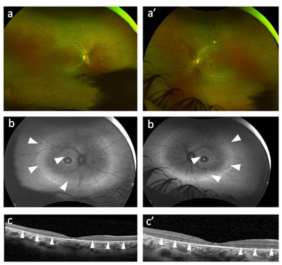
Proband P1 was a 28-year-old Black female affected with an early-onset, non-syndromic retinitis pigmentosa, with symptoms of nyctalopia from 22 years of age (Table 1). At age 28, Snellen BCVA was 20/50 (logMAR 0.4) in the right eye and 20/63 (LogMAR 0.5) in the left eye. Fundus examination revealed bone-spicule pigmentation in the nasal and superior midperiphery and slight attenuation of retinal arterioles without a significant reduction in vascular calibre (Figure 1a). Autofluorescence imaging (FAF) revealed a hypoautofluorescent periphery. In the macular region, a hyperautofluorescent ring was present with patchy hypoautofluorescence in the mid-periphery and reduced autofluorescence in the parafovea (Figure 1b). OCT imaging demonstrated bilateral loss of the ellipsoid zone (EZ, inner segment (IS)/outer segment (OS) junction), extending into the fovea bilaterally (Figure 1c).
Table 1.
Demographic, clinical, and genetic information.
Figure 1.
Multimodal retinal imaging from both eyes of a 28-year-old-female patient with C1QTNF5 c.548G>A; p.G183E. (a,a’) Ultra-widefield pseudocolour fundus photography of the optic disc reveals no clear optic atrophy. Retinal arteries are slightly attenuated, without a significant reduction in vascular calibre. The nasal and superior midperiphery and periphery show bone-spicule pigmentation. The vitreous appears clear, without significant inflammatory cells or opacities. (b,b’) Autofluorescence (AF) Imaging reveals patchy hypoautofluorescent midperiphery bordered by hyperautofluorescent rings (white arrowheads) and deeper hypoautofluorescence in the parafoveal region. (c,c’) Optical Coherence Tomography (OCT) imaging demonstrates thinning of the outer retinal layers, particularly a loss of the ellipsoid zone (EZ), IS/OS junction (white arrowheads). No cystoid macular oedema (CME) can be observed.
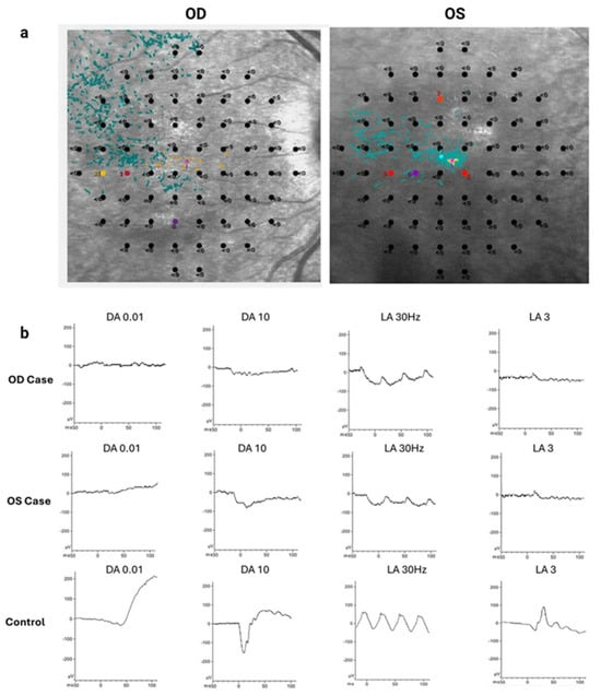
Consistent with photoreceptor degeneration, mesopic microperimetry revealed a complete loss of retinal sensitivity across the macula of P1, with the mean sensitivity threshold recorded as 0 dB (Figure 2a). Photoreceptor function was investigated further using full-field electroretinography. As shown in Figure 2b, scotopic responses were flat in both the right and left eyes across a range of flash stimulation intensities. Photopic responses showed severe attenuation of both single flash and flicker ERG. These data are consistent with a rod–cone dystrophy pattern.
Figure 2.
Microperimetry and electrophysiology from both eyes of a 28-year-old-female patient with C1QTNF5 c.548G>A; p.G183E. (a) Mesopic microperimetry shows severely reduced sensitivity bilaterally across the macula with loss of fixation stability on the right eye and reduced fixation stability on the left eye. . (Red dots: Abnormal values with severe loss of sensitivity; Black dots: Absolute scotoma with 0 dB threshold where the patient cannot perceive the maximum light stimulus) (b) Full field electroretinogram shows flat, dark adapted series and severely attenuated photopic series in both 30Hz flicker and single flash, suggesting a rod–cone dystrophy electrophysiological pattern.
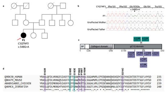
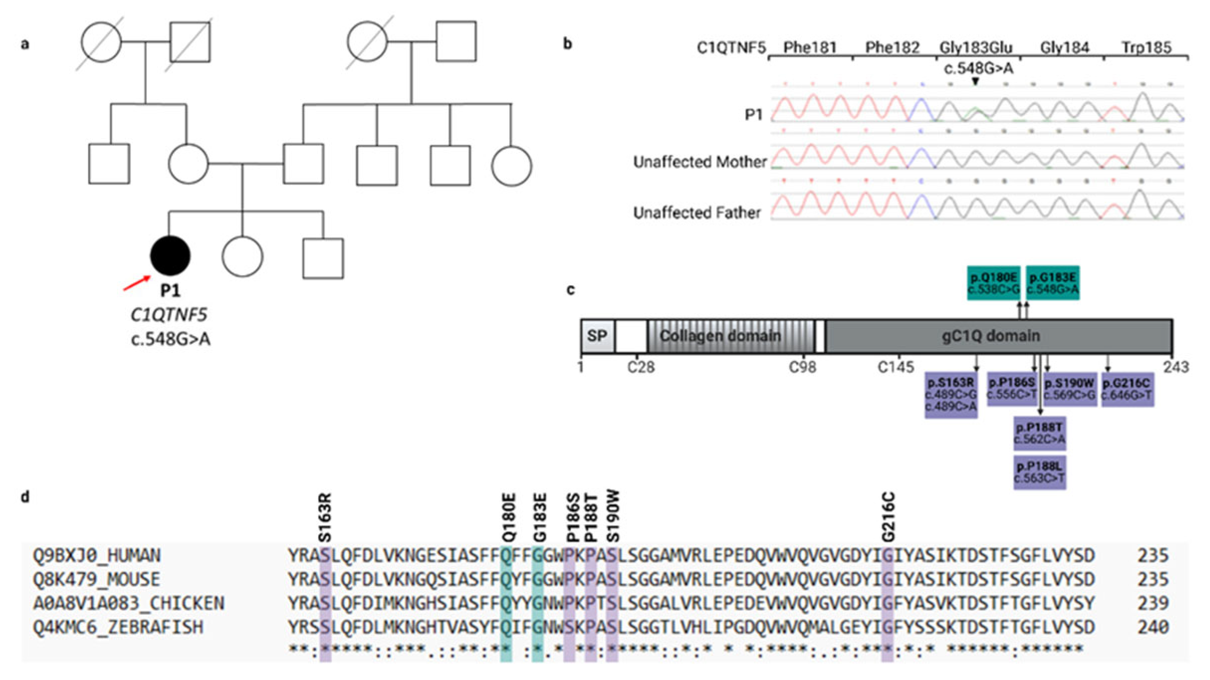
Since there had been no family history of retinal degeneration for proband P1 (Figure 3a), exome sequencing with trio analysis was performed. This identified a novel, likely pathogenic heterozygous missense variant in C1QTNF5, c.548G>A p.(G183E), in P1. The variant had good coverage, was present in 44.9% of the 167 total reads, and was determined to be de novo in both exome and Sanger sequencing data (Figure 3b), with verified parentage (PS2). The variant was not present in gnomAD (https://gnomad.broadinstitute.org/) (PM2_supp), indicating that it is very rare and met pathogenic criteria for AlphaMissense (0.7942) (PP3_supp) with moderately high scores from other tools (CADD 23, REVEL 0.435, BayesDel noAF −0.0438). The affected amino acid, 183, is highly conserved (Figure 3c) and located within the C1q domain of the C1QTNF5 protein. This domain spans amino acids 99–238 (uniprot.org) and includes all previously identified pathogenic variants (PM1) (Figure 3d). This variant meets ACMG/AMP criteria for likely pathogenic with a score of 8 (Table S1).
Figure 3.
Genetic, structural, and evolutionary characterisation of the novel C1QTNF5 c.548G>A; p.G183E variant identified in P1. (a) Pedigree of P1, the only affected individual within the family. Squares and circles denote males and females, respectively. Filled symbols indicate affected individual (P1), open symbols represent unaffected family members, and the red arrow indicates the proband. (b) Sanger sequencing chromatograms confirm the presence of the heterozygous missense variant c.548G>A; p.(G183E) identified via whole-exome sequencing (WES) in the proband and absence in both parents. The black arrow highlights the variant site, showing both wild-type and variant nucleotide peaks in the proband. (c) C1QTNF5 protein domain schematic and mutation clustering within the gC1Q domain of the 243 amino acid protein. SP; signal peptide. Variants resulting in atypical retinal phenotype are shown in teal and known L-ORD pathogenic variants are shown in purple. (d) Cross-species amino acid conservation. Multiple sequence alignment of the C1QTNF5 orthologs across human (Homo sapiens), mouse (Mus musculus), chicken (Gallus gallus), and zebrafish (Danio rerio). The affected amino acid residues (shaded boxes) show strict evolutionary conservation across all four species with * symbols indicating that amino acids are fully conserved, : have strongly similar properties and . weakly similar properties.
3.2. Clinical Presentation and Molecular Diagnosis of Retinal Disease Arising from C1QTNF5 c.538C>G, p.(Q180E)
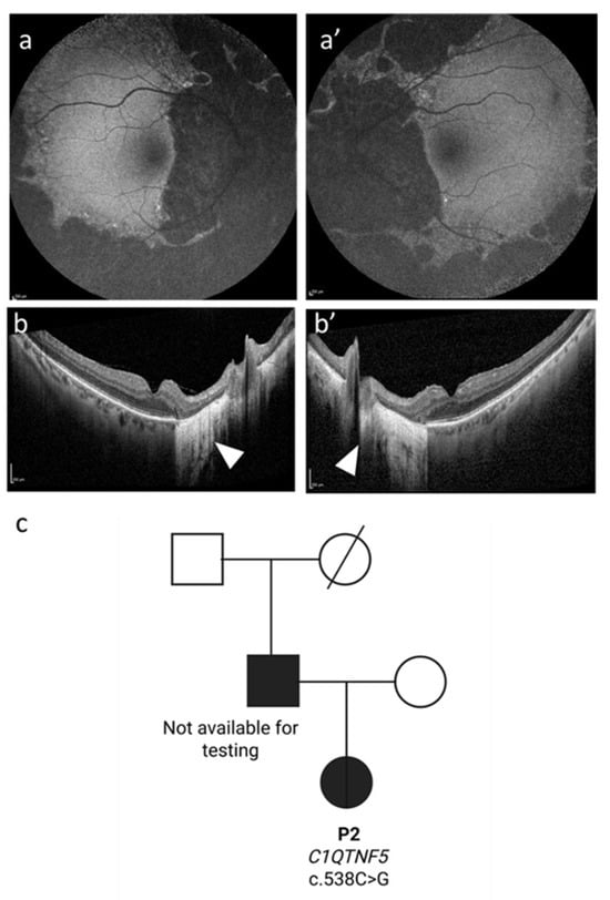
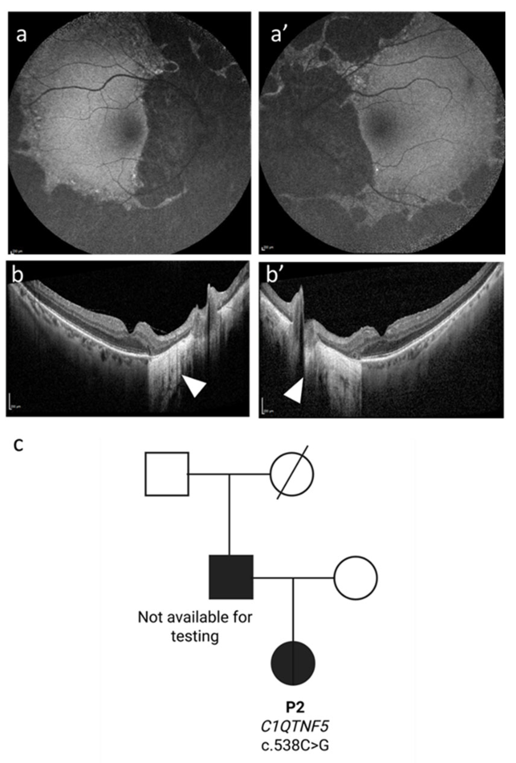
Proband P2 was a female who experienced initial symptoms of mild photophobia and visual field defects at age 47–48 years (Table 1). At age 49 years, BCVA was 20/25 (LogMAR 0.1) bilaterally. At 57 years, BCVA was 20/30 (LogMAR 0.18) in the right eye (after cataract surgery) and 20/25 (LogMAR 0.1) in the left eye. OCT scans showed marked peripheral FAF irregularities in affected areas in this patient (Figure 4a,b). These were consistent with progressing peripheral atrophy with relative sparing of the temporal macula until the late examination at age 57-years. Although not available for genetic testing, her father was also affected with initial symptoms—BCVA 20/20 (LogMAR 0.0) bilaterally and constricted visual fields documented at age 43 years (Figure 4c). This is consistent with an autosomal dominant pattern of inheritance. Family history indicated that the paternal grandfather was not known to be affected and the paternal grandmother died when young.
Figure 4.
Multimodal retinal imaging from both eyes of a 57-year-old-female patient with a C1QTNF5 c.538C>G, p.Q180E variant. (a,a’) Autofluorescence (AF) imaging reveals marked peripapillary and mid-peripheral hypoautofluorescence associated with confluent areas of atrophy. (b,b’) Optical coherence tomography imaging reveals hypertransmission defects associated with peripapillary atrophy (white arrowheads). (c) Affected status of P2 and her father is consistent with autosomal dominant inheritance in this family.
WGS was performed in P2 and a pathogenic C1QTNF5 missense variant c.538C>G, p.(Q180E) was identified. This was previously reported as a pathogenic variant, including in P3–P5 (Family ADRP386) and P6 (BD35) with a diagnosis of adGALCD [5,15]. Our study was therefore extended to re-evaluate family ADRP386, with multiple affected individuals across at least four generations, and P6, who had a father diagnosed with retinitis pigmentosa (Table 1). Pedigrees for ADRP386 and BD35 were reported previously [5]. Haplotype analysis detected shared ancestry for ADRP386 and BD35 [5], but this was not performed for P2.
3.3. Longitudinal Findings of Other Atypical Retinal Phenotype Cases Arising from Pathogenic Variants in C1QTNF5
The classic L-ORD phenotype has been well established by us and others [36,37]. To understand the long-term changes resulting from C1QTNF5 c.538C>G, p.(Q180E), we reviewed previously reported cases including three siblings (P3–5) from family ADRP386 and extended follow-up of P6 from family BD35. Table 1 summarises their demographic, clinical, and genetic information.
For P6, follow-up was extended from 45 years at our last report [5] to 53 years, when the patient was 71-years-old. P3 and her two affected siblings, P4 and P5, had an age of onset ranging from 38–40 years, (Table 1). Photophobia was previously reported to affect P4 and P5 aged 53 and 50 years respectively, 10–14 years after nyctalopia [5]. P6 exhibited retinal alterations and visual field defects at 18 years of age [5], but nyctalopia was not reported until age 48.
Similarities in phenotype existed for siblings P3 (58-year-old female), P4 (56-year-old male), and P5 (53-year-old male) (Supplementary Figures S1–S4). Their age of onset was reported to be around the age of 40 years (Table 1). P4 had some scattered pigmentation on fundus imaging (Supplementary Figures S1a and S2). FAF showed demarcated hypoautofluorescence in the periphery and peripapillary regions sparing the central macula in patients (Supplementary Figures S1b and S2). However, the size and shape of macular sparing varied between individuals, corresponding to various degrees of preserved central visual field (concentric visual field of 10 degrees in P5 to 30 degrees in P3, Supplementary Figure S3). OCT scans showed loss of peripapillary and peripheral macular ellipsoid zone and external limiting membrane and RPE, as demonstrated by marked choroidal hypertransmission (Supplementary Figures S1c and S2). P3, P4, and P5 were found to have a rod–cone dystrophy pattern of response on ffERG with P3 responses being extinct. P4 was also found to have a lattice-like corneal dystrophy (Supplementary Figure S4). These findings were not seen in their siblings. Follow-up in all cases confirmed that over time there was continuous constriction of the residual area of preserved RPE. Bruch membrane thickening, noted in previously described L-ORD phenotypes, was not present. P6 and P2 also had cataract surgery performed (P6, both eyes; P2, right eye) during which long anterior zonules were not seen.
Taken together, individuals with the p.(Q180E) and p.(G183E) variants are phenotypically different from classical L-ORD but all have progressive disease. The p.(Q180E) patients continue to have peripheral and peripapillary confluent areas of foveal sparing atrophy, but patients differ in the severity of atrophy and macular sparing, even amongst siblings. The p.(G183E) variant presents more like a classical rod–cone dystrophy with bone-spicule like pigmentation and with foveal involvement.
3.4. Molecular Modelling of Pathogenic C1QTNF5 Variants
To gain insight into the emerging genotype–phenotype spectrum of C1QTNF5-associated retinal disease, p.G183E, p.Q180E, p.S163R, p.P188T, p.P188L, p.S190W, and p.G216C variants were analysed using variant effect prediction tools available through Ensembl VEP (Table 2). As reported previously for the other variants, predictions of pathogenicity for p.G183E were discordant (AlphaMissense, ClinPred and CADD, likely pathogenic; SIFT, PolyPhen, EVE, tolerated/benign).
Table 2.
Functional impact predictions for C1QTNF5 missense variants generated using Ensembl Variant Effect Predictor.
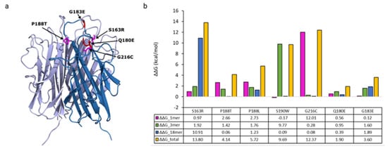
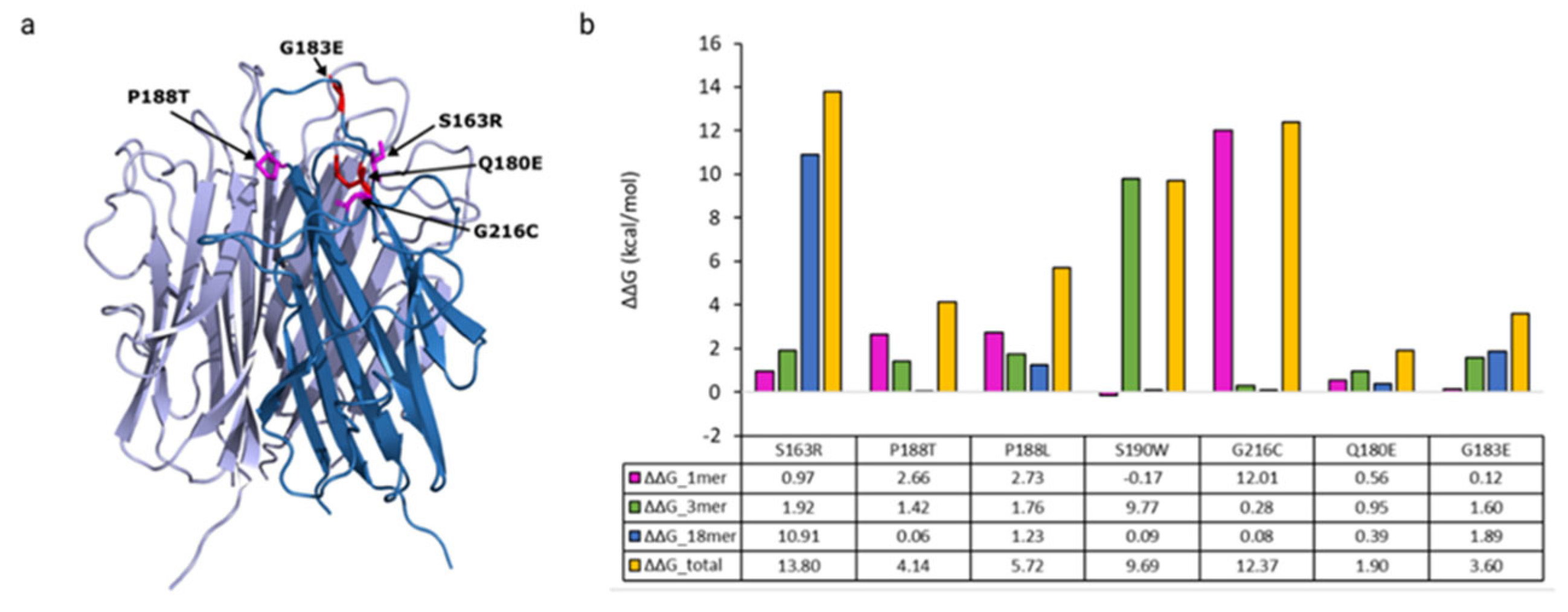
The crystal structure of the gC1q domain of C1QTNF5 and FoldX were used to model monomer stability and trimer and HMW octadecameric C1QTNF5 assemblies. In comparison to the pathogenic variants that cause L-ORD (Figure 5b), p.G183E is predicted to be less destabilising to C1QTNF5 folding and assembly. This amino acid substitution, replacing glycine (G) with negatively charged glutamic acid (E), is predicted to have negligible impact upon folding of the monomer (ΔΔG 0.12 kcal/mol), with additional mild destabilising contributions from trimer assembly (ΔΔG 1.59 kcal/mol) and octadecameric assembly (ΔΔG 1.89 kcal/mol). This is consistent with the location of p.G183E in a surface exposed apical loop where it does not form interface contacts with other subunits, limiting its predicted impact on intermolecular interactions in the trimer or 18-mer (Figure 5a). Together, these contributions give a total ΔΔG of 3.60 kcal/mol relative to the WT protein.
Figure 5.
Structural mapping and in silico thermodynamic stability analysis of C1QTNF5 variants. (a) Three-dimensional structure of the C1QTNF5 trimer (PDB ID: 4NN0). L-ORD pathogenic variants are shown in magenta, and variants resulting in atypical retinal phenotype are shown in red on a single subunit of the trimer, showing spatial clustering and localisation within or close to the apical loop. (b) Predicted free energy changes (ΔΔG, kcal/mol) for protein folding and assembly of the monomer, trimer, and high molecular weight (HMW) species, calculated using FoldX mutagenesis of a single C1QTNF5 subunit.
The p.Q180E variant alters the C-terminal (C’) residue in the first β-strand in the second of two five-stranded anti-parallel β-sheets that form the characteristic jelly-roll fold of the globular head of C1QTNF5 and other members of the C1q family [10]. In silico modelling predicts that substitution of polar, hydrophilic glutamine (Q) by negatively charged glutamic acid (E) will have a moderate impact on total ΔΔG (1.9 kcal/mol) compared to WT C1QTNF5 (Figure 5b). This captures small changes in energetic favourability for protomer folding (0.56 kcal/mol), trimer assembly (0.95 kcal/mol), and higher order assembly (0.39 kcal/mol) when one mutant subunit was included in the model. Evaluation of the total ΔΔG for known L-ORD pathogenic variants (mean ΔΔG = 9.145 kcal/mol, S.D. 4.158) confirmed that L-ORD pathogenic variants are significantly more destabilising than p.Q180E and p.G183E (mean ΔΔG = 2.750 kcal/mol, S.D. 1.202, p = 0.0262, Welch’s two-tailed t-test).
3.5. In Vitro Evaluation of C1QTNF5 Variants Underlying Distinct Phenotypic Manifestations
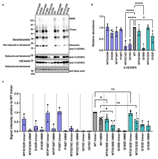
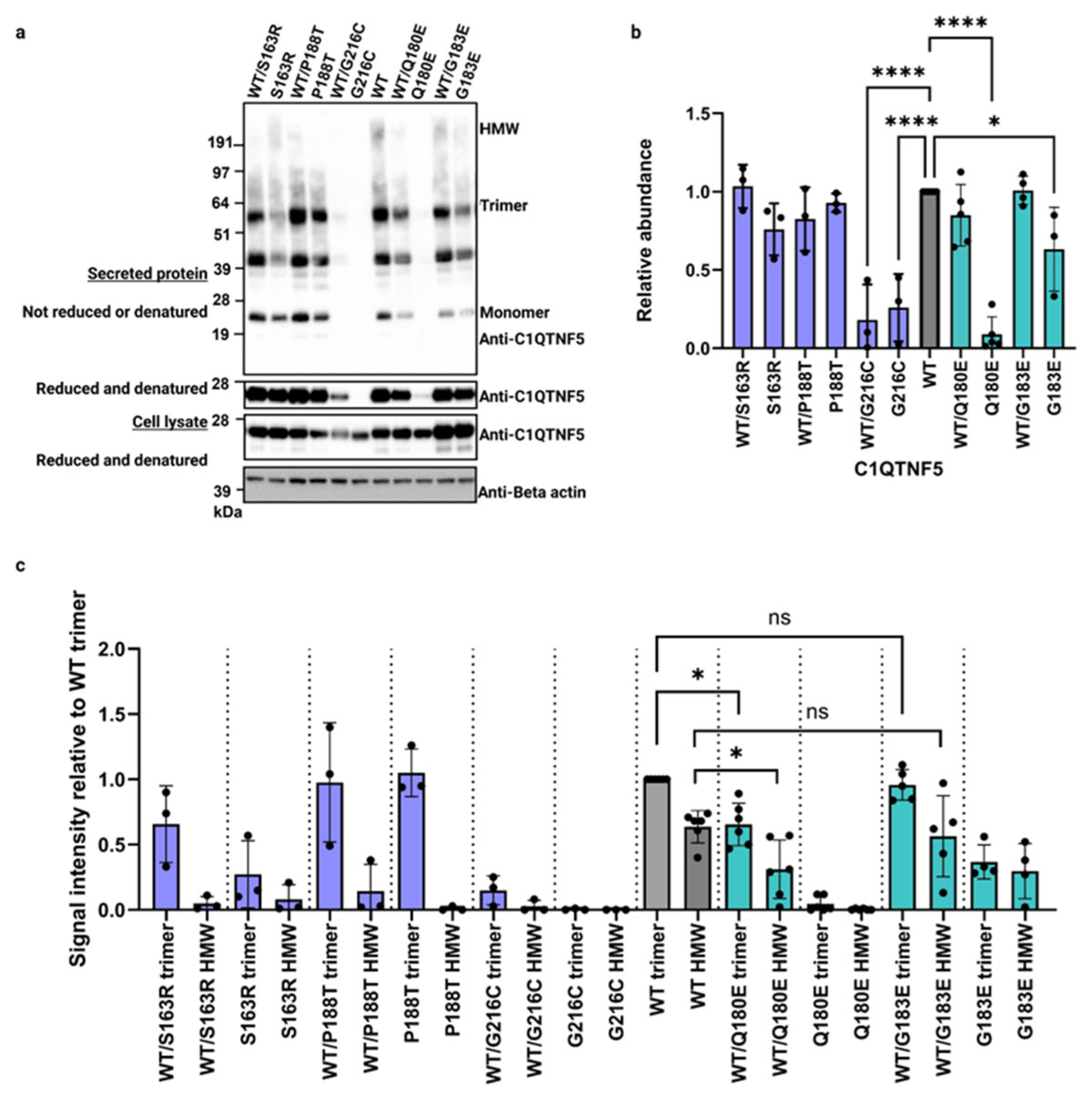
The impact of p.Q180E and p.G183E upon C1QTNF5 expression and stability in vitro was investigated by transient heterologous overexpression of C1QTNF5 in the RPE cell line, hTERT-RPE1. These variants were assessed alongside L-ORD pathogenic variants p.S163R, p.P188T, p.G216C, and WT C1QTNF5. The variant forms of C1QTNF5 were expressed alone and with WT C1QTNF5, to recapitulate the heterozygous state in affected individuals. As shown in Figure 6a, WT variant and co-expressed C1QTNF5 could be detected intracellularly. C1QTNF5 p.G183E, p.S163R, and p.P188T were detected at similar levels to WT protein following examination by polyacrylamide gel electrophoresis under reducing and denaturing conditions. The severe destabilising impact of C1QTNF5 p.G216C caused a highly significant decrease in the intracellular abundance and secretion of C1QTNF5 (Dunnett’s multiple comparison test, p < 0.0001, n = 3, Figure 6b) when this variant was expressed alone or alongside the WT protein (26 kDa). C1QTNF5 p.Q180E also appears to significantly impact intracellular abundance and secretion of C1QTNF5 when expressed alone (p < 0.0001, n = 5, Dunnett’s multiple comparison test), but in striking contrast to p.G216C, there was no significant difference in total C1QTNF5 secreted when p.Q180E is co-expressed with WT C1QTNF5 relative to WT alone (Figure 6a,b). This suggested that when assembled with WT C1QTNF5, this variant is moderately destabilising.
Figure 6.
Stability and multimerisation analysis of novel and pathogenic L-ORD C1QTNF5 variants. (a) Representative Western blot analysis of transiently expressed C1QTNF5 variants expressed alone and with WT C1QTNF5 to model heterozygous state in affected individuals. Cell lysates and secreted protein fractions from hTERT-RPE1 cells expressing wild-type (WT), novel, and pathogenic C1QTNF5 variants were resolved under non-reducing and non-denaturing conditions (to evaluate high molecular weight (HMW) assembly and stability) and reducing conditions (to assess monomeric expression levels). (b) Relative C1QTNF5 protein abundance. Quantification of total protein expression levels for each variant relative to the wild-type control, determined via densitometric analysis of Western blots from a minimum of 3 independent experiments. Statistically significant differences were calculated using Dunnett’s multiple comparisons test (* p < 0.05, **** p < 0.0001). (c) Quantification of C1QTNF5 multimerisation. Assessment of high-molecular-weight and trimer multimer formation for secreted C1QTNF5 variants, quantified using densitometry on non-reducing and non-denaturing Western blots. Data are normalised to the wild-type protein. Statistically significant differences were calculated using Dunnett’s multiple comparisons test (* p < 0.05, **** p < 0.0001, ns not significant).
C1QTNF5 p.G183E and p.Q180E showed a smaller change in ΔΔG (Figure 5b) and AlphaMissense score (Table 2) than known pathogenic L-ORD variants. This suggested that mutant protomers may be able to assemble with WT C1QTNF5 to form trimers and HMW multimers. The relative abundance of secreted trimers and HMW multimers of C1QTNF5 was assessed by densitometric analysis of immunoblots following non-reducing PAGE (Figure 6a,c). In agreement with previous work [2], L-ORD pathogenic variants expressed alone, or in combination with WT, abrogate the formation of HMW C1QTNF5. The p.S163R and p.P188T variants co-expressed with WT show no significant difference in monomer, dimer, or trimer abundance, but the HMW species is severely depleted, while C1QTNF5 p.G216C is largely undetectable in the conditioned media (Figure 6a,c).
The p.G183E variant expressed alone decreases the amount of C1QTNF5 trimer (36% compared to WT trimer) and HMW (47% compared to WT HMW) detected. This destabilising impact of p.G183E appears to be rescued by co-expression with the WT protein where no significant difference in the abundance of trimer (96% of WT, p = 0.9500) or HMW species (88% of WT, p = 0.9957) was identified relative to WT expressed alone (n = 4, Dunnett’s multiple comparison test). The p.Q180E variant alone is undetectable in conditioned media. Upon co-expression with WT C1QTNF5, secretion is partially restored, but abundance remains significantly decreased (trimers, 65% of the WT signal, p = 0.0170; HMW, 49%, p = 0.0410, n = 6, Dunnett’s multiple comparisons test). Immunoblots from all biological replicates used for densitometry are presented in Supplementary Figure S5.
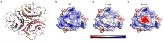
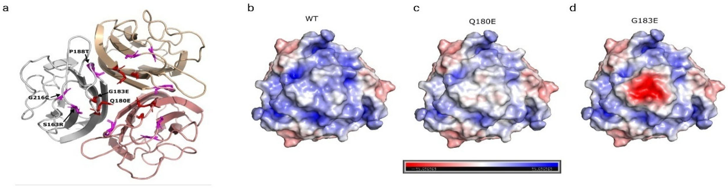
Since p.G183E and p.Q180E co-expressed with WT C1QTNF5 both give rise to secreted, multimeric forms of C1QTNF5, additional in silico analyses were performed. These variants contribute to the formation of a triangular hydrophobic plateau when C1QTNF5 trimers assemble [10] (Figure 7b). The electrostatic potential of homotrimers formed from p.G183E or p.Q180E protomers was compared to the hydrophobic plateau of the WT protein (Figure 7b–d). The hydrophobic plateau would have lower overall positive potential if three p.Q180E subunits were assembled into a trimer. The substitution of p.G183E in each unit of the trimer creates a strongly negative patch in the middle of the hydrophobic plateau. These significantly altered electrostatic potentials on the head of the C1QTNF5 trimer may have a significant impact on protein–protein interactions and C1QTNF5 function.
Figure 7.
The Q180E and G183E missense variants alter the electrostatic surface potential of the trimeric C1QTNF5 complex. (a) Ribbon representation of the trimeric C1QTNF5 protein structure based on the crystal structure (PDB ID: 4NN0). Each monomer is distinguished by a unique colour, and mutated residues are highlighted in stick conformation (L-ORD variants, magenta; atypical, red) with individual labels identifying their positions. (b–d) Electrostatic surface potential of the globular trimeric structure of C1QTNF5 calculated using the APBS Electrostatics plugin in PyMOL. The surface is coloured according to local charge: red indicates negative potential (−5 kT/e), blue indicates positive potential (+5 kT/e), and white represents neutral potential.
4. Discussion
This study identified the genetic cause of retinal degeneration in a sporadic case of early-onset, non-syndromic retinitis pigmentosa as a novel, heterozygous de novo variant in the C1QTNF5 gene, c.548G>A p.(G183E). To our knowledge, this is the first report of a de novo C1QTNF5 variant leading to retinal degeneration, but founder variants in C1QTNF5 were identified in the UK [1] and Belgium [6] previously and were inherited in an autosomal dominant manner. With the identification of C1QTNF5 c.538C>G, p.(Q180E) as the cause of retinal disease in P2, we extend the number of affected families identified in Germany to four [5,15,39]. Although haplotype mapping remains to be performed for this newly reported proband and for another simplex case [39], it was performed for the BD35 and ARDP386 families [5]. This confirmed that the haplotype including the p.Q180E variant is identical by descent which may indicate another founder variant in C1QTNF5. This study also appears to be the first reported case of a C1QTNF5-associated retinopathy in an Afro-Caribbean proband, thus expanding the known demographic and genetic landscape of the disorder beyond European [1,2,3,4,5,6] and Singaporean Chinese [40] ancestry. This newly arising mutation highlights the diagnostic value of parental–child trio sequencing in clinical practice and, together with p.Q180E, reveals a critical hotspot in the gC1Q domain of C1QTNF5 that appears to be intolerant to variation.
The p.G183E variant is located close to the position of previously identified pathogenic variants in C1QTNF5. However, the retinal presentation of this proband was markedly different to that of L-ORD caused by p.S163R, p.P188T, and p.G216C missense changes, where affected individuals first experience nyctalopia in the fifth decade, before characteristic sub-RPE deposit formation, progressive central and then peripheral RPE atrophy, and risk of choroidal neovascularisation causing vision loss in later life [36,37]. P1, heterozygous for the p.(G183E) variant, initially experienced nyctalopia, and retinal phenotype was more similar to an atypical retinitis pigmentosa without marked retinal arteriolar attenuation or optic atrophy. It also differed markedly from the retinal phenotype observed for P2, heterozygous for the p.(Q180E) variant, which was characterised by early peripheral chorioretinal atrophy. This is consistent with the adGALCD-type retinal phenotype that was reported in individuals carrying C1QTNF5 c.538C>G, p.(Q180E) and seen in P3, P4, P5, and P6 [5,15,39]. The identification of these two pathogenic/likely pathogenic variants in C1QTNF5 expand the phenotypic spectrum of the C1QTNF5 locus. A limitation of our study is the small sample size. Although the genotype–phenotype trends observed here are promising, additional unrelated cases carrying these variants are necessary to establish a robust genotype–phenotype correlation. This is especially true for p.(G183E), for which this is the first case report. By confirming that pathogenic variants in C1QTNF5 can drive multiple different disease trajectories, this dismantles the one gene, one disease paradigm linking C1QTNF5 and L-ORD and establishes allelic and phenotypic heterogeneity for C1QTNF5-associated retinal disease.
IRDs exhibit vast genetic and phenotypic heterogeneity, with 533 disease-associated genes and loci identified to date (RetiGene.erdc.info, updated 3/9/2026) [41]. As we report for C1QTNF5, variants within a single gene can drive diverse clinical outcomes, ranging from mild to severe or early-onset blindness to late-onset degeneration, depending on variant type, disease mechanism, and the affected functional domains [42]. This is the first time that this has been explored for C1QTNF5, and resolving these complex genotype–phenotype relationships is critical for accurate diagnosis and prognosis and for guiding future therapeutic strategies.
The amino acids affected by pathogenic C1QTNF5 variants are highly conserved, but when compared to the L-ORD variants, p.G183E and p.Q180E, were less strongly predicted to be pathogenic by tools in the Ensembl VEP and by FoldX mutagenesis. However, both p.G183E and p.Q180E were predicted to destabilise C1QTNF5, with total ΔΔG of 3.60 kcal/mol and 1.9 kcal/mol predicted, respectively, when a single mutant C1QTNF5 subunit was included in the model. Following established thresholds for functional disruption, ΔΔG ≥ 1.5 kcal/mol are considered damaging/destabilising [43,44,45,46], although a more conservative threshold is applied when it contributes to clinical diagnosis. For this, thermodynamic instability is not considered stand-alone evidence for pathogenicity according to ACMG/AMP criteria but must correlate with evolutionary conservation and surpass higher ΔΔG limits. Taken together with the CADD and AlphaMissense VEP presented in Table 2, this study provides additional PP3_supp evidence that p.(G183E) is a likely pathogenic variant.
With a total ΔΔG of 1.9 kcal/mol for inclusion of one p.Q180E subunit in the C1QTNF5 assembly, the FoldX calculated that thermodynamic instability for this variant was lower than in a previous in silico investigation. Kellner et al. [5] predicted that p.Q180E would destabilise folding of the C1QTNF5 protomer (ΔΔG 2.56 kcal/mol) and introduce electrostatic repulsion between interacting protomers, resulting in additive destabilisation of the trimer containing one (ΔΔG 30.5 kcal/mol), two (ΔΔG 64.8 kcal/mol), or three (ΔΔG 101.12 kcal/mol) mutant subunits. Direct comparison of ΔΔG across these studies is not possible because different tools were used for these analyses.
In this study, we provide additional in vitro evidence to support a damaging effect upon C1QTNF5 using transient overexpression in hTERT-RPE1 to assess protein abundance, secretion, and multimerisation. This approach previously uncovered a dominant negative mechanism for the L-ORD variants, where the mutant forms of C1QTNF5 interact with and affect the WT protein to prevent higher order multimerisation [2]. C1QTNF5 p.G183E and p.Q180E appear to have different impacts. The total secreted C1QTNF5 detected for the p.G183E variant was significantly decreased relative to WT protein, although intracellular abundance appeared to be similar. By contrast, the p.Q180E variant was severely destabilised with decreased intracellular abundance and no secreted protein detected. When co-expressed with WT, there was a significant decrease in the amount of C1QTNF5 trimer and HMW form in the conditioned media from cells co-expressing WT/p.Q180E C1QTNF5. These data support the destabilising impact of p.Q180E when multiple affected protomers assemble into a trimer [5], with the introduction of a negatively charged residue resulting in cooperative repulsion and protein instability, with consequently less C1QTNF5 overall. The impact of p.G183E when co-expressed with the WT was less clear, since no such change was observed for WT/p.G183E, where the relative abundance of extracellular HMW and trimeric C1QTNF5 was similar to WT C1QTNF5.
The p.G183E variant is centrally located in an apical loop in the gC1Q domain, positioned at the apex of the globular head [10]. These loops, comprising residues F181-F182-G183-G184-W185-P186, converge in the trimer to form a triangular hydrophobic plateau formed by key residues A162, F179, F181, F182, and Y214 [10]. Hydrophobic interfaces often form the energetic core of transient or stable binding surfaces [47,48]. Missense substitutions that perturb these regions may impair, abolish, or cause abnormal interaction with binding partners without affecting overall protein stability. The L-ORD variant p.S163R abolishes a hydrogen bond with F182, disrupting the plateau and creating a positive charge [10]. Intermolecular hydrogen-bond networks may also be disrupted by alterations in this apical loop, for example, between G184 and P116 and water in adjacent C1QTNF5 multimers when assembled top-to-top [32]. Introduction of p.G183E into the apical loop created a striking change in electrostatic potential in the hydrophobic plateau of the C1QTNF5 trimer, forming a negatively charged patch. This has important implications for C1QTNF5 function, since it will alter the physicochemical properties of an interaction surface which was previously speculated to be required for top-to-top interactions of HMW C1QTNF5 on adjacent cells to facilitate RPE adhesion [10,32], as well as binding to interactors such as HTRA1 [49] and ADIPOR1 [9]. Notably, this shows that the impact of p.G183E is distinct. The alteration in charge and impact upon multimerisation differs from p.S163R, and unlike p.Q180E, trimers and HMW species containing only p.G183E C1QTNF5 are stable and can be detected extracellularly.
The precise consequences of p.G183E and p.Q180E on C1QTNF5 function remains to be determined. A key limitation of our experimental approach is the reliance on transient overexpression in an immortalised human cell-line. Although this system enabled us to evaluate the impact of C1QTNF5 variants, overexpression can result in non-physiological protein kinetics. Future studies utilising endogenously tagged proteins, patient-derived iPSC-RPE, or knock-in models are necessary to validate these findings and to interrogate functional consequences in detail. However, this study reveals an emerging allelic hierarchy of C1QTNF5 dysfunction which results in retinal phenotypic diversity. Variant C1QTNF5 proteins multimerise with wild-type counterparts, but the impact on C1QTNF5 function may vary depending on the nature and position of the variant. In L-ORD, the variants identified to date destabilise the folding or assembly of a HMW complex [2]. This leads to reduced HMW abundance or complete loss of assembly. In contrast, the destabilising impact of p.Q180E is tempered by association with WT to allow secretion and assembly of C1QTNF5 into HMW species, although abundance is diminished and function remains to be determined. The p.G183E variant appears to assemble into trimers and the HMW complex at equivalent levels to WT C1QTNF5, but through altering the electrostatic potential at the apical face of the globular head may impair functional activity by disrupting a critical interaction interface [32]. This seems likely to result in functional impairment, but additional work is required to confirm a dominant-negative mechanism for p.G183E. In keeping with this, and the four-step evaluation framework established by the ClinGen Sequence Variant Interpretation Working Group [50], the functional data generated in this study did not enable us to reach PS3 classification for p.G183E.
These mechanistic differences highlight that not all pathogenic variants act equivalently; rather, their impact depends on whether the variant primarily affects protein stability, intermolecular interactions, or the functional competence of the assembled complex. The distinction between these mechanisms is important when using variant effect prediction or in silico analyses to assess pathogenicity because proteins may retain native folding and multimeric integrity but be functionally defective due to impaired intermolecular interactions. Conversely, variants destabilising multimeric assembly often produce broader defects in protein abundance, solubility, or cellular localisation. Together, these differences in molecular mechanism may help to explain why variants in C1QTNF5 can produce a spectrum of clinical presentations. This highlights the importance of functional validation for novel variant interpretation, ultimately contributing to improved diagnostic accuracy and a deeper understanding of disease biology.
Supplementary Materials
The following supporting information can be downloaded at https://www.mdpi.com/article/10.3390/genes17111375/s1. Table S1: ACMG/AMP Variant Classification Framework for the novel C1QTNF5 c.548G>A; p.Gly183Glu variant identified in P1. Figure S1: Multimodal retinal imaging from the right and left eye of a 56-year-old male patient (P4) carrying a p.Q180E C1QTNF5 variant. Figure S2: Multimodal retinal imaging from the right and left eye of P3, a 58 year-old female patient, and P5, a 53-year-old male patient carrying a p.Q180E C1QTNF5 variant. Figure S3: Kinetic perimetry demonstrates severe bilateral concentric visual-field loss in patients P3, P4, and P5. Figure S4: Colour photographs showing corneal lattice dystrophy in the right and left eye of a 56-year-old male patient (P4) carrying the p.Q180E C1QTNF5 variant. Figure S5: Full Western blots for each independent biological replicate used for quantitative analyses.
Author Contributions
Conceptualization, C.M.S. and S.B.; Methodology, C.M.S., J.A.M., U.K., K.S., S.K., S.B., L.M.R., S.E.S. and E.V.S.; Software, C.M.S. and J.A.M.; Validation, C.M.S., S.B. and S.K.; Formal Analysis, C.M.S., S.K.L., H.K., M.-A.G., V.W. and C.K.; Investigation, C.M.S., S.K.L., N.E.W., L.M.R., S.E.S., H.K., M.-A.G., I.D.O., G.A.F., S.M.A.A., V.W., C.K. and S.B.; Resources, C.M.S., C.D., J.D.P., H.S. and B.H.F.W.; Data Curation, C.M.S., S.B., I.D.O., G.A.F., U.K., K.S. and S.K.; Writing—Original Draft Preparation, C.M.S. and S.B.; Writing—Review and Editing, all authors; Visualization, C.M.S., G.A.F., I.D.O. and S.B.; Supervision, C.M.S., S.B., H.S.; B.H.F.W. and E.V.S.; Project Administration, C.M.S., S.B.; Funding Acquisition, C.M.S.; B.H.F.W., J.A.M., S.B. and E.V.S. All authors have read and agreed to the published version of the manuscript.
Funding
Assessment of variant pathogenicity was supported by a University of Edinburgh Chancellor’s Fellowship Award to CMS. JAM was supported by funding from the Medical Research Council (MRC) Human Genetics Unit core grant (MC_UU_00035/9). Exome sequencing and analysis of P1 was supported by a grant from the National Institutes of Health R01EY015518 (E. V. S.). Whole-genome sequencing of P2 was supported by a grant from the Bavarian Ministry of Science and Arts (Bayerisches Staatsministerium für Wissenschaft und Kunst) to the Bavarian Genomes Network, coordinated by Thomas Meitinger.
Institutional Review Board Statement
The study was conducted in accordance with the Declaration of Helsinki and complied with Health Insurance Portability and Accountability Act (HIPAA) regulations. For data from UCSD, this study was approved by the UCSD IRB board (804279, approved 16 December 2022 and re-approved on 23 July 2026) with no informed consent, beyond standard institutional consent, required for review of retrospective data. For data from MCW, this study was approved by the MCW IRB (PRO00045954) with written informed consent obtained from each individual. For data from University Eye Hospital Tuebingen, this study was approved by the Institutional Review Board of the Medical Faculty of the Eberhard Karls University of Tübingen (project no. 116/2015BO2, last update 9 February 2022). The use of clinical data for research for patients P2 and P6 was approved by the ethic board of the Aerztekammer Nordrhein (ID: 2008367, 3 November 2008). For data from the University of Regensburg for P2, the informed consent process was conducted within the Bavarian Genomes Network under approval from the Ethics Committee of the University of Regensburg (ID: 20-2100-103, last update 19 November 2020).
Informed Consent Statement
Informed consent was obtained from all subjects involved in the study.
Data Availability Statement
The original contributions presented in this study are included in the article/Supplementary Materials. Further inquiries can be directed to the corresponding authors.
Acknowledgments
We would like to thank Isabelle Banke for assistance with family enrolment and Samuel Thompson for help with Sanger sequencing (Medical College of Wisconsin).
Conflicts of Interest
The authors declare no conflicts of interest regarding the present study. U. Kellner: No conflicts of interest. Employer: MVZ Augenärztliches Diagnostik- und Therapiecentrum Siegburg GmbH. Honoraria for lectures: Abbvie GmbH, Germany; Apellis GmbH, Germany; Bayer Vital GmbH, Germany; Heidelberg Engineering GmbH, Germany; Novartis Germany. Monitoring/Advisory boards: Apellis GmbH, Germany; Astellas GmbH, Germany; Bayer Vital GmbH, Germany; Cellerin GmbH, Germany, Chiesi GmbH, Germany; Novartis GmbHGermany; Rhythm Pharmaceuticals Germany GmbH, Roche GmbH, Germany; Sandoz/Hexal AG, Germany. Board Member: Member of the Scientific Board of Pro Retina Germany e.VThe funders had no role in the design of the study; in the collection, analyses, or interpretation of data; in the writing of the manuscript; or in the decision to publish the results.
Abbreviations
The following abbreviations are used in this manuscript:
| L-ORD | Late-Onset Retinal Degeneration |
| IRD | inherited retinal dystrophy |
| WT | wild-type |
| RPE | retinal pigment epithelium |
| C1QTNF5 | C1q-And Tumour Necrosis Factor-Related Protein 5 |
| adGALCD | autosomal dominant gyrate atrophy-like choroidal dystrophy |
| kDa | kilodalton |
| gC1q | globular complement 1q/tumour necrosis factor-like domain |
| HMW | high molecular weight |
| NGS | next-generation sequencing |
| VUS | variant of uncertain significance |
| ACMG/AMP | American College of Medical Genetics and Genomics and Association for Molecular Pathology |
| UCSD | University of California San Diego |
| HIPAA | Health Insurance Portability and Accountability Act |
| IRB | Institutional Review Board |
| BCVA | best corrected visual acuity |
| SD-OCT | spectral-domain optical coherence tomography |
| FAF | fundus autofluorescence |
| MAIA | Macular Integrity Assessment |
| ffERG | full-field electroretinogram |
| FST | full-field stimulus threshold |
| WES | whole exome sequencing |
| WGS | whole genome sequencing |
| MCW | Medical College of Wisconsin |
| VEP | variant effect predictor |
| LDS | lithium dodecyl sulphate |
| PVDF | polyvinylidene difluoride |
| MOPS | 3-(N-morpholino)propanesulfonic acid |
| NFDM | non-fat dried milk |
| PBS-T | phosphate buffered saline with 0.05% Tween |
| HRP | horseradish peroxidase |
| EZ | ellipsoid zone |
| IS | inner segment |
| OS | outer segment |
References
- Hayward, C.; Shu, X.; Cideciyan, A.V.; Lennon, A.; Barran, P.; Zareparsi, S.; Sawyer, L.; Hendry, G.; Dhillon, B.; Milam, A.H.; et al. Mutation in a short-chain collagen gene, CTRP5, results in extracellular deposit formation in late-onset retinal degeneration: A genetic model for age-related macular degeneration. Hum. Mol. Genet. 2003, 12, 2657–2667. [Google Scholar] [CrossRef] [Scilit]
- Stanton, C.M.; Borooah, S.; Drake, C.; Marsh, J.A.; Campbell, S.; Lennon, A.; Soares, D.C.; Vallabh, N.A.; Sahni, J.; Cideciyan, A.V.; et al. Novel pathogenic mutations in C1QTNF5 support a dominant negative disease mechanism in late-onset retinal degeneration. Sci. Rep. 2017, 7, 12147. [Google Scholar] [CrossRef] [Scilit]
- Borooah, S.; Stanton, C.M.; Marsh, J.; Carss, K.J.; Waseem, N.; Biswas, P.; Agorogiannis, G.; Raymond, L.; Arno, G.; Webster, A.R. Whole genome sequencing reveals novel mutations causing autosomal dominant inherited macular degeneration. Ophthalmic Genet. 2018, 39, 763–770. [Google Scholar] [CrossRef] [Scilit]
- Rodríguez-Muñoz, A.; Aller, E.; Jaijo, T.; González-García, E.; Cabrera-Peset, A.; Gallego-Pinazo, R.; Udaondo, P.; Salom, D.; García-García, G.; Millán, J.M. Expanding the Clinical and Molecular Heterogeneity of Nonsyndromic Inherited Retinal Dystrophies. J. Mol. Diagn. 2020, 22, 532–543. [Google Scholar] [CrossRef] [Scilit]
- Kellner, U.; Weisschuh, N.; Weinitz, S.; Farmand, G.; Deutsch, S.; Kortüm, F.; Mazzola, P.; Schäferhoff, K.; Marino, V.; Dell’orco, D. Autosomal Dominant Gyrate Atrophy-Like Choroidal Dystrophy Revisited: 45 Years Follow-Up and Association with a Novel C1QTNF5 Missense Variant. Int. J. Mol. Sci. 2021, 22, 2089. [Google Scholar] [CrossRef] [Scilit]
- De Zaeytijd, J.; Coppieters, F.; De Bruyne, M.; Van Royen, J.; Roels, D.; Six, R.; Van Cauwenbergh, C.; De Baere, E.; Leroy, B.P. Longitudinal phenotypic study of late-onset retinal degeneration due to a founder variant c.562C>A p.(Pro188Thr) in the C1QTNF5 gene. Ophthalmic Genet. 2021, 42, 521–532. [Google Scholar] [CrossRef] [Scilit]
- Wong, G.W.; Krawczyk, S.A.; Kitidis-Mitrokostas, C.; Revett, T.; Gimeno, R.; Lodish, H.F. Molecular, biochemical and functional characterizations of C1q/TNF family members: Adipose-tissue-selective expression patterns, regulation by PPAR-gamma agonist, cysteine-mediated oligomerizations, combinatorial associations and metabolic functions. Biochem. J. 2008, 416, 161–177. [Google Scholar] [CrossRef] [Scilit]
- Lei, X.; Rodriguez, S.; Petersen, P.S.; Seldin, M.M.; Bowman, C.E.; Wolfgang, M.J.; Wong, G.W. Loss of CTRP5 improves insulin action and hepatic steatosis. Am. J. Physiol. Endocrinol. Metab. 2016, 310, E1036–E1052. [Google Scholar] [CrossRef] [Scilit]
- Miyagishima, K.J.; Sharma, R.; Nimmagadda, M.; Clore-Gronenborn, K.; Qureshy, Z.; Ortolan, D.; Bose, D.; Farnoodian, M.; Zhang, C.; Fausey, A.; et al. AMPK modulation ameliorates dominant disease phenotypes of CTRP5 variant in retinal degeneration. Commun. Biol. 2021, 4, 1360. [Google Scholar] [CrossRef] [Scilit]
- Tu, X.; Palczewski, K. Crystal structure of the globular domain of C1QTNF5: Implications for late-onset retinal macular degeneration. J. Struct. Biol. 2012, 180, 439–446. [Google Scholar] [CrossRef] [Scilit]
- Tsao, T.S.; Tomas, E.; Murrey, H.E.; Hug, C.; Lee, D.H.; Ruderman, N.B.; Heuser, J.E.; Lodish, H.F. Role of disulfide bonds in Acrp30/adiponectin structure and signaling specificity. Different oligomers activate different signal transduction pathways. J. Biol. Chem. 2003, 278, 50810–50817. [Google Scholar] [CrossRef] [Scilit]
- Ellingford, J.M.; Barton, S.; Bhaskar, S.; O’SUllivan, J.; Williams, S.G.; Lamb, J.A.; Panda, B.; Sergouniotis, P.I.; Gillespie, R.L.; Daiger, S.P.; et al. Molecular findings from 537 individuals with inherited retinal disease. J. Med. Genet. 2016, 53, 761–767. [Google Scholar] [CrossRef] [Scilit]
- Lin, S.; Vermeirsch, S.; Pontikos, N.; Martin-Gutierrez, M.P.; Varela, M.D.; Malka, S.; Schiff, E.; Knight, H.; Wright, G.; Jurkute, N.; et al. Spectrum of Genetic Variants in the Most Common Genes Causing Inherited Retinal Disease in a Large Molecularly Characterized United Kingdom Cohort. Ophthalmol. Retin. 2024, 8, 699–709. [Google Scholar] [CrossRef] [Scilit]
- Richards, S.; Aziz, N.; Bale, S.; Bick, D.; Das, S.; Gastier-Foster, J.; Grody, W.W.; Hegde, M.; Lyon, E.; Spector, E.; et al. Standards and guidelines for the interpretation of sequence variants: A joint consensus recommendation of the American College of Medical Genetics and Genomics and the Association for Molecular Pathology. Genet. Med. 2015, 17, 405–424. [Google Scholar] [CrossRef] [Scilit]
- Weisschuh, N.; Mazzola, P.; Zuleger, T.; Schaeferhoff, K.; Kühlewein, L.; Kortüm, F.; Witt, D.; Liebmann, A.; Falb, R.; Pohl, L.; et al. Diagnostic genome sequencing improves diagnostic yield: A prospective single-centre study in 1000 patients with inherited eye diseases. J. Med. Genet. 2024, 61, 186–195. [Google Scholar] [CrossRef] [Scilit]
- Reis, L.M.; Atilla, H.; Kannu, P.; Schneider, A.; Thompson, S.; Bardakjian, T.; Semina, E.V. Distinct Roles of Histone Lysine Demethylases and Methyltransferases in Developmental Eye Disease. Genes 2023, 14, 216. [Google Scholar] [CrossRef] [Scilit]
- Karczewski, K.J.; Francioli, L.C.; Tiao, G.; Cummings, B.B.; Alföldi, J.; Wang, Q.; Collins, R.L.; Laricchia, K.M.; Ganna, A.; Birnbaum, D.P.; et al. The mutational constraint spectrum quantified from variation in 141,456 humans. Nature 2020, 581, 434–443. [Google Scholar] [CrossRef] [Scilit]
- Chen, S.; Francioli, L.C.; Goodrich, J.K.; Collins, R.L.; Kanai, M.; Wang, Q.; Alföldi, J.; Watts, N.A.; Vittal, C.; Gauthier, L.D. A genomic mutational constraint map using variation in 76,156 human genomes. Nature 2024, 625, 92–100. [Google Scholar] [CrossRef] [Scilit]
- Rentzsch, P.; Schubach, M.; Shendure, J.; Kircher, M. CADD-Splice-improving genome-wide variant effect prediction using deep learning-derived splice scores. Genome Med. 2021, 13, 31. [Google Scholar] [CrossRef] [Scilit]
- Ioannidis, N.M.; Rothstein, J.H.; Pejaver, V.; Middha, S.; McDonnell, S.K.; Baheti, S.; Musolf, A.; Li, Q.; Holzinger, E.; Karyadi, D.; et al. REVEL: An Ensemble Method for Predicting the Pathogenicity of Rare Missense Variants. Am. J. Hum. Genet. 2016, 99, 877–885. [Google Scholar] [CrossRef] [Scilit]
- Cheng, J.; Novati, G.; Pan, J.; Bycroft, C.; Žemgulytė, A.; Applebaum, T.; Pritzel, A.; Wong, L.H.; Zielinski, M.; Sargeant, T.; et al. Accurate proteome-wide missense variant effect prediction with AlphaMissense. Science 2023, 381, eadg7492. [Google Scholar] [CrossRef] [Scilit]
- Feng, B.J. PERCH: A Unified Framework for Disease Gene Prioritization. Hum. Mutat. 2017, 38, 243–251. [Google Scholar] [CrossRef] [Scilit]
- Tavtigian, S.V.; Harrison, S.M.; Boucher, K.M.; Biesecker, L.G. Fitting a naturally scaled point system to the ACMG/AMP variant classification guidelines. Hum. Mutat. 2020, 41, 1734–1737. [Google Scholar] [CrossRef] [Scilit]
- Bergquist, T.; Stenton, S.L.; Nadeau, E.A.W.; Byrne, A.B.; Greenblatt, M.S.; Harrison, S.M.; Tavtigian, S.V.; O’DOnnell-Luria, A.; Biesecker, L.G.; Radivojac, P.; et al. Calibration of additional computational tools expands ClinGen recommendation options for variant classification with PP3/BP4 criteria. Genet. Med. 2025, 27, 101402. [Google Scholar] [CrossRef] [Scilit]
- McLaren, W.; Gil, L.; Hunt, S.E.; Riat, H.S.; Ritchie, G.R.S.; Thormann, A.; Flicek, P.; Cunningham, F. The Ensembl Variant Effect Predictor. Genome Biol. 2016, 17, 122. [Google Scholar] [CrossRef] [Scilit]
- Alirezaie, N.; Kernohan, K.D.; Hartley, T.; Majewski, J.; Hocking, T.D. ClinPred: Prediction Tool to Identify Disease-Relevant Nonsynonymous Single-Nucleotide Variants. Am. J. Hum. Genet. 2018, 103, 474–483. [Google Scholar] [CrossRef] [Scilit]
- Frazer, J.; Notin, P.; Dias, M.; Gomez, A.; Min, J.K.; Brock, K.; Gal, Y.; Marks, D.S. Disease variant prediction with deep generative models of evolutionary data. Nature 2021, 599, 91–95. [Google Scholar] [CrossRef] [Scilit]
- Eddy, S.R. Where did the BLOSUM62 alignment score matrix come from? Nat. Biotechnol. 2004, 22, 1035–1036. [Google Scholar] [CrossRef] [Scilit]
- Ng, P.C.; Henikoff, S. SIFT: Predicting amino acid changes that affect protein function. Nucleic Acids Res. 2003, 31, 3812–3814. [Google Scholar] [CrossRef] [Scilit]
- Adzhubei, I.; Jordan, D.M.; Sunyaev, S.R. Predicting functional effect of human missense mutations using PolyPhen-2. Curr. Protoc. Hum. Genet. 2013, 76, 7–20. [Google Scholar] [CrossRef] [Scilit]
- UniProt Consortium. UniProt: The Universal Protein Knowledgebase in 2025. Nucleic Acids Res. 2025, 53, D609–D617. [Google Scholar] [CrossRef] [Scilit]
- Tu, X.; Palczewski, K. The macular degeneration-linked C1QTNF5 (S163) mutation causes higher-order structural rearrangements. J. Struct. Biol. 2014, 186, 86–94. [Google Scholar] [CrossRef] [Scilit]
- Delgado, J.; Radusky, L.G.; Cianferoni, D.; Serrano, L. FoldX 5.0: Working with RNA, small molecules and a new graphical interface. Bioinformatics 2019, 35, 4168–4169. [Google Scholar] [CrossRef] [Scilit]
- Dolinsky, T.J.; Czodrowski, P.; Li, H.; Nielsen, J.E.; Jensen, J.H.; Klebe, G.; Baker, N.A. PDB2PQR: Expanding and upgrading automated preparation of biomolecular structures for molecular simulations. Nucleic Acids Res. 2007, 35, W522–W525. [Google Scholar] [CrossRef] [Scilit]
- Jurrus, E.; Engel, D.; Star, K.; Monson, K.; Brandi, J.; Felberg, L.E.; Brookes, D.H.; Wilson, L.; Chen, J.; Liles, K.; et al. Improvements to the APBS biomolecular solvation software suite. Protein Sci. 2018, 27, 112–128. [Google Scholar] [CrossRef] [Scilit]
- Lando, L.; Borooah, S. Late-Onset Retinal Degeneration: Clinical Perspectives. Clin. Ophthalmol. 2022, 16, 3225–3246. [Google Scholar] [CrossRef] [Scilit]
- Li, R.T.H.; Roman, A.J.; Sumaroka, A.; Stanton, C.M.; Swider, M.; Garafalo, A.V.; Heon, E.; Vincent, A.; Wright, A.F.; Megaw, R.; et al. Treatment Strategy With Gene Editing for Late-Onset Retinal Degeneration Caused by a Founder Variant in C1QTNF5. Investig. Ophthalmol. Vis. Sci. 2023, 64, 33. [Google Scholar] [CrossRef] [Scilit]
- Pejaver, V.; Byrne, A.B.; Feng, B.J.; Pagel, K.A.; Mooney, S.D.; Karchin, R.; O’donnell-Luria, A.; Harrison, S.M.; Tavtigian, S.V.; Greenblatt, M.S.; et al. Calibration of computational tools for missense variant pathogenicity classification and ClinGen recommendations for PP3/BP4 criteria. Am. J. Hum. Genet. 2022, 109, 2163–2177. [Google Scholar] [CrossRef] [Scilit]
- Yang-Seeger, D.; Pauleikhoff, L.J.B.; Atiskova, Y.; Thiele, S.; Spitzer, M.S.; Birtel, J. C1QTNF5 missense variant causing autosomal dominant gyrate atrophy-like choroidal dystrophy. Ophthalmic Genet. 2025, 46, 397–400. [Google Scholar] [CrossRef] [Scilit]
- Zhang, P.; Quinodoz, M.; Ong, C.; Ho, J.R.Y.; Tang, R.W.; Tan, T.-E.; Chan, C.M.; Mathur, R.S.; Tay, H.G.; Rivolta, C.; et al. Drusenoid macular dystrophies in Singaporean Chinese: First report of Doyne honeycomb retinal dystrophy and late-onset retinal degeneration from Southeast Asia. Ophthalmic Genet. 2026, 47, 444–450. [Google Scholar] [CrossRef] [Scilit]
- Rivolta, C.; Celik, E.; Kamdar, D.; Cancellieri, F.; Kaminska, K.; Ullah, M.; Barberán-Martínez, P.; Bouckaert, M.; Cortón, M.; Delanote, E.; et al. RetiGene, a comprehensive gene atlas for inherited retinal diseases. Am. J. Hum. Genet. 2025, 112, 2253–2265. [Google Scholar] [CrossRef] [Scilit]
- Stanton, C.M.; Ansari, G.; Pfau, K.; Lipsky, T.; Halachev, M.; Gerasimavicius, L.; Drake, C.; Marsh, J.A.; Hayward, C.; Sumaroka, A.; et al. Widening the Spectrum of Disease Expression due to Heterozygous Variants in EFEMP1. JAMA Ophthalmol. 2026, 144, 915–924. [Google Scholar] [CrossRef] [Scilit]
- Potapov, V.; Cohen, M.; Schreiber, G. Assessing computational methods for predicting protein stability upon mutation: Good on average but not in the details. Protein Eng. Des. Sel. 2009, 22, 553–560. [Google Scholar] [CrossRef] [Scilit]
- Seifi, M.; Walter, M.A. Accurate prediction of functional, structural, and stability changes in PITX2 mutations using in silico bioinformatics algorithms. PLoS ONE 2018, 13, e0195971. [Google Scholar] [CrossRef] [Scilit]
- Bromberg, Y.; Rost, B. Correlating protein function and stability through the analysis of single amino acid substitutions. BMC Bioinform. 2009, 10, S8. [Google Scholar] [CrossRef] [Scilit]
- Gerasimavicius, L.; Liu, X.; Marsh, J.A. Identification of pathogenic missense mutations using protein stability predictors. Sci. Rep. 2020, 10, 15387. [Google Scholar] [CrossRef] [Scilit]
- Tsai, C.J.; Lin, S.L.; Wolfson, H.J.; Nussinov, R. Studies of protein-protein interfaces: A statistical analysis of the hydrophobic effect. Protein Sci. 1997, 6, 53–64. [Google Scholar] [CrossRef] [Scilit]
- Ma, B.; Elkayam, T.; Wolfson, H.; Nussinov, R. Protein-protein interactions: Structurally conserved residues distinguish between binding sites and exposed protein surfaces. Proc. Natl. Acad. Sci. USA 2003, 100, 5772–5777. [Google Scholar] [CrossRef] [Scilit]
- Chekuri, A.; Zientara-Rytter, K.; Soto-Hermida, A.; Borooah, S.; Voronchikhina, M.; Biswas, P.; Kumar, V.; Goodsell, D.; Hayward, C.; Shaw, P.; et al. Late-onset retinal degeneration pathology due to mutations in CTRP5 is mediated through HTRA1. Aging Cell 2019, 18, e13011. [Google Scholar] [CrossRef] [Scilit]
- Brnich, S.E.; Abou Tayoun, A.N.; Couch, F.J.; Cutting, G.R.; Greenblatt, M.S.; Heinen, C.D.; Kanavy, D.M.; Luo, X.; McNulty, S.M.; Starita, L.M.; et al. Recommendations for application of the functional evidence PS3/BS3 criterion using the ACMG/AMP sequence variant interpretation framework. Genome Med. 2020, 12, 3. [Google Scholar] [CrossRef] [Scilit]

Disclaimer/Publisher’s Note: The statements, opinions and data contained in all publications are solely those of the individual author(s) and contributor(s) and not of MDPI and/or the editor(s). MDPI and/or the editor(s) disclaim responsibility for any injury to people or property resulting from any ideas, methods, instructions or products referred to in the content. |
© 2026 by the authors. Licensee MDPI, Basel, Switzerland. This article is an open access article distributed under the terms and conditions of the Creative Commons Attribution (CC BY) license.